The origin, transmission and clinical therapies on coronavirus disease 2019 (COVID-19) outbreak - an update on the status - Fungired
←
→
Page content transcription
If your browser does not render page correctly, please read the page content below
Guo et al. Military Medical Research   (2020) 7:11
https://doi.org/10.1186/s40779-020-00240-0

 REVIEW                                                                                                                                           Open Access

The origin, transmission and clinical
therapies on coronavirus disease 2019
(COVID-19) outbreak – an update on the
status
Yan-Rong Guo1†, Qing-Dong Cao2†, Zhong-Si Hong3†, Yuan-Yang Tan1, Shou-Deng Chen1, Hong-Jun Jin1,
Kai-Sen Tan4, De-Yun Wang4* and Yan Yan1,5*

  Abstract
  An acute respiratory disease, caused by a novel coronavirus (SARS-CoV-2, previously known as 2019-nCoV), the coronavirus
  disease 2019 (COVID-19) has spread throughout China and received worldwide attention. On 30 January 2020, World Health
  Organization (WHO) officially declared the COVID-19 epidemic as a public health emergency of international concern. The
  emergence of SARS-CoV-2, since the severe acute respiratory syndrome coronavirus (SARS-CoV) in 2002 and Middle East
  respiratory syndrome coronavirus (MERS-CoV) in 2012, marked the third introduction of a highly pathogenic and large-scale
  epidemic coronavirus into the human population in the twenty-first century. As of 1 March 2020, a total of 87,137 confirmed
  cases globally, 79,968 confirmed in China and 7169 outside of China, with 2977 deaths (3.4%) had been reported by WHO.
  Meanwhile, several independent research groups have identified that SARS-CoV-2 belongs to β-coronavirus, with highly
  identical genome to bat coronavirus, pointing to bat as the natural host. The novel coronavirus uses the same receptor,
  angiotensin-converting enzyme 2 (ACE2) as that for SARS-CoV, and mainly spreads through the respiratory tract. Importantly,
  increasingly evidence showed sustained human-to-human transmission, along with many exported cases across the globe.
  The clinical symptoms of COVID-19 patients include fever, cough, fatigue and a small population of patients appeared
  gastrointestinal infection symptoms. The elderly and people with underlying diseases are susceptible to infection and prone
  to serious outcomes, which may be associated with acute respiratory distress syndrome (ARDS) and cytokine storm.
  Currently, there are few specific antiviral strategies, but several potent candidates of antivirals and repurposed drugs are
  under urgent investigation. In this review, we summarized the latest research progress of the epidemiology, pathogenesis,
  and clinical characteristics of COVID-19, and discussed the current treatment and scientific advancements to combat the
  epidemic novel coronavirus.
  Keywords: Clinical characteristics, Coronavirus disease 2019 (COVID-19), Origin, SARS-CoV-2, Therapy, Transmission

* Correspondence: entwdy@nus.edu.sg; yanyan35@mail.sysu.edu.cn
†
 Yan-Rong Guo, Qing-Dong Cao and Zhong-Si Hong contributed equally to
this work.
4
 Department of Otolaryngology, Yong Loo Lin School of Medicine, National
University of Singapore, National University Health System, Singapore
119228, Singapore
1
 Guangdong Provincial Key Laboratory of Biomedical Imaging and
Guangdong Provincial Engineering Research Center of Molecular Imaging,
Zhuhai 519000, Guangdong, China
Full list of author information is available at the end of the article

                                      © The Author(s). 2020 Open Access This article is licensed under a Creative Commons Attribution 4.0 International License,
                                      which permits use, sharing, adaptation, distribution and reproduction in any medium or format, as long as you give
                                      appropriate credit to the original author(s) and the source, provide a link to the Creative Commons licence, and indicate if
                                      changes were made. The images or other third party material in this article are included in the article's Creative Commons
                                      licence, unless indicated otherwise in a credit line to the material. If material is not included in the article's Creative Commons
                                      licence and your intended use is not permitted by statutory regulation or exceeds the permitted use, you will need to obtain
                                      permission directly from the copyright holder. To view a copy of this licence, visit http://creativecommons.org/licenses/by/4.0/.
                                      The Creative Commons Public Domain Dedication waiver (http://creativecommons.org/publicdomain/zero/1.0/) applies to the
                                      data made available in this article, unless otherwise stated in a credit line to the data.
Guo et al. Military Medical Research (2020) 7:11 Page 2 of 10
Background variety of CoVs, including SARS-CoV-like and MERS-
In December 2019, a cluster of pneumonia cases, caused by CoV-like viruses [11–13]. Upon virus genome sequen-
a newly identified β-coronavirus, occurred in Wuhan, cing, the COVID-19 was analyzed throughout the
China. This coronavirus, was initially named as the 2019- genome to Bat CoV RaTG13 and showed 96.2% overall
novel coronavirus (2019-nCoV) on 12 January 2020 by genome sequence identity [8], suggesting that bat CoV
World Health Organization (WHO). WHO officially and human SARS-CoV-2 might share the same ancestor,
named the disease as coronavirus disease 2019 (COVID- although bats are not available for sale in this seafood
19) and Coronavirus Study Group (CSG) of the Inter- market [14]. Besides, protein sequences alignment and
national Committee proposed to name the new coronavirus phylogenetic analysis [15] showed that similar residues
as SARS-CoV-2, both issued on 11 February 2020. The of receptor were observed in many species, which pro-
Chinese scientists rapidly isolated a SARS-CoV-2 from a vided more possibility of alternative intermediate hosts,
patient within a short time on 7 January 2020 and came such as turtles, pangolin and snacks.
out to genome sequencing of the SARS-CoV-2 [1]. As of 1 Human-to-human transmission of SARS-CoV-2 occurs
March 2020, a total of 79,968 cases of COVID-19 have mainly between family members, including relatives and
been confirmed in mainland China including 2873 deaths friends who intimately contacted with patients or incuba-
[2]. Studies estimated the basic reproduction number (R0) tion carriers. It is reported [16] that 31.3% of patients re-
of SARS-CoV-2 to be around 2.2 [3], or even more (range cent travelled to Wuhan and 72.3% of patients contacting
from 1.4 to 6.5) [4], and familial clusters of pneumonia [5] with people from Wuhan among the patients of non-
outbreaks add to evidence of the epidemic COVID-19 residents of Wuhan. Transmission between healthcare
steadily growing by human-to-human transmission. workers occurred in 3.8% of COVID-19 patients, issued
by the National Health Commission of China on 14 Feb-
Origin and transmission of SARS-CoV-2 ruary 2020. By contrast, the transmission of SARS-CoV
The SARS-CoV-2 is a β-coronavirus, which is enveloped and MERS-CoV is reported to occur mainly through
non-segmented positive-sense RNA virus (subgenus sar- nosocomial transmission. Infections of healthcare workers
becovirus, Orthocoronavirinae subfamily) [6]. Corona- in 33–42% of SARS cases and transmission between pa-
viruses (CoV) are divided into four genera, including α−/ tients (62–79%) was the most common route of infection
β−/γ−/δ-CoV. α- and β-CoV are able to infect mammals, in MERS-CoV cases [17, 18]. Direct contact with inter-
while γ- and δ-CoV tend to infect birds. Previously, six mediate host animals or consumption of wild animals was
CoVs have been identified as human-susceptible virus, suspected to be the main route of SARS-CoV-2 transmis-
among which α-CoVs HCoV-229E and HCoV-NL63, sion. However, the source(s) and transmission routine(s)
and β-CoVs HCoV-HKU1 and HCoV-OC43 with low of SARS-CoV-2 remain elusive.
pathogenicity, cause mild respiratory symptoms similar
to a common cold, respectively. The other two known Genome structure and key viral factors
β-CoVs, SARS-CoV and MERS-CoV lead to severe and Isolated from a COVID-19 pneumonia patient, a worker
potentially fatal respiratory tract infections [7]. It was in the Wuhan seafood market, the complete genome of
found that the genome sequence of SARS-CoV-2 is Wuhan-Hu-1 coronavirus (WHCV), one strain of SARS-
96.2% identical to a bat CoV RaTG13, whereas it shares CoV-2, is 29.9 kb [14]. While SARS-CoV and MERS-CoV
79.5% identity to SARS-CoV. Based on virus genome se- have positive-sense RNA genomes of 27.9 kb and 30.1 kb,
quencing results and evolutionary analysis, bat has been respectively [19]. It has been shown that the genome of
suspected as natural host of virus origin, and SARS- CoVs contains a variable number (6–11) of open reading
CoV-2 might be transmitted from bats via unknown frames (ORFs) [20]. Two-thirds of viral RNA, mainly lo-
intermediate hosts to infect humans. It is clear now that cated in the first ORF (ORF1a/b) translates two polypro-
SARS-CoV-2 could use angiotensin-converting enzyme teins, pp1a and pp1ab, and encodes 16 non-structural
2 (ACE2), the same receptor as SARS-CoV [8], to infect proteins (NSP), while the remaining ORFs encode
humans (upper panel, Fig. 1). accessory and structural proteins. The rest part of virus
genome encodes four essential structural proteins, includ-
Epidemiology − reservoirs and transmission ing spike (S) glycoprotein, small envelope (E) protein,
The epidemic of unknown acute respiratory tract infec- matrix (M) protein, and nucleocapsid (N) protein [21],
tion broke out first in Wuhan, China, since 12 December and also several accessory proteins, that interfere with the
2019, possibly related to a seafood market. Several stud- host innate immune response. Wu et al. [14] have recently
ies suggested that bat may be the potential reservoir of performed deep meta-transcriptomic sequencing on
SARS-CoV-2 [9, 10]. However, there is no evidence so WHCV, which contained 16 predicted NSP. WHCV
far that the origin of SARS-CoV-2 was from the seafood exhibits some genomic and phylogenetic similarity to
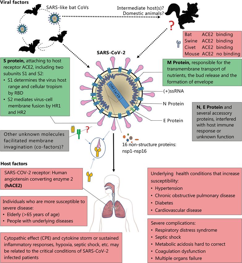
market. Rather, bats are the natural reservoir of a wide SARS-CoV, particularly in the S-glycoprotein gene andGuo et al. Military Medical Research (2020) 7:11 Page 3 of 10 Fig. 1 Viral and host factors that influence the pathogenesis of SARS-CoV-2. Bats are the reservoir of a wide variety of coronaviruses, including severe acute respiratory syndrome coronavirus (SARS-CoV) -like viruses. SARS-CoV-2 may originate from bats or unknown intermediate hosts and cross the species barrier into humans. Virus-host interactions affect viral entry and replication. Upper panel: Viral factor. SARS-CoV-2 is an enveloped positive single-stranded RNA (ssRNA) coronavirus. Two-thirds of viral RNA, mainly located in the first open reading frame (ORF 1a/b), encodes 16 non-structure proteins (NSPs). The rest part of the virus genome encodes four essential structural proteins, including spike (S) glycoprotein, small envelope (E) protein, matrix (M) protein, and nucleocapsid (N) protein, and also several accessory proteins. S glycoprotein of SARS-CoV-2 binds to host cell receptors, angiotensin-converting enzyme 2 (ACE2), that is a critical step for virus entry. The possible molecules facilitated membrane invagination for SARS-CoV-2 endocytosis are still unclear. Other virus proteins may contribute to pathogenesis. Host factors (Lower panel) can also influence susceptibility to infection and disease progression. The elderly and people with underlying disease are susceptible to SARS-CoV-2 and tend to develop into critical conditions. RBD, receptor-binding domain; HR1, heptad repeats 1; HR2, heptad repeats 2
Guo et al. Military Medical Research   (2020) 7:11                                                              Page 4 of 10

receptor-binding domain (RBD), indicating the capability        the virion-containing vesicles fuse with the plasma mem-
of direct human transmission. Compared with the known           brane to release the virus.
SARS-CoV and MERS-CoV genome, SARS-CoV-2 is                       Because the binding of SARS-CoV-2 Spike (S) glyco-
closer to the SARS-like bat CoVs in terms of the whole          protein and ACE2 receptor is a critical step for virus
genome sequence. Most genomic encoded proteins of               entry, virus-receptor binding affinity is under intensive
SARS-CoV-2 are similar to SARS-CoVs, as well as exist           study through different approaches. Systematic detection
certain differences. At the protein level, there are no         of β-CoV receptors showed that human cells expressing
amino acid substitutions that occurred in NSP7, NSP13,          ACE2, but not human Dipeptidyl peptidase-4 (DPP4) or
envelope, matrix, or accessory proteins p6 and 8b, except       APN (Aminopeptidase N), were enhanced entry of
in NSP2, NSP3, spike protein, underpinning subdomain,           SARS-CoV-2 [37]. While, another study showed that S-
i.e., RBD [22]. Another recent research suggested [23] that     protein and ACE2 binding efficiency is 10- to 20- fold
the mutation in NSP2 and NSP3 play a role in infectious         higher than that of SARS-CoV, evidenced by Cryo-EM
capability and differentiation mechanism of SARS-CoV-2.         Structure of the SARS-CoV-2 Spike in the prefusion
This provokes people to explore the difference of the host      conformation [38]. For SARS-CoV, the cleavage of tri-
tropism and transmission between SARS-CoV-2 and                 mer S protein is triggered by the cell surface-associated
SARS-CoV or conduct further investigations on the po-           transmembrane protease serine 2 (TMPRSS2) [39] and
tential therapeutic targets. Zhang et al. [24] analyzed the     cathepsin [40], while the possible molecules facilitated
genotypes of COVID-19 in different patients from several        membrane invagination for SARS-CoV-2 endocytosis are
provinces and found that SARS-CoV-2 had been mutated            still unclear. Up to the date this review paper was pre-
in different patients in China. Although the degree of di-      pared, reports showed that the SARS-CoV-2 may readily
versification of SARS-CoV-2 is smaller than the mutation        transmit, while cause less serious human infection rather
of H7N9 avian influenza [25]. Tang et al. [26] conducted a      than human SARS-CoV. Based on the latest WHO re-
population genetic analyses of 103 SARS-CoV-2 genomes           port, the number of infected people (over 80,000
and classified out two prevalent evolvement types of            globally, updated on 1 March 2020). The global outbreak
SARS-CoV-2, L type (~ 70%) and S type (~ 30%). The              may due to the following factors: firstly, the unknown
strains in L type, derived from S type, are evolutionarily      pneumonia outbroke at the time of China Spring Festi-
more aggressive and contagious. Thus, virologists and epi-      val, when the mass population flowing. Secondly, more
demiologists need to closely monitor the novel corona-          detailed molecular mechanisms of viral binding and
virus, in order to inspect the virulence and epidemic.          entry manners await to be elucidated, which may ham-
                                                                per the development of targeted therapy. Thirdly, avail-
Coronavirus replication and pathogenesis                        able data suggested that the SARS-CoV-2 may be less
ACE2, found in the lower respiratory tract of humans, is        virulent than the SARS-CoV and MERS-CoV, with the
known as cell receptor for SARS-CoV [27] and regulates          currently analyzed mortality of COVID-19 is 3.4%, lower
both the cross-species and human-to-human transmission          than death rate of SARS (9.6%) and MERS (around
[28]. Isolated from the bronchoalveolar lavage fluid (BALF)     35%), respectively [19]. Thus, the potential mechanisms
of a COVID-19 patient, Zhou et al. [8] have confirmed that      for human-to-human transmission and pathogenic
the SARS-CoV-2 uses the same cellular entry receptor,           mechanisms of the SARS-CoV-2 are under extensively
ACE2, as SARS-CoV. The virion S-glycoprotein on the sur-        studied.
face of coronavirus can attach to the receptor, ACE2 on the
surface of human cells [29]. S glycoprotein includes two sub-   Clinical characteristics
units, S1 and S2 [30]. S1 determines the virus-host range and   As an emerging acute respiratory infectious disease,
cellular tropism with the key function domain − RBD, while      COVID-19 primarily spreads through the respiratory
S2 mediates virus-cell membrane fusion by two tandem do-        tract, by droplets, respiratory secretions, and direct con-
mains, heptad repeats 1 (HR1) [31] and HR2 [32]. After          tact [41] for a low infective dose [42]. Otherwise, it has
membrane fusion, the viral genome RNA is released into the      been reported a SARS-CoV-2 was isolated from fecal
cytoplasm, and the uncoated RNA translates two polypro-         swabs of a severe pneumonia patient on 10 February
teins, pp1a and pp1ab [33], which encode non-structural         2020 from a critical case in the Fifth Affiliated Hospital,
proteins, and form replication-transcription complex (RTC)      Sun Yat-Sen University, Guangdong, China. Likewise,
in double-membrane vesicle [34]. Continuously RTC repli-        Zhang et al. [43] have found the presence of SARS-CoV-
cate and synthesize a nested set of subgenomic RNAs [35],       2 in fecal swabs and blood, indicating the possibility of
which encode accessory proteins and structural proteins.        multiple routes transmission. ACE2 protein presents in
Mediating endoplasmic reticulum (ER) and Golgi [36], newly      abundance on lung alveolar epithelial cells and entero-
formed genomic RNA, nucleocapsid proteins and envelope          cytes of small intestine remarkably [44], which may help
glycoproteins assemble and form viral particle buds. Lastly,    understand the routes of infection and disease
Guo et al. Military Medical Research (2020) 7:11 Page 5 of 10
manifestations. Based on current epidemiological investi- factor-α (TNF-α) increase, indicating the immune status
gation, the incubation period is 1–14 days, mostly 3–7 of patients. The data showed that ICU patients had
days. And the COVID-19 is contagious during the la- higher plasma levels of IL-2, IL-7, IL-10, granulocyte
tency period [45]. It is highly transmissible in humans, colony-stimulating factor (GCSF), 10 kD interferon-
especially in the elderly and people with underlying dis- gamma-induced protein (IP-10), monocyte chemo-
eases. The median age of patients is 47–59 years, and attractant protein-1 (MCP-1), macrophage inflammatory
41.9–45.7% of patients were females [16, 41, 46]. As it is protein 1-α (MIP-1α), and TNF-α [48].
designated SARS-CoV-2, COVID-19 patients presented Moreover, the CT imaging showed that computed tom-
certainly similar symptoms, such as fever, malaise, and ography on the chest was ground-glass opacity (56.4%)
cough [47]. Most adults or children with SARS-CoV-2 and bilateral patchy shadowing (51.8%) [16], sometimes
infection presented with mild flu-like symptoms and a with a rounded morphology and a peripheral lung distri-
few patients are in critical condition and rapidly develop bution, analyzed from the patients in the Fifth Affiliated
acute respiratory distress syndrome, respiratory failure, Hospital, Sun Yat-Sen University [55]. Clinicians have
multiple organ failure, even deaths [48]. been aware that, a part of confirmed patients appeared the
normal CT image presentations. The diagnostic sensitivity
Diagnostic criteria of radiologic is limited, so it is necessary to verify with
The viral research institution in China has conducted clinical symptoms and virus RNA detections.
preliminary identification of the SARS-CoV-2 through
the classical Koch’s postulates and observing its morph- Complications and clinical outcomes
ology through electron microscopy [49]. So far, the Based on the current information, most patients had a
golden clinical diagnosis method of COVID-19 is nucleic good prognosis, while a few patients were in critical condi-
acid detection in the nasal and throat swab sampling or tion, especially the elderly and those with chronic under-
other respiratory tract samplings by real-time PCR and lying diseases. As of 1 March 2020, a total of 79,968
further confirmed by next-generation sequencing. confirmed cases, including 14,475 (18.1%) with severe ill-
ness, and 2873 deaths (3.5%) in mainland China had been
Clinical symptoms reported by WHO [2]. Complications included acute re-
A recent study led by Prof. Nan-Shan Zhong’s team, by spiratory distress syndrome (ARDS), arrhythmia, shock
sampling 1099 laboratory-confirmed cases, found that [46], acute kidney injury, acute cardiac injury, liver dys-
the common clinical manifestations included fever function and secondary infection [48]. The poor clinical
(88.7%), cough (67.8%), fatigue (38.1%), sputum produc- outcome was related to disease severity. The disease tends
tion (33.4%), shortness of breath (18.6%), sore throat to progress faster in elderly people, with the median num-
(13.9%), and headache (13.6%) [16]. In addition, a part of ber of days from the occurrence of the first symptoms to
patients manifested gastrointestinal symptoms, with death shorter among people aged 65 years or more [56,
diarrhea (3.8%) and vomiting (5.0%). The clinical mani- 57]. Similar to H7N9 patients [58], the elderly male with
festations were in consistence with the previous data of comorbidities and ARDS showed a higher death risk. Add-
41, 99, and 138 patients analysis in Hubei province [46, itionally, more than 100 children were infected, with the
48, 50]. Fever and cough were the dominant symptoms youngest being 30 h after birth [59]. Neonates and the eld-
whereas upper respiratory symptoms and gastrointes- erly need more attention and care due to their immature
tinal symptoms were rare, suggesting the differences in or weak immune system.
viral tropism as compared with SARS-CoV [51], MERS-
CoV [52], and influenza [53]. The elderly and those with Host immune response and immunopathology
underlying disorders (i.e., hypertension, chronic ob- The immune response is vital for the control and reso-
structive pulmonary disease, diabetes, cardiovascular dis- lution of CoV infections, while it can also lead to immu-
ease), developed rapidly into acute respiratory distress nopathogenesis, associated with the immune response out
syndrome, septic shock, metabolic acidosis hard to cor- of control. The S proteins of Coronavirus binds to the
rect and coagulation dysfunction, even leading to the host cells by ACE2, fusing to the membrane and release
death [48] (lower panel, Fig. 1). the viral RNA. The viral RNAs, as pathogen-associated
In laboratory examination results, most patients had molecular patterns (PAMPs), are detected by the pattern
normal or decreased white blood cell counts, and lym- recognition receptors (PRRs). Usually, Toll-like receptor
phocytopenia [16, 54]. But in the severe patients, the (TLR) 3, TLR7, TLR8, and TLR9 sense viral RNA and
neutrophil count, D-dimer, blood urea, and creatinine DNA in the endosome [60, 61]. The viral RNA receptor
levels were higher significantly, and the lymphocyte retinoic-acid inducible gene I (RIG-I) [62], cytosolic recep-
counts continued to decrease. Additionally, inflamma- tor melanoma differentiation-associated gene 5 (MDA5)
tory factors (interleukin (IL)-6, IL-10, tumor necrosis and nucleotidyltransferase cyclic GMP-AMP synthaseGuo et al. Military Medical Research (2020) 7:11 Page 6 of 10
(cGAS) [63] are responsible for the recognition of viral Remdesivir (GS-5734) is a 1′-cyano-substituted adeno-
RNA and DNA in the cytoplasm. These complex signal- sine nucleotide analog prodrug and shows broad-
ling recruit adaptors, including TIR-domain-containing spectrum antiviral activity against several RNA viruses.
adaptor protein including IFN-β (TRIF), mitochondrial Based on the data collected from in vitro cell line and
antiviral-signalling protein (MAVS) [64] and stimulator of mouse model, remdesivir could interfere with the NSP12
interferon genes protein (STING) [65] to trigger down- polymerase even in the setting of intact ExoN proofread-
stream cascades molecules, involving adaptor molecule ing activity [76]. Remdesivir has been reported to treat
MyD88 and lead to the activation of the transcription fac- the first US case of COVID-19 successfully [77]. Chloro-
tor nuclear factor-κB (NF-κB) and interferon regulatory quine is a repurposed drug with great potential to treat
factor 3 (IRF3) and the production of type I Interferons COVID-19. Chloroquine has been used to treat malaria
(IFN-α /β) and a series of pro-inflammatory cytokines for many years [78], with a mechanism that is not well
[66]. Hence, virus-cell interactions produce a diverse set understood against some viral infections. Several pos-
of immune mediators against the invading virus [67]. In- sible mechanisms are investigated: Chloroquine can in-
nate immunity is needed in a precise regulation to elimin- hibit pH-dependent steps of the replication of several
ate the virus, otherwise will result in immunopathology. A viruses [79], with a potent effect on SARS-CoV infection
few plasma cytokines and chemokines were observed and spread [80]. Moreover, chloroquine has immuno-
ascended in COVID-19 patients, including IL-1, IL-2, IL- modulatory effects, suppressing the production/release
4, IL-7, IL-10, IL-12, IL-13, IL-17, GCSF, macrophage of TNF-α and IL-6. It also works as a novel class of
colony-stimulating factor (MCSF), IP-10, MCP-1, MIP-1α, autophagy inhibitor [81], which may interfere with viral
hepatocyte growth factor (HGF), IFN-γ and TNF-α [48, infection and replication. Several studies have found that
68, 69]. Of note, an anatomy report of COVID-19 pneu- chloroquine interfered with the glycosylation of cellular
monia corpse [70] indicated that COVID-19 caused an receptors of SARS-CoV [80] and functioned at both
inflammatory response in the lower airway and led to lung entry and at post-entry stages of the COVID-19 infec-
injury. Collectively, the virus particles invade the respira- tion in Vero E6 cells [82]. A combination of remdesivir
tory mucosa firstly and infect other cells, triggering a and chloroquine was proven to effectively inhibit the re-
series of immune responses and the production of cyto- cently emerged SARS-CoV-2 in vitro.
kine storm in the body, which may be associated with the Scientists previously confirmed that the protease in-
critical condition of COVID-19 patients. hibitors lopinavir and ritonavir, used to treat infection
with human immunodeficiency virus (HIV) [83], could
Treatment of COVID-19 improve the outcome of MERS-CoV [84] and SARS-
Current therapies CoV [85] patients. It has reported that β-coronavirus
Given the lack of effective antiviral therapy against viral loads of a COVID-19 patient in Korea significantly
COVID-19, current treatments mainly focused on symp- decreased after lopinavir/ritonavir (Kaletra®, AbbVie,
tomatic and respiratory support according to the Diagno- North Chicago, IL, USA) treatment [86]. Additionally,
sis and Treatment of Pneumonia Caused by COVID-19 clinicians combined Chinese and Western medicine
(updated to version 6) issued by National Health Commis- treatment including lopinavir/ritonavir (Kaletra®), arbi-
sion of the People’s Republic of China [71]. Nearly all dol, and Shufeng Jiedu Capsule (SFJDC, a traditional
patients accepted oxygen therapy, and WHO recom- Chinese medicine) and gained significant improvement
mended extracorporeal membrane oxygenation (ECMO) in pneumonia associated symptoms in Shanghai Public
to patients with refractory hypoxemia [72]. Rescue treat- Health Clinical Center, China [87].The other antiviral
ment with convalescent plasma and immunoglobulin G drugs include nitazoxanide, favipiravir, nafamostat, and
[73] are delivered to some critical cases according to their so on (See Table 1 for details).
conditions.
Conclusions
Antiviral treatments The outbreak of COVID-19 swept across China rapidly
Based on the experience of fighting the epidemic SARS- and has spread to 85 countries/territories/areas outside
CoV and MERS-CoV previously, we may learn some les- of China as of 5 March 2020 [2]. Scientists have made
sons for some treatment strategies against coronavirus progress in the characterization of the novel coronavirus
[74]. Antiviral drugs and systemic corticosteroid treat- and are working extensively on the therapies and vac-
ment commonly used in clinical practice previously, in- cines against the virus. We have summarized the current
cluding neuraminidase inhibitors (oseltamivir, peramivir, knowledge of SARS-CoV-2 as follows: Firstly, the emer-
zanamivir, etc), ganciclovir, acyclovir, and ribavirin, as ging pneumonia, COVID-19, caused by SARS-CoV-2,
well as methylprednisolone [46, 75] for influenza virus, exhibits strong infectivity but less virulence, compared
are invalid for COVID-19 and not recommended. to SARS and MERS, in terms of morbidity and mortality.Guo et al. Military Medical Research (2020) 7:11 Page 7 of 10
Table 1 Common and potent antiviral drugs
Status Drugs Action mode Anti-infective mechanism Target diseases Ref.
Approved Lopinavir/ Protease inhibitors Inhibiting HIV-1 protease for protein cleavage, HIV/AIDS, SARS, MERS [83–85]
Ritonavir resulting in non-infectious, immature viral particles
Approved, Chloroquine 9-aminoquinolin Increasing endosomal pH, immunomodulating, Malaria, autoimmune [79–82]
Investigational, autophagy inhibitors disease
Vet approved
Experimental Remdesivir Nucleotide analogue Interfering with virus post-entry Ebola, SARS, MERS [76, 88, 89]
(GS-5734) prodrug
(A wide array of RNA viruses)
Investigational Nafamostat Synthetic serine Prevents membrane fusion by reducing the Influenza, MERS, Ebola [90, 91]
protease inhibitor release of cathepsin B; anticoagulant activities
Approved Ribavirin Synthetic guanosine Interfering with the synthesis of viral mRNA HCV, SARS, MERS [92–94]
nucleoside (a broad-spectrum activity against several
RNA and DNA viruses)
Approved Oseltamivir Neuraminidase Inhibiting the activity of the viral neuraminidase Influenza viruses A [95, 96]
inhibitor enzyme, preventing budding from the host cell,
viral replication, and infectivity
Approved Penciclovir/ Nucleoside analog A synthetic acyclic guanine derivative, resulting HSV, VZV [97]
Acyclovir in chain termination
Approved, Ganciclovir Nucleoside analog Potent inhibitor of the Herpesvirus family AIDS-associated [98]
Investigational including cytomegalovirus cytomegalovirus
infections
Investigational Favipiravir Nucleoside analog: Acting on viral genetic copying to prevent its Ebola, influenza [99–101]
(T-705) Viral RNA reproduction, without affecting host cellular A(H1N1)
polymerase inhibitor RNA or DNA synthesis
Approved, Nitazoxanide Antiprotozoal agent Modulating the survival, growth, and proliferation A wide range of viruses [102–104]
Investigational, Vet of a range of extracellular and intracellular including human/animal
approved protozoa, helminths, anaerobic and coronaviruses
microaerophilic bacteria, viruses
HIV Human immunodeficiency virus, AIDS Acquired immune deficiency syndrome, SARS Severe acute respiratory syndrome, MERS Middle East
respiratory syndrome, HCV Hepatitis C virus, HSV Herpes simplex virus, VZV Varicella-zoster virus
Originating from reservoir of bats and unknown inter- inadequate protection, as well as overwork, frustration
mediate hosts, SARS-CoV-2 binds to ACE2 with high af- and exhaustion [105]. Chinese Government and author-
finity as a virus receptor to infect humans. Secondly, the ities have launched psychological intervention, and we
susceptible population involves the elderly and people sincerely hope that Chinese people and other countries
with certain underlying medical conditions, which re- overcome the epidemic as fast as possible.
quires more attention and care. Thirdly, so far, the sup-
porting treatments, combined with potent antiviral Abbreviations
ACE2: Angiotensin-converting enzyme 2; APN: Aminopeptidase N;
drugs, such as remdesivir, chloroquine, or lopinavir/rito- ARDS: Acute respiratory distress syndrome; BALF: Bronchoalveolar lavage
navir, have been conducted with definite effect on treat fluid; cGAS: Cyclic GMP-AMP synthase; CoV: Coronavirus; COVID-
COVID-19 patients, while solid data from more clinical 19: Coronavirus disease 2019; CSG: Coronavirus Study Group;
DPP4: Dipeptidyl peptidase-4; E protein: Envelope protein;
trials are needed. However, questions remain vague and ECMO: Extracorporeal membrane oxygenation; ER: Endoplasmic reticulum;
more studies are urgent to explore the transmission and GCSF: Granulocyte colony-stimulating factor; HGF: Hepatocyte growth factor;
pathogenicity mechanism of the emerging coronavirus. HR1: Heptad repeats 1; HR2: Heptad repeats 2; IFN: Interferon; IL: Interleukin;
IP-10: 10 kD interferon-gamma-induced protein; IRF3: Interferon regulatory
To make clear the evolutionary path from the original factor 3; M protein: Matrix protein; MAVS: Mitochondrial antiviral-signalling
host to cross-species transmission so as to potentially protein; MCP-1: Monocyte chemoattractant protein-1; MCSF: Macrophage
limit the transmission to naïve animals or humans. In colony-stimulating factor; MDA5: Melanoma differentiation-associated gene
5; MERS-CoV: Middle East respiratory syndrome coronavirus; MIP-
addition, to uncover the mystery of the molecular mech- 1α: Macrophage inflammatory protein 1-α; N protein: Nucleocapsid protein;
anism of viral entry and replication, which provides the NF-κB: Nuclear factor-κB; NSP: Non-structure protein; ORF: Open reading
basis of future research on developing targeted antiviral frame; PAMP: Pathogen-associated molecular pattern; PRR: Pattern
recognition receptor; RBD: Receptor-binding domain; RIG-I: Retinoic-acid
drugs and vaccines. inducible gene I; RTC: Replication-transcription complex; S protein: Spike
Given more than 80% of patients are confirmed in glycoprotein; SARS-CoV: Severe acute respiratory syndrome coronavirus;
Hubei province, the hospitals and medical workers in STING: Stimulator of interferon genes protein; TLR: Toll-like receptor;
TMPRSS2: Transmembrane protease serine 2; TNF-α: Tumor necrosis factor α;
Hubei are facing and bearing enormous pressure and se- TRIF: TIR-domain-containing adaptor protein including IFN-β; WHCV: Wuhan-
vere challenge, including a high risk of infection and Hu-1 coronavirus; WHO: World Health OrganizationGuo et al. Military Medical Research (2020) 7:11 Page 8 of 10
Acknowledgements 6. Zhu N, Zhang D, Wang W, Li X, Yang B, Song J, et al. A novel coronavirus
We thank Drs. Hong Shan, Jin-Yu Xia, Zhong-He Li, Jing Liu, Ming-Xing from patients with pneumonia in China, 2019. N Engl J Med. 2020;382(8):
Huang, Xi Liu, Fei Xiao, Shao-Lin Li, Xiao-Feng Li, Xiao-Hua Wang, Xiu-Juan 727–33.
Qu and Ms. Yan Nan from library, from the Fifth Affiliated Hospital, Sun Yat- 7. Yin Y, Wunderink RG. MERS, SARS and other coronaviruses as causes of
Sen University, for their sharing the critical information on theranostics of pneumonia. Respirology. 2018;23(2):130–7.
COVID-19 patients and literature searching. 8. Zhou P, Yang XL, Wang XG, Hu B, Zhang L, Zhang W, et al. A pneumonia
outbreak associated with a new coronavirus of probable bat origin. Nature.
Authors’ contributions 2020. https://doi.org/10.1038/s41586-020-2012-7.
DYW and YY conceived this review; YRG, QDC, ZH, YYT, SDC, HJJ performed 9. Giovanetti M, Benvenuto D, Angeletti S, Ciccozzi M. The first two cases of
the literature review and wrote the paper; KST, DYW, YY, YRG helped with 2019-nCoV in Italy: where they come from? J Med Virol. 2020:1–4. https://
the review and writing of the paper; all authors read and approved the final doi.org/10.1002/jmv.25699 [Epub ahead of print].
manuscript. 10. Paraskevis D, Kostaki EG, Magiorkinis G, Panayiotakopoulos G, Sourvinos G,
Tsiodras S. Full-genome evolutionary analysis of the novel corona virus
(2019-nCoV) rejects the hypothesis of emergence as a result of a recent
Funding recombination event. Infect Genet Evol. 2020;79:104212.
This study was supported by grants awarded to YY by the National Natural 11. Hampton T. Bats may be SARS reservoir. JAMA. 2005;294(18):2291.
Science Foundation of China (81870019), Guangdong Provincial Natural Science 12. Banerjee A, Kulcsar K, Misra V, Frieman M, Mossman K. Bats and
Foundation (2018A030313554), National Key R&D Program of China coronaviruses. Viruses. 2019;11(1):E41. https://doi.org/10.3390/v11010041.
(2018YFC0910601); DYW by the National Medical Research Council, Singapore
13. Li W, Shi Z, Yu M, Ren W, Smith C, Epstein JH, et al. Bats are natural
(NMRC/CIRG/1458/2016); KST is a recipient of fellowship support from European
reservoirs of SARS-like coronaviruses. Science. 2005;310(5748):676–9.
Allergy and Clinical Immunology (EAACI) Research Fellowship 2019.
14. Wu F, Zhao S, Yu B, Chen YM, Wang W, Song ZG, et al. A new coronavirus
associated with human respiratory disease in China. Nature. 2020. https://
Availability of data and materials doi.org/10.1038/s41586-020-2008-3 [Epub ahead of print].
The data and materials used during the current review are all available in 15. Liu Z, Xiao X, Wei X, Li J, Yang J, Tan H, et al. Composition and divergence
this review. of coronavirus spike proteins and host ACE2 receptors predict potential
intermediate hosts of SARS-CoV-2. J Med Virol. 2020. https://doi.org/10.
Ethics approval and consent to participate 1002/jmv.25726 [Epub ahead of print].
Not applicable. 16. Guan WJ, Ni ZY, Hu Y, Liang WH, Ou CQ, He JX, et al. Clinical characteristics
of coronavirus disease 2019 in China. N Engl J Med. 2020. https://doi.org/10.
1056/NEJMoa2002032.
Consent for publication 17. Chowell G, Abdirizak F, Lee S, Lee J, Jung E, Nishiura H, et al. Transmission
Not applicable. characteristics of MERS and SARS in the healthcare setting: a comparative
study. BMC Med. 2015;13:210.
Competing interests 18. Kang CK, Song KH, Choe PG, Park WB, Bang JH, Kim ES, et al. Clinical and
The funder had no role in study design, data collection, and analysis, epidemiologic characteristics of spreaders of middle east respiratory
decision to publish, or preparation of the manuscript. The authors declare syndrome coronavirus during the 2015 outbreak in Korea. J Korean Med Sci.
that they have no competing interests. 2017;32(5):744–9.
19. de Wit E, van Doremalen N, Falzarano D, Munster VJ. SARS and MERS:
Author details recent insights into emerging coronaviruses. Nat Rev Microbiol. 2016;14(8):
1
Guangdong Provincial Key Laboratory of Biomedical Imaging and 523–34.
Guangdong Provincial Engineering Research Center of Molecular Imaging, 20. Song Z, Xu Y, Bao L, Zhang L, Yu P, Qu Y, et al. From SARS to MERS,
Zhuhai 519000, Guangdong, China. 2Department of Cardiothoracic Surgery, thrusting coronaviruses into the spotlight. Viruses. 2019;11(1):E59. https://
the Fifth Affiliated Hospital, Sun Yat-Sen University, Zhuhai 519000, doi.org/10.3390/v11010059.
Guangdong, China. 3Center of Infectious Disease, the Fifth Affiliated Hospital, 21. Cui J, Li F, Shi ZL. Origin and evolution of pathogenic coronaviruses. Nat
Sun Yat-Sen University, Zhuhai 519000, Guangdong, China. 4Department of Rev Microbiol. 2019;17(3):181–92.
Otolaryngology, Yong Loo Lin School of Medicine, National University of 22. Wu A, Peng Y, Huang B, Ding X, Wang X, Niu P, et al. Genome composition
Singapore, National University Health System, Singapore 119228, Singapore. and divergence of the novel coronavirus (2019-nCoV) originating in China.
5
Center for Interventional Medicine, the Fifth Affiliated Hospital, Sun Yat-Sen Cell Host Microbe. 2020. https://doi.org/10.1016/j.chom.2020.02.001 [Epub
University, Zhuhai 519000, Guangdong, China. ahead of print].
23. Angeletti S, Benvenuto D, Bianchi M, Giovanetti M, Pascarella S, Ciccozzi M.
Received: 29 February 2020 Accepted: 9 March 2020 COVID-2019: the role of the nsp2 and nsp3 in its pathogenesis. J Med Virol.
2020. https://doi.org/10.1002/jmv.25719.
24. Zhang L, Shen FM, Chen F, Lin Z. Origin and evolution of the 2019 novel
References coronavirus. Clin Infect Dis. 2020. https://doi.org/10.1093/cid/ciaa112 [Epub
1. Lu R, Zhao X, Li J, Niu P, Yang B, Wu H, et al. Genomic characterisation and ahead of print].
epidemiology of 2019 novel coronavirus: implications for virus origins and 25. Wu D, Zou S, Bai T, Li J, Zhao X, Yang L, et al. Poultry farms as a source of
receptor binding. Lancet. 2020;395(10224):565–74. avian influenza a (H7N9) virus reassortment and human infection. Sci Rep.
2. WHO. Coronavirus disease (COVID-2019) situation reports. 2020. https:// 2015;5:7630.
www.who.int/emergencies/diseases/novel-coronavirus-2019/situation- 26. Tang X, Wu C, Li X, Song Y, Yao X, Wu X, et al. On the origin and
reports. Accessed 5 Mar 2020. continuing evolution of SARS-CoV-2. Natl Sci Rev. 2020. https://doi.org/10.
3. Riou J, Althaus CL. Pattern of early human-to-human transmission of Wuhan 1093/nsr/nwaa036.
2019 novel coronavirus (2019-nCoV), December 2019 to January 2020. Euro 27. Jia HP, Look DC, Shi L, Hickey M, Pewe L, Netland J, et al. ACE2 receptor
Surveill. 2020;25(4):2000058. https://doi.org/10.2807/1560-7917.ES.2020.25.4. expression and severe acute respiratory syndrome coronavirus infection
2000058. depend on differentiation of human airway epithelia. J Virol. 2005;79(23):
4. Liu Y, Gayle AA, Wilder-Smith A, Rocklov J. The reproductive number of 14614–21.
COVID-19 is higher compared to SARS coronavirus. J Travel Med. 2020. 28. Wan Y, Shang J, Graham R, Baric RS, Li F. Receptor recognition by novel
https://doi.org/10.1093/jtm/taaa021. coronavirus from Wuhan: an analysis based on decade-long structural
5. Chan JF, Yuan S, Kok KH, To KK, Chu H, Yang J, et al. A familial cluster of studies of SARS. J Virol. 2020. https://doi.org/10.1128/JVI.00127-20 [Epub
pneumonia associated with the 2019 novel coronavirus indicating person- ahead of print].
to-person transmission: a study of a family cluster. Lancet. 2020;395(10223): 29. Tortorici MA, Veesler D. Structural insights into coronavirus entry. Adv Virus
514–23. Res. 2019;105:93–116.Guo et al. Military Medical Research (2020) 7:11 Page 9 of 10
30. Zhang N, Jiang S, Du L. Current advancements and potential strategies in the 54. Kui L, Fang YY, Deng Y, Liu W, Wang MF, Ma JP, et al. Clinical characteristics
development of MERS-CoV vaccines. Expert Rev Vaccines. 2014;13(6):761–74. of novel coronavirus cases in tertiary hospitals in Hubei Province. Chin Med
31. Xia S, Zhu Y, Liu M, Lan Q, Xu W, Wu Y, et al. Fusion mechanism of 2019- J. 2020. https://doi.org/10.1097/CM9.0000000000000744 [Epub ahead of
nCoV and fusion inhibitors targeting HR1 domain in spike protein. Cell Mol print].
Immunol. 2020. https://doi.org/10.1038/s41423-020-0374-2. 55. Chung M, Bernheim A, Mei X, Zhang N, Huang M, Zeng X, et al. CT imaging
32. Yu F, Du L, Ojcius DM, Pan C, Jiang S. Measures for diagnosing and treating features of 2019 novel coronavirus (2019-nCoV). Radiology. 2020;200230.
infections by a novel coronavirus responsible for a pneumonia outbreak https://doi.org/10.1148/radiol.2020200230. [Epub ahead of print].
originating in Wuhan, China. Microbes Infect. 2020. https://doi.org/10.1016/j. 56. Wang W, Tang J, Wei F. Updated understanding of the outbreak of 2019
micinf.2020.01.003 [Epub ahead of print]. novel coronavirus (2019-nCoV) in Wuhan, China. J Med Virol. 2020;92(4):
33. de Wilde AH, Snijder EJ, Kikkert M, van Hemert MJ. Host factors in 441–7.
coronavirus replication. Curr Top Microbiol Immunol. 2018;419:1–42. 57. Yang X, Yu Y, Xu J, Shu H, Xia J, Liu H, et al. Clinical course and outcomes of
34. Sawicki SG, Sawicki DL. Coronavirus transcription: a perspective. Curr Top critically ill patients with SARS-CoV-2 pneumonia in Wuhan, China: a single-
Microbiol Immunol. 2005;287:31–55. centered, retrospective, observational study. Lancet Respir Med. 2020.
35. Hussain S, Pan J, Chen Y, Yang Y, Xu J, Peng Y, et al. Identification of novel https://doi.org/10.1016/s2213-2600(20)30079-5.
subgenomic RNAs and noncanonical transcription initiation signals of 58. Gao HN, Lu HZ, Cao B, Du B, Shang H, Gan JH, et al. Clinical findings in 111
severe acute respiratory syndrome coronavirus. J Virol. 2005;79(9):5288–95. cases of influenza a (H7N9) virus infection. N Engl J Med. 2013;368(24):2277–85.
36. Perrier A, Bonnin A, Desmarets L, Danneels A, Goffard A, Rouille Y, et al. The 59. Wang J, Qi H, Bao L, Li F, Shi Y, National Clinical Research Center for Child H,
C-terminal domain of the MERS coronavirus M protein contains a trans- et al. A contingency plan for the management of the 2019 novel coronavirus
Golgi network localization signal. J Biol Chem. 2019;294(39):14406–21. outbreak in neonatal intensive care units. Lancet Child Adolesc Health. 2020.
37. Letko M, Marzi A, Munster V. Functional assessment of cell entry and https://doi.org/10.1016/S2352-4642(20)30040-7 [Epub ahead of print].
receptor usage for SARS-CoV-2 and other lineage B betacoronaviruses. Nat 60. Alexopoulou L, Holt AC, Medzhitov R, Flavell RA. Recognition of double-
Microbiol. 2020. https://doi.org/10.1038/s41564-020-0688-y. stranded RNA and activation of NF-kappaB by toll-like receptor 3. Nature.
38. Song W, Gui M, Wang X, Xiang Y. Cryo-EM structure of the SARS 2001;413(6857):732–8.
coronavirus spike glycoprotein in complex with its host cell receptor ACE2. 61. Wu J, Chen ZJ. Innate immune sensing and signaling of cytosolic nucleic
PLoS Pathog. 2018;14(8):e1007236. acids. Annu Rev Immunol. 2014;32:461–88.
39. Millet JK, Whittaker GR. Host cell proteases: critical determinants of 62. Yoo JS, Kato H, Fujita T. Sensing viral invasion by RIG-I like receptors. Curr
coronavirus tropism and pathogenesis. Virus Res. 2015;202:120–34. Opin Microbiol. 2014;20:131–8.
40. Simmons G, Gosalia DN, Rennekamp AJ, Reeves JD, Diamond SL, Bates P. 63. Wu J, Sun L, Chen X, Du F, Shi H, Chen C, et al. Cyclic GMP-AMP is an
Inhibitors of cathepsin L prevent severe acute respiratory syndrome endogenous second messenger in innate immune signaling by cytosolic
coronavirus entry. P Natl Acad Sci USA. 2005;102(33):11876–81. DNA. Science. 2013;339(6121):826–30.
41. Li Q, Guan X, Wu P, Wang X, Zhou L, Tong Y, et al. Early transmission 64. Seth RB, Sun L, Ea CK, Chen ZJ. Identification and characterization of MAVS,
dynamics in Wuhan, China, of novel coronavirus-infected pneumonia. N a mitochondrial antiviral signaling protein that activates NF-kappaB and IRF
Engl J Med. 2020. https://doi.org/10.1056/NEJMoa2001316 [Epub ahead 3. Cell. 2005;122(5):669–82.
of print]. 65. Ishikawa H, Barber GN. STING is an endoplasmic reticulum adaptor that
42. Lee PI, Hsueh PR. Emerging threats from zoonotic coronaviruses-from SARS facilitates innate immune signalling. Nature. 2008;455(7213):674–8.
and MERS to 2019-nCoV. J Microbiol Immunol Infect. 2020. https://doi.org/ 66. Kawai T, Akira S. The role of pattern-recognition receptors in innate
10.1016/j.jmii.2020.02.001 [Epub ahead of print]. immunity: update on toll-like receptors. Nat Immunol. 2010;11(5):373–84.
43. Zhang W, Du RH, Li B, Zheng XS, Yang XL, Hu B, et al. Molecular and 67. Takeuchi O, Akira S. Innate immunity to virus infection. Immunol Rev. 2009;
serological investigation of 2019-nCoV infected patients: implication of 227(1):75–86.
multiple shedding routes. Emerg Microbes Infect. 2020;9(1):386–9. 68. Chen C, Zhang XR, Ju ZY, He WF. Advances in the research of cytokine
44. Hamming I, Timens W, Bulthuis ML, Lely AT, Navis G, van Goor H. Tissue storm mechanism induced by Corona Virus Disease 2019 and the
distribution of ACE2 protein, the functional receptor for SARS coronavirus. A corresponding immunotherapies. Zhonghua Shaoshang Zazhi. 2020;36(0):
first step in understanding SARS pathogenesis. J Pathol. 2004;203(2):631–7. E005.
45. Jin YH, Cai L, Cheng ZS, Cheng H, Deng T, Fan YP, et al. A rapid advice 69. Liu Y, Zhang C, Huang F, Yang Y, Wang F, Yuan J, et al. 2019-novel
guideline for the diagnosis and treatment of 2019 novel coronavirus (2019- coronavirus (2019-nCoV) infections trigger an exaggerated cytokine
nCoV) infected pneumonia (standard version). Mil Med Res. 2020;7(1):4. response aggravating lung injury. 2020. http://www.chinaxiv.org/abs/202
46. Wang D, Hu B, Hu C, Zhu F, Liu X, Zhang J, et al. Clinical characteristics of 002.00018. Accessed 18 Feb 2020.
138 hospitalized patients with 2019 novel coronavirus-infected pneumonia 70. Liu Q, Wang R, Qu G, Wang Y, Liu P, Zhu Y, et al. General anatomy report of
in Wuhan, China. JAMA. 2020. https://doi.org/10.1001/jama.2020.1585 [Epub novel coronavirus pneumonia death corpse. J Forensic Med. 2020;36(1):19–21.
ahead of print]. 71. National Health Commission of the People’s Republic of China. Diagnosis
47. Poutanen SM, Low DE, Henry B, Finkelstein S, Rose D, Green K, et al. and Treatment of Pneumonia Caused by 2019-nCoV (version 6). 2020.
Identification of severe acute respiratory syndrome in Canada. N Engl J http://www.gov.cn/zhengce/zhengceku/2020-02/19/content_5480948.htm.
Med. 2003;348(20):1995–2005. Accessed 18 Feb 2020.
48. Huang C, Wang Y, Li X, Ren L, Zhao J, Hu Y, et al. Clinical features of 72. WHO. Clinical management of severe acute respiratory infection when novel
patients infected with 2019 novel coronavirus in Wuhan, China. Lancet. coronavirus (nCoV) infection is suspected. https://www.who.int/publications-
2020;395(10223):497–506. detail/clinical-management-of-severe-acute-respiratory-infection-when-novel-
49. Lu H, Stratton CW, Tang YW. Outbreak of pneumonia of unknown etiology coronavirus-(ncov)-infection-is-suspected. Accessed 28 Jan 2020.
in Wuhan, China: the mystery and the miracle. J Med Virol. 2020;92(4):401–2. 73. Chen L, Xiong J, Bao L, Shi Y. Convalescent plasma as a potential therapy
50. Chen N, Zhou M, Dong X, Qu J, Gong F, Han Y, et al. Epidemiological and for COVID-19. Lancet Infect Dis. 2020. https://doi.org/10.1016/s1473-
clinical characteristics of 99 cases of 2019 novel coronavirus pneumonia in 3099(20)30141-9.
Wuhan, China: a descriptive study. Lancet. 2020;395(10223):507–13. 74. Zumla A, Chan JF, Azhar EI, Hui DS, Yuen KY. Coronaviruses - drug discovery
51. Lee N, Hui D, Wu A, Chan P, Cameron P, Joynt GM, et al. A major outbreak and therapeutic options. Nat Rev Drug Discov. 2016;15(5):327–47.
of severe acute respiratory syndrome in Hong Kong. N Engl J Med. 2003; 75. Li H, Wang YM, Xu JY, Cao B. Potential antiviral therapeutics for 2019 Novel
348(20):1986–94. Coronavirus. Chin J Tuberc Respir Dis. 2020;43(0):E002.
52. Assiri A, Al-Tawfiq JA, Al-Rabeeah AA, Al-Rabiah FA, Al-Hajjar S, Al-Barrak A, 76. Agostini ML, Andres EL, Sims AC, Graham RL, Sheahan TP, Lu X, et al.
et al. Epidemiological, demographic, and clinical characteristics of 47 cases Coronavirus susceptibility to the antiviral remdesivir (gs-5734) is mediated
of Middle East respiratory syndrome coronavirus disease from Saudi Arabia: by the viral polymerase and the proofreading exoribonuclease. mBio. 2018;
a descriptive study. Lancet Infect Dis. 2013;13(9):752–61. 9(2):e00221–18. https://doi.org/10.1128/mBio.00221-18.
53. Wang H, Xiao X, Lu J, Chen Z, Li K, Liu H, et al. Factors associated with 77. Holshue ML, DeBolt C, Lindquist S, Lofy KH, Wiesman J, Bruce H, et al. First
clinical outcome in 25 patients with avian influenza a (H7N9) infection in case of 2019 novel coronavirus in the United States. N Engl J Med. 2020.
Guangzhou, China. BMC Infect Dis. 2016;16(1):534. https://doi.org/10.1056/NEJMoa2001191 [Epub ahead of print].Guo et al. Military Medical Research (2020) 7:11 Page 10 of 10
78. Aguiar ACC, Murce E, Cortopassi WA, Pimentel AS, Almeida M, Barros DCS, 102. Rossignol JF. Nitazoxanide: a first-in-class broad-spectrum antiviral agent.
et al. Chloroquine analogs as antimalarial candidates with potent in vitro Antivir Res. 2014;110:94–103.
and in vivo activity. Int J Parasitol Drugs Drug Resist. 2018;8(3):459–64. 103. Cao J, Forrest JC, Zhang X. A screen of the NIH clinical collection small
79. Savarino A, Boelaert JR, Cassone A, Majori G, Cauda R. Effects of chloroquine molecule library identifies potential anti-coronavirus drugs. Antivir Res. 2015;
on viral infections: an old drug against today's diseases? Lancet Infect Dis. 114:1–10.
2003;3(11):722–7. 104. Rossignol JF. Nitazoxanide, a new drug candidate for the treatment of
80. Vincent MJ, Bergeron E, Benjannet S, Erickson BR, Rollin PE, Ksiazek TG, et al. Middle East respiratory syndrome coronavirus. J Infect Public Health. 2016;
Chloroquine is a potent inhibitor of SARS coronavirus infection and spread. 9(3):227–30.
Virol J. 2005;2:69. 105. Kang L, Li Y, Hu S, Chen M, Yang C, Yang BX, et al. The mental health of
81. Golden EB, Cho HY, Hofman FM, Louie SG, Schonthal AH, Chen TC. medical workers in Wuhan, China dealing with the 2019 novel coronavirus.
Quinoline-based antimalarial drugs: a novel class of autophagy inhibitors. Lancet Psychiatry. 2020;7(3):e14.
Neurosurg Focus. 2015;38(3):E12.
82. Wang M, Cao R, Zhang L, Yang X, Liu J, Xu M, et al. Remdesivir and
chloroquine effectively inhibit the recently emerged novel coronavirus
(2019-nCoV) in vitro. Cell Res. 2020. https://doi.org/10.1038/s41422-020-
0282-0 [Epub ahead of print].
83. Cvetkovic RS, Goa KL. Lopinavir/ritonavir: a review of its use in the
management of HIV infection. Drugs. 2003;63(8):769–802.
84. Arabi YM, Asiri AY, Assiri AM, Aziz Jokhdar HA, Alothman A, Balkhy HH, et al.
Treatment of Middle East respiratory syndrome with a combination of
lopinavir/ritonavir and interferon-β1b (MIRACLE trial): statistical analysis plan
for a recursive two-stage group sequential randomized controlled trial.
Trials. 2020;21(1):8.
85. Chu CM, Cheng VC, Hung IF, Wong MM, Chan KH, Chan KS, et al. Role of
lopinavir/ritonavir in the treatment of SARS: initial virological and clinical
findings. Thorax. 2004;59(3):252–6.
86. Lim J, Jeon S, Shin HY, Kim MJ, Seong YM, Lee WJ, et al. Case of the index
patient who caused tertiary transmission of COVID-19 infection in Korea: the
application of lopinavir/ritonavir for the treatment of COVID-19 infected
pneumonia monitored by quantitative RT-PCR. J Korean Med Sci. 2020;35(6):e79.
87. Wang Z, Chen X, Lu Y, Chen F, Zhang W. Clinical characteristics and
therapeutic procedure for four cases with 2019 novel coronavirus pneumonia
receiving combined Chinese and Western medicine treatment. Biosci Trends.
2020. https://doi.org/10.5582/bst.2020.01030 [Epub ahead of print].
88. Tchesnokov EP, Feng JY, Porter DP, Gotte M. Mechanism of inhibition of
ebola virus rna-dependent rna polymerase by remdesivir. Viruses. 2019;11(4):
E326. https://doi.org/10.3390/v11040326.
89. Lo MK, Feldmann F, Gary JM, Jordan R, Bannister R, Cronin J, et al.
Remdesivir (GS-5734) protects African green monkeys from Nipah virus
challenge. Sci Transl Med. 2019;11(494):eaau9242. https://doi.org/10.1126/
scitranslmed.aau9242.
90. Hsieh HP, Hsu JT. Strategies of development of antiviral agents directed
against influenza virus replication. Curr Pharm Des. 2007;13(34):3531–42.
91. Nishimura H, Yamaya M. A synthetic serine protease inhibitor, Nafamostat
Mesilate, is a drug potentially applicable to the treatment of ebola virus
disease. Tohoku J Exp Med. 2015;237(1):45–50.
92. PAAASLD-IDSA H Guidance & Panel. Hepatitis C guidance 2018 update:
AASLD-IDSA recommendations for testing, managing, and treating hepatitis
C virus infection. Clin Infect Dis. 2018;67(10):1477–92.
93. Tsang K, Zhong NS. SARS: pharmacotherapy. Respirology. 2003;8(Suppl 1):
S25–30.
94. Arabi YM, Shalhoub S, Mandourah Y, Al-Hameed F, Al-Omari A, Al Qasim E,
et al. Ribavirin and interferon therapy for critically ill patients with middle
east respiratory syndrome: a multicenter observational study. Clin Infect Dis.
2019. https://doi.org/10.1093/cid/ciz544 [Epub ahead of print].
95. McQuade B, Blair M. Influenza treatment with oseltamivir outside of labeled
recommendations. Am J Health. 2015;72(2):112–6.
96. Jefferson T, Jones M, Doshi P, Spencer EA, Onakpoya I, Heneghan CJ.
Oseltamivir for influenza in adults and children: systematic review of clinical
study reports and summary of regulatory comments. BMJ. 2014;348:g2545.
97. Shiraki K. Antiviral drugs against alphaherpesvirus. Adv Exp Med Biol. 2018;
1045:103–22.
98. Al-Badr AA, Ajarim TDS. Ganciclovir. Profiles Drug Subst Excip Relat
Methodol. 2018;43:1–208.
99. Furuta Y, Gowen BB, Takahashi K, Shiraki K, Smee DF, Barnard DL. Favipiravir (T-
705), a novel viral RNA polymerase inhibitor. Antivir Res. 2013;100(2):446–54.
100. Goldhill DH, Te Velthuis AJW, Fletcher RA, Langat P, Zambon M, Lackenby A,
et al. The mechanism of resistance to favipiravir in influenza. P Natl Acad Sci
USA. 2018;115(45):11613–8.
101. Cardile AP, Warren TK, Martins KA, Reisler RB, Bavari S. Will there be a cure
for Ebola? Annu Rev Pharmacol. 2017;57:329–48.You can also read