The Paris endoscopic classification of superficial neoplastic lesions: esophagus, stomach, and colon
←
→
Page content transcription
If your browser does not render page correctly, please read the page content below
The Paris endoscopic classification of superficial
neoplastic lesions: esophagus, stomach, and colon
November 30 to December 1, 2002
Participants in the Paris Workshop
Paris, France

    An international group of endoscopists, surgeons,             type 0 - superficial polypoid, flat/depressed, or
and pathologists gathered in Paris for an inten-                    excavated tumors
sive workshop designed to explore the utility and                 type 1 - polypoid carcinomas, usually attached on
clinical relevance of the Japanese endoscopic clas-                 a wide base
sification of superficial neoplastic lesions of the GI            type 2 - ulcerated carcinomas with sharply de-
tract. This report summarizes the conclusions of the                marcated and raised margins
workshop and proposes a general framework for the                 type 3 - ulcerated, infiltrating carcinomas without
endoscopic classification of superficial lesions of                 definite limits
the esophagus, stomach, and colon. The clinical rele-             type 4 - nonulcerated, diffusely infiltrating carci-
vance of this classification is demonstrated in tables              nomas
that show the relative proportion of each subtype in              type 5 - unclassifiable advanced carcinomas
the esophagus, stomach, and colon, assessing the
                                                                    In summary, the macroscopic appearance of
risk of submucosal invasion and the risk of lymph
                                                                gastric cancer is distributed in 6 types (0-5) in the
node metastases.
                                                                JGCA classification. Type 0 with its subtypes adapted
    In the esophagus, stomach, and colon, neoplastic
                                                                to endoscopic appearance includes both noninvasive
lesions of the digestive tract are called ‘‘superficial’’ at
                                                                neoplasia and cancer, which can be confirmed by
endoscopy when the endoscopic appearance suggests
                                                                pathologic analysis. The classification of gastric
either a small cancer or a noninvasive neoplastic
                                                                ‘‘superficial’’ neoplasia was promptly applied to
lesion (dysplasia/adenoma). If invasive, ‘‘superficial’’
                                                                esophageal tumors,4 and later, when the incidence
tumors correspond to the T1 stage of the TNM
                                                                of colorectal cancer increased in Japan, to large bowel
classification, in which invasion is limited to the
                                                                tumors as well.
mucosa and submucosa. ‘‘Superficial’’ tumors are
                                                                    Many endoscopists, particularly in the West,
nonobstructive, usually are asymptomatic, and
                                                                considered the Japanese classification, with its
often are detected as an incidental finding or by
                                                                numerous divisions for esophagus, stomach, and
screening.
                                                                colon, to be a ‘‘botanical hobby,’’ too complex for
    In Japan, neoplastic lesions of the stomach with
                                                                practical use. Western endoscopists tend to base
a ‘‘superficial’’ endoscopic appearance are classified
                                                                treatment decisions largely on the size and the
as subtypes of ‘‘type 0.’’1,2 The term ‘‘type 0’’ was
                                                                location of the tumor and on the histology of biopsy
chosen to distinguish the classification of ‘‘super-
                                                                specimens. However, Japanese endoscopists have
ficial’’ lesions from the Borrmann classification,
                                                                found that the endoscopic classification of a lesion can
proposed in 1926 for ‘‘advanced’’ gastric tumors,
                                                                be an important determinant of when endoscopic
which included types 1 to 4.3 Within type 0, there
                                                                therapy should be applied. In choosing therapy,
are polypoid and non-polypoid subtypes. The non-
                                                                endoscopic appearance may be supplemented by
polypoid subtypes include lesions with a small
                                                                other endoscopic criteria, including EUS and EMR,
variation of the surface (slightly elevated, flat, and
                                                                to evaluate lifting of the lesion during endoscopy and
slightly depressed) and excavated lesions. The
                                                                to obtain a large pathology specimen. In patients at
Japanese Gastric Cancer Association (JGCA) also
                                                                increased risk for surgery, EMR may be the primary
added a type 5 for unclassifiable advanced tumors.
                                                                treatment, supplemented as needed by ablation
The complete modification for gastric tumors
                                                                treatments, such as electrocoagulation or photody-
becomes:
                                                                namic therapy.
                                                                    Skepticism of the value of endoscopic classification
                                                                of superficial neoplastic lesions has been further
Copyright Ó 2003 by the American Society for Gastrointestinal   encouraged by East/West differences in pathology
Endoscopy                        0016-5107/2003/$30.00 + 0      classification of intramucosal neoplasia. The recent
                                  PII: S0016-5107(03)02159-X    Vienna classification5 has, to some extent, resolved

VOLUME 58, NO. 6 (SUPPL), 2003                                                    GASTROINTESTINAL ENDOSCOPY         S3
Paris Workshop Participants                                The Paris endoscopic classification of superficial neoplastic lesions

Table 1. Revised Vienna classification of epithelial           polyp-cancer sequence established by Muto, Bussey,
neoplasla for esophagus, stomach, and colon5,13               and Morson in 19756 is still valid. Chromoendoscopy
Negative for IEN
                                                              is much less useful for polypoid than for non-polypoid
Indefinite for IEN                                            lesions, and detailed endoscopic analyses of the
Low-grade IEN                                                 morphology of a polyp are less helpful in the
  Adenoma/dysplasia                                           prediction of invasive malignancy than is the gross
High-grade neoplasia (intraepithelial                         evaluation of size or expansion of the stalk. There-
  or intramucosal)
  Adenoma/dysplasia                                (4-1)
                                                              fore, routine chromoendoscopy at colonoscopy car-
  Noninvasive carcinoma                            (4-2)      ried out to detect adenomas often is considered to
  Suspicious for invasive carcinoma                (4-3)      have little value in the West.7 Consequently, small
  Intramucosal carcinoma (lamina                   (4-4)      non-polypoid (flat) adenomas (noninvasive neopla-
    propria invasion)                                         sia) or even carcinomas may go undetected.
Submucosal carcinoma
                                                                 The East and West points of view are now much
  IEN, Intraepithelial neoplasia.                             closer. Asian, European, and American patholo-
                                                              gists5,8-10 proposed a consensus histopathologic clas-
these differences in the use of the terminology of            sification in 3 major groups for intramucosal
dysplasia, adenoma, early cancer, and advanced                neoplasia: noninvasive low grade, noninvasive high
cancer. Feedback from the analysis of the pathology           grade, and cancer with invasion of the lamina propria
specimen is critical to teaching endoscopic diagnosis         (Table 1). This consensus, adopted in Vienna, has
in Japan and leads to continuing education that helps         been published in a recent supplement of Gastroin-
move endoscopists along the learning curve. From              testinal Endoscopy.11 Merging endoscopic and path-
a Japanese perspective, Western endoscopists tend             ologic terminologies will use the potential
to lack attention to endoscopic detail in obtaining           advantages of each of them. The Vienna classifica-
images and descriptions of superficial lesions. While         tion, adopted (in part) in the recent World Health
this may be, in part, because of differences in               Organization (WHO) classification of digestive tu-
endoscopes and ancillary techniques such as chro-             mors,12 has been slightly modified, with improved
moendoscopy, there is the sense that Western endo-            agreement scores and therapeutic relevance.13-15
scopists do not appreciate and underuse precise                  With respect to macroscopic morphology, the
endoscopic description, which can be of great value           existence of small, but potentially malignant, non-
in assessing depth of invasion and in deciding                polypoid lesions in the large bowel is now acknowl-
treatment.                                                    edged; however, the importance of their role as
   The distinct East and West points of view on the           precursors of advanced cancer is still unclear in
importance of endoscopic description developed                Western populations.7,16,17 Last, but not least, the
during the second half of the twentieth century. In           increasing incidence of neoplasia in Barrett’s esoph-
Japan, the high burden of gastric cancer encouraged           agus (where non-polypoid precursors play a major
early detection, at first with endoscopy and then with        role) has stimulated the interest of Western special-
double contrast radiology. Because flat precursors            ists in improving the detection, description, and
play an almost exclusive role in gastric carcinogen-          classification of non-polypoid dysplastic lesions.
esis, early endoscopic detection required extreme
rigor during the endoscopic procedure. Additional
techniques, such as chromoendoscopy and magni-                      TERMINOLOGY AND DEFINITIONS
fication, also were developed to help identify subtle         Superficial neoplasia at endoscopy
lesions. Meanwhile, improved gastroscopes were                  A neoplastic lesion is called ‘‘superficial’’ when its
made in Japan. These instruments, with improved               endoscopic appearance suggests that the depth of
optics, were initially tested at leading Japanese             penetration in the digestive wall is not more than into
medical centers.                                              the submucosa, i.e., there is no infiltration of the
   In Japan, the approach to early detection of               muscularis propria. In the esophagus, neoplasia
neoplastic lesions in the esophagus and later in the          develops in the stratified squamous epithelium or in
colon continued along similar lines to those of gastric       a metaplastic columnar mucosa (Barrett’s esopha-
cancer. Some highly skilled endoscopists limit their          gus). Distal to the esophagus, neoplasia develops in
practice to a single organ. At the same time the              the columnar mucosa in the stomach. A distinction is
Japanese were concentrating on gastric carcinoma,             made between tumors located at the cardia and
many other countries were emphasizing the pre-                tumors distal to the cardia (sub-cardiac tumors).
vention of colorectal cancer. Polypoid precursors play        Tumors at the esophagogastric junction include
a much greater role in large bowel neoplasia, and the         adenocarcinoma in the distal esophagus and at the

S4   GASTROINTESTINAL ENDOSCOPY                                                           VOLUME 58, NO. 6 (SUPPL), 2003
The Paris endoscopic classification of superficial neoplastic lesions                                    Paris Workshop Participants

Table 2. Neoplastic lesions with ‘‘superficial’’
morphology

                                                                        Diagram 1. Schematic representation of the major variants of
                                                                        type 0 neoplastic lesions of the digestive tract: polypoid (Ip
                                                                        and Is), non-polypoid (IIa, IIb, and II c), non-polypoid and
                                                                        excavated (III). Terminology as proposed in a consensus
                                                                        macroscopic description of superficial neoplastic lesions.15

                                                                        At endoscopy, slightly elevated lesions are easily
cardia. Tumors of the large bowel (colon and rectum)                    misclassified as sessile (polypoid subtype). This
are described in a single group.                                        distinction is more reliable on pathologic examina-
   ‘‘Superficial’’ neoplasia includes neoplastic lesions                tion of an operative specimen, in which it is possible
with no invasion in the lamina propria and carci-                       to compare the height of the lesion with the full
noma with invasion of the lamina propria and a depth                    thickness of the normal mucosa. Some elevated
of penetration limited to the mucosa (stomach and                       lesions may reach a large (>10 mm) lateral diameter
esophagus) or the submucosa (large bowel). The                          without increasing their height or protrusion above
name ‘‘early cancer’’ suggests a localized tumor with                   the mucosa. In the colon, these are called ‘‘lateral
potential for complete cure after complete resection,                   spreading type.’’ In the case of depressed lesions, the
i.e., a low risk for lymph node metastases. Non-                        entire thickness of the mucosa in the lesion is often
neoplastic lesions of the columnar epithelium (juve-                    less than that of the adjacent mucosa. Some elevated
nile or, in the large bowel, hyperplastic polyps) also                  lesions have a central depression. When there is
have a ‘‘superficial’’ morphology. Hyperplastic polyps                  a shallow depression at the top of an elevated lesion,
have little or no potential for transformation to                       which is still more elevated than the surrounding
neoplastic lesions, but serrated adenomas are un-                       normal mucosa, the depressed portion of the lesion is
common, noninvasive neoplastic lesions, combining                       called ‘‘relatively depressed.’’
neoplastic cells and a serrated structure.
Polypoid and non-polypoid neoplastic lesions                            Metaplasia
   The distinctive characters of polypoid and non-                         Metaplasia is the transformation of an epithelium
polypoid lesions are summarized in Table 2 and                          to another type of epithelium with distinct morphol-
Diagram 1, and illustrated in Diagrams 2 to 11.                         ogy and function. Intestinal metaplasia in the
   A polypoid neoplastic lesion protrudes above the                     esophagus and stomach is classified as complete
surrounding surface at endoscopy. In the operative                      (type I) or incomplete (type II or III). Intestinal
specimen, the height of the lesion is more than double                  metaplasia type I is largely composed of absorptive
the thickness of the adjacent mucosa. In peduncu-                       cells with a well-defined brush border, some goblet
lated polyps, the base is narrow; in sessile polyps, the                cells, and occasional Paneth cells. Intestinal meta-
base and the top of the lesion have the same diameter.                  plasia type II and III are characterized by columnar
Intermediate and broad-based forms are called semi-                     intermediate cells and goblet cells that secrete
pedunculated (Isp); they should be managed just as                      sialomucin (type II) or sulfomucin (type III). In the
sessile polyps.                                                         distal esophagus, metaplasia is composed of 3
   Non-protruding or non-polypoid neoplastic lesions                    distinct types of epithelium, distributed in a patch-
include ulcers and the so-called flat lesions. In                       work or mosaic pattern: cardiac or junctional-type
the latter situation, the lesion, compared with the                     epithelium, where glands are composed almost
adjacent mucosa, is either slightly elevated, or                        entirely of mucus-secreting cells; oxyntic-type epi-
completely flat, or depressed (absolutely depressed).                   thelium, where parietal and chief cells are present;

VOLUME 58, NO. 6 (SUPPL), 2003                                                              GASTROINTESTINAL ENDOSCOPY             S5
Paris Workshop Participants                                       The Paris endoscopic classification of superficial neoplastic lesions

                                                                     Diagram 3. Neoplasia in the columnar epithelium (Barrett’s
                                                                     esophagus, stomach, colon, and rectum): types 0-II elevated
                                                                     (IIa), completely flat (IIb), or depressed (IIc). In the transverse
                                                                     section, the lesion is compared with closed cups of a biopsy
                                                                     forceps (2.5 mm); the dotted arrow passes above the top of
Diagram 2. Neoplasia in the columnar epithelium (Barrett’s           the IIa lesion. In the frontal view, the elevated, flat, or
esophagus, stomach, colon, and rectum): types 0-I: pedun-            depressed zones of the mucosa are presented in distinct
culated (Ip) or sessile (Is) in transverse section. In 0-Is the      shading.
protrusion of the lesion (dark) is compared with the height of
the closed cups of a biopsy forceps (2.5 mm); the dotted arrow
passes under the top of the lesion. m, mucosa, mm,
muscularis mucosae; sm, submucosa.

Diagram 4. Neoplasia in the columnar epithelium (Barrett’s esophagus, stomach, colon, and rectum): combined types 0-IIa and
0-IIc. In the transverse section, the lesion is compared with the closed cups of a biopsy forceps (2.5 mm). In the frontal view, the
elevated and depressed zones are presented in distinct shading. A, Types IIc + IIa: elevated area in a depressed lesion. B, Types
IIa + IIc: depressed area in an elevated lesion. Two variants are shown in transverse section and frontal view. In variant 2, the
depressed area at the top does not reach the level of the surrounding mucosa; this is a relatively depressed lesion.

intestinal type epithelium, often called ‘‘specialized               ‘‘depressed adenoma’’ are accepted and commonly
intestinal metaplasia.’’ In the stomach, intestinal                  used for discrete lesions. Low-grade or high-grade
metaplasia is associated with chronic gastritis and                  intraepithelial neoplasia, without invasion into the
Helicobacter pylori infection. In both sites there also              lamina propria also is called adenomatous or dys-
can be pancreatic metaplasia.                                        plastic epithelium. In Asian countries, both types of
                                                                     lesions are called adenoma in the stomach21-25 or in
                                                                     the large bowel,26-30 with a distinction between
Adenoma and dysplasia                                                polypoid, flat, and depressed adenomas. In the
   In Western countries, a noninvasive neoplastic                    Vienna consensus classification for intramucosal
and benign lesion of the columnar epithelium is                      neoplasia, the terms adenoma and dysplasia are
called an ‘‘adenoma’’ when protruding (polypoid) and                 both replaced by ‘‘intraepithelial neoplasia.’’
a ‘‘dysplasia’’ when flat or depressed (non-polyp-                       The morphology of an area of intraepithelial
oid),18-20 although the terms ‘‘flat adenoma’’ and                   neoplasia has an impact on the prognosis; a higher

S6   GASTROINTESTINAL ENDOSCOPY                                                                  VOLUME 58, NO. 6 (SUPPL), 2003
The Paris endoscopic classification of superficial neoplastic lesions                                  Paris Workshop Participants

risk of progression to cancer is associated with de-
pressed lesions.
   The name ‘‘de novo’’ cancer applies to small (often
less than 5 mm), flat or depressed cancerous lesions,
when there are no adenomatous glands in the opera-
tive specimen, suggesting that the carcinoma did not
develop from an adenomatous or dysplastic pre-
cursor.

The histopathologic classification of neoplasia
   A consensus classification of the progression of                     Diagram 5. Neoplasia in the columnar epithelium (Barrett’s
                                                                        esophagus and stomach): type 0-III excavated. In the
neoplasia in the digestive mucosa was proposed after
                                                                        stomach, the bottom of the lesion is non-neoplastic. In
the Vienna Workshop5 and revised recently,13 as
                                                                        Barrett’s esophagus, the neoplastic area covers the entire
shown in Table 1. The classification applies to                         surface of the lesion.
stratified squamous epithelium and to columnar
epithelium (Barrett’s esophagus, stomach, large
bowel). In the absence of invasion into the lamina                      risk of nodal invasion is nil in this situation, and there
propria of the mucosa, noninvasive neoplastic lesions                   is a tendency in the West to avoid the terminology
are classified by the degree of intraepithelial neo-                    ‘‘carcinoma’’ for lesions without submucosal invasion,
plasia into two groups: low grade and high grade.                       because they are completely cured with local excision.
   The interobserver variation in the distinction                       Beyond this stage, all neoplastic lesions with invasion
between low-grade dysplasia and indefinite for                          of the submucosa are called invasive carcinoma.
dysplasia/intraepithelial neoplasia and also between                        The TNM classification. Before treatment, with
‘‘negative’’ or ‘‘indefinite’’ for dysplasia/intraepithe-               the help of diagnostic tests and procedures, the tumor
lial neoplasia is large; but variation is much less with                is staged according to the TNM classification; the
the diagnosis of high-grade dysplasia compared with                     depth of tumor invasion in the bowel wall corre-
other grades. High-grade intraepithelial neoplasia,                     sponds to the T of the classification. In the esophagus,
with severe nuclear changes and architectural                           the stomach, and the large bowel, the endoscopist
complexity, equivalent to carcinoma, has also been                      classifies the morphology of ‘‘superficial’’ neoplastic
called ‘‘carcinoma in situ.’’                                           lesions (intraepithelial neoplasia and carcinoma) in
   Site variations in the terminology. In the                           the variants of type 0. The pathologist classifies the
stratified squamous epithelium of the esophagus,                        histology of the tumor in the groups of the Vienna
high-grade intraepithelial neoplasia, intraepithelial                   classification of neoplasia.
carcinoma, and in situ carcinoma, are equivalent                            When an operative specimen is available, the
names. When there is invasion of the lamina propria                     depth of invasion is classified by the pathologist
of the mucosa, the lesion is called micro-invasive or                   according to the T of the p-TNM classification (‘‘p’’ is
intramucosal carcinoma.                                                 postoperative). In the esophagus and the stomach,
   In the columnar epithelium of Barrett’s esopha-                      intraepithelial tumors with no invasion of the lamina
gus, stomach, and large bowel, lesions with high-                       propria (p-Tis), are called carcinoma in situ and are
grade intraepithelial neoplasia and no invasion of the                  not included in tumor registries. In the esophagus
lamina propria have been called intramucosal carci-                     and in the stomach, intramucosal carcinoma with
noma in Japan and high-grade dysplasia in Western                       invasion of the lamina propria is called p-T1m;
countries. Most of the divergence disappears when                       carcinoma with invasion of the submucosa is called
the Vienna consensus classification is used. In the                     p-T1sm. In the large bowel, the terms p-Tm and p-Tis
revised version of the classification, the lesions called               usually are avoided in the West because they have no
intramucosal carcinoma in the East and high-grade                       clinical relevance regarding survival, and they are
dysplasia in the West become subdivisions of the                        classified as high-grade intraepithelial neoplasia.
same group (Group 4; see Table 1).                                      When there is invasion of the submucosa, the tumor
   The consensus terminology makes a distinction                        is p-T1sm. This double histologic and TNM classifi-
between high-grade intramucosal neoplasia with no                       cation is presented in the recent edition of the WHO
invasion of the lamina propria and high-grade intra-                    classification.12 In summary, a superficial carcinoma
mucosal neoplasia with invasion of the lamina                           in the digestive mucosa will be classified as p-Tis,
propria. The latter is called intramucosal carcinoma                    p-Tm (esophagus, stomach) or p-Tsm (esophagus,
in the esophagus or stomach. In the large bowel, the                    stomach, colon).

VOLUME 58, NO. 6 (SUPPL), 2003                                                            GASTROINTESTINAL ENDOSCOPY           S7
Paris Workshop Participants                                        The Paris endoscopic classification of superficial neoplastic lesions

Diagram 6. Neoplasia in the columnar epithelium (Barrett’s esophagus, stomach, colon, and rectum): in the transverse view, the
lesion is compared with the height of the closed cups of a biopsy forceps (2.5 mm). In the frontal view, the elevated, depressed, or
relatively depressed zones of the mucosa are presented in distinct shading. A, Combined types 0-III and 0-IIc. Type III + IIc: a large
excavated lesion in a depressed zone. Type IIc + III: a small excavated zone in a depressed lesion. B, Combined types 0-Is and 0-
IIc. Type IIc + Is: the dotted arrow passes under the elevated zone. Type Is + IIc: the depressed zone is more elevated than the
adjacent mucosa; this is a relatively depressed lesion.

    METHODOLOGY FOR CLASSIFICATION                                    abnormal areas of the stomach and the colon is indigo
Endoscopic detection and chromoendoscopy                              carmine solution (0.5%-1%), a contrast stain. Chro-
   Recent models of videoendoscopes meet the re-                      moendoscopy with indigo carmine helps in the
quirements for the acquisition of a high-quality                      distinction between non-neoplastic (hyperplastic) or
digital image in terms of resolution, color reproduc-                 neoplastic lesions in the large bowel. Indigo carmine
tion, contrast, and structure enhancement. The                        dye spraying, which is practiced routinely in Ja-
primary step in diagnosis is to identify the presence                 pan,33,34 has been used in the West,35-39 but is still
of an area of the mucosa slightly discolored (more                    uncommon.40 Methylene blue chromoendoscopy has
pale or more red), an irregular microvascular                         been used for the detection of intestinal metaplasia41-
                                                                      46
network, or a slight elevation or depression.                            in the esophagus and the stomach and has been
   The second step in diagnosis is based on chro-                     used in the large bowel by spraying a 0.1% solution in
moendoscopy, to help in the meticulous description of                 successive segments. In a recent randomized study,
the lesion. Chromoendoscopy should be readily avail-                  this procedure was applied to the surveillance of
able and should be performed when a target lesion                     patients with ulcerative colitis. An increased yield of
has been detected. The routine use of endoscopic dyes                 non-polypoid neoplastic lesions was obtained in the
to improve the imaging of a focal lesion does not mean                group of patients evaluated with chromoendoscopy
that a systematic application covering the entire                     with magnification endoscopy.47 Magnification op-
mucosal surface must be performed in every case.                      tics were believed to be a major factor of improved
Diffuse staining to increase the yield of detection has,              efficacy.48 The endoscopic application of dilute acetic
however, been proposed in those at high risk of                       acid has been proposed as a useful agent in studying
neoplasia (e.g., familial colorectal cancer or ulcera-                the architecture of the metaplastic mucosa in
tive colitis).                                                        Barrett’s esophagus.49,50
   A variety of agents have been proposed for                            Video and still-picture recording of lesions de-
chromoendoscopy. Iodine solution (1.5%-2%), a vital                   tected at endoscopy has been simplified with the
stain, is the basic agent used for the stratified                     digital equipment available in the modern endoscopy
squamous epithelium of the esophagus.31,32 Neo-                       unit. Such recordings have proven helpful during
plastic areas remain unstained (negative stain), in                   follow-up. The selection of the most representative
contrast to the dark brown positive stain of the                      image for each lesion, a routine practice in Japan,
normal epithelium. The dye most commonly used on                      also has been a stimulant for the precise description

S8   GASTROINTESTINAL ENDOSCOPY                                                                   VOLUME 58, NO. 6 (SUPPL), 2003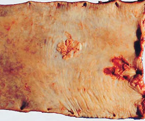
The Paris endoscopic classification of superficial neoplastic lesions                                    Paris Workshop Participants

of the lesion. Routine image recording for all pro-
cedures recently has been included in the guide-
lines of the European Society of Gastrointestinal
Endoscopy51 and is practiced at many institutions
worldwide.

Endoscopic classification in type 0
   During endoscopy, the assessment of the mor-
phology of a superficial neoplastic lesion in the
digestive mucosa is based on quantitative and
qualitative criteria. At first, the size of the lesion
and its diameter are quantified as precisely as
                                                                        Diagram 7. Depth of invasion of the submucosa in the
possible, preferably by using a graduated gauge.
                                                                        columnar epithelium (Barrett’s esophagus, stomach, colon,
Then the morphology is classified into one of the 5
                                                                        and rectum) assessed in the specimen obtained after surgery.
types of the Japanese-Borrmann classification for                       Depth of submucosal invasion is divided into two groups:
advanced cancer or in type 0 if the appearance of the                   superficial (sm1) and deep (sm2) with respect to a cutoff limit
lesion is compatible with a superficial lesion (mucosa                  determined on a micrometric scale (500 l in the stomach,
or submucosa). At this stage, the macroscopic                           1000 l in the colon).
classification is decided only from the gross appear-
ance. The classification should not be influenced by                    there is a sharp discontinuity in the epithelial layer,
any previous information and should not be modified                     and the muscularis mucosae is interrupted. A
by the findings of the pathologist. This means that                     consensus macroscopic description of superficial
the superficial pattern at endoscopy may be invali-                     neoplastic lesions has been published recently
dated by the results of the pathology. A lesion with                    (Table 2).15
a type 0 endoscopic appearance may turn out to                             Mixed types associate two distinct types of
be an advanced cancer on pathology in the p-TNM                         morphology. The pattern consisting of an elevation
classification, or the reverse. In the Japanese                         (IIa) and a depression (IIc) is easily diagnosed at
studies, most superficial endoscopic lesions are                        endoscopy. However, the exact placement of this
classified according to subtypes of type 0; this applies                mixed type in the Japanese classification requires
to squamous cell carcinoma in the esophagus,52-55                       a precise evaluation of the morphology, and there is
and to adenocarcinoma in the gastric cardia,56 in the                   room for interobserver disagreement because the
distal stomach,57-67 and in the colon and the                           relative surface of each type is not the only factor
rectum.68-73 The Japanese classification has some-                      relevant to prognosis. A depressed lesion with
times been used by Western investigators for neo-                       elevated borders or a central elevation is classified
plastic lesions in Barrett’s esophagus74 and also in                    as type 0-IIc + IIa. An elevated lesion with a central
the large bowel (with the cooperation of Japanese                       depression at its top is classified as type 0-IIa + IIc.
experts).16,17,75-77                                                    This mixed type includes relatively depressed
  Type 0 lesions are classified in 3 distinct groups:                   lesions in which depressed areas do not reach below
    type 0-I, polypoid                                                  the level of the normal mucosa. As a rule, type
    type 0-II, non-polypoid and nonexcavated                            IIa + IIc lesions have a poorer prognosis, with a risk
    type 0-III, non-polypoid with a frank ulcer                         of large invasion in the submucosa than all other
                                                                        types of lesions, including the IIa pattern. Other
The subgroups I and II are again segmented.                             mixed types and site variations of the classification
   Type 0-I includes two variants:                                      are described; this results in excessive complexity
    pedunculated (0-Ip)                                                 (Tables 3-13, pages S15-S17).
    sessile (0-Is)
                                                                        Hints for application of the classification
  Type 0-II includes 3 variants:
    slightly elevated (0-IIa)                                              In a pragmatic and simple approach, it is man-
    completely flat (0-IIb)                                                 datory to classify superficial lesions routinely in
    slightly depressed without ulcer (0-IIc)                                at least one of the 5 major types: 0-I, 0-IIa, 0-
                                                                            IIb, 0-IIc, 0-III, shown in Table 2 and Diagram
The distinction between a depressed (0-IIc) and an                          1. The relative proportions of each type differ
excavated or ulcerated lesion (0-III) is readily made                       in the esophagus, the stomach, and the large
in the operative specimen. In the excavated lesion,                         bowel.

VOLUME 58, NO. 6 (SUPPL), 2003                                                              GASTROINTESTINAL ENDOSCOPY             S9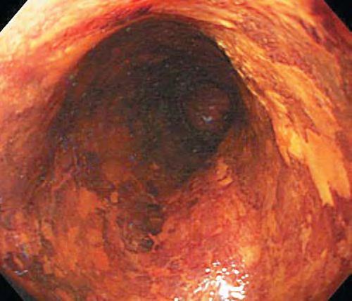
Paris Workshop Participants                                     The Paris endoscopic classification of superficial neoplastic lesions

Diagram 8. Depth of invasion of the submucosa in the columnar epithelium, assessed for the clinical relevance of EMR and for the
risk of nodal metastases. Group 1 (m and sm1): EMR is possible. Group 2 (sm2): surgical treatment is preferred. A, Barrett’s
esophagus and stomach: the cutoff limit between sm1 and sm2 is 500 l. B, Colon and rectum: the cutoff limit between sm1 and
sm2 is 1000 l.

   Polypoid 0-I lesions can be divided into type 0-Ip                The same qualitative and quantitative scale is
    and type 0-Is (pedunculated and sessile). In the                   used for the classification of neoplasia in the
    absence of clinical relevance, an intermediate                     columnar mucosa of Barrett’s esophagus, stom-
    type 0-Isp (semipedunculated) is not necessary;                    ach, and large bowel. The scale applies as well
    such lesions are managed as type Is lesions.                       for neoplasia in the duodenum or the small
   Special attention is attached to depressed type                    intestine. The principal variations in the mor-
    0-IIc lesions. The distinction between a de-                       phology of type 0 neoplastic lesions are shown in
    pressed (0-IIc) and ulcerated lesion (0-III)                       Diagrams 1-6.
    during endoscopy is based upon the depth of                       In the esophagus, neoplasia in the stratified
    the depression and the analysis of the epithelial                  squamous epithelium is classified in identical
    surface in the depressed area. Superficial                         major subtypes but with a distinct quantitative
    erosions in a depressed lesion involve only the                    scale. The standard of comparison is a single
    most superficial layers. In the ulcerated lesion,                  cup of the opened biopsy forceps. Lesions pro-
    there is loss of the mucosa and often of the                       truding above the level of the cup (approxi-
    submucosa. In the large bowel, type 0-IIc                          mately 1.2 mm) are classified as 0-Is. The depth
    lesions, even of small diameter, are often at                      of depressed lesions is compared with half the
    a more advanced stage of neoplasia, with deeper                    level of a single cup (approximately 0.6 mm).
    invasion than the other types.                                     The morphology of type 0 neoplastic lesions in
   With small and elevated neoplastic lesions, the                    the stratified epithelium of the esophagus is
    respective classification in the polypoid sessile                  shown in Diagram 9.
    type 0-Is or in the non-polypoid elevated type 0-
    IIa is made easier by placing a biopsy forceps                 Lesions included in the classification
    next to the lesion as a calibrating gauge. This                   The classification of type 0 neoplastic lesions
    standard applies to the height of the lesion and               applies to carcinomas, benign intraepithelial neo-
    not to its diameter. Lesions protruding above                  plasia, whether low grade or high grade, and also to
    the level of the closed jaws of the biopsy forceps             non-neoplastic lesions that are capable of harboring
    (approximately 2.5 mm) are classified as 0-Is;                 a neoplastic component (e.g., hyperplastic polyps).
    lesions protruding below this level are classified                A number of morphologic alterations of the
    as 0-IIa.                                                      mucosal surface associated with inflammation are
   When there is a depression at the center of a                  only listed as risk factors and are not included in the
    neoplastic lesion, its level is compared with that             type 0 classification. This applies to the inflammatory
    of the adjacent mucosa. The lesion is classified               lesions of esophagitis, associated with squamous cell
    as absolutely depressed when the level of                      cancer in parts of Asia78,79 and to chronic gastritis
    depression is lower than the surface of the                    secondary to H pylori associated with gastric cancer.
    adjacent mucosa and as relatively depressed                    The rule also applies to specialized intestinal meta-
    when the depression is still higher than the                   plasia in Barrett’s esophagus, a known risk factor for
    surface of the adjacent mucosa; this applies                   cancer80-83 and to intestinal metaplasia (in associa-
    particularly to the large bowel.                               tion with inflammation and atrophy) in the stomach.

S10   GASTROINTESTINAL ENDOSCOPY                                                               VOLUME 58, NO. 6 (SUPPL), 2003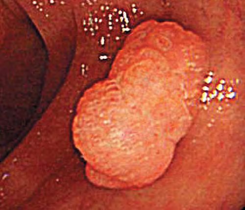
The Paris endoscopic classification of superficial neoplastic lesions                                    Paris Workshop Participants

Although there is a distinct endoscopic appearance
of intestinal metaplasia when using chromoendo-
scopy or magnification endoscopy, this morphologic
appearance is not included in the type 0 classification.
   In the stomach, a majority of lesions with low-
grade intraepithelial neoplasia never progress to
cancer, while high-grade, noninvasive intraepithe-
lial neoplasia progresses to cancer much more
readily.84 Studies with biomarkers confirm that
neoplastic lesions in the cardia have a distinct
natural history as compared with those in the distal
stomach.85 Specific immunohistochemistry charac-
teristics are attributed to intestinal metaplasia at the
esophagogastric junction86-89 and in the stomach.90
Adenomas of the stomach are rare and most will not                      Diagram 9. Squamous cell neoplasia in the esophagus.
                                                                        Adapted scale of thickness in transverse section for the major
progress to cancer.91 Isolated non-neoplastic hyper-
                                                                        subtypes: polypoid, non-polypoid, and excavated. The m and
plastic polyps rarely undergo neoplastic transforma-                    sm are represented as a single layer. The protruding lesions
tion, but this does occur in gastric polyposis.92                       are compared with one open cup of a biopsy forceps (1.2
Serrated adenomas are rare in the stomach.93                            mm); the dotted arrow passes above the top of the Is and IIa
   In the large bowel, non-neoplastic polyps are                        lesions. The depressed lesions are compared with half the
frequent. In the material collected for the National                    height of an open cup.
Polyp Study in the United States, 8.5% of the patients
were excluded for this reason.94 Most non-neoplastic
hyperplastic polyps are not protruding, and their                       a greater likelihood of early p53 and delayed K-ras
slightly elevated appearance would be classified as                     mutations.112-120 Serrated adenomas, classified as
a type 0-IIa. Aberrant crypt foci are considered to be                  neoplastic lesions, may show biomarkers similar to
the earliest precursors of colorectal neoplasia, but                    non-polypoid neoplastic lesions.121-126
progression to macroscopic neoplastic lesions proba-
bly is rare.95-97 In fact, aberrant crypt foci, which can               Endoscopic staging
be detected on magnification endoscopy as small                            The morphology of a type 0 neoplastic lesion has
protrusions, can be considered to be the most                           predictive value for depth of invasion into the
diminutive examples of type 0 lesions.                                  digestive tract wall, providing an ‘‘endoscopic stag-
   In the large bowel, the correlation between the                      ing.’’ Thus, endoscopic descriptive morphology can
macroscopic appearance and the histology of super-                      assist in treatment decisions involving endoscopic
ficial neoplastic lesions has been reviewed recently.98                 resection or surgery. The primary role for endoscopic
Many Japanese experts are convinced that there are                      staging is to predict the risk of submucosal invasion
two major pathways for neoplastic lesions, stressing                    and the associated risk of nodal metastases.
the role of non-polypoid precursors 99-105 and the                         For a type 0-I lesion, diameter is a reliable pre-
faster rate of progression of depressed lesions.106-107                 dictive criterion. The risk of submucosal invasion
However, this also can be interpreted as differences                    increases with the diameter. On the other hand, with
in terminology, because small but high-grade adeno-                     type 0-II lesions, the morphologic subtypes have
mas in the West may be called ‘‘de novo carcinomas’’                    greater importance. Invasion of the submucosa is
in Japan.                                                               more frequent in depressed lesions (IIc).
   In the Western interpretation, a small adenoma                          The less than perfect reliability of endoscopic
with high-grade dysplasia may evolve rapidly into                       staging can be improved by EUS, particularly with
a small flat invasive carcinoma that loses evidence of                  high frequency probes (20 MHz). Both endoscopic
its adenomatous origin. This is likely a major route in                 morphologic staging and EUS have their limits.
hereditary non-polyposis colon cancer (HNPCC).                          Endoscopy tends to understage superficial lesions,
There is a recent trend in the Western studies108-111                   and EUS tends to overstage them. When the two
to accept a broader spectrum of progression for                         methods agree, the predictive value is high.58
neoplastic lesions, including the role of non-polypoid
lesions and the concept of de novo cancer. Indeed,                      The specimen collected after EMR
there are likely numerous pathways at the molecular                        In the endoscopy unit, the single tissue specimen
level. Molecular biology also suggests that non-                        obtained after en bloc mucosectomy should be gently
polypoid lesions have a distinct evolution with                         stretched. The deep margin of the specimen can be

VOLUME 58, NO. 6 (SUPPL), 2003                                                            GASTROINTESTINAL ENDOSCOPY              S11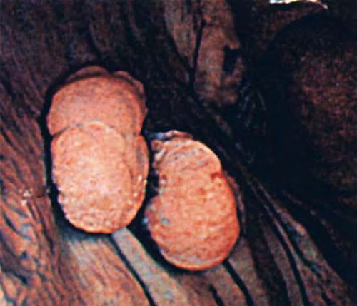
Paris Workshop Participants                                  The Paris endoscopic classification of superficial neoplastic lesions

marked with dye or India ink. The specimen should                  In surgically resected specimens from the stomach
then be pinned on cardboard or a similar soft, porous           and the colon, the semiquantitative evaluation of the
material with the mucosal surface up and placed in              invasion in the submucosa (divided in two or 3
neutral formalin.                                               sectors) is less and less used. Currently, the quanti-
   For lesions removed with a piecemeal technique,              tative micrometric measure is the common guideline
the endoscopist should, if possible, reconstruct the            for the specimens issued from a surgical or an
complete surface of the lesion on cardboard from the            endoscopic resection. The categorization of cancer
fragments. The surface of the fixed specimen should             invasion as superficial or deep in the submucosa is
be examined and photographed. A more detailed                   determined by an organ-specific limit fixed at
examination, by using crystal violet staining and               a certain depth. With the quantitative method of
stereomicroscopy, may prove helpful in correlative              measurement, sm1 means ‘‘less invasive than the
studies of the surface between pathology and magni-             cutoff limit,’’ and sm2 means ‘‘deeper than the cutoff
fication endoscopy.                                             limit.’’ In Japan, the pathologists have established
                                                                distinct empirical cutoff limits for columnar neo-
The specimen (surgery or EMR) in the pathology                  plasia in the stomach128 and columnar neoplasia in
laboratory                                                      the large bowel.129,130 Application of these categories
   In the pathology laboratory, the specimen is                 to surgical specimens is the only way to achieve
withdrawn from the fixative and pre-cut in parallel             comparability between EMR and surgical resections.
fragments, 2 mm in width for EMR specimens and 5
to 6 mm in width for surgical specimens. The margins            Invasion of the submucosa in squamous cell
in the adjacent normal mucosa are included for                  neoplasia of the esophagus
analysis in the serial histologic sections. The pathol-            In the esophageal mucosa, 3 distinct layers are
ogist evaluates the histology (ideally according to the         described; they correspond, respectively, to the
Vienna classification) and assesses the degree of               epithelium (m1), the lamina propria (m2), and the
differentiation of the tumor, the depth of invasion,            muscularis mucosae itself (m3). In the operative
and the completeness of excision.                               specimen where the full thickness of the wall
   The resection is complete if the margins of the              (including muscularis propria) is available, the sub-
specimen are free from tumor tissue on serial                   mucosa is arbitrarily divided in 3 successive sectors
sections; this concerns the proximal and the distal             of equivalent thickness (sm1, sm2, sm3), as shown in
margins of surgically resected specimens and all the            Diagram 10. This precise subdivision into 6 layers
margins (vertical and lateral) on EMR specimens.                has been proposed because the risk of nodal metas-
   Depth of cancer invasion into the submucosa is               tases increases from nil to high with the depth of
a critical factor in predicting the risk of nodal               invasion in the successive layers of the mucosa and
metastases in superficial tumors of the digestive               submucosa. The correspondence between depth of
mucosa. This applies to prognosis after a segmental             invasion in a complete transverse section of the
surgical resection and guides treatment after EMR.              esophageal wall and the most appropriate treatment
The risk of nodal metastasis is relatively low when             is shown in Diagram 11a. Cancer invading only the
cancer invasion is limited to the superficial sub-              superficial levels (m1 + m2) usually can be treated
mucosa, and significantly higher when invasion                  successfully with EMR. Invasion into deep levels
reaches the deep submucosa (Tables 15-18, page S18).            (sm2 + sm3) usually requires surgery for a cure.
   In surgically resected specimens from the esopha-            Middle level invasion (m3 + sm1) requires balancing
gus, the muscularis propria is present, and the full            clinical factors with surgery, preferred when the
thickness of the submucosa is available. This allows            performance status of the patient is high. However,
a reliable, semiquantitative evaluation of the depth of         in a specimen obtained after EMR, the full thickness
tumor invasion in the submucosa, divided into 3                 of the submucosa is not available, and this division is
sectors of equivalent thickness: sm1, sm2, and sm3.             not valid. Therefore, invasion in the submucosa is
However, in EMR specimens, the complete sub-                    measured with a micrometric scale, from the bottom
mucosa is not available, and the semiquantitative               of the mucosal layer (i.e., the lower layer of the
evaluation of the depth of invasion is not fully reliable.      muscularis mucosae). In Japan, an empirical cutoff
The only precise method is a quantitative micro-                value has been adopted. When cancer invasion of the
metric measure in microns (l) of the depth of inv-              submucosa is less than 200 l, the risk of nodal
asion, measured from the bottom of the mucosa (i.e.,            metastases is small, and EMR can be considered
the lower layer of the muscularis-mucosae). The risk            safe.127 The cutoff limit is shown in Diagram 11b. In
of nodal metastasis is assumed to be low when the               this situation, the quantitative scale should be used
depth of invasion is less than a determined cutoff.127          instead of describing the layers as sm1 and sm2.

S12   GASTROINTESTINAL ENDOSCOPY                                                            VOLUME 58, NO. 6 (SUPPL), 2003
The Paris endoscopic classification of superficial neoplastic lesions Paris Workshop Participants
Diagram 10. Depth of invasion of squamous cell neoplasia in
the esophagus. Mucosal carcinoma is divided into 3 groups:
m1 or intraepithelial, m2 or micro-invasive (invasion through
the basement membrane), m3 or intramucosal (invasion to
the muscularis mucosae). The depth of invasion in the
submucosa is divided into 3 sections of equivalent thickness:
superficial (sm1), middle (sm2), and deep (sm3).
Invasion of the submucosa in columnar
neoplasia of the stomach and the large bowel
Diagram 11. Depth of invasion of squamous cell neoplasia in
For adenocarcinomas in Barrett’s esophagus and the esophagus adapted for relevance to EMR and the risk of
the stomach, subdivisions also are proposed to nodal metastases. A, Full-thickness specimen. Group 1 (m
stratify depth of invasion in the submucosa by using and sm1): EMR is possible. Group 3 (sm2 and 3): surgical
the same quantitative method for operative and EMR treatment. Group 2 (m3 and sm1): uncertain indications. B,
specimens. In EMR specimens from Barrett’s esoph- Specimen obtained after EMR: the cutoff limit between sm1
agus or the stomach, the cutoff value for invasion into and sm2 is 200 l EMR is adequate for sm1.
the submucosa is 500 l (Diagram 8a). The rationale
for this value is that when the depth of invasion is less
than 500 l into the submucosa (sm1), the risk of nodal Contribution of magnifying endoscopy
metastases is low128 and endoscopic treatment can be In videoendoscopy, charge-coupled device (CCD)
considered adequate (Table 17). On the contrary, chips with high pixel density provide high-resolution
surgery is preferable when invasion is more than images. In recent instruments, magnification is
500 l deep (sm2). available by using either an optical zoom (330-380)
In the colon, the risk of nodal metastases is low or a combined optical and electronic magnification.
when cancer invasion of the submucosa is limited to Zoom magnification is used selectively on target
the most superficial third and extends laterally to lesions, because at maximum magnification focal
less than 50% of the width of the mucosal lesion. length is short and depth of field is small. The
Nodal metastases frequently occur, associated with magnified image can be further improved by elec-
lesions, with massive invasion of the submucosa, tronic modification, which results in structural
either laterally in the superficial third or in deeper enhancement of the surface. The electronic enhance-
invasion reaching the middle or the lower third. ment involves a selective amplification of the in-
Some investigators use, as in the esophagus, tensity of some wavelengths of light reflected from
a semiquantitative evaluation of invasion depth in the mucosal surface and collected by the CCD. A
the colonic submucosa in 3 sectors (1, 2, 3) of simplified spectroscopic method, narrow band imag-
equivalent thickness and in 3 groups (a, b, c) for ing, also is in development. In this system, the
lateral extent in the superficial layer. A limited incident light is restricted to narrow bands in the 3
invasion corresponds to sm1a and sm1b, and a mas- basic colors (red, blue, green) to obtain distinct
sive invasion to sm1c, sm2, and sm3. In EMR images (deep, intermediate, and superficial), which
specimens from the large bowel, the cutoff value adop- are superimposed, resulting in increased relief.
ted for quantitative micrometric measure is 1000 l130 Magnifying endoscopy has two distinct applica-
(Diagram 8b). An EMR can be considered safe when tions: the analysis of the surface architecture of the
invasion of the submucosa is less than 1000 l. epithelium (pit pattern) with the help of a contrast
VOLUME 58, NO. 6 (SUPPL), 2003 GASTROINTESTINAL ENDOSCOPY S13Paris Workshop Participants The Paris endoscopic classification of superficial neoplastic lesions
dye (chromoendoscopy by using indigo carmine), and lesion with its corresponding histopathology. In
the analysis of the vascular network in transparency series from endoscopy units, the pathology compo-
across translucent unstained epithelium.46,49,131-151 nent is the weak point, while endoscopic description
Contrast magnification endoscopy has been used for is the weak point in series from a surgical unit or
esophageal squamous cell cancer in Japan,147,148,151 a pathology laboratory. The relative proportions of
for Barrett’s esophagus in Japan and in the each type and subtype vary according to the histology
West,46,49,131-134 for the stomach in Japan,135-138 of the epithelium (stratified squamous vs. columnar),
and for the large bowel in Japan139-144 and the the organ (esophagus vs. stomach or colon), and the
West.47,145,146 A recent randomized trial144 has recruitment of the cases (mass screening vs. oppor-
confirmed the efficacy of this technique for differen- tunistic cases). Tables 3-8 show the distribution of
tiating non-neoplastic lesions that do not require the morphologic subtypes; Tables 9-14 show data on
treatment from neoplastic lesions. the depth of invasion in mucosa or submucosa. Tables
Interpretation of the surface pit pattern with 15-18 show data on the frequency of nodal invasion in
magnification is easier in the large bowel than in superficial neoplastic lesions. Endoscopic images of
the stomach because of gastric inflammation associ- the mucosal surface and of superficial neoplastic
ated with the high prevalence of H pylori in many lesions type 0 are presented in Figures 1 to 95,
populations. In the large bowel mucosa, distinct classified by morphologic appearance.
types of pit patterns have been described for normal
mucosa and for non-neoplastic and neoplastic lesions
(low-grade or high-grade intraepithelial neoplasia). Neoplastic lesions in columnar epithelium
In carcinoma, the surface pattern is either irregular Barrett’s esophagus. Available data on the
or amorphous. In addition, magnification may be morphology of superficial neoplastic lesions in Bar-
helpful in the discrimination of hyperplastic polyps rett’s esophagus are scarce; type 0-II lesions are the
and serrated adenomas, the latter showing an most frequent (70%), but depressed type lesions (0-
alternating pattern or regular pits (non-neoplastic IIc) are uncommon (Table 4). The endoscopic mor-
areas) and cerebriform crests (neoplastic areas).71 phology of a target lesion has poor reliability in the
Magnification in mucosal transparency explores prediction of invasive or noninvasive neoplasia
the microvascular network across the translucent because multiple foci are frequent and cancer with
unstained epithelium and requires no contrast invasion of the submucosa may occur near an area
dye.147-151 This technique is based on the evaluation with noninvasive epithelial neoplasia.
of the changes in caliber (dilatation) and shape of Esophagogastric junction and cardia. Neo-
neoplastic vessels. In the esophagus, this evaluation plastic lesions at the esophagogastric junction and
can contribute to the differentiation of squamous the cardia tend to be considered in a single group in
cell neoplasia from inflammation and also to the Japan, but the cardia is often associated with
suspicion of carcinoma with invasion into the sub- Barrett’s mucosa in Europe and North America.
mucosa.148,151 The distribution of morphologic variants in type 0 is
The classification presented in this text is based on the same as in Barrett’s esophagus. The proportion of
the technology of standard endoscopy. However, depressed (0-IIc) lesions is less than in the sub-
progress in image processing may offer different cardiac stomach.56
perspectives.152 Magnifying endoscopy has potential Sub-cardiac stomach. Most superficial neo-
to be used routinely in the upper digestive tract in plastic lesions in the sub-cardiac stomach are type
Barrett’s esophagus. In the large bowel, magnifica- 0-II, and most of them (70%-80%) are depressed (0-
tion may potentially be used routinely for the IIc). Type 0-I (polypoid) lesions are rare, as well as
differentiation between non-neoplastic and neoplas- type 0-III (ulcerated) (Tables 5 and 8). Polypoid
tic lesions (optical biopsy), and for the distinction adenomas are rare precursors of invasive cancer.
between intramucosal lesions and lesions with in- Flat or slightly depressed areas of low-grade or
vasion of the deep submucosa. high-grade noninvasive intraepithelial neoplasia
often are called flat or depressed adenomas in the
ELEMENTS FOR A CONSENSUS CLASSIFICATION Japanese studies.21,25 The global risk of submuco-
Endoscopic morphology of subtypes sal invasion in type 0-II lesions is just under 40%
within type 0 in the large series from 1990 to 1999, reported by
In the Japanese studies, there are several large the National Cancer Center Hospital in Tokyo
series showing the distribution and variations in the (Table 10, page S16) or in the results from the
morphology of superficial neoplastic lesions type 0. national mass screening campaign in 1997 (Table
Some series include the endoscopic description of the 14). The figure is still lower (19%) in the series
S14 GASTROINTESTINAL ENDOSCOPY VOLUME 58, NO. 6 (SUPPL), 2003The Paris endoscopic classification of superficial neoplastic lesions Paris Workshop Participants
Distribution of Morphologic Subtypes
Table 3. Squamous epithelium of the Table 4. Barrett’s esophagus—morphology*
esophagus—morphology*
N %
n %
0-I 21
0-I 16 Ip + Is 14
Ip + Is 262 0-IIa,b 61
0-IIa,b 34 IIa 17
IIa 303 IIb 23
IIb 221 0-IIc 13
0-IIc 45 IIc 3
IIc 707 IIa + IIc 6
0-III 5 0-III 5
III 69 III 3
Total 1562 Total 66
*Distribution (numbers and percentages) of major macroscopic *Distribution (numbers and percentages) of major macroscopic
categories within type 0. Multicenter analysis conducted in Japan categories within type 0. Single endoscopy series (high-grade
in 143 institutions: 1562 lesions with pathology confirmation in neoplasia in 3 lesions and intramucosal carcinoma in 63
the operative specimen (unclassified in 290 other lesions).53 lesions).74
Table 5. Stomach—morphology* Table 6. Colon—morphology*
n % n %
0-I 3 0-I 57
0-I 66 Ip + Is 5455
0-IIa,b 17 0-IIa,b 39
0-IIa 356 IIa + IIb 3674
0-IIb 9 0-IIc 4
0-IIc 78 IIc 404
0-IIc 1486 0-III 0
0-IIc + IIa 21 III 0
0-IIa + IIc 132
Total 9533
0-III 2
0-IIc + 0-III 15 Note: Lesions described as lateral spreading type were in-
0-III 13 cluding in type 0-IIa.
Total 2098 *Distribution (numbers and percentages) of major macroscopic
categories within type 0. Endoscopy series (9533 lesions in the
*Distribution (numbers and percentages) of major macroscopic period 1985-1996) from Akita Red Cross Hospital.68
categories within type 0. Surgical series (2098 lesions in the
period 1990-1999) with pathology confirmation from National Table 8. Stomach compared to
Cancer Center Hospital in Tokyo (unclassified in 3 other lesions.) colon—morphology*
(From M. Sasako, unpublished data presented at Paris workshop.)
Stomach Colon
Table 7. Colon—morphology*
n % n %
n %
0-I 6 79
0-I 50 Ip + Is 213 1768
Ip 1303 0-IIa,b 17 13
Is 504 IIa 488 296
0-IIa,b 44 IIb 42 3
IIa 1604 0-IIc 70 7
IIb 33 IIc 1717 39
0-IIc 5 IIc + IIa 118 —
IIc, IIc + IIa 60 IIa + IIc 415 127
IIa + IIc 97 0-III 7Paris Workshop Participants The Paris endoscopic classification of superficial neoplastic lesions
Depth of Invasion
Table 9. Squamous epithelium of the Table 10. Stomach—depth of invasion*
espophagus—depth of invasion*
N8 total N8 m N8 sm % sm
m1 + m2 m3 + sm1 sm2 + sm3
0-I
n (%) n (%) n (%)
0-I 66 28 38 57%
0-I 0-IIa,b
Ip + Is 11 (4) 44 (16) 207 (79) 0-IIa 356 254 102 29%
0-IIa,b 0-IIb 10 8 2 20%
IIa 62 (20) 94 (31) 147 (48) 0-IIc
IIb 152 (69) 36 (16) 33 (15) 0-IIc 1488 931 557 37%
0-IIc 0-IIc + IIa 19 10 9 47%
IIc 256 (39) 245 (34) 206 (27) 0-IIa + IIc 132 46 86 65%
0-III 0-III
III 2 (3) 9 (13) 58 (84) 0-IIc + III 15 9 6 40%
Total 483 (31) 428 (27) 651 (41) Total 2086 1286 800 38%
Note: The depth of invasion is divided into 3 groups: superficial *Depth of invasion into the mucosa (m) or submucosa (sm) in
(2/3 of the mucosa of (m1 + m2); intermediate (last layer of the neoplastic lesions, type 0, with pathology control, treated by
mucosa + first layer of the submucosa or m3 + sm1); deep (2/3 of surgery or endoscopic mucosectomy (2086 lesions in the period
the submucosa or sm2 + sm3). 1990-1999) in the National Cancer Center Hospital in Tokyo.
*Depth of invasion into the mucosa (m) or submucosa (sm) with (From M. Sasako, unpublished data from Paris workshop.)
reference to major macroscopic categories within type 0. A
multicenter analysis conducted in Japan in 143 institutions:
1562 lesions with pathology confirmation in the operative Table 11. Stomach—depth of invasion*
specimen.53
2000-2001
N8 cancer m 382 (81%)
N8 cancer sm 89 (19%)
from 2000 to 2001, where all cases were treated by Total 471
EMR, reported by the National Cancer Center
*Depth of invasion, into the mucosa or submucosa, in neo-
Hospital in Tokyo. The endoscopic morphology of
plastic lesions type 0, with pathology confirmation, treated only
a type 0-II lesion has a predictive value for the risk by endoscopic mucosectomy (471 lesions) in the National Cancer
of submucosal invasion. The risk is higher for type Center Hospital in Tokyo. (From M. Sasako, unpublished data
0-I and combined type 0-IIa + IIc, and lower for from Paris workshop.)
type 0-IIb lesions. Concerning clinical relevance,
non-depressed (type 0-IIa or 0-IIb) lesions with
a diameter less than 2.0 cm can be treated safely cial’’ neoplastic lesions. Most type 0-II lesions are
by endoscopy; the safety limit is lowered to 1.0 cm elevated and non-depressed (type 0-IIa), and the flat
for all types of lesions with a depressed (0-IIc) type (type 0-IIb) is extremely rare. Depressed lesions
morphology. (type 0-IIc) also are rare (4%-5% of all lesions). Is
Large bowel. Many non-neoplastic lesions of the there a difference between East and West observa-
large bowel mucosa with a superficial and non- tions of the morphologic distribution of superficial
depressed morphology are hyperplastic polyps. In neoplastic lesions in the large bowel? This seems
the absence of magnification, the endoscopic pre- unlikely. Different proportions suggest possible mis-
diction of their nature is not always easy, justifying classification between type 0-Is and type 0-IIa lesions
a tissue sample for histologic analysis. Some polyps in less specialized units.
with a surface suggesting a hyperplastic polyp have In Japan, in the national mass screening cam-
a neoplastic component (adenoma). These are now paign based on the fecal occult blood test, the
classified as serrated adenomas by histology. In proportion of lesions type 0-Ip or 0-Is is the same
Western countries, most superficial neoplastic le- (about 80%) as in the West 67 (Table 8) and is lower
sions of the large bowel (80% or more) have a polypoid than in the series from the Niigata Hospital (50%).
morphology, while the frequent non-neoplastic Among type 0-I lesions, the proportion of those
hyperplastic polyps have a ‘‘flat’’ morphology. In classified as pedunculated (type 0-Ip) is much smaller
Japan, the proportion of polypoid neoplastic lesions is (652/1768 = 37%) in the mass screening series from
lower in the series published by specialized units Japan67 than in the series from the Niigata Hospital
such as the Akita Red Cross Hospital (Table 6) or the (1303/1843 = 70%) (Table 7), suggesting a biased
Niigata Hospital (Table 7). In these series, non- evaluation of protrusion of sessile lesions in the
polypoid lesions represent up to 50% of all ‘‘superfi- endoscopic image.
S16 GASTROINTESTINAL ENDOSCOPY VOLUME 58, NO. 6 (SUPPL), 2003You can also read