The Recessive Epigenetic swellmap Mutation Affects the Expression of Two Step II Splicing Factors Required for the Transcription of the Cell ...
←
→
Page content transcription
If your browser does not render page correctly, please read the page content below
The Plant Cell, Vol. 17, 1994–2008, July 2005, www.plantcell.org ª 2005 American Society of Plant Biologists

The Recessive Epigenetic swellmap Mutation Affects the
Expression of Two Step II Splicing Factors Required for the
Transcription of the Cell Proliferation Gene STRUWWELPETER
and for the Timing of Cell Cycle Arrest in the Arabidopsis Leaf

Nicole K. Clay and Timothy Nelson1
Department of Molecular, Cellular, and Developmental Biology, Yale University, New Haven, Connecticut 06520-8104

Generally, cell division can be uncoupled from multicellular development, but more recent evidence suggests that cell cycle
progression and arrest is coupled to organogenesis and growth. We describe a recessive mutant, swellmap (smp), with
reduced organ size and cell number. This defect is partially compensated for by an increase in final cell size. The mutation
causes a precocious arrest of cell proliferation in the organ primordium and possibly reduces the rate of cell division there. The
mutation proved to be an epigenetic mutation (renamed smpepi) that defined a single locus, SMP1, but affected the expression
of both SMP1 and a second very similar gene, SMP2. Both genes encode CCHC zinc finger proteins with similarities to step II
splicing factors involved in 39 splice site selection. Genetic knockouts demonstrate that the genes are functionally redundant
and essential. SMP1 expression is associated with regions of cell proliferation. Overexpression of SMP1 produced an increase
in organ cell number and a partial decrease in cell expansion. The smpepi mutation does not affect expression of eukaryotic cell
cycle regulator genes CYCD3;1 and CDC2A but affects expression of the cell proliferation gene STRUWWELPETER (SWP)
whose protein has similarities to Med150/Rgr1-like subunits of the Mediator complex required for transcriptional activation.
Introduction of SWP cDNA into smpepi plants fully restored them to wild-type, but the expression of both SMP1 and SMP2 were
also restored in these lines, suggesting a physical interaction among the three proteins and/or genes. We propose that step II
splicing factors and a transcriptional Mediator-like complex are involved in the timing of cell cycle arrest during leaf
development.

INTRODUCTION                                                                 during the growth and development of the leaf organ primordium,
                                                                             for example, there are no fixed patterns of cell division, but rather
Plant cells do not move and are surrounded by a rigid cell wall,             a stochastic gradient of cell division arrest from the distal tip to
and for this reason, cell division rates and patterns were thought           the base of the leaf (Donnelly et al., 1999).
to be directly responsible for generating new structures during                 The accumulating evidence of cell division–independent
development. However, despite genetic manipulation of either                 mechanisms of development has lead to a long debate on the
cell proliferation or cell expansion, the resulting organs and/or            actual function or necessity of cell division in plant development
organisms often attain the normal size (Hemerly et al., 1995;                (reviewed in Doonan, 2000). However, more recent evidence has
Smith et al., 1996; Cleary and Smith, 1998; Jones et al., 1998;              assigned developmental significance to cell cycle progression
Wang et al., 2000; De Veylder et al., 2001; Autran et al., 2002).            and exit. For example, experiments using cell cycle inhibitors and
They may consist of fewer but larger cells, or more numerous but             in particular DNA synthesis inhibitors have shown that, unlike
smaller cells, suggesting that cell division can be uncoupled from           cytokinesis, cell cycle progression is coupled to meristem
iterative plant development. Similarly, in animals, generally,               growth and patterning (Grandjean et al., 2004). Also, the over-
changes in cell size can be compensated for by changes in cell               expression of the D-type cyclin CYCD3;1, unlike most other cell
number to maintain the final size of an organism, suggesting                 cycle proteins, inhibited several cell differentiation pathways in
a lack of correlation between cell division and the formation of             the leaf, indicating an important role in leaf development (Dewitte
complex structures during development (reviewed in Day and                   et al., 2003). More specifically, CYCD3;1 overexpression re-
Lawrence, 2000; Weinkove and Leevers, 2000). Furthermore,                    duced the proportion of cells in the G1 phase of the cell cycle,
                                                                             suggesting that cell cycle exit at the G1 phase is required for
                                                                             proper execution of differentiation in the leaf (Dewitte et al.,
1 To  whom correspondence should be addressed. E-mail timothy.               2003). Similarly, in mammals, cell cycle exit has been shown
nelson@yale.edu; fax 203-432-5711.                                           to be required for skeletal myogenesis (Skapek et al., 1995;
The author responsible for distribution of materials integral to the         Zachsenhaus et al., 1996; Guo and Walsh, 1997) and lens fiber
findings presented in this article in accordance with the policy described   cell differentiation (Zhang et al., 1998). Most likely, there exists
in the Instructions for Authors (www.plantcell.org) is: Timothy Nelson
(timothy.nelson@yale.edu).
                                                                             a subtle and close interplay between growth/organogenesis and
Article, publication date, and citation information can be found at          cell cycle progression/exit, and this relationship is defined by
www.plantcell.org/cgi/doi/10.1105/tpc.105.032771.                            developmental context.
Splicing Factors in Cell Cycle Arrest 1995 The relationship among D-type cyclins, G1 phase cell cycle cyclin-dependent kinases and D-cyclins. This would involve two exit, and leaf cell differentiation is intriguing because in all transcription factors, E2F and Dpa, which, also in plants, are eukaryotes, the G1 phase seems to be a major checkpoint in involved in the activation of the cell cycle genes and in the the decision to stop or to continue cell proliferation. In plants, like maintenance of meristematic competence (De Veylder et al., in animals, the initiation of cell division probably involves the 2002). During cell differentiation, the opposite process is thought inactivation of the retinoblastoma protein by the appropriate to occur (i.e., a reactivation of the retinoblastoma blocks the cells Figure 1. General Plant Phenotype of the smp Mutant. (A) to (E) Cleared leaves viewed under dark-field illumination. The wild type is in (A), and smp is in (B) to (E). (D) is a close-up of the veins in smp. Arrowheads point to axialization defects in veins. Arrows point to discontinuities in the venation pattern. (F) and (G) Leaf shape and size of 2-week-old plants grown on soil. The wild type (left in [F]) and smp ([G] and right in [F]). (H) Leaf rosettes of 4-week-old wild-type (left) and several smp (right) plants. (I) Fully-grown wild-type (left) and smp (right) plants. (J) and (K) Inflorescences of the wild type (J) and smp (K). (L) and (M) Mature green wild-type (left) and smp (right) siliques were viewed under bright-field illumination (M) or cleared and viewed under dark-field illumination (L) to look at seed size and number. (N) and (O) Week-old smp seedlings containing a transgenic copy of SMP gene and exhibiting a partially restored phenotype (C1) or a wild-type phenotype (C2) were grown on agar media, and the outgrowths of the first two leaves were compared with those of wild-type (top row) and untransgenic smp (bottom row) seedlings. (P) Leaf shape and size of 2- to 3-week-old wild-type (left) and C1 (right) plants.
1996     The Plant Cell

in G1, inducing cell differentiation) (Ach et al., 1997). Much more        pointed venation pattern, both of which were reflected in the leaf
work needs be done to uncover the players and mechanisms                   shape and size (Figures 1A to 1H). This phenotype was com-
regulating cell proliferation in organs and/or organisms.                  pletely penetrant and affected all leaf-like organs except for
   In a screen for leaf development mutants, we identified a reces-        cotyledons. Besides the reduction in leaf organ size (less than
sive mutant, swellmap (smp), with reduced leaf organ size and cell         half that of the wild type for all rosette leaves; Figure 1F), there
number. This defect is partially compensated for by an increase            were pronounced serrations at the marginal teeth, and the veins
in final cell size. The recessive mutation proved to be epigenetic         themselves were not properly axialized; that is, the vein cell files
and defined a single locus, SMP1, that was differently methylated          were not aligned from end to end in a smooth, tight bundle and
but affected the expression of both SMP1 and a second very                 instead had spaces between them (Figure 1D). Frequently, there
similar gene, SMP2, both of which encode putative step II splic-           were discontinuities in the midvein at the leaf tip, and the polar
ing factors, which are involved in 39 splice site selection. Genetic       ends of vein cells did not join near the leaf margins (Figures 1B,
knockouts demonstrate that the genes are functionally redun-               1C, and 1E). The mutant phenotype was first evident at the
dant and developmentally essential. SMP1 expression is asso-               seedling stage by the retarded outgrowth of the first pair of
ciated with regions of cell proliferation in lateral organs, and           leaves and slightly reduced root growth (Figure 1N). Fully grown
overexpression of SMP1 similarly reduced leaf organ size but in-           smp plants were reduced in stature, had narrower and/or shorter
creased cell number. The smpepi mutation affects the transcription         organs on the whole, and were fairly infertile, producing only a few
of the cell proliferation gene STRUWWELPETER (SWP), whose                  siliques (seed-bearing pods) that contain fewer but larger seeds
protein has similarities to Med150/Rgr1-like subunits of the               (Figures 1I, 1L, and 1M). Moreover, a few smp floral meristems
Mediator complex, which is required for transcriptional activation,        produced clustered floral buds (Figure 1K), which were spaced
suggesting that SWP is the direct target of SMP1 and/or SMP2.              normally later in development.
                                                                              Because of their conspicuous and developmentally familiar
                                                                           nature, leaves were used to study the more cellular aspects of the
RESULTS                                                                    smp mutation. Transverse sections through smp leaves revealed
                                                                           that the leaves were wider and that aside from vascular cells,
smp Mutation Affects Cell Number and Size                                  which were normally sized, all other leaf cells were fewer in
                                                                           number and larger in size (Figure 2D). Cell numbers were
A single allele of the smp mutant was identified from a mutant             quantified through transverse sections of three fully grown leaves
screen by a reduced amount of leaf venation and a narrow,                  from the second pair of leaves to arise from each genotype

Figure 2. Leaf Cell Number and Size Are Affected in the smp Mutant.

(A) and (B) Scanning electron micrographs of the adaxial surface of fully expanded rosette leaves. (A) Wild-type; (B) smp. White bar on the bottom
right ¼ 10 mm.
(C) and (D) Transverse sections through fully expanded rosette leaves. (C) Wild-type; (D) smp. Bar ¼ 50 mm.
Splicing Factors in Cell Cycle Arrest 1997 (vascular cells were excluded), and the mean cell number in the layers (particularly the palisade layer) seen later on in mature mutant (95 cells, SD of 8.3; 0.11 mm2) was found to be 45.2% of leaves (Figure 2D). that in the wild type (210.2 cells, SD of 5; 0.11 mm2). Similarly, To see if smp’s effect on cell proliferation extended beyond scanning electron micrographs of smp leaves confirmed that the leaves, we examined the structure of the inflorescence shoot epidermal layer consisted of fewer but larger cells (Figure 2B) and apical meristem (SAM), the formation of floral organs at the that this increase in cell size was not enough to fully restore final meristem’s periphery, and the structure of the inflorescence stem organ size to that of the wild type. Thus, the smaller leaf size in the just below the youngest silique. Longitudinal sections through mutant was due to a reduction in the number of cells per leaf, and smp SAMs indicated that the organization into layers (at least for this reduction was partially compensated for by an increase the first two layers) was not disrupted, although the layers in final cell size. Interestingly, although the longitudinal vascular consisted of fewer meristematic cells (Figure 9C). Floral organ pattern was severely reduced, the transectional vascular pattern formation appeared normal, albeit with reduced cell numbers (the arrangement of xylem and phloem) was normal except that (Figure 9C). The same is true for the stem (data not shown). the veins were slightly thicker. To determine if the reduced cell number in smp leaves was due SMP1 and SMP2 Encode Putative Step II Splicing Factors to a contraction of the cell proliferation phase or to a reduction in the rate of division during organ development, transverse sec- A map-based cloning strategy was used to isolate the gene. tions of leaf primordia were taken at various time points during Genetic analysis of F2 progeny indicated that the smp mutant the cell proliferation phase of leaf development. Meristematic phenotype segregated as a single recessive locus. The smp cells appear as small, densely stained cells in contrast with mutation was mapped initially between simple sequence length expanding vacuolated cells. Relative to the wild type, mutant leaf polymorphic markers nga111 and AthGENEA on the bottom of primordia had significantly more cells that were vacuolated and chromosome 1 and then finely mapped to a region on BAC larger sized, suggesting that those cells had prematurely exited F1E22 that spanned five predicted genes. Sequencing of all five the cell proliferation phase (Figures 3D to 3F) Also, the sections genes in the mutant background did not reveal any detectable revealed large intercellular spaces developing within the organ molecular lesion or point mutation. One of the genes (At1g65660) (Figures 3G to 3I), suggesting that meristematic cells were not encodes a CCHC zinc finger protein that is 22% identical to the proliferating fast enough to keep up with the expanding epider- yeast SLU7 protein, a step II splicing factor involved in 39 splice mal cell layer (i.e., a retarded rate of cell division) or that a cell wall site selection (Figure 4B; Frank and Guthrie, 1992). Constitutive defect led to such a schizogenous-like separation of tissues. expression of a single locus of the wild-type transgene fully Whatever the cause of these large intercellular spaces, their complemented the smp mutation in at least two independent development accounts for the disorganization of the inner cell lines (Figure 1O). Furthermore, expression of this gene by its Figure 3. Precocious Arrest of Cell Proliferation in Leaf Primordia of the smp Mutant. Transverse sections through leaf primordia of 7- to 9-d-old wild-type ([A] to [C]) and smp ([D] to [I]) seedling were arranged in developmental order from (A) to (C) and from (D) to (F). (G) and (H) are serial sections of the same plant. Arrowheads point to large intercellular spaces. Bar ¼ 50 mm.
1998     The Plant Cell

Figure 4. SMP1 Encodes a Putative Step II Splicing Factor.

(A) Genomic structure of the SMP1 and SMP2 genes. Black boxes indicate exons. Arrows indicate direction of transcription. SALK T-DNA insertions are
indicated for the smp1 and smp2 insertion alleles.
(B) Alignment of the deduced amino acid sequences of SMP1, AT4G37120 (SMP2), yeast SLU7 (Frank and Guthrie, 1992), and human hSLU7 (Chua and
Reed, 1999). The CCHC domain is underlined. Shared amino acids are shaded.

native promoter (810 bp of upstream sequence) gave partial                  that in BT002797. Thus, SMP1 coding sequence consists of
rescue during the first week of growth and then a full rescue               10 exons that span a 2.6-kb genomic region (Figure 4A).
hereafter (Figures 1N and 1P). The constitutive expression of                  The CCHC zinc finger motif (CX2CX4HX4C), also known as
a C-terminal translational fusion of the putative SMP gene to               retroviral-type zinc finger, is found in the nucleocapsid pro-
smGFP (SMP:GFP) also fully rescued the mutant in at least two               teins of RNA retroviruses (e.g., Moloney murine leukemia virus
independent lines. Thus, At1g65660 was identified as the SMP                [Shinnick et al., 1981], Rous sarcoma virus [Schwartz et al.,
gene and is very similar to another unlinked gene, At4g37120                1983], and human immunodeficiency virus [Wain-Hobson
(Figure 4B; 90% identity on an amino acid level). Hereafter, we             et al., 1985]); in transposable elements (e.g., Drosophila Copia
will refer to At1g65660 and At4g37120 as SMP1 and SMP2,                     [Mount and Rubin, 1985] and yeast Ty element 3-2 [Hansen et al.,
respectively.                                                               1988]); and in developmental proteins (e.g., Drosophila Nanos
   There was a discrepancy between the sequence of the full-                [Curtis et al., 1997], human CNBP [Rajavashisth et al., 1989],
length SMP1 cDNA in GenBank (accession number BT002797)                     and Caenorhabditis elegans glH-1 [Roussell and Bennett, 1993]).
and the genomic sequence of the predicted SMP1 coding region                All of the above have been demonstrated to bind to single-
in that the cDNA sequence in BT002797 contained an additional               stranded nucleic acids. Based on the sequence similarity to
exon further upstream of the predicted start codon and a single             the yeast step II splicing factor SLU7 outside the zinc finger
base pair change in the fourth exon. To confirm that the                    motif, SMP1 and SMP2 most likely bind RNA (Figure 4B). SMP1
sequence in BT002797 is correct, SMP1 cDNA was indepen-                     and SMP2 contain no other recognizable motif or targeting signal
dently isolated from wild-type RNA, and its sequence matched                peptide.
Splicing Factors in Cell Cycle Arrest     1999

Figure 5. SMP1 Is Hypomethylated in smpepi.

(A) Restriction maps of the SMP1 locus. Black boxes indicate exons, and arrowheads indicate recognition sites recognized by methylation-insensitive
restriction enzyme MboI and methylation-sensitive enzymes BglII, HaeIII, NcoI, Sau3AI, HpaII, and MspI. Gray arrowheads indicate sites found to be
methylated.
(B) DNA gel blots of digested DNA from wild-type and homozygous smpepi plants were hybridized with either 32P-labeled probe A (bottom) or
32P-labeled probe B (top). Note the presence and abundance of 3.127-kb band in the MspI digest of wild-type DNA, the reduction of 3.127-kb band in

the MspI digest of smpepi, and the reduction of 2.265-kb band in HpaII digest of smpepi.

Epigenetic Mutation Defines One Locus but Affects                           (2.6%). Two of the four wild-type-looking plants were complete
the Expression of Two Loci                                                  revertants; their selfed progeny segregated 3:1 wild type to
                                                                            mutant phenotype (10/36 ¼ 27.8% and 8/33 ¼ 24.2%). The
No molecular lesions or base pair changes were found in the                 partial revertants had segregation rates of 12/28 (43%) and
coding and upstream sequences of the SMP1 gene in the smp                   7/17 (41%).
mutant, leading us to explore epigenetic changes in the region.                To determine if the smp allele was generated by an epimuta-
Epigenetic phenomena in general exhibit genetic instability. For            tion, DNA methylation-sensitive restriction analysis was per-
example, the clark kent-3 epigenetic allele of SUPERMAN exhib-              formed on wild-type and mutant DNA, and the digested DNA
its a reversion rate of 17/586 (2.9%; Jacobsen and Meyerowitz,              was probed with gene-specific probes to determine the overall
1997). Similarly, smp has a spontaneous reversion rate of 4/151             methylation profile of the SMP1 locus in the whole plant.
2000 The Plant Cell Restriction endonucleases HpaII and MspI recognize the same the 2nd exon may be responsible for the mutant phenotype. sequence, CCGG, but differ in their sensitivity to methylation. Because the mutant allele was most likely generated by an MspI will not cut if the outer cytosine is methylated, and HpaII will epimutation, it was renamed smp1epi. not cut if either of the two cytosines is methylated. When the DNA Two additional alleles (smp1-1 and smp2-1) were identified gel blots were probed with a promoter fragment (Figure 5A; probe that contain a T-DNA insertion in the third intron of the SMP1 A), bands of expected size appeared, suggesting that those sites gene and third exon of the SMP2 gene, respectively (Figure 6A; in the promoter were unmethylated and that the digests were Alonso et al., 2003). RT-PCR results confirmed that SMP1 and complete. When the DNA gel blots were probed with a coding SMP2 transcripts were undetectable in smp1-1 and smp2-1 fragment (Figure 5A; probe B), bands larger than expected were homozygous plants, respectively (Figure 6B), indicating that both found in both the digests, suggesting that cytosine methylation alleles are null. Surprisingly, plants homozygous for either in- existed at two CCGG sites in the coding sequence, more sertion allele looked wild-type (Figure 7A), suggesting that the specifically, the 2nd exon (Figure 5B). The presence and abun- genes have redundant functions and similar or overlapping dance of a 3.127-kb band in the MspI digest of wild-type DNA expression patterns. Furthermore, RT-PCR results indicated and the reduction of the same band in the MspI digest of smpepi that the transcripts of both SMP1 and SMP2 were reduced in DNA indicated that the outer cytosines at both CCGG sites were smp1epi homozygous plants (Figure 6B), suggesting that the hypomethylated in the mutant. Conversely, the reduction of a smp1epi mutation induced transcriptional silencing of both SMP1 2.265-kb band in HpaII digest of smpepi compared with that of and SMP2 genes. the wild type indicated that the inner cytosines were hypermeth- To recapitulate the smpepi mutant phenotype, we crossed ylated in the mutant. Together, the DNA gel blots suggest that homozygous smp1-1 plants to homozygous smp2-1 plants, (1) both cytosines of CCGG sites in the 2nd exon of SMP1 gene identified wild-type-looking F2 plants that were homozygous are heavily methylated in the wild type and that (2) those cytosines for one mutation and heterozygous for the other, allowed them to are differently methylated in mutant plants. Methylation was not self-pollinate, grew the F3 on agar plates, and looked for double detected in either the wild type or mutant allele using methylation- mutants that should make up one-quarter of the population. All sensitive restriction endonucleases BglII, BstB1, ClaI, HaeIII, F3 plants looked wild-type, and PCR genotyping of individual NcoI, PstI, PvuI, PvuII, and Sau3AI (Figures 5A and 5B; data not plants indicated that although both mutations segregated nor- shown), suggesting that the methylation pattern at CCGG sites in mally in the F2 population (n ¼ 40), no F3 was found to be Figure 6. SMP1 mRNA Expression Analyses. (A) RNA gel blot with 10 mg of total RNA was hybridized with a 32P-labeled SMP1 probe corresponding to the second exon of the cDNA, SWP probe corresponding to the 39 end of the cDNA, CDC2A probe, or CYCD3;1 probe. Methylene blue–stained 28S RNA was the loading control. C2 refers to an smpepi plant that contained a transgenic copy of the SMP1 gene and exhibited a wild-type phenotype. OX1 and OX2 refer to independent SMP1- overexpressing lines. SMP1:GFP refers to a wild-type plant that contains a transgenic copy of a C-terminal translational fusion of the SMP1 gene to the smGFP gene. (B) Total RNA from wild-type, smpepi, smp1-1, and smp2-1 plants was reverse transcribed and PCR amplified using primers to SMP1, SMP2, and SWP cDNAs, and where applicable, to a region upstream of the SALK T-DNA insertion site. RT-PCR products were run on an ethidium bromide–stained gel. PCR-amplified eIF4A was the loading control.
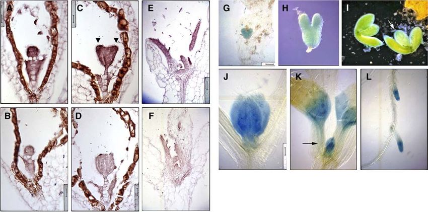
Splicing Factors in Cell Cycle Arrest     2001

                                                                               (Figure 8L). In general, SMP1 expression is associated with
                                                                               regions of cell proliferation in lateral organs.

                                                                               Overexpression of SMP1 Affects Cell Number and Size

                                                                               To look at SMP1 function, we overexpressed the SMP1 gene
                                                                               using the constitutive 35S promoter of Cauliflower mosaic virus
                                                                               (35S CaMV). We characterized five lines that contained either the
                                                                               35S CaMVp:SMP1 or 35S CaMVp:SMP1:smGFP construct and
                                                                               gave a similar dominant heritable phenotype. SMP1 transcript
                                                                               levels in three of those lines were examined, and the observed
                                                                               phenotype was associated with an overproduction of SMP1
                                                                               mRNA of the expected size (Figure 6A; ;2 kb). In SMP1:GFP
                                                                               plants, a single band of ;2.7 kb was observed, consistent with
                                                                               the expected size of the mRNA (Figure 6A). Many smaller
                                                                               discrete bands were also detected, suggesting posttranscrip-
                                                                               tional silencing of that transcript.
                                                                                  Overall, SMP1 overexpression reduced leaf organ size and
Figure 7. SMP1 and SMP2 Are Functionally Redundant and Essential.              stem internodal length. All five SMP1-overexpressing lines gen-
                                                                               erated plants exhibiting clustered siliques (Figure 9B), which
(A) Wild-type, smp1-1, and smp2-1 plants, respectively.
                                                                               were slightly crinkled and contained fewer but larger seeds (data
(B) and (C) Siliques (seed-bearing pods) taken from plants homozygous
                                                                               not shown). Three of those lines also produced a few plants that
for smp1-1 and heterozygous for smp2-1 were cleared and viewed under
dark-field illumination. The silique on the left in (B) is wild-type and was
                                                                               were phenotypically more severe and resembled severe dwarfs.
used for comparison.                                                           More specifically, their inflorescences were very reduced in
                                                                               stature and produced smaller cauline leaves (Figure 9C).
                                                                                  Because a similar reduction in leaf organ size was observed for
homozygous for both mutations (n ¼ 39), suggesting that the                    both SMP1-overexpressing plants and smpepi mutant plants,
double mutant was not viable. We examined the siliques taken                   histological analyses were used to compare their cell size and
from F2 plants and found that approximately one-quarter to one-                number relative to that of the wild type. Sections through cauline
half of the seeds were missing (Figures 7B and 7C), suggesting                 leaves of phenotypically severe SMP1-overexpressing plants
that the double mutant was not viable at the gametophytic and/or               indicated that the leaves were thinner and their cells were more
embryo stage. Because reduced expression of SMP1 and SMP2                      numerous but smaller per area than those observed in the
in the smpepi mutant resulted in fertility defects, the observed               wild type (Figures 10B and 10E). Cell numbers were quantified
lethality of the double mutant further indicated that the function-            through transverse sections of three fully grown cauline leaves
ally redundant genes are essential.                                            from each genotype (vascular cells were excluded), and the
                                                                               mean cell number in SMP1-overexpressing leaves (220.3 cells,
                                                                               SD of 11.9; 0.11 mm2) was found to be 110.2% of that in the wild

SMP1 Expression Is Associated with Regions of Cell                             type (200 cells, SD of 1.2; 0.11 mm2). Inflorescence shoot apices
Proliferation in Lateral Organs                                                were also examined, and scanning electron micrographs of the
                                                                               epidermal layer of the third or fourth stem internode from the
The expression pattern of SMP1 was visualized by RNA in situ                   youngest silique indicated that SMP1 overexpression interfered
hybridizations as well as histochemical staining of plants con-                with cell elongation. The overall pattern of cell expansion was
taining a transgenic copy of the SMP1 promoter driving the                     complex, but a few interspersed cells were shorter and wider
b-glucuronidase (GUS) reporter gene. SMP1 mRNA expression                      (Figure 9E).
was found throughout early globular-staged embryos (Figure
8A), and expression intensified in the cotyledon primordia of                  Regulatory Role in Expression of the Cell Proliferation
heart-staged embryos (Figure 8B). The broad expression pattern                 Gene SWP
persisted through embryogenesis (Figures 8G and 8H) until the
bent cotyledon stage where SMP1 promoter activity increased in                 Several characterized Arabidopsis thaliana genes are thought to
procambial cells (Figure 8I). After germination, SMP1 mRNA                     play a role in cell proliferation in lateral organs. The SWP gene, for
expression was found throughout leaf primordia but not in the                  example, is involved in defining the duration of the cell pro-
SAM (Figure 8C). SMP1 promoter activity was also found                         liferation phase in the leaf primordium without affecting cell
throughout very young leaf primordia (Figure 8J) and as the leaf               division rates (Autran et al., 2002). The loss-of-function and gain-
matured, was turned off in a tip-to-base manner (Figure 8K). This              of-function phenotypes of SWP resemble those of SMP1 and/or
pattern of GUS expression overlapped with the general pattern                  SMP2; that is, they have similar effects on leaf organ size as well
of cell proliferation and presaged the basipetal wave of cell                  as leaf cell number and cell size. However, they have different
maturation that would flow from leaf tip to leaf base. This pattern            leaf morphologies (i.e., the extent of leaf serration), which may be
of GUS expression also holds true for lateral root primordia                   due to differences in ecotypic background. More importantly,
2002 The Plant Cell Figure 8. SMP1 Expression Is Associated with Regions of Cell Proliferation. (A) to (F) Longitudinal sections were hybridized with antisense ([A], [C], and [E]) or sense ([B], [D], and [F]) dioxigenin-labeled SMP1 RNA probe. (A) and (B) Globular-staged embryos. Bar ¼ 16 mm. (C) and (D) Heart-staged embryos. Arrowheads point to cotyledon primordia. Bar ¼ 16 mm. (E) and (F) Developing leaves and leaf primordia of week-old seedlings. Bar ¼ 50 mm. (G) to (L) Wild-type plants containing a transgenic copy of the SMP promoter-driven GUS reporter gene were stained for GUS activity and viewed under bright-field illumination unless noted otherwise. (G) Heart-staged embryo. Bar ¼ 50 mm. (H) Torpedo-staged embryo. Bar ¼ 31.25 mm. (I) Bent cotyledon-staged embryos viewed under dark-field illumination because of saturated GUS staining under bright-field illumination. Arrow points to stronger GUS staining in the procambial strands of the cotyledons. Bar ¼ 100 mm. (J) Developing leaves of a 7-d-old seedling. Bar ¼ 66 mm. (K) Developing leaves and leaf primordia of a 9-d-old seedling. Arrow points to GUS-stained leaf primordia. Bar ¼ 100 mm. (L) GUS-stained lateral root primordia of a week-old seedling. Bar ¼ 100 mm. unlike smpepi, the swp mutation affects the maintenance of the exhibited a wild-type phenotype (Figure 11A). RT-PCR results SAM. The SWP gene encodes a protein with similarities to confirmed that two of these restored lines expressed a wild-type Med150/Rgr1-like subunits of the Mediator complex, which is level of fully spliced SWP transcript and more so than the line that required for RNA polymerase II recruitment at target promoters in exhibited the mutant phenotype (Figures 11B and 11C). Surpris- response to specific transcriptional activators. Given the strong ingly, transcript levels of SMP1 and SMP2 were also increased in phenotypic similarities between smpepi and swp plants and all three lines (Figure 11B). It is highly unlikely that all three lines their effects on the duration of the cell proliferation phase, SWP are complete revertants of the smpepi mutation (reversion rate is expression level was examined by RNA gel blot analysis and only 2.6%), and given SWP’s putative role in transcriptional RT-PCR and was found to be undetectable in smpepi plants activation, it is more likely that the concomitant increase of all but normal in SMP1-overexpressing plants and in smp1-1 and three transcripts by the introduction of the SWP cDNA suggests smp2-1 insertion lines (Figures 6A and 6B), suggesting that SWP a physical interaction among their proteins and/or genes as well transcription depends on the expression of SMP1 and/or SMP2 as a connection between their methylation status and transcrip- and that the reduction of SWP transcription is responsible for tional status. much of the smpepi phenotype. Interestingly, we found two CCGG sites in the 5th and 6th To demonstrate that the primary regulatory role for both exons of the SWP gene that were highly methylated in both wild- putative step II splicing factors, SMP1 and SMP2, is in the type and smpepi plants (Figure 12). Methylation was not detected transcription of SWP, a construct containing the full-length SWP in either the wild-type or smpepi allele using methylation-sensitive cDNA driven by the 35S CaMV promoter was introduced into restriction endonucleases BglII, BstB1, ClaI, HaeIII, NcoI, PstI, smpepi mutant plants. Because of the moderate infertility of these PvuI, PvuII, and Sau3AI (Figure 12B; data not shown). Further- plants, only four independent lines were obtained, three of which more, similar to the methylation pattern found in the SMP1 gene,
Splicing Factors in Cell Cycle Arrest       2003

Figure 9. Internodal Cell Elongation Affected in SMP1-Overexpressing Lines.

(A) to (C) Shoot architecture. (A) Wild-type; (B) and (C) SMP1-overexpressing lines.
(D) and (E) Scanning electron micrographs of the third or fourth stem internode from the youngest silique. (D) Wild-type; (E) SMP1-overexpressing line.
White bar at the bottom right ¼ 10 mm.

the outer and inner cytosines at those sites in the SWP gene were             DISCUSSION
differently methylated in the mutant (Figure 12B).
    Another well-characterized Arabidopsis gene involved in                   The recessive smpepi allele defines a single locus because the
defining the duration of the cell proliferation phase in the                  introduction of wild-type SMP1 genomic DNA fully rescued
leaf primordium is AINTEGUMENTA (ANT). Inactivation and                       smpepi homozygous plants. Also, smpepi is a loss-of-function
overexpression of this transcription factor reduced and in-                   allele because the transcript level of SMP1 (and SMP2) is
creased the final cell number in lateral organs, respectively,                reduced. Several lines of evidence indicate that the smpepi allele
without affecting cell division rates (Mizukami and Fischer, 2000).           is an epi-allele; it is recessive, exhibits genetic instability, is
However, there was no compensatory decrease in cell size in                   associated with a different methylation pattern of two sites in the
ANT overexpression plants; instead, final organ size was in-                  2nd exon of the SMP1 gene, affects the transcription of its sister
creased. Furthermore, ANT overexpression activated ectopic                    gene, and resembles a weaker version of the smp1 smp2 double
expression of CYCD3;1, which encodes a D-cyclin important for                 mutant. SMP1 is one of a few Arabidopsis genes known to be
the initiation of cell division at the G1 phase in leaves (Mizukami           methylated in the wild-type condition. Typically, methylation is
and Fischer, 2000). ANT and CYCD3;1 expressions were exam-                    positively correlated with transcriptional gene silencing, but
ined by RNA gel blot analysis and were found to be unchanged in               there are notable exceptions in plants and animals. For example,
smpepi and SMP1-overexpressing plants (Figure 6A; data not                    in the case of the maize (Zea mays) paramutagenic B9 and
shown), suggesting that the role of SMP1 and SMP2 in cell pro-                paramutable B-1 alleles, the high-expressing B-1 allele is meth-
liferation regulation in leaves does not involve ANT nor CYCD3;1              ylated more than the low-expressing B9 allele (Stam et al., 2002).
at the RNA level.                                                             In mouse, the maternal-specific methylation of the imprinted
    Aside from D-cyclins, the other major regulators of eukaryotic            lgf2r gene is necessary for expression (Stöger et al., 1993). In the
cell cycle initiation are cyclin-dependent kinases. In Arabidopsis,           case of smpepi, the methylation pattern is more complex.
the CDC2A gene encodes a functional cyclin-dependent kinase                      Transcriptional gene silencing is typically associated with
homolog that is expressed in all plant meristems (Martinez et al.,            promoter methylation; however, we have found no methylation
1992; Hemerly et al., 1993) and, when inactivated, reduces cell               differences at multiple sites in the promoter. Interestingly, in the
proliferation in leaves while increasing cell size (Hemerly et al.,           case of SMP1-overexpressing lines, SMP1 transcripts appeared
1995). CDC2A expression also was found to be unchanged                        to undergo posttranscriptional gene silencing, which is typically
in smpepi and SMP1-overexpressing plants (Figure 6A), sug-                    associated with methylation of the coding sequence.
gesting that at least at the RNA level, SMP1 and/or SMP2                         It would appear that the methylation status of certain cytosines
acts downstream of CYCD3;1 and CDC2A in cell proliferation                    in the SMP1 gene is responsible for transcriptional silencing of
regulation.                                                                   not only the SMP1 gene but also the unlinked SMP2 gene. If that
2004 The Plant Cell Figure 10. Overexpression of SMP1 Affects Cell Number and Size. (A) and (B) Transverse sections through fully expanded wild-type (A) and SMP1-overexpressing (B) cauline leaves. Bar ¼ 62.5 mm. (C) to (E) Longitudinal sections through the primary inflorescence shoot apices of smpepi (C), wild-type (D), and SMP1-overexpressing (E) plants. Arrowheads point to the pedicel of a developing flower. Bar ¼ 50 mm. is the case, then it resembles other trans-silencing phenomena, (e.g., Prp3p, Prp8p, Prp22p, and Cef1p; Shea et al., 1994; which usually involve the presence of repeats as a trigger Hwang and Murray, 1997; McDonald et al., 1999). Some splicing mechanism. For example, in Arabidopsis, the inverted repeat factors have been found to be specific for certain cellular RNAs of one pai locus was shown to trigger methylation of all other pai (e.g., Prp17p on TUB1 and TUB3 intron splicing; Chawla et al., homologs (Luff et al., 1999). Also, in maize, the tandem direct 2003). Because of the localization of SMP1 mRNA expression to repeats in the promoter of the paramutagenic B9 allele was regions of cell proliferation in lateral organs and of the smpepi shown to trigger methylation of the paramutable B-1 allele (Stam mutation’s effect on the duration of the cell proliferation phase et al., 2002). However, there are no detectable repeat structures there, most likely SMP1 and SMP2 affect the correct 39 splicing in the promoter and coding sequences of both SMP1 and SMP2 of certain target pre-mRNA transcripts that direct cell prolifera- genes, so it remains unclear how the methylation pattern of at tion in lateral organs. One of the functional targets of SMP1 and least two sites in the SMP1 gene was able to affect expression of SMP2 is the transcription of SWP, and it may be the primary the unlinked SMP2 gene. Also, there are no known transposons target because transgenic expression of a fully spliced SWP in the vicinity of SMP1 that can trigger epigenetic modifications, cDNA fully restored the smpepi mutant to wild-type. That it also so it remains unclear how epi-alleles in general and the smpepi caused a concomitant increase in the expression of SMP1 and allele in particular are generated. Finally, further mystery sur- SMP2 suggests some interaction among the three proteins and/ rounds the similar methylation patterns found at CCGG sites in or genes in transcriptional activation and methylation status. the coding regions of the SMP1 and SWP genes because no Finally, the loss of function of both SMP1 and SMP2 do not sequence similarities were found between the two genes in the adversely affect the transcription and transcript processing of vicinity of the CCGG sites. cell cycle regulators CYCD3;1 and CDC2A, which had been SMP1 and SMP2 encode a CCHC zinc finger protein with shown to be involved in cell cycle progression in leaves. similarities to step II splicing factors. Step II splicing factors are Because plant growth and organ formation do not involve cell responsible for the selection of correct 39 splice sites, and some migration or for the most part cell death, the final organ cell had been characterized in other systems to be nonessential and number mostly depends on two factors: (1) the number of cells play a role in the efficient progression through cell cycle tran- initially recruited to the organ primordium from the meristem and sitions (e.g., Cdc40p; Vaisman et al., 1995; Boger-Nadjar et al., (2) the proliferation of the meristematic cells in the developing 1998). In fact in yeast, many splicing factors were defined by organ. Cell proliferation itself can be regulated at two levels: (1) mutations that had been uncovered in cell cycle mutant screens the duration of the cell proliferation phase and (2) the rate of cell
Splicing Factors in Cell Cycle Arrest        2005

                                                                                 organ shape nor size, suggesting that some intrinsic patterning
                                                                                 mechanism(s) senses the final shape and size and regulates
                                                                                 them by altering the other parameter (Hemerly et al., 1995; Smith
                                                                                 et al., 1996; Cleary and Smith, 1998; Jones et al., 1998; Wang
                                                                                 et al., 2000; De Veylder et al., 2001; Autran et al., 2002). However,
                                                                                 several examples suggest that such compensatory mechanisms
                                                                                 might not always be activated. For example, overexpression of
                                                                                 ANT increases not only cell proliferation but also final organ size,
                                                                                 and it does this through activation of ectopic CYCD3;1 expres-
                                                                                 sion (Mizukami and Fischer, 2000). Also, overexpression of E2Fa
                                                                                 and Dpa, which encode transcription factors involved in the
                                                                                 activation of cell cycle genes, induces extra cell divisions but also
                                                                                 severely inhibits overall plant growth in Arabidopsis (De Veylder
                                                                                 et al., 2002). Kim and Sinha (2003) suggested that timing may be
                                                                                 the critical third dimension that determines whether compensa-
                                                                                 tory mechanisms come into play or not. It may be that these
                                                                                 mechanisms are activated when cell proliferation or expansion is
Figure 11. Genetic Interaction between SMP1, SMP2, and SWP.                      altered downstream of cell cycle genes. In the case of SMP1 and
                                                                                 SMP2, their effect on the phase duration of cell proliferation in
(A) smpepi plants containing a transgenic copy of SWP cDNA and
exhibiting a wild-type phenotype (left; shown is line 3) or smpepi mutant
                                                                                 lateral organs is sensed by some intrinsic patterning mecha-
phenotype (right; shown is line 4).                                              nism(s); the reduced activity of both genes or the increased
(B) Total RNA from wild-type, smpepi, and four independent smpepi lines          expression of SMP1 produces fewer but larger cells or more
containing a transgenic copy of SWP cDNA were reverse transcribed and            numerous but smaller cells per area, respectively.
PCR amplified using primers to SMP1, SMP2, and SWP. RT-PCR
products were run on an ethidium bromide–stained gel. PCR-amplified
eIF4A was the loading control. 1, 2, and 3 refer to lines that exhibit a wild-
                                                                                 METHODS
type phenotype, and 4 refers to a line that exhibits smpepi phenotype.
(C) RNA gel blot with 10 mg of total RNA was hybridized with a
32P-labeled SWP probe corresponding to the 39 end of the cDNA.                   Plant Materials and Growth Conditions
Methylene blue–stained rRNAs were the loading control. Numbers refer
                                                                                 Arabidopsis thaliana ecotypes Columbia (Col-0) and Landsberg carrying
to transgenic lines.
                                                                                 the erecta mutation (Ler) were used for comparison with mutant plants
                                                                                 and for crosses. Seeds were grown under constant white light (;300 mE
                                                                                 m2 s1) either on 0.75% agar media consisting of MS basal salts (Sigma-
division. These variables are not necessarily coupled; for exam-
                                                                                 Aldrich, St. Louis, MO), Haughn and Somerville (1986) nutrient solution,
ple, overexpression of the cyclin-dependent kinase inhibitor                     0.5 g/L Mes, and 10 g/L sucrose or on soil (3:1 mix of Metro-Mix 200 to
KRP1 or KRP2 reduced the rate of cell division in young leaves                   vermiculite; Scotts, Marysville, OH).
without affecting the moment of cell cycle start and arrest (Wang
et al., 2000; De Veylder et al., 2001). In these lines, a similar
                                                                                 Mutant Isolation
uncoupling of cell growth from cell division was observed (fewer
but larger cells). In the case of the smaller leaves of the smpepi               A visual screen for venation pattern mutants was performed on
mutant, the reduced leaf cell number is attributed to a precocious               diepoxybutane-mutagenized M2 Arabidopsis (ecotype Col-0) seeds.
arrest of cell proliferation, which was inferred from the level of cell          Briefly, hydrated seeds were incubated in 18 mM diepoxybutane (Sigma-
                                                                                 Aldrich) for 4 h and washed extensively before planting (M1 generation).
vacuolation and cell number of similarly aged leaf primordial.
                                                                                 M2 seeds, the progeny of self-fertilized M1 plants, were pooled from
   The KRP-overexpressing lines also exhibit a similar leaf mor-
                                                                                 every 10 M1 line and screened ;2 weeks after germination. One to two
phology change (pronounced serration phenotype at the leaf                       rosette leaves from each M2 plant were fixed in 3:1 ethanol:acetic acid,
teeth), which has been attributed to the continuous high expres-                 dehydrated in an ethanol series, cleared in Hemo-De (Fisher Scientific,
sion of mitotic cyclins (CYCB1;1 and CYCA2;1) at the leaf teeth                  Fairlawn, NJ), mounted on slides with 2:1 Permount (Fisher Scientific) to
and in the surrounding vasculature during late leaf development                  xylene, and viewed under dark-field optics for alterations in the venation
when they are turned off in other parts of the leaf (Van Lijsebettens            pattern. Putative venation pattern mutants were confirmed by a second-
and Clarke, 1998; Burssens et al., 2000). Mitotic cyclin expres-                 ary screen and backcrossed twice to wild-type Col-0 plants for further
sion reduces KRP’s inhibitory effect on cell division activity. The              phenotypic characterization.
smpepi mutation may similarly be less effective at inhibiting cell
division activity in the veins that extend to the teeth because                  Histochemical Localization of GUS Activity and
vascular cells were least affected by the smpepi mutation.                       Histological Analyses
   In animals, generally, changes in cell size can be compensated                Plant tissues were stained for GUS activity overnight at 378C in GUS
for by changes in cell number to maintain the final size of an                   buffer, 20% methanol, and 0.5 mg/mL 5-bromo-4-chloro-3-indolyl-
organism (reviewed in Day and Lawrence, 2000; Weinkove and                       b-D-glucuronidase as described by Malamy and Benfey (1997). For obser-
Leevers, 2000). Similarly, for plants, alterations in either cell                vation of whole mounts, both stained and unstained tissue were fixed,
proliferation or cell expansion alone generally do not affect final              cleared, and mounted on slides as described above. Specimens were
2006 The Plant Cell
Figure 12. SWP Is Hypomethylated in smpepi.
(A) Restriction map of SWP locus. Black boxes indicate exons, and arrowheads indicate recognition sites of methylation-sensitive enzymes BglII, HaeIII,
NcoI, HpaII, and MspI. Gray arrowheads indicate sites found to be methylated.
(B) DNA gel blots of digested DNA from wild-type (W) and smpepi (M) plants were hybridized with either 32P-labeled probe A (left) or 32P-labeled probe B
(right). Note the presence and abundance of 6.262-kb band in both HpaII digests and in the MspI digest of wild-type DNA. Also, all three bands (3.274,
2.585, and 2.265 kb) were reduced in the HpaII digest of smpepi.
examined with a Zeiss Axiophot microscope (Oberkochen, Germany). smpepi mutation, and DNA from ;188 mutant plants were used with
Tissues used for RNA in situ hybridizations and scanning electron Cereon’s insertion/deletion and single-nucleotide polymorphism markers
microscopy were fixed overnight at 48C in FAA (3.7% formaldehyde, (Jander et al., 2002) to finely map the smpepi mutation to a region spanned
50% ethanol, and 5% acetic acid). For histological analysis, fixed by a single BAC.
dehydrated specimens were embedded in Spurr’s media (EM Sciences, The SMP1 coding sequence along with 810 bp of upstream sequence
Ft. Washington, PA) or in Paraplast Plus (Fisher Scientific). Plastic and 160 bp of downstream sequence were PCR amplified and subcloned
sections (2 mm) were cut on a Sorvall MT2-B ultramicrotome (Newtown, into XbaI/SacI sites of the PJIM19 (BAR) binary vector. The SMP1 coding
CT) with glass knives and were stained briefly with 1% toluidine blue. sequence was subcloned into BamHI/SacI sites of the PJIM19 (KAN)
Paraffin sections (8 mm) were cut on a model 820 microtome (Arthur H. binary vector downstream of the 35S CaMV promoter. Upstream se-
Thomas Co., Philadelphia, PA). Specimens were prepared for scanning quence (180 bp) was subcloned into XbaI/BamHI sites of the PBI101
electron microscopy by overnight fixation in 3:1 ethanol:glacial acetic binary vector (Clontech, Palo Alto, CA) upstream of the GUS gene. The
acid, dehydration through an ethanol series, and critical-point drying SMP1 coding sequence (minus the stop codon) was PCR amplified and
with liquid carbon dioxide in a Polaron pressure chamber (Watford, subcloned into XbaI/StuI sites of the PJIM19smGFP binary vector in
Hertfordshire, UK). Dried samples were mounted on stubs, sputter frame and upstream of the smGFP gene (minus the start codon). All four
coated with gold-palladium alloy for 30 s or with gold for 2 min, examined constructs were sequenced for errors and introduced into wild-type and/
with an ISI SS-40 scanning electron microscope (Santa Clara, CA), and or smpepi plants via the Agrobacterium tumefaciens–mediated floral dip
photographed on Polaroid Polapan 53 film (Waltham, MA). method (Clough and Bent, 1998), and transformants were selected on
agar media containing 15 mg/mL BASTA or 50 mg/mL kanamycin.
Genetic Mapping and Plant Transformation Vector Construction
DNA Gel Blot Analysis
Plants homozygous for the smpepi mutation were crossed to wild-type Ler
plants to generate an F2 mapping population. DNA from 20 mutant plants Two micrograms of genomic DNA was digested with BglII, BstBI, ClaI,
were used with simple sequence length polymorphic markers (Bell and HaeIII, MboI, NcoI, PstI, PvuI, PvuII, Sau3AI, HpaII, or MspI for 6 h,
Ecker, 1993; Ponce et al., 1998) to map the chromosomal location of the electrophoresed in 1.5% agarose gel, transferred to ZetaProbe GTSplicing Factors in Cell Cycle Arrest 2007
blotting membrane (Bio-Rad, Hercules, CA), UV cross-linked, and baked Received March 18, 2005; revised May 9, 2005; accepted May 12, 2005;
for 2 h at 808C. Blots were hybridized to 32P-labeled DNA probes published June 3, 2005.
overnight at 428C, washed twice at 558C for 10 min in 13 SSC/1%
SDS, and exposed to Eastman Kodak X-OMAT AR film (Rochester, NY)
at 708C.
REFERENCES
Ach, R.A., Durfee, T., Miller, A.B., Taranto, P., Hanley-Bowdoin, L.,
Total RNA Isolation and RNA Gel Blot Analysis
Zambryski, P., and Gruissem, W. (1997). RRB1 and RRB2 encode
Total RNA was isolated from the aerial part of 38-d-old plants with maize retinoblastoma-related proteins that interact with a plant D-type
TRIzol (Gibco BRL, Cleveland, OH) according to the manufacturer’s cyclin and geminivirus replication protein. Mol. Cell. Biol. 17,
instructions. Ten micrograms of total RNA was electrophoresed in 1.2% 5077–5086.
formaldehyde-agarose gel, transferred to ZetaProbe GT blotting mem- Alonso, J.M., et al. (2003). Genome-wide insertional mutagenesis of
brane (Bio-Rad), UV cross-linked, and baked for 2 h at 808C. Blots were Arabidopsis thaliana. Science 301, 653–657.
stained with 0.02% methylene blue/0.3 M sodium acetate, pH 5.2, for Autran, D., Jonak, C., Belcram, K., Beemster, G.T.S., Kronenberger,
3 min, and destained with 20% ethanol for 10 to 15 min, and the resulting J., Grandjean, O., Inzé, D., and Traas, J. (2002). Cell numbers
rRNA bands were visualized with a Gel Doc 2000 (Bio-Rad). Blots were and leaf development in Arabidopsis: A functional analysis of the
then hybridized to 32P-labeled DNA probes overnight at 428C, washed at STRUWWELPETER gene. EMBO J. 21, 6036–6049.
558C for 30 min in 13 SSC/1% SDS and then in 0.53 SSC/2.5% SDS, and Bell, C.J., and Ecker, J.R. (1993). Assignment of 30 microsatellite loci
exposed to Eastman Kodak X-OMAT AR film at 708C. to the linkage map of Arabidopsis. Genomics 19, 137–144.
Boger-Nadjar, E., Vaisman, N., Ben-Yehuda, S., Kassir, Y., and
Kupiec, M. (1998). Efficient initiation of S-phase in yeast requires
RT-PCR Cdc40p, a protein involved in pre-mRNA splicing. Mol. Gen. Genet.
Two micrograms of total RNA was reverse-transcribed with 200 units of 260, 232–241.
Superscript II (Invitrogen, Carlsbad, CA). The resulting cDNA:RNA hybrids Burssens, S., de Almeida Engler, J., Beeckman, T., Richard, C.,
were treated with 10 units of DNase I (Roche, Indianapolis, IN) for 30 min Shaul, O., Ferreira, P., Van Montagu, M., and Inzé, D. (2000).
at 378C, purified on Qiaquick PCR column (Qiagen, Valencia, CA), and Developmental expression of the Arabidopsis thaliana CycA2;1 gene.
used as template to PCR amplify SMP1 (40 cycles), SMP2 (40 cycles), Planta 211, 623–631.
SWP (40 cycles), and eIF4A (39 cycles). PCR conditions are as follows: Chawla, G., Sapra, A.K., Surana, U., and Vijayraghavan, U. (2003).
948C for 15 s, 528C for 15 s, and 728C for 20 s. PCR products (350 to Dependence of pre-mRNA introns on PRP17, a non-essential splicing
440 bp) were electrophoresed in 1.5% agarose gel and visualized with a factor: Implications for efficient progression through cell cycle tran-
Gel Doc 2000. Primer sequences for SMP1 are 59-GACCATAGGA- sitions. Nucleic Acids Res. 31, 2333–2343.
AGCAAATTGA-39 and 59-AAGATCTATCACACGATGGT-39; for SMP2 Chua, K., and Reed, R. (1999). Human step II splicing factor hSlu7
are 59-GATCACAGGAAGAAATTAGAA-39 and 59-ACGATCTACCACAT- functions in restructuring the spliceosome between the catalytic steps
GACGGT-39; and for SWP are 59-TCTGCTCTTGTTGGTCGAG-39 and of splicing. Genes Dev. 13, 841–850.
59-TGATAAGAACCTGTCAGCAA-39. Clay, N.K., and Nelson, T. (2002). VH1: A provascular-specific receptor
kinase that influences leaf cell patterns in Arabidopsis. Plant Cell 14,
2707–2722.
RNA in Situ Hybridization Cleary, A.L., and Smith, L.G. (1998). The Tangled1 gene is required for
SMP1 cDNA (nucleotides 439 to 640 relative to ATG) was PCR amplified spatial control of cytoskeletal arrays associated with cell division
with primers containing engineered T7 and T3 RNA polymerase promoter during maize leaf development. Plant Cell 10, 1875–1888.
sites and used as template to generate dioxigenin-labeled sense and Clough, S.J., and Bent, A.F. (1998). Floral dip: A simplified method
antisense RNA probes. In situ hybridizations and labeling reactions were for Agrobacterium-mediated transformation of Arabidopsis thaliana.
performed as described by Jackson (1991) with some modifications, Plant J. 16, 735–743.
which were described by Clay and Nelson (2002). Curtis, D., Treiber, D.K., Tao, F., Zamore, P.D., Williamson, J.R., and
Lehmann, R. (1997). A CCHC metal-binding domain in Nanos is
essential for translational regulation. EMBO J. 16, 834–843.
Cell Counts Day, S.J., and Lawrence, P.A. (2000). Measuring dimensions: The
regulation of size and shape. Development 127, 2977–2987.
For each genotype, transverse sections through a total of three adult
De Veylder, L., Beeckman, T., Beemster, G.T.S., de Almeida Engler,
leaves were used for cell counts per defined area. The defined area
J., Ormenese, S., Maes, S., Naudts, M., Van der Schueren, E.,
encompassed a region just right or left of the midvein, and the average of
Jacqmard, A., Engler, G., and Inzé, D. (2002). Control of prolifera-
two serial sections was used to calculate the mean cell number per area
tion, endoreduplication and differentiation by Arabidopsis E2Fa-Dpa
for each leaf. This average was then used to calculate the mean cell num-
transcription factor. EMBO J. 21, 1360–1368.
ber per area for all three leaves.
De Veylder, L., Beeckman, T., Beemster, G.T.S., Krols, L., Terras, F.,
Landrieu, I., Van der Schueren, E., Maes, S., Naudts, M., and Inzé,
ACKNOWLEDGMENTS D. (2001). Functional analysis of cyclin-dependent kinase inhibitors of
Arabidopsis. Plant Cell 13, 1653–1667.
Sequencing analysis was performed by the HHMI Biopolymer/Keck Dewitte, W., Riou-Khamlichi, C., Scofield, S., Healy, J.M.S.,
Foundation Biotechnology Resource Lab (Yale University, New Haven, Jacqmard, A., Kilby, N.J., and Murray, J.A.H. (2003). Altered cell
CT). We thank James A. Sullivan for his generous gift of the PJIM19 cycle distribution, hyperplasia, and inhibited differentiation in Arabi-
vectors. This work was supported by National Science Foundation dopsis caused by the D-type cyclin CYCD3. Plant Cell 15, 79–92.
Grants IBN-0114648 and IBN-0416731 to T.N. Donnelly, P.M., Bonetta, D., Tsukaya, H., Dengler, R.E., and Dengler,2008 The Plant Cell N. (1999). Cell cycling and cell enlargement in developing leaves of Mizukami, Y., and Fischer, R.L. (2000). Plant organ size control: Arabidopsis. Dev. Biol. 215, 407–419. AINTEGUMENTA regulates growth and cell numbers during organo- Doonan, J. (2000). Social controls on cell proliferation in plants. Curr. genesis. Proc. Natl. Acad. Sci. USA 97, 942–947. Opin. Plant Biol. 3, 482–487. Mount, S.M., and Rubin, G.M. (1985). Complete nucleotide sequence Frank, D., and Guthrie, C. (1992). An essential splicing factor, SLU7, of the Drosophila transposable element copia: Homology between mediates 39 splice site choice in yeast. Genes Dev. 6, 2112–2124. copia and retroviral proteins. Mol. Cell. Biol. 5, 1630–1638. Grandjean, O., Vernoux, T., Laufs, P., Belcram, K., Mizukami, Y., and Ponce, M.R., Quesada, V., and Micol, J.L. (1998). Rapid discrimination Traas, J. (2004). In vivo analysis of cell division, cell growth, and of sequences flanking and within T-DNA insertions in the Arabidopsis differentiation at the shoot apical meristem in Arabidopsis. Plant Cell genome. Plant J. 14, 497–501. 16, 74–87. Rajavashisth, T.B., Taylor, A.K., Andalibi, A., Svenson, K.L., and Guo, K., and Walsh, K. (1997). Inhibition of myogenesis by multiple Lusis, A.J. (1989). Identification of a zinc finger protein that binds to cyclin-Cdk complexes. Coordinate regulation of myogenesis and cell the sterol regulatory element. Science 245, 640–643. cycle activity at the level of E2F. J. Biol. Chem. 272, 791–797. Roussell, D.L., and Bennett, K.L. (1993). glh-1, a germ-line putative Hansen, L.J., Chalder, D.L., and Sandmeyer, S.B. (1988). Ty3, a yeast RNA helicase from Caenorhabditis, has four zinc fingers. Proc. Natl. retrotransposon associated with tRNA genes, has homology to animal Acad. Sci. USA 90, 9300–9304. retroviruses. Mol. Cell. Biol. 3, 5245–5256. Schwartz, D.E., Tizard, R., and Gilbert, W. (1983). Nucleotide Haughn, G.W., and Somerville, C. (1986). Sulfonylurea-resistant sequence of rous sarcoma virus. Cell 32, 853–869. mutants of Arabidopsis thaliana. Mol. Gen. Genet. 204, 430–434. Shea, J.E., Toyn, J.H., and Johnston, L.H. (1994). The budding yeast Hemerly, A., de Almeida Engler, J., Van Montagu, M., Engler, G., U5 snRNP Prp8 is a highly conserved protein which links RNA splicing Inzé, D., and Ferreira, P. (1995). Dominant negative mutants of the with cell cycle progression. Nucleic Acids Res. 22, 5555–5564. Cdc2 kinase uncouple cell division from iterative plant development. Shinnick, T.M., Lerner, R.A., and Sutcliffe, J.G. (1981). Nucleotide EMBO J. 14, 3925–3936. sequence of Moloney murine leukaemia virus. Nature 342, 543–548. Hemerly, A.S., Ferreira, P.C.G., de Almeida Engler, J., Van Montagu, Skapek, S.X., Rhee, J., Spicer, D.B., and Lassar, A.B. (1995). M., Engler, G., and Inzé, D. (1993). cdc2a expression in Arabidopsis Inhibition of myogenic differentiation in proliferating myoblasts by thaliana is linked with competence for cell division. Plant Cell 5, cyclin D1-dependent kinase. Science 267, 1022–1024. 1711–1723. Smith, L.G., Hake, S., and Sylvester, A.W. (1996). The tangled-1 Hwang, L.H., and Murray, A.W. (1997). A novel yeast screen for mitotic mutation alters cell division orientations throughout maize leaf devel- arrest mutants identifies DOC1, a new gene involved in cyclin pro- opment without affecting leaf shape. Development 122, 481–489. teolysis. Mol. Biol. Cell 10, 1877–1887. Stam, M., Belele, C., Dorweiler, J.E., and Chandler, V.L. (2002). Jackson, D. (1991). In-situ hybridisation in plants. In Molecular Plant Differential chromatin structure within a tandem array 100 kb up- Pathology: A Practical Approach, D.J. Bowles, J.S.J. Gurr, and M. stream of the maize b1 locus is associated with paramutation. Genes McPherson, eds (Oxford: Oxford University Press), pp. 163–174. Dev. 16, 1906–1918. Jacobsen, S.E., and Meyerowitz, E.M. (1997). Hypermethylated Stöger, R., Kubicka, P., Liu, C.-G., Kafri, T., Razin, A., Cedar, H., and SUPERMAN epigenetic alleles in Arabidopsis. Science 277, Barlow, D.P. (1993). Maternal-specific methylation of the imprinted 1100–1103. mouse lgf2r locus identifies the expressed locus as carrying the Jander, G., Norris, S.R., Rounsley, S.D., Bush, D.F., Levin, I.M., and imprinted signal. Cell 73, 61–71. Last, R.L. (2002). Arabidopsis map-based cloning in the post-genome Vaisman, N., Tsouladze, A., Robzyk, K., Ben-Yehuda, S., Kupiec, M., era. Plant Physiol. 129, 440–450. and Kassir, Y. (1995). The role of Saccharomyces cerevisiae Cdc40p Jones, A.M., Im, K.-H., Savka, M.A., Wu, M.-J., DeWitt, G., Shillito, in DNA replication and mitotic spindle formation and/or maintenance. R., and Binns, A.N. (1998). Auxin-dependent cell expansion mediated Mol. Gen. Genet. 247, 123–136. by overexpressed auxin-binding protein 1. Science 282, 1114–1117. Van Lijsebettens, M., and Clarke, J. (1998). Leaf development in Kim, M., and Sinha, N. (2003). Regulating shapes and sizes. Dev. Cell 4, Arabidopsis. Plant Physiol. Biochem. 36, 47–60. 441–447. Wain-Hobson, S., Sonigo, P., Danos, O., Cole, S., and Alizon, M. Luff, B., Pawlowski, L., and Bender, J. (1999). An inverted repeat (1985). Nucleotide sequence of the AIDS virus, LAV. Cell 40, 9–17. triggers cytosine methylation of identical sequences in Arabidopsis. Wang, H., Zhou, Y., Gilmer, S., Whitwill, S., and Fowke, C.L. (2000). Mol. Cell 3, 505–511. Expression of the plant cyclin-dependent kinase inhibitor ICK1 affects Malamy, J.E., and Benfey, P.N. (1997). Organization and cell differen- cell division, plant growth and morphology. Plant J. 24, 613–623. tiation in lateral roots of Arabidopsis thaliana. Development 124, Weinkove, D., and Leevers, S.J. (2000). The genetic control of organ 33–44. growth: Insights from Drosophila. Curr. Opin. Genet. Dev. 10, 75–80. Martinez, M.C., Jørgensen, J.-E., Lawton, M.A., Lamb, C.J., and Zachsenhaus, E., Jiang, Z., Chung, D., Martin, J.D., Phillips, R.A., Doerner, P.W. (1992). Spatial pattern of cdc2 expression in relation to and Gallie, B.L. (1996). pRb controls proliferation, differentiation, and meristem activity and cell proliferation during plant development. death of skeletal muscle cells and other lineages during embryogen- Proc. Natl. Acad. Sci. USA 89, 7360–7364. esis. Genes Dev. 10, 3051–3064. McDonald, W.H., Ohi, R., Smelkova, N., Frendewey, D., and Gould, Zhang, P., Wong, C., DePinho, R.A., Harper, J.W., and Elledge, S.J. K.L. (1999). Myb-related fission yeast cdc5p is a component of a 40S (1998). Cooperation between the Cdk inhibitors p27(KIP1) and snRNP-containing complex and is essential for pre-mRNA splicing. p57(KIP2) in the control of tissue growth and development. Genes Mol. Cell. Biol. 19, 5352–5362. Dev. 12, 3162–3167.
You can also read