THE ROLE OF CANNABINOID RECEPTORS, G ALPHA Z, AND B CELL RECEPTOR IN LYMPHOMA PATHOBIOLOGY WITH FOCUS ON CHEMOTAXIS - From Department of ...
←
→
Page content transcription
If your browser does not render page correctly, please read the page content below
From Department of Laboratory Medicine, Division of Pathology
          Karolinska Institutet, Stockholm, Sweden

    THE ROLE OF CANNABINOID
RECEPTORS, G ALPHA Z, AND B CELL
     RECEPTOR IN LYMPHOMA
  PATHOBIOLOGY WITH FOCUS ON
          CHEMOTAXIS

                        Magali Merrien

                       Stockholm 2021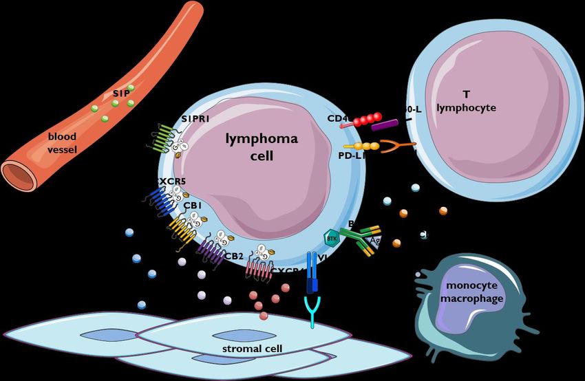
All previously published papers were reproduced with permission from the publisher.
Published by Karolinska Institutet.
Printed by Universitetsservice US-AB, 2021
© Magali Merrien, 2021
ISBN 978-91-8016-083-4
Cover picture illustrates the release of a lymphoma cell out from its microenvironment –
Painting by Josiane Jannès.
The role of cannabinoid receptors, G alpha z, and B cell
receptor in lymphoma pathobiology with focus on
chemotaxis
THESIS FOR DOCTORAL DEGREE (Ph.D.)

By

Magali Merrien

The thesis will be defended in public at 4U, Entréplan, Alfred Nobels Allé 8, Huddinge, 22nd
January 2021, 10:00am.

Principal Supervisor:                          Opponent:
Professor Birgitta Sander                      Associate Professor Guillermo Velasco
Karolinska Institutet                          Complutense University, Madrid
Department of Laboratory Medicine              Department of Biochemistry and Molecular
Division of Pathology                          Biology

Co-supervisor(s):                              Examination Board:
Professor emerita Eva Kimby                    Docent Daniel Molin
Karolinska Institutet                          Uppsala University
Department of Medicine                         Department of Immunology, Genetics and
                                               Pathology

Professor Edvard Smith                         Professor Christopher Fowler
Karolinska Institutet                          Umeå Universty
Department of Laboratory Medicine              Department of Integrative Medical Biology
Division of Clinical Research Center (KFC)

PhD Agata M. Wasik                             Docent Jonas Fuxe
Karolinska Institutet                          Karolinska Institutet
Department of Laboratory Medicine              Department of Laboratory Medicine
Division of Pathology                          Division of Pathology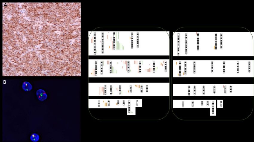
To my family, and my friends that became family.

“It is imperfection - not perfection - that is the end result of the program written into that
formidably complex engine that is the human brain, and of the influences exerted upon us by
the environment and whoever takes care of us during the long years of our physical,
psychological and intellectual development.”

- Rita Levi-Montalcini -
POPULAR SCIENCE SUMMARY

Lymphoma is a blood cancer of B lymphocytes, the cells that are an important component of
our immune system involved in producing antibodies. Malignant B lymphocytes are dividing
and proliferating excessively without becoming fully mature, and spreading to other tissues.
When lymphoma cells are in tissues, such as lymph nodes and bone marrow, they can disturb
the development of other blood cells and lead to complications.

In tissues, lymphoma cells interact with surrounding non-malignant cells, helping the
malignant cells to hide and survive from therapies. Some mechanisms used in these interactions
have already been found, and various treatments exploit these features to force the malignant
cells to leave that pro-survival environment and reach the blood circulation. In the blood,
malignant cells become more sensitive to external factors, such as chemotherapy. However,
not all patients respond to these treatment strategies and more knowledge is necessary to
improve the outcome. This thesis focuses on important mechanisms involved in the cancer
pathobiology.

One of my research projects explored the way lymphoma cells from different tissue origin
(blood and lymph node) interact with and attach to non-malignant cells, in a co-culture in vitro
system. This system could be useful to test lymphoma cells from patients before deciding on
the treatment, in order to improve the efficacy of therapy from the start. This might decrease
the risks of relapse and/or the development of aggressive disease.

During my PhD, we discovered a protein, G alpha z, to be highly expressed in mantle cell
lymphoma compared with healthy B lymphocytes. G alpha z conveys signals from cell surface
bound receptors to inside the cell and might play a role in the localization of malignant cells in
tissues. I also show in this thesis that another molecule, 2-AG, has a similar impact on
lymphoma cells. 2-AG is a ligand to the cannabinoid receptors, which are best known for being
expressed in neurons where they mediate the effects of marijuana. Cannabinoid receptors and
their ligands are dysregulated in many malignancies including lymphoma. Here, we provide
evidence that 2-AG attracts the lymphoma cells.

Finally, THC and CBD administration to lymphoma patients modulates levels of blood
leukocytes and is probably inducing the migration of lymphoma cells and normal lymphocytes
away from the blood into the tissues. These effects are unwanted during lymphoma therapy
since the lymphoma cells thrive in lymph nodes and bone marrow where they receive
protection from chemotherapy.

In summary, these studies have provided new information on factors that regulate the migration
of lymphoma cells from blood to tissues and might pave way for increased understanding of
novel mechanisms involved in the interaction between lymphoma cells and cells of the tissue
microenvironment.
ABSTRACT

Mantle cell lymphoma (MCL) and chronic lymphocytic leukaemia (CLL) are two incurable B
cell malignancies, with an overall survival of 5 to 8 years and 6 to 10 years, respectively.
Therapies are available but are often very aggressive, and patients relapse due to minimal
residual disease. Minimal residual disease is defined by the presence of few malignant cells
that escaped from therapy, mainly due to the survival signals provided by non-malignant cells
from the tissue environment, in lymph nodes and in bone marrow. Alternative and targeted
therapies are under investigation to increase patient overall survival and to reduce the risks of
relapses. However, some patients do not respond to these treatments, as malignant cells develop
mechanisms that prevent the drug efficacy. Many factors have already been depicted to
contribute to MCL pathogenesis, and in this thesis, a new potential actor in MCL pathobiology
is described, the protein G alpha z (Gαz).

The gene encoding for Gαz, GNAZ is overexpressed in most MCL cases compared to B
lymphocytes from reactive lymph nodes. It was found that GNAZ expression correlates with
lymphocytosis, and inversely correlates with the cannabinoid receptor type 1 previously
described as a receptor potentially involved in the egress and/or retention of MCL cells within
the tissue. Although the downregulation of GNAZ did not affect cell survival, proliferation or
chemotaxis in vitro, its potential role in MCL pathobiology is of interest and needs further
investigation.

Moreover, we characterize a co-culture in vitro system of MCL cell lines with mesenchymal
stromal cells that permitted to identify differentially expressed genes between cells from
different tissue origin. The JeKo-1 MCL cell line from peripheral blood origin, utilized the
BCR signalling pathway to adhere to stromal cells, while the Rec-1 MCL cell line from lymph
node origin did not, which conferred resistance to BCR targeted therapies. This system could
be useful for testing patient samples to determinate a potential resistance before treatment
decision.

Finally, the endocannabinoid system has been previously identified as dysregulated in both
MCL and CLL. Here, we provide a new role of the endogenous cannabinoid 2-
arachidonoylglycerol in chemotaxis of malignant B cells, regulated by both cannabinoid
receptors type 1 and type 2. This endocannabinoid did not only induce chemotaxis by itself but
also modulated the chemotaxis towards the chemokine CXCL12. In addition, a single
administration of the natural cannabinoids, THC and CBD, in lymphoma patients promoted the
redistribution of malignant cells from peripheral blood, and also affected non-malignant
immune cells in blood. This potential negative effect of cannabinoids on the immune cells
should be taken into consideration, knowing that around 25% of cancer patients use
cannabinoids to alleviate symptoms and side effects from therapy.
LIST OF SCIENTIFIC PAPERS

  I. Mundt F,* Merrien M,* Nygren L, Sutton LA, Christensson B, Wahlin BE,
     Rosenquist R, Sander B, Wasik AM.
     Expression of GNAZ, encoding the Gαz protein, predicts survival in mantle
     cell lymphoma.
     Br J Haematol. 2019 May;185(4):708-712. doi: 10.1111/bjh.15810. Epub 2019
     Feb 20. PMID: 30788840.

 II. Sadeghi L,* Arvidsson G,* Merrien M, Wasik AM, Görgens A, Smith CIE,
     Sander B, Wright AP.
     Differential B-Cell Receptor Signaling Requirement for Adhesion of Mantle
     Cell Lymphoma Cells to Stromal Cells.
     Cancers (Basel). 2020 May 2;12(5):1143. doi: 10.3390/cancers12051143.
     PMID: 32370190; PMCID: PMC7281289.

 III. Magali Merrien, Agata M. Wasik, Christopher M. Melén, Kristina Sonnevi,
      Henna-Riikka Junlén, Birger Christensson, Björn E. Wahlin and Birgitta
      Sander.
      2-arachidonoylglycerol modulates the CXCL12-mediated chemotaxis in
      mantle cell lymphoma and chronic lymphocytic leukemia.
      Manuscript.

IV. Christopher M. Melén*, Magali Merrien*, Agata M. Wasik, Georgios
    Panagiotidis, Olof Beck, Kristina Sonnevi, Henna-Riikka Junlén, Birger
    Christensson, Birgitta Sander**, Björn Engelbrekt Wahlin.**
    A single dose of cannabinoids transiently affects malignant B cells and CXCR4
    expression in patients with indolent B-cell leukemia.
    Manuscript.

* first authors equal contribution
** last authors equal contribution
CONTENTS
1 Introduction of the research field .................................................................................... 1
1.1 B cell development ................................................................................................ 1
1.2 Downstream signalling of BCR ............................................................................ 5
1.3 B cell malignancies................................................................................................ 6
1.3.1 Mantle Cell Lymphoma ............................................................................ 7
1.3.2 Chronic Lymphocytic Leukaemia ............................................................ 9
1.4 Lymphoma microenvironment............................................................................11
1.4.1 The B Cell Receptor ................................................................................11
1.4.2 Non-malignant cells ................................................................................12
1.4.3 Chemokines and adhesion molecules .....................................................14
1.4.4 Cannabinoid receptors and ligands .........................................................16
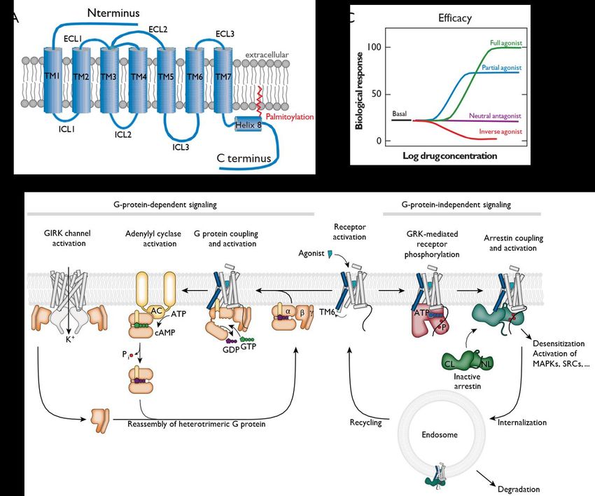
1.5 G protein and G protein coupled receptors .........................................................19
1.5.1 G protein coupled receptors ....................................................................19
1.5.2 G proteins ................................................................................................21
1.5.3 CXCR4 signalling ...................................................................................22
1.5.4 Signalling through the cannabinoid receptors ........................................23
1.6 The endocannabinoid system ..............................................................................23
1.6.1 Cannabinoid receptors.............................................................................23
1.6.2 Ligands to cannabinoid receptors ...........................................................25
1.6.3 Enzymes ..................................................................................................30
1.6.4 Other receptors binding cannabinoids ....................................................32
1.7 Medical use of cannabinoids ...............................................................................32
2 Research aims ................................................................................................................35
2.1 Overall aim ..........................................................................................................35
2.2 Specific aims........................................................................................................35
3 Aspects on the methodologies used ..............................................................................37
3.1 Ethical considerations..........................................................................................37
3.2 Cell lines and patients material ...........................................................................38
3.2.1 MCL cell lines and stromal cells ............................................................38
3.2.2 Patient material ........................................................................................38
3.3 B cells enrichment from patient blood samples..................................................39
3.4 Gene silencing using siRNA and electroporation method .................................39
3.5 Protein expression ...............................................................................................40
3.5.1 Western blotting ......................................................................................40
3.5.2 Flow cytometry analysis .........................................................................40
3.6 Chemotaxis and adhesion assays ........................................................................40
3.6.1 Cells staining ...........................................................................................40
3.6.2 Boyden chamber assay for chemotaxis assessment ...............................40
3.6.3 Co-culture for adhesion assay .................................................................41
3.6.4 Read-out ..................................................................................................41
3.7 Bioinformatic tools ..............................................................................................42
4 Results and discussion ...................................................................................................43
4.1 Paper I ..................................................................................................................43
4.1.1 Results .....................................................................................................43
4.1.2 Discussion ...............................................................................................44
4.2 Paper II .................................................................................................................45
4.2.1 Results .....................................................................................................45
4.2.2 Discussion ...............................................................................................46
4.3 Paper III ...............................................................................................................474.3.1 Results ..................................................................................................... 47
4.3.2 Discussion ............................................................................................... 48
4.4 Paper IV ............................................................................................................... 49
4.4.1 Results ..................................................................................................... 49
4.4.2 Discussion ............................................................................................... 50
5 Conclusions ................................................................................................................... 51
6 Points of perspective ..................................................................................................... 53
7 Acknowledgements ....................................................................................................... 55
8 References ..................................................................................................................... 61LIST OF ABBREVIATIONS 2-AG 2-Arachidonoylglycerol BCR B Cell Receptor BM Bone Marrow BTK Bruton’s Tyrosine Kinase C Constant CARD11 Caspase Recruitment Domain family member 11 CB Cannabinoid receptor CBD Cannabidiol CD Cluster of Differentiation CFSE Carboxyfluorescein Succinimidyl Ester CLL Chronic Lymphocytic Leukaemia cMCL Classical Mantle Cell Lymphoma D Diversity del Deletion DAG Diacylglycerol ECM Extracellular Matrix ERK1/2 Extracellular signal-Regulated Kinases 1/2 FAAH Fatty Acid Amide Hydrolase FAK Focal Adhesion Kinase FBS Foetal Bovine Serum FISH Fluorescence In Situ Hybridization GC Germinal Centre GPCR G Protein Coupled Receptor GPR G Protein Receptor HUVEC Human Umbilical Vein Endothelial Cells ICAM-1 Intercellular Adhesion Molecule-1 Ig Immunoglobulin IGHV Immunoglobulin Heavy chain Variable region IL Interleukin IP3 Inositol (1,4,5)-triphosphate J Joining Kv Voltage-gated Potassium channel LN Lymph Node MAGL Monoacylglycerol Lipase MAPK Mitogen-Activated Protein Kinase MCL Mantle Cell Lymphoma M-CLL Mutated Chronic Lymphocytic Leukaemia MHC Major Histocompatibility Complex MSC Mesenchymal Stem/Stromal Cell NAPE-PLD N-Acyl-Phosphatidyl-Ethanolamine Phospholipase D NFκB Nuclear Factor κB NHL Non-Hodgkin Lymphoma nnMCL Non-nodal Mantle Cell Lymphoma PB Peripheral Blood PCR Polymerase Chain Reaction PD-1 Programmed cell Death protein 1 PD-L-1 Programmed cell Death protein Ligand 1 PI3K Phosphoinositide 3-Kinase PIP3 Phosphatidylinositol-3,4,5-triPhosphate PKC Protein Kinase C PLCγ2 Phospholipase Cγ2 PPAR Peroxisome Proliferator-Activated Receptor R-CHOP Rituximab-Cyclophosphamide Doxorubicin Vincristine Prednisone RFI Relative Fold Increase S1PR Sphingosine-1-Phosphate Receptor siRNA Small interfering RNA STAT3 Signal Transducer and Activator of Transcription 3 THC ∆9-Tetrahydrocannabinol TNF Tumour Necrosis Factor TRPV1 Transient Receptor Potential Vanilloid type-1 U-CLL Unmutated Chronic Lymphocytic Leukaemia V Variable VCAM-1 Vascular Cell Adhesion Molecule-1 VLA-4 Very Late Activation antigen 4
1 INTRODUCTION OF THE RESEARCH FIELD
In this thesis, molecular and mechanistic pathways are investigated in two B lymphoid
malignancies: mantle cell lymphoma (MCL) and chronic lymphocytic leukaemia (CLL), with
the aim to increase knowledge on their pathogenesis and pathobiology. To understand how
these cancers arise, it is important to first describe the development of a normal B lymphocyte
(Figure 1).
1.1 B CELL DEVELOPMENT
The immune system is divided in two types of responses: the innate immune response and the
adaptive immune response. The innate response is the fast first response to foreign bodies,
germs or tissue injury but is a non-specific and short-term response. The adaptive response on
the other hand, will produce a specific and long-lasting response, with a memory for already
encountered pathogens that will facilitate and accentuate the next response (Abbas et al, 2014).
The adaptive immune response consists of two main parts: the humoral immunity and the cell-
mediated immunity. B lymphocytes are responsible for the humoral immunity, due to their
production of proteins called antibodies. Antibodies are formed by a combination of
immunoglobulin chains (described below) and specifically target antigens that come from the
pathogens. Only fully mature B lymphocytes will be able to produce efficient antibodies after
encountering antigens.
During development and maturation, B lymphocytes go through several steps of
differentiation. Each step is characterized by changes in: i) cell morphology, ii) cluster of
differentiation (CD), which are used for the recognition of cell surface molecules and for
immunophenotyping of cells, iii) the immunoglobulin (Ig) heavy and light chains that form the
B cell receptor (BCR) and secreted antibodies (Burger et al, 2018, DeFranco 2015). It is a
dynamic process involving different stimuli and checkpoints (Melchers 2015, Perez-Andres et
al, 2010).
After birth, B lymphocytes start their development in the bone marrow (BM) from a
haematopoietic stem cell, characterized by expression of CD34 and CD45 (pan-leukocyte
marker). The CD34+/CD45+ stem cell first differentiates into an early stage called pro-B cell,
that expresses the B lymphocyte marker CD19, which will be expressed during the whole
development until latest stage of plasma blast when it will be lost. The next step of the
development is the pre-B cell, characterized by the Ig surface expression due to the
rearrangement by DNA break and repair mechanism to assemble exons from variable (V),
diversity (D) and joining (J) genes of the Ig heavy chain (Lieber 2009). This VDJ arrangement
forms the antigen recognition and binding part of the Ig. Additionally, the association of the
Lambda5 and VpreB surrogate light chains will form the pre-BCR. At that stage, pre-B cells
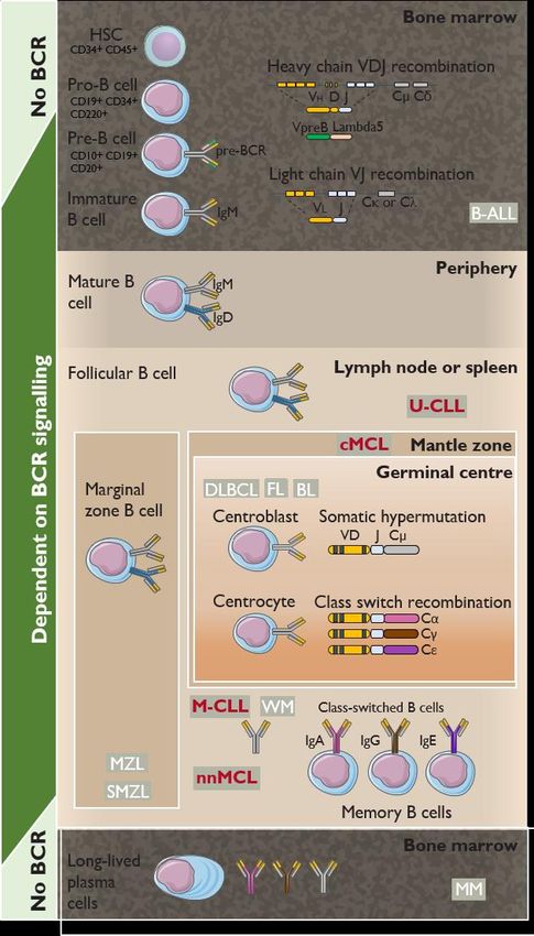
1have lost CD34 but have attained CD20, another B cell marker, in addition to CD45 and CD19. Once the pre-BCR matures into a fully functioned BCR with heavy (VDJ segments and constant region Cµ region) and light chains (VJ combination coupled with Cκ or Cλ), the pre- B cell becomes an immature B cell, expressing IgM at the cell surface (Figure 1). Immature B cells that will come out of the BM, acquire also surface IgD due to alternative splicing that will assemble the VDJ segments with a different constant region C gene (here from Cµ to Cδ) and become fully mature B cells (Abbas, et al 2014). The maturation process of B lymphocytes in BM requires the expression of the chemokine receptor CXCR4 and the cannabinoid receptor (CB) type 2. Indeed, knock-out studies in mice showed that the absence of CXCR4 resulted in impaired lymphopoiesis and reduced haematopoiesis in BM (Ma et al, 1998), and that the absence of CB2 led to a defective retention of immature B cells in BM, thus an accumulation in the blood instead (Pereira et al, 2009). The mature but naïve B cells encounter pathogens in follicles of secondary lymphoid organs such as lymph nodes (LN), spleen or Peyer’s patches in small intestine. The entry and maintenance of immune cells into the lymphoid organs are driven by the presence of chemokines such as CXCL12, CXCL13, CCL19 and CCL21, secreted by stromal cells. Chemokines are low-molecular weight proteins that have a chemo-attractive effect on cells that express the respective receptor, in this case: CXCR4, CXCR5 and CCR7 (Okada et al, 2002). In the secondary lymphoid organs, B cells become activated after antigen binding to their BCR, and the process from naïve mature B cells to fully activated competent plasma or memory cells mostly occurs in the germinal centres (GC) of these organs. It can also take place outside GC, so called extrafollicular B cell activation (Chappell et al, 2012). Germinal centres are important histological structures that also involve the presence of chemokines, and that lymphocytes express the receptors CXCR4, CXCR5 and CCR7. Knock- out of these receptors results in altered LN and spleen GC structures due to the impaired migration of lymphocytes in those areas (Förster et al, 1996, Förster et al, 1999, Müller et al, 2002). The sphingosine-1-phosphate receptors (S1PR) 1 and 2 are also participating in the presence of B lymphocytes in GC. B lymphocytes downregulate S1PR1, which is an egress receptor binding to its ligand S1P in high concentration in peripheral blood (PB), and instead express at the cell surface S1PR2 that inhibits the migration of cells, therefore keeping them in GC (reviewed in (Cyster et al, 2012)). Cells that do not encounter antigen, meaning that the BCR is not activated, will upregulate S1PR1 and egress from the LN (Arnon et al, 2013). The B cell in the GC proliferates in the “dark zone” of the GC first and acquire somatic mutations in the variable region of the heavy and light chains (hypermutation), in order to possess a wide variety of BCR to recognize as many antigens as possible. The proliferating and activated B cells are called centroblasts. Once the centroblasts have proliferated, they will arrive at the “light zone” where follicular dendritic cells will display the specific antigen for high affinity selection of the specific B cell clone that recognizes that antigen. In addition, the B lymphocyte will interact with T lymphocytes via different mechanisms, including the binding of CD40 to CD40 ligand on T lymphocyte, and the major histocompatibility complex (MHC) 2
II to T cell receptor. These stimuli induce the clonal expansion and Ig class switch resulting in
the expression of a new heavy chain isotype (Cα, Cγ or Cε). These clones express IgA, IgG
and IgE, which provides other functions of the Ig such as Fc-receptor binding, localization to
mucosal areas (IgA), and binding to mast cells and eosinophils (IgE), therefore activating a
relevant type of immune response (Abbas, et al 2014, Cyster et al, 2019).

These mature cells expressing different types of Ig are smaller in size and are called
centrocytes. The centrocytes will leave the GC to become memory B cell or antibody secreting
plasma cells, which are both long lived cells (Reviewed in (Cyster and Allen 2019, LeBien et
al, 2008)).

During this development process, checkpoints are in place to ensure that the BCR recognizes
antigens with sufficient strength, and on the other hand, that the BCR does not recognize self-
antigens that would damage the surrounding cells and lead to autoimmune diseases. In such
cases, the B cell will go through another round of Ig genes rearrangement or be eliminated by
apoptosis (Melchers 2015).

                                                                                               3
Figure 1. Representation of key steps of the B cell development and different B-cell lymphoma that can arise from cells at the different stage of their differentiation. Abbreviations: BL, Burkitt lymphoma; cMCL, classical mantle cell lymphoma; DLBCL, diffuse large B cell lymphoma; FL, follicular lymphoma; HSC, haematopoietic stem cell; MCL, mantle cell lymphoma; M-CLL, mutated chronic lymphocytic leukaemia; MM, multiple myeloma; MZL, marginal zone lymphoma; nnMCL, non-nodal mantle cell lymphoma; SMZL, splenic marginal zone lymphoma; U-CLL, unmutated chronic lymphocytic leukaemia; WM, Waldenstrom macroglobulinaemia (adapted with permission from Burger, J.A. & Wiestner, A. (2018) Targeting B cell receptor signalling in cancer: preclinical and clinical advances. Nat Rev Cancer, 18, 148-167). 4
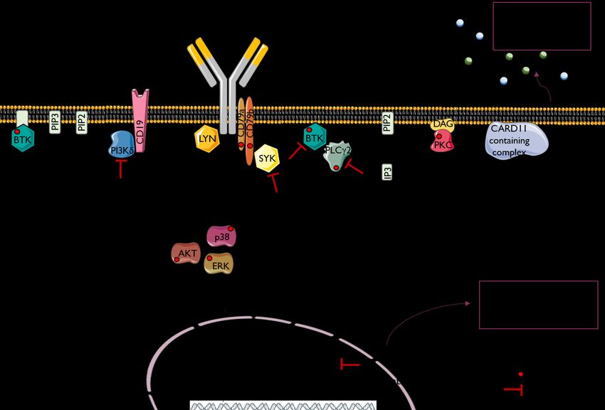
1.2    DOWNSTREAM SIGNALLING OF BCR
Signalling through the BCR is essential for B cell survival and proliferation, and knocking
down the BCR results in cell apoptosis (Lam et al, 1997). After binding of antigen, the BCR
encounters a conformational change that will activate the SRC protein tyrosine kinases family,
such as LYN. These kinases will phosphorylate the two signal transduction proteins CD79A
and CD79B that are coupled to the BCR intracellular domain. This will recruit SYK protein
that will transduce the signal by activating the BCR co-receptor CD19 together with other
adaptor proteins, and recruiting phosphoinositide 3-kinase (PI3K) to the plasma membrane.
There, PI3K activation will generate the second messenger phosphatidylinositol-3,4,5-
triphosphate (PIP3) that is important for the activation of the Bruton’s tyrosine kinase BTK and
its downstream target phospholipase Cγ2 (PLCγ2). Several signalling cascades are then
activated, including calcium mobilization and activation of protein kinase C (PKC) and
CARD11 containing complex, which lead to activation of the transcription factor NFκB. NFκB
will induce the release of cytokines, such as CCL3 and CCL4, and activate pathways involved
in survival (Sasaki et al, 2016), cell adhesion (Spaargaren et al, 2003) and migration (de Gorter
et al, 2007). Several other signalling pathways are also activated upon BCR stimulation, such
as the serine/threonine kinase AKT and the mitogen-activated protein kinases (MAPK)
ERK1/2 and p38, all regulating cell proliferation and survival (Reviewed in (Burger and
Wiestner 2018, DeFranco 2015, Efremov et al, 2020, Seda et al, 2015); Figure 2).

Figure 2. Scheme representing the main signalling molecules involved upon BCR activation (inspired and adapted
with permission from Jerkeman, M. et al., (2017) Targeting of B-cell receptor signalling in B-cell malignancies.
Journal of internal medicine).

                                                                                                              5
1.3 B CELL MALIGNANCIES
B cell malignancies can arise from any steps of the B cell development, as highlighted in Figure
1. It can be divided in two types: Hodgkin and non-Hodgkin (NHL) lymphoma. Mantle cell
lymphoma (MCL) and chronic lymphocytic leukaemia (CLL) are NHLs, both with
characteristics of a mature B cell, expressing CD5 and IgM at the cell surface. They are two
lymphoma entities with very similar characteristics but also some disparities, summarized in
Table 1 below.
Table 1. Characteristics of MCL and CLL.
MCL CLL
CD5+ CD19+
Immunophenotype
CD23-; CD20+ (bright) CD23+; CD20+ (dim)
100 cases per year in Sweden 500 cases per year in Sweden
Incidence
(in Europe 1-2 per 100,000 person) (4 per 100,000 person)
Prevalence 2-10% NHL
Survival 5 to 8 years 6 to >10 years
Median age 65-year-old 72-year-old
Male:Female ratio 3:1 2:1
rituximab + chemotherapy combination
Therapy
ibrutinib idelalisib, ibrutinib, venetoclax
t(11;14) tri(12), del(13q)
Frequent genetic
aberrations somatic mutation on ATM and TP53 genes together with del(11q) and/or
del(17p)
BCR dependent on the BCR signalling
SOX11 SOX11+ SOX11- SOX11-
IGHV IGHV mutated, IGHV
unmutated, TP53 mutation, unmutated, IGHV mutated,
IGHV mutation
classical nodal non-nodal shorter longer survival
MCL leukemic cases survival
CB1
CB1 CB1 negative overexpression in 50% cases
overexpression
CB2 overexpression in 100% cases overexpression in 90-95% cases
61.3.1 Mantle Cell Lymphoma
Mantle cell lymphoma (MCL) is aggressive and incurable lymphoma with a median survival
of only 5-8 years (Abrahamsson et al, 2014, Jain et al, 2019, Nygren et al, 2012). MCL cells
have the morphological characteristics of mantle zone B cells.
1.3.1.1 Genetic characteristics
The first genetic aberration that defines MCL is the chromosomal translocation
t(11;14)(q13;q32), which occurs at the pre-B cell stage, when the V(D)J segments are being
joined to form the Ig (Küppers et al, 2001, Nadeu et al, 2020). This translocation places the
gene encoding for cyclin D1 under the control of the IGH enhancer, leading to cyclin D1
overexpression, which promotes transition from G1 to S phase in the cell cycle. Since more
than 95% of MCL tumours express cyclin D1, while normal lymphocytes and most other
lymphomas are cyclin D1 negative, immunohistochemical staining for cyclin D1 (Figure 3A)
is part of the diagnostic work-up and the fluorescence in situ hybridization (FISH) method for
detecting the translocation (Li et al, 1999) (Figure 3B). However, carrying this chromosomal
translocation isn’t sufficient to develop MCL as cells with the t(11;14) have been detected by
sensitive PCR method in 1-7% of healthy individuals without consequences (Hirt et al, 2004,
Lecluse et al, 2009). Other genetic aberrations are therefore necessary and happen later in the
B cell development, such as deletion or mutations of ATM (Campo et al, 2015), which is
important for DNA damage recognition. Other additional cellular functions are dysregulated
in MCL, including defects in DNA-repair, cell cycle regulation and apoptosis (reviewed in
(Jares et al, 2012)). SOX11 has also become a diagnostic marker since it is expressed in 90%
of MCL cases and is important for the distinction from CLL (described in the next section) in
the rare cyclin D1 negative MCL cases (Wasik et al, 2015a).
1.3.1.2 Clinical presentation
MCL is usually first detected in LN, but BM involvement is frequent, and the disease can also
involve the spleen or gastro-intestinal tract. Two subtypes of MCL can be distinguished:
classical/conventional nodal MCL (cMCL) and leukemic non-nodal MCL (nnMCL)
(Swerdlow et al, 2016).
cMCL is characterized by SOX11 expression and it is believed that the cells have not entered
the GC (reviewed in (Ghia et al, 2017)). Instead, these cells locate in the mantle zone, and do
not experience rearrangement of the variable region of Ig heavy chain (IGHV), and therefore
present an unmutated IGHV gene (>98% homology with the germline sequence). This subtype
can evolve into a more aggressive variant (blastoid MCL), which is characterized by a larger
cell size, high proliferation (Ki67+), TP53 mutations and multiple genetic alterations (Beà et
al, 1999). TP53 can also be mutated in the cMCL, which is then associated to worse outcome
((Hernandez et al, 1996) and reviewed in (Sander et al, 2016)).
7nnMCL was first classified as atypical CLL, because of the leukemic presentation rather than LN engagement, and presence of mutated IGHV genes (Orchard et al, 2003). However, as the malignant cells in the blood carry the t(11,14), which is the hallmark of MCL, the CLL diagnosis was revised. It is now well known that nnMCL has a distinct genomic signature, with fewer chromosomal alterations than cMCL ((Fernàndez et al, 2010, Royo et al, 2012); Figure 3C). Based on these findings, a molecular assay has recently been developed by Clot et al. to identify and discriminate between cMCL and nnMCL in situations where clinical and biological characteristics were not sufficient to determine the subtype of MCL (Clot et al, 2018). Among the identified genes, the cannabinoid receptor type 1 (CNR1 encoding for the protein CB1, described in more details in sections 1.4.4.1 and 1.6) is described as highly expressed in cMCL and at a low expression level in nnMCL. This molecular assay could be used in the decision of the treatment strategy, as patients with the indolent nnMCL form do not require treatment until symptoms, but most cMCL, also those with leukemic presentation will need therapy. Figure 3. Characteristics of MCL. A. Cyclin D1 staining in MCL case visualized by positive staining (brown) detected in the nuclei. The staining is normally variable with both weakly and strongly positive cells (original magnification x10). B. FISH staining using probes targeting chromosome 14 (green) and chromosome 11 (red), yellow signal represents gene chromosomal fusion (original magnification x63). C. chromosomal alterations differences between cMCL and nnMCL. (A and B were kindly provided by Prof. Birgitta Sander; C was obtained with permission from Fernàndez, V. et al., (2010) Genomic and gene expression profiling defines indolent forms of mantle cell lymphoma. Cancer Res, 70, 1408-1418.). 8
1.3.1.3 Treatment options
The conventional treatment for MCL is R-CHOP regimen, which consists of rituximab
antibody targeting CD20 together with a chemotherapy combination (cyclophosphamide,
doxorubicin, vincristine and prednisone). Today, most young patients are treated with an
intensified R-CHOP alternating with high dose of cytarabine, followed by stem cell rescue, so
called autologous stem cell transplantation. This options has been reported to increase the
overall survival for young and fit patients (Dreyling et al, 2005, Eskelund et al, 2016, Geisler
et al, 2008, Kolstad et al, 2017). Recently, targeting the BCR signalling pathway became a new
treatment option (e.g. BTK inhibitors ibrutinib or acalabrutinib). Also inhibitors of Bcl-2 and
cyclin-dependent kinases are under investigation (reviewed in (Jerkeman et al, 2017)).
However, these targeted therapies are still given mostly to relapsed MCL patients (Dreyling et
al, 2018).

Although nnMCL has a longer survival of 7-10 years, compared with 3-5 years for cMCL,
MCL is still an incurable disease.

1.3.2 Chronic Lymphocytic Leukaemia
Chronic lymphocytic leukaemia (CLL) is a common subtype of lymphoma/leukaemia. It is a
heterogenous disease which results in various clinical outcome (Hallek 2019).

1.3.2.1 Genetic characteristics
CLL is characterized by four main chromosomal abnormalities: deletion of long arm of
chromosome 11 (del(11q)), which deletes ATM gene involved in DNA damage detection, like
for MCL; trisomy 12 (tri12); del(13q) which deletes the micro-RNA miR15a and miR16-1 that
are normally silencing the gene encoding for the anti-apoptotic Bcl-2 protein (Calin et al, 2002,
Cimmino et al, 2005); and deletion of TP53 on the short arm of chromosome 17 (del(17p),
(Döhner et al, 2000), Figure 4). All these alterations promote cell survival and proliferation. A
worse prognosis is seen when del(11q) and del(17p) are combined with somatic mutations in
the remaining ATM and TP53 genes (Zenz et al, 2010). Twenty percent of CLL cases, however,
do not carry any of these four genetic aberrations (Döhner, et al 2000).

The mutation status of IGHV gene is also a prognostic factor in CLL, with unmutated IGHV
(U-CLL) associated to a more aggressive disease course (Hamblin et al, 1999, Oscier et al,
2002). Gene sequencing analysis of the IGHV gene is now considered part of the diagnostic
routine for CLL (Davi et al, 2020, International CLL-IPI working group. 2016, Langerak et al,
2011, Rosenquist et al, 2017) and because it has been done already in a large proportion of
cases, it is now possible to evaluate and get a risk stratification of CLL disease course and
overall survival according to IGHV mutation status (International CLL-IPI working group.
2016). The mutation status of IGHV and the presence of specific sequences help to predict
therapy responses and outcome (Fischer et al, 2017, Sutton et al, 2017).

                                                                                               9
Figure 4. The most common genetic alterations in CLL (blue box represents loss of gene and orange box overexpression of the gene; inspired by Hallek, M., Shanafelt, T.D. & Eichhorst, B. (2018) Chronic lymphocytic leukaemia. Lancet, 391, 1524-1537). 1.3.2.2 Clinical presentation As for MCL, CLL cells proliferate in BM and in secondary lymphoid organs, but also accumulate in PB. For a CLL diagnosis, the threshold of B lymphocyte count is at least 5x10⁹ cells per litre of blood. CLL is a heterogenous disease, that can present as a pre-leukemic form (small cell lymphocytic lymphoma), an indolent form, or be progressive and resistant to therapy. The severity of the disease is measured according to the Rai and Binet staging system, including the assessment of symptoms, clinical parameters such as anaemia, thrombocytopenia, lymphadenopathy (Hallek et al, 2018), presence of genetic aberrations and as mentioned above the IGHV mutation status. Interestingly, CLL cells that are found in the PB are less activated compared with cells in BM and LN as the gene signature was enriched for the NFκB pathway in CLL cells from LN (Burger and Wiestner 2018, Herishanu et al, 2011). 1.3.2.3 Treatment options The standard therapy for CLL includes a chemotherapy cocktail, mostly fludarabine and cyclophosphamide, combined with anti-CD20 antibody rituximab. However, targeted therapy is more used in CLL, especially the BTK irreversible inhibitor ibrutinib (reviewed in (Smith 2017)). Other signalling pathways inhibitors such as the PI3K inhibitor (idelalisib), the Bcl-2 inhibitor (venetoclax) or SYK inhibitors have shown promising results (reviewed in (Jerkeman, et al 2017)). 10
In both MCL and CLL, disease progression and relapse after treatment are very common, due
to resistances to treatment (Ondrisova et al, 2020), which can be intrinsic or acquired (Zhao et
al, 2017). Resistances can arise when malignant cells try to escape by mutating the region in
the drug-binding sites to avoid recognition from it. For instance, after ibrutinib, a point mutation
(BTK C481S) reduces the binding affinity of ibrutinib for BTK. Other types of mutations have
been described such as gain-of-function mutations on PLCγ2 gene, which allows a BCR
signalling independent of BTK activation (Woyach et al, 2014).

Relapses are common also in patients with minimal residual disease. It is hypothesised that
minimal residual disease is due to a few lymphoma cells that reside in tissues, supported by
non-malignant cells in the microenvironment and receiving survival signals that make them
resistant to therapies (Burger et al, 2011, Kurtova et al, 2009, Medina et al, 2012).

1.4   LYMPHOMA MICROENVIRONMENT
Like many cancer types, lymphoma cells need the signals and interactions with surrounding
non-malignant cells and other components, called the microenvironment, to survive and
proliferate. One striking clue about the importance of the microenvironment came from studies
that demonstrated that primary MCL or CLL cells survived up to several months in co-culture
with stromal cells, compared with just few days when cultured alone (Medina, et al 2012,
Panayiotidis et al, 1996). Several actors are involved in the communication between lymphoma
cells and non-malignant cells in the tissue microenvironment, and the main ones are described
below and presented in Figure 5.

1.4.1 The B Cell Receptor
The BCR signalling is a key-player in both MCL and CLL. The initial role of the BCR in a
normal B lymphocyte is to recognize the antigen that will stimulate the cells into proliferation
and differentiation processes in order to produce antibodies. The malignant B cells are utilizing
the same mechanisms and pathways through the BCR for survival and proliferation.

Ibrutinib is a drug that inhibits BCR signalling by targeting the downstream tyrosine kinase
BTK. Ibrutinib irreversibly blocks BTK, which causes the egress of tumour cells out from the
tissue into the blood stream in both CLL and MCL (Chang et al, 2013), confirming the
communication between malignant and non-malignant cells via the BCR.

Indeed, in the malignant context, the BCR is continuously active and sometimes over-
stimulated as a result of the following mechanisms: i) binding of auto-antigen from the
surrounding cells in the microenvironment, ii) acquirement of activating mutations in the BCR
downstream signalling molecules, iii) antigen-independent tonic activation of the BCR
(reviewed in (Burger et al, 2014, Burger and Wiestner 2018)).

                                                                                                 11
1.4.1.1 Auto-antigen stimulation In CLL, it has been discovered that approximately one third of patients share a similar IGHV sequence, specifically the antigen recognition domain (complementarity-determining region 3; (Agathangelidis et al, 2012)). These subsets of BCR are called stereotypes. This stereotypy can be explained by the recognition of antigens that are not commonly recognized by normal B lymphocytes (low frequency of these IGHV sequences normally) as they are auto-antigens present in the microenvironment, but specifically selected by CLL cells. In MCL the same phenomenon has been reported but with different IGHV gene sequence profile compared to CLL (Hadzidimitriou et al, 2011, Sutton et al, 2013). 1.4.1.2 Activating mutations Mutations in different proteins involved in the BCR signalling pathway can occur (e.g. mutations in the gene encoding for CD79B) and lead to continuous activation of the pathway together with inhibition of the negative feedback loop that should control the BCR stimulation. However, these mutations are rare in MCL and CLL (reviewed in (Burger and Wiestner 2018)). 1.4.1.3 Tonic antigen-independent BCR signalling The third way of BCR activation is the tonic signalling, which occurs independently from antigen binding. It is mediated by PI3K pathway, and serves for the normal B lymphocytes as a survival signal when no antigen is binding (reviewed in (Rickert 2013)). Malignant B cells also use this signalling pathway, increasing the signalling by the activation of important protein kinases or the downregulation of PI3K inhibitor protein (Burger et al, 2013, Merolle et al, 2018). 1.4.1.4 Tissue localization Gene pathway analysis in both MCL and CLL demonstrated that the BCR and downstream NFκB signalling are enriched, especially within the LN, compared with PB, where there is little interaction with other cells (Herishanu, et al 2011, Saba et al, 2016). NFκB signalling regulates cell survival and proliferation (reviewed in (Burger and Wiestner 2018, Efremov, et al 2020, Ghia, et al 2017)). Indeed, non-malignant cells in the microenvironment, especially in LNs, (e.g. lymphoma-associated macrophages) provide signals that activate BCR-signalling (reviewed in (Ten Hacken et al, 2016)). 1.4.2 Non-malignant cells MCL and CLL cells in tissues interact with the surrounding cells, especially other immune cells that are communicating with normal B cells to develop immune responses upon antigen encountering (Cyster and Allen 2019). Malignant cells can reside in BM or other lymphoid tissues such as LN, where different cell types provide support for the tumour cells. The role of the non-malignant cells is very complex and still needs to be investigated further. 12
1.4.2.1 Bone marrow stromal cells
In BM, the stromal cells (also called mesenchymal stem cells, MSCs) predominantly support
CLL and MCL survival by secreting chemokines and expressing essential surface molecules
(Honczarenko et al, 2006, Li et al, 2016). Importantly, the communication between the
malignant cell and the non-malignant BM MSCs has to involve direct contact (Lagneaux et al,
1998) and is bi-directional (Ding et al, 2009). Indeed, malignant cells are attracted to MSCs,
bind to them and have the capacity to migrate beneath them, all due to the expression of
adhesion molecules and chemokine receptors that recognize chemokines secreted by MSCs
(Burger et al, 1999, Kurtova, et al 2009, Medina, et al 2012). In return, signalling pathways
such as ERK1/2, AKT and NFκB are activated in MSCs after interaction with CLL cells (Ding,
et al 2009, Lutzny et al, 2013).

In addition, MCL and CLL primary cells co-cultured with BM MSCs induce the secretion of
several soluble factors such as interleukin IL-6 (Zhang et al, 2012) and chemokines CCL3 and
CCL4 (Zucchetto et al, 2010), activating different signalling pathways that all promote
survival.

Altogether these close interactions activate survival pathways like ERK1/2 (Kurtova, et al
2009) and anti-apoptotic proteins, such as Bcl-2 (Lagneaux et al, 1999, Lwin et al, 2009, Lwin
et al, 2007), making lymphoma cells protected from therapy by MSCs.

1.4.2.2 Lymph node microenvironment
Compared with BM, the LN microenvironment has been more investigated in haematological
malignancies. It is composed of a wide variety of cells, due to the fact that it is the location for
presentation of antigen, activation, proliferation and maturation of antigen-specific B
lymphocytes. It is also where CLL and MCL proliferate the most, compared to BM and PB
(Herishanu, et al 2011, Saba, et al 2016).

T lymphocytes are an important part of the LN microenvironment. In MCL, the amount of T
lymphocytes found in LN biopsies is lower in the aggressive form of disease, and a high ratio
of CD4+/CD8+ T cells is correlated to longer overall survival in MCL (Nygren et al, 2014).
This suggests that the aggressive forms of MCL are less dependent on the microenvironment
signals.

In CLL, CD8+ T lymphocytes are enriched in blood (Herrmann et al, 1982), and cytotoxic
CD4+ T cells are attracted in LN microenvironment by CLL cells that secrete cytokines
(Hartmann et al, 2016). Interaction between B and T lymphocytes results in proliferation
signals in B cells via for instance CD40-CD40L interaction (Abbas, et al 2014, Castillo et al,
2000).

The presence of CD4+ T lymphocytes in LNs are related to a good patient outcome in MCL
(Nygren, et al 2014) and in CLL (Nunes et al, 2012), due to their anti-tumoral role (Dobrzanski
2013). However, malignant cells develop a way to exhaust T cells. Indeed, once the CD4+ T
cells interact with CLL cells, they lose their ability to form immunological synapse (Ramsay

                                                                                                 13
et al, 2012). The immunological synapse consists of changes in cytoskeleton of T cells in contact with the antigen presenting B cell via the MHC-II, or with the tumour cell via self- antigen, which is supposed to activate the T cell for cytotoxic actions (Houghton et al, 2004, Nassef Kadry Naguib Roufaiel et al, 2015). Nevertheless, this effect is inhibited by the presence of programmed cell death protein 1 (PD) ligand PD-L-1 on MCL/CLL cells (Allahmoradi et al, 2017, Wang et al, 2013). PD-L-1, which is a negative regulator of T cells in normal condition to avoid auto-immune responses (Jin et al, 2011), is then recognized by the receptor PD-1 on T cells, providing signals that abolish their function of tumour cell lysis (Wang, et al 2013). Monocytes, macrophages and nurse like cells attract malignant cells into lymph nodes and other lymphoid organs by secreting chemokines (Burger et al, 2000). In addition, these non- malignant cells are attracted by malignant cells via chemokines, such as CCL3 and CCL4 (Zucchetto, et al 2010). 1.4.3 Chemokines and adhesion molecules Many different chemokines, cytokines and other molecules, their respective receptors, as well as adhesion molecules are involved in the communication between the non-malignant cells and CLL/MCL cells, but only the most relevant ones to this thesis are described here. 1.4.3.1 Chemokine and egress receptors CXCR4, CXCR5 and CCR7 are chemokine receptors expressed in normal B lymphocytes, and they are important for their development, maturation and differentiation. These chemokine receptors are part of the superfamily of G protein coupled receptors (GPCR), and are overexpressed in MCL (Corcione et al, 2004, Kurtova, et al 2009) and CLL (Burger, et al 1999, Till et al, 2002). Malignant cells use essentially the same mechanisms as the non-malignant B cells, to promote their survival and proliferation, while diminishing the effects of the opposite mechanisms such as apoptosis. In a healthy setting, CCR7 ligand (CCL21) is responsible for the entry of lymphocytes into LN, CXCR5 ligand (CXCL13) for the positioning in follicular areas in LN and CXCR4 ligand (CXCL12) for the entry and maintenance of B cells in BM. In lymphoma, the expression of these receptors and ligands is also different according to the tissue compartments, in favour of cell retention (Middle et al, 2015). This is explained by the lymphoma tissue microenvironment secreting these ligands, creating a gradient and attracting cells expressing the receptors, therefore playing a major role in homing of lymphoma cells into tissues. 14
In MCL and CLL, all three chemokine receptors (CXCR4, CXCR5 and CCR7) are upregulated
at the cell surface (reviewed in (Burger and Ford 2011, Burger and Gribben 2014)). In CLL,
the high levels of CCR7 and CXCR4 could at least partially be explained by the enhanced
recycling of the receptors compared with non-malignant B lymphocytes (Patrussi et al, 2015).

Additionally, there is a balance, or unbalance in the case of malignancy, of expression between
the chemokine receptors that induce the retention of cells in tissues, and S1PR1 which promotes
their egress (Cyster and Schwab 2012, Patrussi, et al 2015).

S1PR1 is involved in the egress of normal B lymphocytes from LN into PB, where its ligand,
the biolipid S1P, is in high concentration. S1PR1 is expressed in most MCL cases (Nishimura
et al, 2010), with a different expression level observed in different tissue of origin derived MCL
cell lines (Sadeghi et al, 2020). S1PR1 gene is also found mutated in 8% of MCL cases,
inducing in most cases the reduction of S1PR1 expression, correlating with advanced stages of
the disease and promoting the retention of cells within the tissue (Wasik et al, 2018). In CLL,
the expression of S1PR1 is often impaired, and its surface expression is even more reduced in
vitro upon several stimuli like BCR stimulation with anti-IgM antibody or CD40-CD40L
interaction (Borge et al, 2014, Till et al, 2015).

Both CXCR4 and CXCR5 were shown to be involved in the migration of malignant cells
beneath MSCs, thus protecting them from therapies (Burger, et al 1999, Kurtova, et al 2009).
CXCR4 expression is important for the malignant cell survival as it was demonstrated that
culturing MCL cell lines with its ligand CXCL12 increased survival compared with medium
alone, and that knock-down of CXCR4 reduced cell proliferation (Chen et al, 2016b). In
addition, CXCR4 expression is dynamic, allowing the cells to recirculate from LN to PB and
vice-versa (Chen et al, 2016a). CXCR7 is also a receptor for CXCL12 but is mostly involved
in cell adhesion rather than migration (Burns et al, 2006) and is not expressed in leukocytes
(Berahovich et al, 2010).

Because all interactions are dynamic, cross-activation of specific downstream signalling
pathways happens, as for instance the phosphorylation and activation of BTK by CXCL12 in
CLL (Montresor et al, 2018, Nore et al, 2000), involving both CXCR4 and BTK signalling
pathways in adhesion of cells. In MCL, we found that these signalling pathways are differently
activated in different cell lines (Sadeghi, et al 2020). Interaction between BCR and CXCR4
signalling is also suggested by the fact that CLL and MCL cells are released in PB after
ibrutinib treatment (Chang, et al 2013), due to the inhibition of surface CXCR4 expression
(Chen, et al 2016a).

Also, BCR signalling results in secretion of CCL3 and CCL4 from CLL, which will further
attract non-malignant immune cells such as monocytes and T cells as mentioned earlier (Burger
et al, 2009).

                                                                                               15
1.4.3.2 Adhesion molecules Adhesion molecules are participating in cell-to-cell adhesion, as well as cell-to-extracellular matrix (ECM) contacts. They are part of the process of homing and retention of B lymphocytes into lymphoid tissue. There are several types of adhesion molecules, categorized based on their structures. Integrins are transmembrane receptors composed of two subunits: α and β. Different α and β subunits can be paired together, creating a variety of different integrins. The very late activation antigen (VLA-4, also known as CD49d) is a α4β1 integrin expressed on leukocytes. It can recognize ECM components such as fibronectin and vascular cell adhesion molecule-1 (VCAM-1). CD49d is also expressed at the cell surface of MCL and CLL cells. This integrin is part of the mechanism used by non-malignant and malignant lymphocytes to migrate beneath the MSCs (Burger et al, 2001, Kurtova, et al 2009, Miyake et al, 1992). In malignancies, VLA-4 is therefore associated with nodal and extra-nodal involvement (Strati et al, 2017, Terol et al, 1999) and worse prognosis due to drug resistance (Kurtova, et al 2009, Shanafelt et al, 2008). Another adhesion molecule of importance is ICAM-1, expressed mainly on stromal cells but also on some leukocytes, and in some of MCL and CLL cases (Jacob et al, 1999, Molica et al, 1995). ICAM-1 is upregulated upon adhesion to stromal cells in co-culture in vitro system (Sadeghi, et al 2020), validating the fact that its expression is higher in MCL and CLL samples from LN compared with PB (Arvidsson et al, 2018). 1.4.4 Cannabinoid receptors and ligands 1.4.4.1 Cannabinoid receptors Gene expression analysis comparing LN biopsies from MCL patients and non-malignant cells from reactive tonsils and LNs described for the first time the overexpression of cannabinoid receptors (CB) type 1 and type 2 in MCL (described in section 1.6.1; (Ek et al, 2002, Islam et al, 2003)). Screening of different NHL subtypes identified CB1 and CB2 overexpressed in most of the B cell lymphoma subtypes included in the study such as CLL, marginal zone, follicular and diffuse large B cell lymphomas (Gustafsson et al, 2008, Rayman et al, 2007). The mRNA expression of CBs had already been identified in human immune cells in the 1990s (Bouaboula et al, 1993, Galiegue et al, 1995) after the publication of earlier reports describing effects of the CB ligands from the marijuana plant (delta-9-tetrahydrocannabinol, THC) in immune cells function, hypothesizing that the receptors should then be expressed on those cells. Both CB1 (encoded by CNR1 gene) and CB2 (encoded by CNR2 gene) mRNA expression could be quantified in lymphoid organs and immune cell subtypes. CNR2 is there expressed at higher levels compared to CNR1, up to 100-fold increase, and the highest expression levels for both receptors were found in tonsil, and B lymphocytes (Galiegue, et al 1995). 16
In MCL and CLL, the range of expression measured by quantitative PCR is wide for CNR1
(relative fold increase (RFI) compared to non-malignant B cells from 0.63 to nearly 5000) and
CNR2 (RFI from 1.06 to nearly 200), with CNR1 reaching the levels of CNR2 for some cases,
if not higher (Wasik et al, 2014). Protein expression was also confirmed in patient samples and
in MCL cell lines (Gustafsson, et al 2008, Islam, et al 2003). CNR1 has two splice variants in
addition to the full-length, which causes impaired binding to endocannabinoids (Ryberg et al,
2005). However, only splice variant CNR1a and not CNR1b seems to be expressed in MCL,
and at very low levels since it was not detected on the first round of PCR but instead on the
second round of PCR performed on the product from the first round (Gustafsson, et al 2008).

The role of the CBs in lymphoma is not yet deciphered. However, analysis of CB expression
and clinical data from a study in our group showed that low CNR1 levels correlated to
lymphocytosis (>5x10⁹ lymphocytes/litre of blood) (Wasik, et al 2014). CNR1 was also found
at lower levels in indolent (low progression of disease) MCL (Fernàndez, et al 2010), and
confirmed recently from gene expression analysis comparing cMCL and nnMCL profiles, in
which CNR1 is downregulated in leukemic nnMCL compared with cMCL (Clot, et al 2018).
Therefore, it is hypothesized that CB1 might be involved in homing, retention and/or egress of
the malignant B-cells from the tissue to the PB.

In CLL, low CNR1 cases displayed more frequently unmutated IHGV genes (U-CLL), and low
CNR1 expression levels correlated to better overall survival (Freund et al, 2016).

1.4.4.2 Endogenous ligands to cannabinoid receptors
In addition, endogenous ligands to cannabinoid receptors (endocannabinoids, described in
further detail in chapter 1.6) are released on demand by MSCs in BM (Kose et al, 2018).
Endocannabinoids might play a role in the tumour microenvironment as their expression levels
are found dysregulated in plasma of cancer patients compared with healthy individuals, as well
as at the site of tumour in mice models (Sailler et al, 2014, Zhang et al, 2016).

                                                                                            17
You can also read


