The role of imaging in predicting the development of rheumatoid arthritis - De Gruyter
←
→
Page content transcription
If your browser does not render page correctly, please read the page content below
R H E U M AT O L O G Y A N D I M M U N O L O G Y R E S E A R C H
Review • DOI: 10.2478/rir-2021-0007• 2(1) • 2021 • 27–33
The role of imaging in predicting the development of
rheumatoid arthritis
Ho So, Isaac Cheng, Lai-Shan Tam*
Department of Medicine and Therapeutics, The Prince of Wales Hospital, The Chinese University of Hong Kong, Shatin, Hong Kong, China
Received January 4, 2021 accepted February 19, 2021
Abstract
Rheumatoid arthritis (RA) remains a chronic debilitating disease with a significant negative societal impact, despite
the expanding landscape of treatment options. This condition is often preceded by a phase of systemic autoimmunity
with circulating autoantibodies, elevated pro-inflammatory cytokines, or subtle structural changes. The capability of
identifying individuals in the preclinical phase of RA disease makes a “preventive window of opportunity” possible.
Much recent work has focused on the role of imaging modalities including ultrasound (US), magnetic resonance imaging
(MRI), and high-resolution peripheral quantitative computer tomography (HR-pQCT) in identifying at-risk individuals
with or without early joint symptoms for the development of inflammatory arthritis. This article will review the evidence
and discuss the challenges as well as opportunities of proactive risk assessment by imaging in RA.
Keywords
rheumatoid arthritis • ultrasound • magnetic resonance imaging • high-resolution peripheral quantitative computer tomography
Introduction by sensitive imaging techniques, namely, ultrasound (US),
magnetic resonance imaging (MRI), and high-resolution
Rheumatoid arthritis (RA) is a common chronic systemic peripheral quantitative CT (HR-pQCT). US can be regarded
inflammatory condition characterized by persistent synovitis as an extension of the clinical examination in real-time,
and bone erosions. The uncontrolled disease can lead to joint whereas the primary advantage of MRI is the possibility
destruction, functional disability, decreased quality of life, to visualize bone marrow abnormality. They both have no
cardiopulmonary complications, and a shortened lifespan.[1–6] ionizing radiation and can be used during pregnancy. While
The outcomes of patients with RA have been revolutionized MRI is limited by its long examination time and high cost,
by early diagnosis and aggressive treatment strategy based the main drawback of US is its operator dependency.[12] HR-
on the treat-to-target approach.[7, 8] However, RA remains pQCT is a novel three-dimensional (3D) imaging technique for
a lifelong incurable disease associated with the burden of detailed bone microstructure analysis. With an isotropic voxel
long-term therapy and debilitating disease flares for most size of 61 or 82 mm, it is capable of offering high-resolution
patients. It also carries substantial socioeconomic costs. imaging (100 or 142 mm, respectively) at the peripheral
[9]
Currently, therapy aims to achieve clinical remission.[10] sites.[13] It was originally designed to assess volumetric bone
With the development of effective targeted therapies, future mineral density (vBMD) and microarchitectural abnormalities
ambitions will be either to prevent RA or to achieve drug-free in the distal tibia and radius. In the past decade, HR-pQCT
remission, effectively a cure. All these are only possible if we has been increasingly applied to study local anabolic (e.g.,
can identify the robust predictors of progressive disease in osteophytes and enthesiophytes) and catabolic (e.g., erosions)
at-risk individuals and intervene early. bone changes and joint space parameters, mainly in the
metacarpophalangeal (MCP) joints in patients with arthritis. In
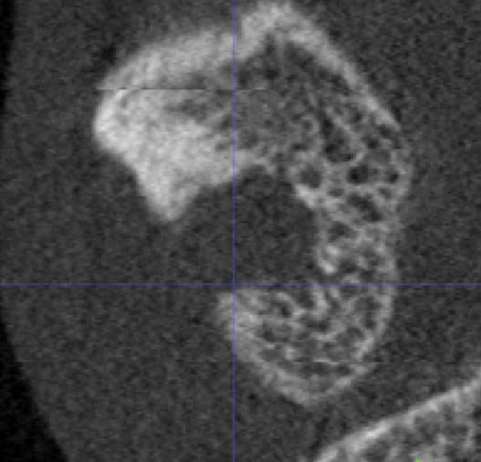
Synovitis and bone loss are the hallmarks of RA. They are patients with RA, it exhibited higher sensitivity compared with
crucial in the pathogenesis, diagnosis, and prognosis of the other imaging modalities and has been regarded as the gold
disease. It is traditionally believed that synovitis promotes the standard for detecting bone erosions (Figure 1).[14] The juxta-
release of pro-inflammatory cytokines, which subsequently and intraarticular vBMD and microarchitectural abnormalities in
activate osteoclasts and enhance bone resorption at RA can also be ascertained by HR-pQCT.[15, 16] Unfortunately,
vulnerable anatomical sites leading to bone loss and thus only extremities can be scanned at the moment, due to the
joint damage.[11] This concept has been challenged by recent limitation of the gantry size.
findings that bone changes or tendinitis could occur very
early in the course of RA, even in the preclinical phases of The potential to identify the subclinical features, which are
the disease. All these abnormalities can now be detected predictors for the future development of RA by imaging,
Address for correspondence:
*Lai-Shan Tam, Department of Medicine and Therapeutics, The Prince of Wales
Hospital, The Chinese University of Hong Kong, Shatin, Hong Kong , China.
E-mail: lstam@cuhk.edu.hk
27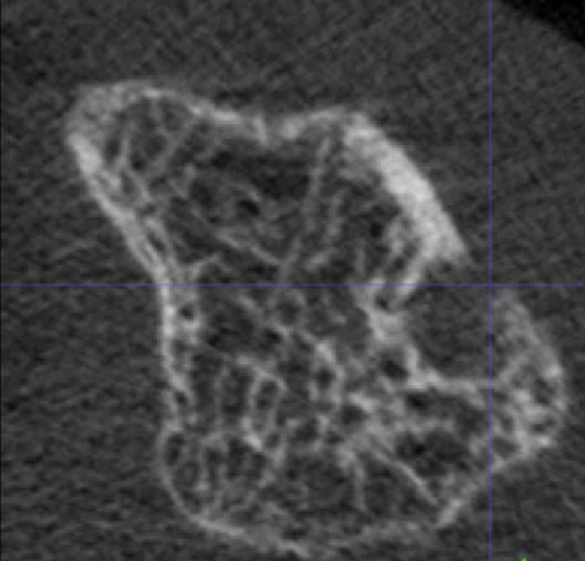
RHEUMATOLOGY AND IMMUNOLOGY RESEARCH
Review • DOI: 10.2478/rir-2021-0007• 2(1) • 2021 • 27–33

   A.

               Axial                                       Sagittal                                 Coronal

  B.                                                                                              c.

               Before segmentation                         After segmentation                           Erosion

          Figure 1. Example of erosion identification and quantification on HRpQCT
Figure 1. Example of erosion identification and quantification on HR-pQCT. (A) Identifying erosion in the axial, sagittal, and coronal planes;
          A. Identifying erosion in axial, sagittal and coronal plane; B. Example of segmentation of
(B) Example of segmentation of remaining bone; and (C) Erosion area quantified. HR-pQCT, high-resolution peripheral quantitative computer
tomography.
          remaining bone; C. Erosion area quantified
raises the opportunity to prevent disease development                    without any statistical analysis. Table 1 summarizes the
or progression in these individuals. Studying these early                sample size, follow-up duration, patient characteristics, and
structural changes could also improve our understanding of               main results of the studies identified.
the pathogenic mechanism of inflammatory arthritis. In this
review, we aim to summarize and discuss the recent literature,           Ultrasound
covering the use of US, MRI, and HR-pQCT in predicting the
development of RA in at-risk individuals.                                US can sensitively detect RA changes such as early bone
                                                                         erosions, subclinical synovitis (manifested as synovial
Methods                                                                  thickening and/or abnormal power Doppler signal), and
                                                                         tenosynovitis.[17] In a cohort of 136 anti-cyclic citrullinated
Articles included in this review were searched using                     peptide    antibody     (ACPA)-positive    individuals     with
the PubMed platform. Full-text English-language article                  musculoskeletal symptoms but no clinical synovitis, the
searches were conducted using combinations of items,                     presence of intraarticular power Doppler signal and erosion
including “ultrasound,” “MRI,” “Magnetic resonance imaging,”             on US over any of the 32 joints (wrists, MCP joints, proximal
“computed       tomography,”    “high-resolution  peripheral             interphalangeal joints, and metatarsophalangeal [MTP]
quantitative CT,” “rheumatoid arthritis,” “predict,” and                 joints) was strongly (both P < 0.001) associated with the
“prediction.” The search results were supplemented by                    development of inflammatory arthritis after a median follow-
reference citations from notable reviews on this topic. The              up of 18.3 months.[18] In another seropositive arthralgia
search strategy was done till 31 August 2020. A narrative                cohort (n = 163) with a median follow-up of 12 months,
review of findings from the literature search was performed              baseline synovial thickening was detected in 30% of

28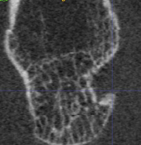
RHEUMATOLOGY AND IMMUNOLOGY RESEARCH
                                                                                                          Review • DOI: 10.2478/rir-2021-0007• 2(1) • 2021 • 27–33

Table 1. Summary of evidence on various imaging modalities in predicting the development of inflammatory arthritis

               First author      Sample size   Duration of follow-up     Subjects recruited                        Main results
  US           Nam JL18          136           Median 18.3 months        Musculoskeletal symptoms,                 Doppler signal and erosion over hand and
                                                                         ACPA positive,                            foot joints associated with development of
                                                                         no clinical synovitis                     inflammatory arthritis
               Van Beers-Tas     163           Median 12 months          Arthralgia,                               Synovial thickening of hand joints
               MH19                                                      RF or ACPA positive,                      associated with development of clinical
                                                                         no clinical arthritis                     arthritis
               Filer A20         58            18 months                 Clinical synovitis at least one joint,    Synovial thickening of wrists and MCPJ,
                                                                         symptom duration
RHEUMATOLOGY AND IMMUNOLOGY RESEARCH
Review • DOI: 10.2478/rir-2021-0007• 2(1) • 2021 • 27–33
autoantibodies, US-detected synovial thickening was also study (n = 31) on a similar population of undifferentiated
shown to be the only predictor of evolution to RA, among arthritis with a median follow-up of 15 months, wrist synovitis
other clinical variables including inflammatory markers, after and erosions were associated with the final diagnosis of
a mean follow-up of 18 months.[22] Of note, a recent study in RA.[29] In another study of 150 patients with recent-onset
ACPA-positive patients with arthralgia but no clinical synovitis arthralgia clinically suspected to progress to RA over time as
revealed that bone erosion on US was predictive of the judged by rheumatologists (clinically suspect arthralgia), MRI
development into inflammatory arthritis.[23] This is by far the inflammation over hands and feet as reflected by synovitis,
largest study (n = 400) with the longest follow-up duration bone marrow edema, and tenosynovitis was independently
(median 41.4 months). The most intriguing findings were that associated with arthritis development after a median follow-
the prevalence of bone erosion was significantly higher in up of 6.3 months.[30] It was subsequently found that adding
the 5th MTP joints than in the MCP joints, and the presence feet to hands MRI did not increase the accuracy of predicting
of bone erosion in more than one joint was the strongest arthritis development in patients with arthralgia.[31] Compared
imaging predictor (odd ratio = 10.6) for the development of with subclinical inflammation, the clinical value of MRI-
inflammatory arthritis. To conclude, US-defined synovial detected bone erosions might be more doubtful. In a large
thickening, power Doppler signal, tenosynovitis, and bone cohort of patients with joint pain but no clinically overt arthritis
erosion over peripheral joints appear to have predictive value (n = 490), although MRI erosion scores were higher in ACPA-
for inflammatory arthritis. positive than negative patients and were correlated with
subclinical inflammation, they were deemed not independently
On the contrary, there are some important considerations to predictive of inflammatory arthritis development.[32] Erosion
be borne in mind before indiscriminate use of US in at-risk scores were associated with arthritis development, but not
populations. First, the subclinical inflammation detectable by after adjustments for age and subclinical inflammation. In
US might be a late feature in the development of inflammatory sum, synovitis, tenosynovitis, and bone marrow edema over
arthritis. Serial US assessments in a cohort of ACPA-positive the hands detected by MRI could predict the development of
at-risk individuals showed that synovial thickening or Doppler inflammatory arthritis.
signal developed just directly before the occurrence of
clinical synovitis.[24] It was hypothesized that there was a late Due to the relatively long scanning time, limited access, and
increase in inflammatory burden before the development of lack of specificity, the use of MRI is generally recommended
arthritis as a result of a “second hit” immunogenic trigger in only in difficult patient cases at least for the management of
the at-risk individuals after a period of stability. The narrow early arthritis.[33] It is noteworthy that the commonly used MRI
window between the detection of US abnormalities and scoring system, OMERACT RA magnetic resonance imaging
clinical arthritis might not allow any meaningful intervention. scoring (RAMRIS) system, was not developed for diagnostic
Second, US acquisition protocol, definitions of pathology, purposes, but for outcome measures in clinical trials.[34]
and scoring systems varied among studies and centers.
Therefore, unified internationally recognized scoring systems HR-pQCT
should be used, such as the one endorsed by the European
League Against Rheumatism (EULAR)/Outcome Measures HR-pQCT studies on individuals with or without joint
in Rheumatology Clinical Trials (OMERACT).[25, 26] Lastly, symptoms before the diagnosis of RA are scanty. In a cross-
it is also not clear which and how many joints need to be sectional study, asymptomatic ACPA-positive individuals
imaged for optimum predictive accuracy. Comprehensive US (n = 15) had reduced bone mineral density and worsened
protocols which include most joints could take up to 60 min microarchitecture over the metacarpal heads compared
and may not be practical in most clinical settings.[20] with ACPA-negative healthy controls (n = 15) on HR-
pQCT.[35] Although no major difference between the two
MRI groups regarding the number and size of bone erosions
could be shown, intraarticular bone loss appeared to occur
MRI can detect subclinical inflammation and bone erosion, in the preclinical phases of RA as reflected by the impaired
which are indicative of RA.[27] In an early study of 129 microarchitecture in the ACPA-positive individuals. In a
patients with undifferentiated arthritis as determined longitudinal case-control study, although the baseline number
by rheumatologists, contrast MRI-proven synovitis and and size of erosions over metacarpal heads on HR-pQCT in
bone edema or erosion over hand joints in conjunction ACPA-positive patients with arthralgia (n = 29) were similar to
with autoantibodies were found to be useful in predicting the healthy controls (n = 29), both parameters worsened only
progression to RA at 1 year.[28] The positive predictive value in the patient group after 1 year.[36, 37] Out of the 22 patients
of bone edema plus ACPA positivity was 100%. In a smaller with long-term follow-up, 10 developed RA (RA progressors)
30RHEUMATOLOGY AND IMMUNOLOGY RESEARCH
Review • DOI: 10.2478/rir-2021-0007• 2(1) • 2021 • 27–33
Table 2. Comparison of ultrasound (US), magnetic resonance imaging (MRI) and high-resolution peripheral quantitative CT (HR-pQCT):
advantages and disadvantages
US MRI HR-pQCT
Advantages Can visualizes structures in real-time Can visualizes bone marrow edema Very high resolution (RHEUMATOLOGY AND IMMUNOLOGY RESEARCH
Review • DOI: 10.2478/rir-2021-0007• 2(1) • 2021 • 27–33
over the MTP5 joint were also noted to be both specific and targeted treatments aiming at preventing joint damage or
sensitive for RA.[44] A high-resolution imaging examining this even RA disease from occurring. Further detailed imaging
area for changes could be used to risk-stratify individuals may also be provocative for mechanistic researches in RA
presented with joint symptoms. 3D US technology is reported to better understand how systemic autoimmunity ultimately
to be more sensitive than conventional US, while the translates into an inflammatory joint disease. With the ever-
second-generation HR-pQCT can offer even higher image advancing musculoskeletal imaging technology and targeted
resolution and allow feet scanning.[45] The ability to identify pharmacological treatments, the two “holy grails” of RA
the earliest abnormalities is of paramount importance for the management—disease prevention and cure—may not be
implementation of any prompt, appropriate, and cost-effective out-of-reach.
Conflict of Interest
None Declared.
References
[1] Young A, Dixey J, Cox N, et al. How does Functional Disability Rheumatoid Arthritis. J Autoimmun. 2020;110:102400.
in Early Rheumatoid Arthritis (RA) Affect Patients and their [12] Carstensen SMD, Terslev L, Jensen MP, et al. Future Use of
Lives? Results of 5 Years of Follow-Up in 732 Patients from Musculoskeletal Ultrasonography and Magnetic Resonance
the Early RA Study (ERAS). Rheumatology. 2000;39:60311. Imaging in Rheumatoid Arthritis. Curr Opin Rheumatol.
[2] Toussirot E. Predictive Factors for Disability as Evaluated 2020,32:264–272.
by the Health Assessment Questionnaire in Rheumatoid [13] Klose-Jensen R, Tse JJ, Keller KK, et al. High-Resolution
Arthritis: A Literature Review. Inflamm Allergy Drug Targets. Peripheral Quantitative Computed Tomography for Bone
2010;9:519. Evaluation in Inflammatory Rheumatic Disease. Front Med
[3] Kiltz U, van der Heijde D. Health-Related Quality of Life in (Lausanne). 2020;7:337.
Patients with Rheumatoid Arthritis and in Patients with [14] Figueiredo CP, Perez MO, Sales LP, et al. HR pQCT In
Ankylosing Spondylitis. Clin Exp Rheumatol. 2009;27:S10811. Vivo Imaging of Periarticular Bone Changes in Chronic
[4] John H, Kitas G, Toms T, et al. Cardiovascular Co-Morbidity Inflammatory Diseases: Data from Acquisition to Impact
in Early Rheumatoid Arthritis. Best Pract Res Clin Rheumatol. on Treatment Indications. Modern Rheumatol. 2020, DOI:
2009;23:7182. 10.1080/14397595.2020.1804669.
[5] Wang D, Zang J, Lau J, et al. Mechanisms of Lung Disease [15] Zhu TY, Griffith JF, Qin L, et al. Structure and Strength of the
Development in Rheumatoid Arthritis. Nat Rev Rheumatol. Distal Radius in Female Patients with Rheumatoid Arthritis: A
2019;15:581–596. Case-Control Study. J Bone Miner Res. 2013;28:794–806.
[6] Zochling J, Braun J. Mortality in Rheumatoid Arthritis and [16] Zhu TY, Griffith JF, Qin L, et al. Alterations of Bone Density,
Ankylosing Spondylitis. Clin Exp Rheumatol. 2009;27:S12730. Microstructure, and Strength of the Distal Radius in Male
[7] Nell VP, Machold KP, Eberl G, et al. Benefit of Very Early Patients with Rheumatoid Arthritis: A Case-Control Study with
Referral and Very Early Therapy with Disease-Modifying Anti- HR-pQCT. J Bone Miner Res 2014;29:2118–2129.
Rheumatic Drugs in Patients with Early Rheumatoid Arthritis. [17] Wakefield RJ, Balint PV, Szkudlarek M, et al. Musculoskeletal
Rheumatology. 2004;43:906–914. Ultrasound Including Definitions for Ultrasonographic
[8] Klarenbeek NB, Güler-Yüksel M, van der Kooij SM, et al. The Pathology. J Rheumatol 2005;32:2485–2487.
Impact of Four Dynamic, Goal-Steered Treatment Strategies [18] Nam JL, Hensor EM, Hunt L, et al. Ultrasound Findings Predict
on the 5-Year Outcomes of Rheumatoid Arthritis Patients in Progression to Inflammatory Arthritis in Anti-CCP Antibody-
the Best Study. Ann Rheum Dis. 2011;70:1039–1046. Positive Patients without Clinical Synovitis. Ann Rheum Dis.
[9] Zhu TY, Li EK, Tam LS. Societal Costs of Rheumatoid Arthritis 2016;75:2060–2067.
in Hong Kong: A Prevalence-Based Cost-of-Illness Study. [19] Van Beers-Tas MH, Blanken AB, Nielen MMJ, et al. The Value
Rheumatology. 2011;50:1293-1301. of Joint Ultrasonography in Predicting Arthritis in Seropositive
[10] Smolen JS, Landewe RBM, Bijlsma JWJ, et al. EULAR Patients with Arthralgia: A Prospective Cohort Study. Arthritis
Recommendations for the Management of Rheumatoid Arthritis Res Ther. 2018;19:279.
with Synthetic and Biological Disease-Modifying Antirheumatic [20] Filer A, de Pablo P, Allen G, et al. Utility of Ultrasound Joint
Drugs: 2019 Update. Ann Rheum Dis. 2020;79:685-699. Counts in the Prediction of Rheumatoid Arthritis in Patients
[11] Scherer HU, Haupl T, Burmester GR. The Etiology of with Very Early Synovitis. Ann Rheum Dis. 2011;70:500–507.
32RHEUMATOLOGY AND IMMUNOLOGY RESEARCH
Review • DOI: 10.2478/rir-2021-0007• 2(1) • 2021 • 27–33
[21] Sahbudin I, Pickup L, Nightingale P, et al. The Role of Ultrasound- Suspect Arthralgia? A Longitudinal Study. Scand J Rheumatol.
Defined Tenosynovitis and Synovitis in the Prediction of Rheumatoid 2020;49:461–467.
Arthritis Development. Rheumatology. 2018;57:1243–1252. [33] Combe B, Landewe R, Daien CI, et al. 2016 Update of the
[22] Zufferey P, Rebella C, Benaima C, et al. Ultrasound can EULAR Recommendations for the Management of Early
be Useful to PRedict an Evolution Towards Rheumatoid Arthritis. Ann Rheum Dis. 2017;76:948–959.
Arthritis in Patients with Inflammatory Polyarthralgia without [34] Østergaard M, Peterfy C, Conaghan P, et al. OMERACT
Anticitrullinated Antibodies. Joint Bone Spine 2017;84:299–303. Rheumatoid Arthritis Magnetic Resonance Imaging Studies.
[23] Di Matteo A, Mankia K, Duquenne L, et al. Ultrasound Erosions Core set of MRI Acquisitions, Joint Pathology Definitions,
in the Feet Best Predict Progression to Inflammatory Arthritis and the OMERACT RA-MRI Scoring System. J Rheumatol.
in Anti-CCP Positive at-Risk Individuals without Clinical 2003;30:1385–1386.
Synovitis. Ann Rheum Dis. 2020;79:901–907. [35] Kleyer A, Finzel S, Rech J, et al. Bone Loss Before the Clinical
[24] Pentony P, Mankia K, Hensor EM, et al. SAT0107 Sequential Onset of Rheumatoid Arthritis in Subjects with Anticitrullinated
Ultrasound Shows A Late Increase in Inflammatory Burden in Protein Antibodies. Ann Rheum Dis. 2014;73:854–860.
Anti-CCP Positive Patients with Non-Specific Musculoskeletal [36] Keller KK, Thomsen JS, Stengaard-Pedersen K, et al. Local
Symptoms Just Before Progression to Inflammatory Arthritis. Bone Loss in Patients with Anti-Citrullinated Peptide Antibody and
Ann Rheum Dis. 2018;77(Suppl 2):916. Arthralgia, Evaluated with High-Resolution Peripheral Quantitative
[25] D’Agostino MA, Terslev L, Aegerter P, et al. Scoring Ultrasound Computed Tomography. Scand J Rheumatol. 2018;47:110–116.
Synovitis in Rheumatoid Arthritis: A EULAR-OMERACT [37] Keller KK, Thomsen JS, Stengaard-Pedersen K, et al. One-
Ultrasound Taskforce-Part 1: Definition and Development of Year Progression of Erosive Disease in Patients with Anti-
A Standardised, Consensus-Based Scoring System. RMD Citrullinated Peptide Antibodies and Arthralgia. Joint Bone
Open. 2017;3:e000428. Spine. 2020;87:181–183.
[26] Terslev L, Naredo E, Aegerter P, et al. Scoring Ultrasound [38] Simon D, Kleyer A, Bui CD, et al. Micro-Structural Bone
Synovitis in Rheumatoid Arthritis: A EULAR-OMERACT Changes are Associated with Broad-Spectrum Autoimmunity
Ultrasound Taskforce-Part 2: Reliability and Application to and Predict the Onset of Rheumatoid Arthritis. Arthrit
Multiple Joints of A Standardised Consensusbased Scoring Rheumatol. 2020. doi: 10.1002/art.41229.
System. RMD Open. 2017;3:e000427. [39] Kleyer A, Krieter M, Oliveira I, et al. High Prevalence of
[27] Østergaard M, Pedersen SJ, Døhn UM. Imaging in Rheumatoid Tenosynovial Inflammation Before Onset of Rheumatoid
Arthritis: Status and Recent Advances for Magnetic Resonance Arthritis and its Link to Progression to RA — A Combined MRI/
Imaging, Ultrasonography, Computed Tomography and CT Study. Semin Arthritis and Rheum. 2016;46:143–150.
Conventional Radiography. Clin Rheumatol. 2008;22:1019–1044. [40] Emery P, Durez P, Dougados M, et al. Impact of Tcell
[28] Tamai M, Kawakami A, Uetani M, et al. A Prediction Rule for Costimulation Modulation in Patients with Undifferentiated
Disease Outcome in Patients with Undifferentiated Arthritis Inflammatory Arthritis or Very Early Rheumatoid Arthritis: A
Using Magnetic Resonance Imaging of the Wrists and Finger Clinical and Imaging Study of Abatacept (the ADJUST Trial).
Joints and Serologic Autoantibodies. Arthritis Care Res. Ann Rheum Dis. 2010;69:510–516.
2009;61:772–778. [41] Burgers LE, Allaart CF, Huizinga TWJ, et al. Brief Report:
[29] Ji L, Li G, Xu Y, et al. Early Prediction of Rheumatoid Arthritis Clinical Trials Aiming to Prevent Rheumatoid Arthritis Cannot
by Magnetic Resonance Imaging in the Absence of Anti-Cyclic Detect Prevention without Adequate Risk Stratification: A Trial
Citrullinated Peptide Antibodies and Radiographic Erosions in of Methotrexate Versus Placebo in Undifferentiated Arthritis as
Undifferentiated Inflammatory Arthritis Patients: A Prospective an Example. Arthrit Rheumatol. 2017;69:926–931.
Study. Int J Rheum Dis. 2015;18:859–865. [42] Gerlag DM, Safy M, Maijer KI, et al. Effects of B-Cell Directed
[30] Van Steenbergen HW, Mangnus L, Reijnierse M et al. Therapy on the Preclinical Stage of Rheumatoid Arthritis: The
Clinical Factors, Anticitrullinated Peptide Antibodies and MRI- PRAIRI Study. Ann Rheum Dis. 2019;78:179–185.
Detected Subclinical Inflammation in Relation to Progression [43] Hulsmans HM, Jacobs JW, van der Heijde DM, et al. The
from Clinically Suspect Arthralgia to Arthritis. Ann Rheum Dis. Course of Radiologic Damage During the First Six Years of
2016;75:1824–1830. Rheumatoid Arthritis. Arthrit Rheumatol. 2000;43:1927–1940.
[31] Boer AC, Wouters F, Dakkak YJ, et al. Improving the [44] Zayat AS, Ellegaard K, Conaghan PG, et al. The Specificity of
Feasibility of MRI in Clinically Suspect Arthralgia for Prediction Ultrasound-Detected Bone Erosions for Rheumatoid Arthritis.
of Rheumatoid Arthritis by Omitting Scanning of the Feet. Ann Rheum Dis. 2015;74:897–903.
Rheumatology. 2020;59:1247–1252. [45] Lai KL, Chen DY, Chen YH, et al. Assessment of Wrist
[32] Wouters F, Matthijssen XME, Boeters DM, et al. Do Magnetic Joint Inflammation in Patients with Rheumatoid Arthritis by
Resonance Imaging-Detected Erosions Predict Progression Quantitative Two- and Three-Dimensional Power Doppler
to Rheumatoid Arthritis in Patients Presenting with Clinically Ultrasonography. Clin Exp Rheumatol. 2014;32:674–679.
33You can also read