The Role of Temperature in COVID-19 Disease Severity and Transmission Rates - Preprints.org
←
→
Page content transcription
If your browser does not render page correctly, please read the page content below
Preprints (www.preprints.org) | NOT PEER-REVIEWED | Posted: 5 May 2020 doi:10.20944/preprints202005.0070.v1
The Role of Temperature in COVID-19
Disease Severity and Transmission Rates
Dominique Kang1, Clifford Ellgen1
1
Pacific Theoretical Physics and Mathematics Research, Pasadena, CA, USA
Correspondence: cellgen@alumni.caltech.edu
Highlights
● SARS-CoV-2 loses infectivity at normal core body temperature (37°C), but viral stability may
increase substantially with small decreases in temperature proximate to 37°C.
● Even air as warm as 25°C cools URT surfaces to several degrees below body temperature,
which may make the URT exceptionally conducive to SARS-CoV-2 replication. Increased
URT viral growth may contribute to higher transmission rates and more severe disease.
● Low core body temperature may enable more rapid SARS-CoV-2 growth, resulting in more
severe disease. This may help explain higher case fatality rates among men and elderly
people.
© 2020 by the author(s). Distributed under a Creative Commons CC BY license.
Preprints (www.preprints.org) | NOT PEER-REVIEWED | Posted: 5 May 2020 doi:10.20944/preprints202005.0070.v1
Abstract
Air temperature and body temperature may influence COVID-19 disease severity and transmission
rates. In vitro data indicate that SARS-CoV-2 loses infectivity at normal core body temperature
(37°C); however, small reductions in temperature proximate to 37°C may result in substantially
increased viral stability. If these results are representative of viral decay rates in vivo, then cooler
temperatures in the body may enable more rapid viral growth. Breathing cool air—even as warm as
25°C—cools upper respiratory tract (URT) surfaces to several degrees below body temperature,
and these lower temperatures may make the URT exceptionally conducive to SARS-CoV-2
replication. Increased URT viral load may enable more effective transmission. Additionally, because
SARS-CoV-2 infection may frequently begin in the URT before spreading through the body, an
increased rate of viral replication in the URT early in the disease course may result in more rapid
progression of disease, potentially causing more severe adverse outcomes. Core body temperature
may also be a factor in disease severity, as lower core body temperatures may enable more rapid
viral growth. The significance of air temperature and body temperature to disease severity and
transmission rates may inform preventative measures and post-exposure prophylaxis treatments
for COVID-19.
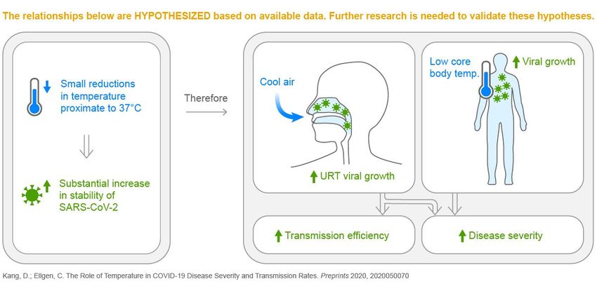
Graphical Abstract
The relationships pictured are hypothesized based on available data. Further research is needed
to validate these hypotheses.
Preprints (www.preprints.org) | NOT PEER-REVIEWED | Posted: 5 May 2020 doi:10.20944/preprints202005.0070.v1
Introduction
The infectivity of viruses is affected by changes in temperature, and the human coronavirus
SARS-CoV-2, which causes coronavirus disease 2019 (COVID-19), is no exception. The stability of
SARS-CoV-2 at temperatures close to normal core body temperature suggests that temperature may
play a particularly significant role in the transmission and severity of COVID-19. In vitro data
indicate that viral stability may be particularly sensitive to fluctuations around 37°C. Variation of
temperatures in the body and between individuals may therefore result in substantially different
rates of viral growth.
A variety of factors can influence temperatures in the body. Air temperature stands out as
particularly relevant because it can directly influence temperatures in the URT, an area that plays a
key role in the transmission and pathogenesis of COVID-19. Additionally, core body temperatures
vary between individuals and may also influence disease severity. This review examines the roles of
air temperature and core body temperature in the transmission and severity of COVID-19.
Effect of Temperature on Viral Viability
● SARS-CoV-2 loses infectivity at normal core body temperature (37°C), but viral stability may
increase substantially with small decreases in temperature proximate to 37°C.
As with other viruses, the stability of SARS-CoV-2 is affected by temperature. Higher temperatures
increase the rate of viral decay.1 Differences in viral stability at temperatures proximate to body
temperature are of particular interest because there is variation in temperature in the body, as well
as variation in core body temperature between individuals. These variations may contribute to
different rates of viral growth in the body, potentially having implications for transmission rates
and patient outcomes.
While SARS-CoV-2 is extremely stable at room temperature (20-25°C), laboratory data indicate
reduced stability at normal core body temperature (37°C).1,2 A study found that SARS-CoV-2
incubated in virus transport medium for 1 day at 22°C had only a 0.5-log unit reduction in
infectious titer; at 37°C, there was a 3.5-log unit reduction in infectious titer after 1 day.1 Higher
temperatures were found to result in more rapid viral decay.
No data are yet available on the stability of SARS-CoV-2 at temperatures slightly below 37°C, but
such data are available for SARS-CoV-1, the virus that causes severe acute respiratory syndrome
(SARS). SARS-CoV-1 is much more stable a few degrees below body temperature than it is at body
temperature. A study found that incubating dried virus at 33°C on a nonporous surface at a relative
humidity of 95% for 24 hours resulted in a 1-log unit reduction in infectious titer, while incubation
at 38°C resulted in a 3.4-log unit reduction.3 The substantial difference in viral stability between
33°C and 38°C is particularly significant to viral replication in vivo because temperatures in the
body can fluctuate within this temperature range.4,5 The same study furthermore found that
incubation of SARS-CoV-1 at 22-25°C for 24 hours resulted in a 0.75-log reduction in infectious
Preprints (www.preprints.org) | NOT PEER-REVIEWED | Posted: 5 May 2020 doi:10.20944/preprints202005.0070.v1
titer. Stated differently, there is only a 0.25-log unit difference in reduction of infectious titer
between 22°C and 33°C over 1 day; but, between 33°C and 38°C, there is a 2.4-log unit difference.
This indicates that the reduction in viral stability is not linear with increases in temperature; rather,
the impact of a temperature change on viral infectivity is greatly affected by the temperature at
which that temperature change occurs. In particular, variations in temperature between 22 and
33°C have minute impacts on viral decay, while small decreases in temperature proximate to 38°C
result in a large increase in viral stability.
In vitro studies indicate that SARS-CoV-1 and SARS-CoV-2 demonstrate similarity in viral stability at
a wide range of temperatures, including room temperature2 and body temperature1,3 (Table 1). The
similarity in viral stability at these temperatures suggests the possibility of similar stability at other
temperatures, as well. Like SARS-CoV-1, SARS-CoV-2 may become substantially more stable as
temperatures decrease from 38°C to 33°C and, at 33°C, stability may be comparable to that at room
temperature. This potential heat sensitivity based on SARS-CoV-1 data can be considered along
with the aforementioned laboratory data on SARS-CoV-2: the rate of viral decay of SARS-CoV-2 is
relatively rapid at 37°C1; viral stability may increase substantially as temperatures decrease from
37°C to 33°C; and, at 33°C, SARS-CoV-2 may be extremely stable, approximately as stable as it is at
room temperature. The difference in viral decay rate between 37°C and 33°C may be about 2 to
3-log units per day for virus incubated in solution in vitro. If SARS-CoV-2 is sensitive to heat in this
way and if the rate of viral decay is similar in vivo, then viral growth in vivo could be substantially
impacted by fluctuations in body temperature. In particular, the rate of viral growth could increase
substantially as temperatures in the body fall below 37°C. This is relevant to disease progression of
COVID-19 because parts of the body—and for many individuals, the entire body—frequently fall
below 37°C.
Additional research may be necessary to determine whether the rates of viral decay in vitro are
similar to those that would occur in vivo. The remainder of this review assumes that in vitro data
are representative of in vivo viral decay rates and explores the possible impacts on transmission
and disease severity.Preprints (www.preprints.org) | NOT PEER-REVIEWED | Posted: 5 May 2020 doi:10.20944/preprints202005.0070.v1
Table 1. Comparison of stability of SARS-CoV-1 and SARS-CoV-2 at various temperatures.
Temperature Virus Condition Reduction in Study
range infectious titer
(log10)
Room Both 21-23°C in aerosols and Similar under all Van
temperature SARS-CoV-1 & on various surfaces circumstances Doremalen et
SARS-CoV-2 tested al2
SARS-CoV-1 1 day at 22-25°C; dried 0.75±0.5 Chan et al3
virus on a nonporous
surface at a relative
humidity of 40-50%
(found to produce
similar results to virus
in solution for the
conditions tested)
SARS-CoV-2 1 day at 22°C; incubated 0.5±0.05 Chin et al1
in virus transport
medium
Core body SARS-CoV-1 1 day at 38°C; dried 3.4±0.5 Chan et al3
temperature virus on a nonporous
surface at a relative
humidity of 95% (found
to produce similar
results to virus in
solution for the
conditions tested)
SARS-CoV-2 1 day at 37°C; incubated 3.2±0.05 Chin et al1
in virus transport
mediumPreprints (www.preprints.org) | NOT PEER-REVIEWED | Posted: 5 May 2020 doi:10.20944/preprints202005.0070.v1
Figure 1. Infectivity of SARS-CoV-1 at different temperatures on a nonporous surface with relative
humidity >95% (which was found to produce similar results to virus in solution). Stability of
SARS-CoV-1 at 38°C is similar to that of SARS-CoV-2 at 37°C, and stability of SARS-CoV-1 at 28°C is
similar to that of SARS-CoV-2 at 22-25°C. (The differences in temperatures of these comparisons is a
result of the limited set of temperatures that have been tested; the differences do not imply
dissimilarity in stability.) It is possible that stability of SARS-CoV-1 and SARS-CoV-2 is also similar
near 33°C. Figure by Chan et al, 2011.3Preprints (www.preprints.org) | NOT PEER-REVIEWED | Posted: 5 May 2020 doi:10.20944/preprints202005.0070.v1
Effect of Air Temperature on Disease Severity and
Transmission Rates
Impact of Air Temperature on URT Viral Growth
● Even air as warm as 25°C cools URT surfaces to several degrees below body temperature,
which may make the URT exceptionally conducive to SARS-CoV-2 replication.
The surfaces of the URT are often in a temperature range that may correspond to relative
SARS-CoV-2 stability. When breathing air below body temperature, the temperature of the
airstream is warmed by the body until it reaches body temperature, and this process cools the
airways. Breathing air at 25°C (the upper end of room temperature) lowers the nasal valve area to
30°C and the nasopharynx to 34°C.4 The airstream ordinarily rises to within 0.5°C of body
temperature before reaching the trachea,6 making lower temperatures unique to the URT.
This relatively small change in temperature may have a substantial impact on the rate of viral
growth in the URT. As described previously, viral stability may increase substantially as
temperature falls below 37°C, and, at 33°C, SARS-CoV-2 may be extremely stable. Furthermore,
SARS-CoV-2 likely replicates effectively in URT epithelium: SARS-CoV-2 uses the SARS-CoV-1
receptor human angiotensin-converting enzyme 2 (ACE2) to obtain entry into host cells, which
suggests the two viruses may target similar cells7,8; and in vitro data indicate that SARS-CoV-1
replicates productively on the nasal epithelium.9 High URT viral loads have moreover been
observed in COVID-19 patients.10,11 The relatively cool temperature of URT surfaces may therefore
make the nasal epithelium an exceptionally suitable environment for SARS-CoV-2 replication and
spread.
COVID-19 patients often develop fever,12,13 and this heat may suppress viral growth. However, fever
does not heat URT surfaces as effectively as it does other areas of the body. Inhaled air begins at
ambient temperature at the nostrils and converges to the patient’s body temperature as it moves
through the airway.4 As such, URT surfaces are cooled by the inhalation of air below body
temperature, even in a feverish patient.
Impact of Air Temperature on Transmission Rates
● By contributing to increased URT viral load, cooler air temperatures may enable more efficient
transmission.
The increase in URT viral growth enabled by cooler air temperatures may be relevant to COVID-19
transmission rates, as higher viral load in the URT could enable more efficient transmission. The
primary mode of transmission of COVID-19 is currently thought to be direct contact of mucous
membranes (eyes, nose, mouth) with infectious respiratory droplets and through exposure to
fomites.14–16 Higher viral load in the URT could result in higher viral titer in respiratory droplets
expelled by an infected host. Higher URT viral load may also increase efficiency of transmission viaPreprints (www.preprints.org) | NOT PEER-REVIEWED | Posted: 5 May 2020 doi:10.20944/preprints202005.0070.v1
fomites. Fomites can become contaminated through contact with body secretions from infected
hosts, and higher URT viral load could result in an increased concentration of virus in the initial
inoculum. The increased viral titer on fomite surfaces could enable more efficient transmission to
naive hosts. In addition, increased initial titer of inoculum prolongs viral survival on fomite
surfaces, increasing the duration of time over which those fomites would remain contaminated.17
This hypothesized relationship of URT viral load and transmission rate is supported by data from
an animal study: A study on ferrets infected with SARS-CoV-2 found that high URT virus titers
resulted in efficient transmission to naive ferrets by direct contact and contributed to airborne
transmission.18
We previously hypothesized that cooler air temperatures increase SARS-CoV-2 viral replication in
the URT. If this is true, then one would expect temperature and transmission rates to be inversely
correlated. Available data are consistent with this: Some studies have found that increases in
outdoor temperature are associated with modest but significant decreases in COVID-19
transmission rates.19–23 Unfortunately, these studies must be interpreted with caution as they face
issues with poor data quality and confounding factors.24,25 Furthermore, studies on outdoor
temperature are of limited use in evaluating this hypothesis because they do not necessarily
account for the actual air temperature breathed by a given population: In developed countries,
people spend the vast majority of their time in temperature-controlled environments,26,27 and, in
these countries, the majority of person-to-person transmission of respiratory viruses occurs
indoors.28 Indoor air temperature may be relevant to transmission because comfortable indoor
temperatures of 20-25°C are well within the temperature range that may correspond to increased
SARS-CoV-2 replication in the URT. It could be valuable to study the relationship between the air
temperature breathed by a population—which may be indoor or outdoor air temperature—and
COVID-19 transmission rate.
When interpreting the studies on outdoor air temperature and transmission rates, one may
conjecture that higher temperatures decreased viral survival on fomites by increasing the rate of
viral decay, thereby contributing to decreases in transmission. While this would certainly be the
case at some temperatures, the rate of viral decay is only trivially affected by increases in
temperature until temperature is sufficiently high. Increasing the temperature from 4°C to 22°C
results in a difference of ≤0.6-log unit viral reduction per day for SARS-CoV-2 incubated in virus
transport medium.1 If stability of SARS-CoV-2 is similar to SARS-CoV-1, the rate of viral decay would
remain almost completely unchanged until temperatures exceed 33°C. Three of the environmental
studies that found inverse correlation between transmission rates and outdoor air temperature
considered only temperatures ≤22°C.20,21,23 SARS-CoV-2 is extremely stable in this range. An
increased rate of viral decay on fomites due to increased outdoor air temperature is therefore
unlikely to have contributed substantially to the observed decrease in transmission rates in these
three studies. By contrast, breathing air at any temperature affects URT temperature and may
thereby contribute to URT viral load and transmission rate.Preprints (www.preprints.org) | NOT PEER-REVIEWED | Posted: 5 May 2020 doi:10.20944/preprints202005.0070.v1
Impact of Air Temperature on Disease Severity
● Due to the significance of URT viral growth in the pathogenesis of COVID-19, temperature of
air inhaled may impact COVID-19 disease severity.
While much remains to be understood about the pathogenesis of COVID-19, available data indicate
that the rate of viral replication in the URT—especially during early stages of disease—may have a
significant impact on disease severity. The progression of COVID-19 may be represented by three
increasingly severe phases: (1) acute viral replication, wherein SARS-CoV-2 establishes infection
and replicates primarily in the respiratory tract, typically causing mild symptoms (i.e. dry cough,
headache, fever); (2) pulmonary involvement, wherein pneumonia develops and hypoxia may
ensue; and (3) hyperactive immune response, wherein excessive cytokine production contributes
to immune system dysregulation and severe adverse outcomes occur (i.e. acute respiratory distress
syndrome [ARDS], systemic inflammatory response syndrome [SIRS], cardiac failure).29,30 The first
phase is most relevant when considering the significance of URT infection in disease progression.
The relevance of URT infection may be elucidated by the pathogenic similarity of COVID-19 and
SARS. The pathological features of the two diseases bear notable resemblance to each other.31,32 As
current understanding is that both causal coronaviruses obtain entry into host cells utilizing ACE2,
it is reasonable to assume that these two viruses infect similar cells.7,8 While differences between
the two diseases are nontrivial, available data suggest that the two viruses may follow a similar
path of viral growth and spread through the respiratory system in the initial phase of the disease
course.
With SARS-CoV-1, replication typically first occurs in upper airway epithelial cells, and virus
subsequently spreads to the lower respiratory tract,33 infecting tracheobronchial and alveolar
epithelial cells.34 Viral spread to alveoli has been hypothesized to occur via airway surfaces: In vitro
data indicate that SARS-CoV-1 replicates productively on the nasal and tracheobronchial epithelium
(especially on ciliated epithelial cells) and sheds over time onto the luminal surface of the
epithelium.9 The abundance of ACE2 on luminal surfaces of conducting airway epithelium may
enable considerable and progressive viral growth and spread throughout the airways.7–9,34
Current evidence suggests that SARS-CoV-2 also infects airway epithelial cells early in the disease
course: In confirmed COVID-19 patients, high viral titers are frequently observed in the upper and
lower respiratory tracts early in infection.10,11 Given SARS-CoV-2 affinity for ACE2, the abundance of
ACE2 on airway epithelium may support progressive viral growth and spread throughout the
airways, as has been hypothesized to occur with SARS-CoV-1. These data suggest that SARS-CoV-2
may similarly establish infection in the URT and progressively replicate and spread through the
airway epithelia.
We previously hypothesized that breathing cool air—even as warm as 25°C—may make the URT
exceptionally conducive to SARS-CoV-2 replication. The postulated pathogenesis suggests that
breathing cool air—especially during early stages of infection—may contribute to more rapid
escalation in disease severity. Data on viral load are consistent this hypothesis: URT viral load isPreprints (www.preprints.org) | NOT PEER-REVIEWED | Posted: 5 May 2020 doi:10.20944/preprints202005.0070.v1
strongly correlated with adverse outcomes and has been hypothesized to be a predictor of disease
severity.10 The nasopharyngeal viral load of severe COVID-19 cases has been found to be about 60
times higher than that of mild cases.10
The stability of SARS-CoV-2 at the cool temperatures of the URT and its relative instability at
normal core body temperature also suggest the possibility that the URT serves as a viral reservoir
that enables continual reinfection of proximate airway surfaces, supporting repeated cycles of viral
replication and spread through the airways. This would indicate that viral growth in the URT
remains pathogenically significant even after infection has spread beyond the URT.
Conversely, warmer air temperatures may suppress viral replication in the URT. Given the role of
URT infection in the pathogenesis of COVID-19, reducing the rate of viral replication in the URT
during the early stages of establishment of COVID-19 may slow disease progression. Some have
hypothesized that suppressing viral growth during the early stages of establishment of COVID-19
(i.e. prior to peak viral load and severe symptoms) may reduce duration of symptoms and prevent
progression of severity.29,35,36
Effect of Body Temperature on Disease Severity
● Low core body temperature may enable more rapid viral growth, which may contribute to
more severe disease.
The potentially substantial increase in SARS-CoV-2 stability with decreases in temperatures
proximate to 37°C may have implications for individuals with low core body temperatures. Lower
core body temperatures may support more rapid viral growth and thereby contribute to higher
viral load, which is strongly correlated with disease severity.10,11 If this is the case, one would expect
to see an increase in disease severity among individuals with unusually low body temperature.
While we are unaware of any direct analysis of the relationship between these two factors, existing
data are consistent with this possibility.
Emerging evidence indicate that the case fatality rate of COVID-19 is associated with age, sex, and
comorbidities, including hypertension, diabetes, and cardiovascular disease.37–39 Higher case fatality
rates among men has been hypothesized to be caused by sex-based immunological differences and
gendered differences in behavior that produce comorbities (e.g. drinking and smoking).40 Higher
case fatality rates among elderly people may be related to higher prevalence of comorbidities and
age-related changes in the immune system.35,41 Another relevant factor in case fatality rate may be
body temperature. There are substantially more men and elderly people with unusually low body
temperatures than there are women and younger people, respectively. A large cross-sectional study
found that mean body temperature is 0.17°C lower in men than in women, with the mean body
temperature being 0.19°C lower in 80+ year old women than in 20-29 year old women, and 0.15°C
lower in 80+ year old men than in 20-29 year old men.5 While these differences in mean body
temperature are small, the relatively greater number of men and elderly with unusually low body
temperature may be considerable: Measured body temperatures appear to be approximatelyPreprints (www.preprints.org) | NOT PEER-REVIEWED | Posted: 5 May 2020 doi:10.20944/preprints202005.0070.v1
normally distributed (Figure 2), and there is a significant multiplier effect that occurs when
comparing two normal distributions with similar variance but slightly different mean.
To demonstrate this multiplier effect, assume the body temperatures of two populations A and B
can be represented with two normal distributions (Figure 3). Let A have a mean of 36.4°C, and let B
have a mean that is 0.2°C lower, 36.2°C. Let both A and B have a standard deviation of 0.6°C. (These
values were selected to be similar to measured values from the aforementioned study on body
temperatures, but limitations in the data provided by the study prevent us from estimating precise
values.5 As such, these numbers should be considered merely an example.) Let’s say we want to
know the number of people with unusually low body temperatures which—for the purposes of this
example—we arbitrarily define as ≤35.5°C, which is 1.5 standard deviations below the mean of
distribution A. The frequency of body temperatures ≤35.5°C is 4.8% in population A and 9.1% in
population B. Stated differently, given a standard deviation of 0.6°C, a 0.2°C decrease in mean body
temperature results in a 90% increase in the number of people with unusually low body
temperatures of ≤35.5°C. The difference in frequency is even more pronounced as temperatures
decrease: The frequency of body temperatures ≤35.0°C is 0.6% in population A and 1.5% in
population B— a 150% increase.
Referring again to measured temperatures,5 the distributions appear to be approximately normally
distributed with similar variances but slightly different means. This suggests that the number of
people with low body temperature is considerably greater among men than women and among
elderly people than young people. A calculation from the study supports this conjecture: Frequency
of temperaturesPreprints (www.preprints.org) | NOT PEER-REVIEWED | Posted: 5 May 2020 doi:10.20944/preprints202005.0070.v1
Figure 2. Distribution of measured body temperatures by age and sex. Adapted from figures by Waalen
& Buxbaum, 2011.5
Figure 3. Distributions of body temperatures in two hypothetical populations, A and B. The mean of A
is 36.4°C, and the mean of B is 36.2°C. Both A and B have a standard deviation of 0.6°C. Unusually low
body temperatures are arbitrarily defined as ≤35.5°C (indicated by the dotted line), which is 1.5
standard deviations below the mean of A.Preprints (www.preprints.org) | NOT PEER-REVIEWED | Posted: 5 May 2020 doi:10.20944/preprints202005.0070.v1
Concluding Remarks and Future Directions
The heat sensitivity of SARS-CoV-2 suggests that cooler air temperatures and lower body
temperatures may enable increased rates of viral growth in the body and thereby contribute to
increased transmission rates and disease severity.
In vitro data indicate that SARS-CoV-2 loses infectivity at normal core body temperature (37°C),
but—like SARS-CoV-1—viral stability may increase substantially with small decreases in
temperature proximate to 37°C. Further research on how viral decay of SARS-CoV-2 is affected by
small changes in temperature within a range that occurs in the body (i.e. ~30-40°C) would help
elucidate the potential effect of temperature on viral growth in vivo. Such experiments should
incubate virus in a medium that produces viral decay rates comparable to those which occur in vivo.
Even air as warm as 25°C cools URT surfaces to several degrees below body temperature, which
may make the URT exceptionally conducive to SARS-CoV-2 replication. Animal studies may be
useful in testing this hypothesis: Animals inoculated with SARS-CoV-2 could be kept at various air
temperatures, and URT viral load could be tracked and analyzed for correlation with air
temperature.
Because increased URT viral load likely enables more effective transmission, lower air
temperatures may contribute to increased transmission of COVID-19. Several studies have analyzed
correlation between outdoor air temperature and transmission rates; however, these studies are of
limited use in evaluating the hypotheses proposed in this review because they do not account for
indoor air temperature. People in developed nations spend the majority of their time in
temperature-controlled environments, and even comfortable temperatures (e.g. 20-25°C) cool the
URT sufficiently to potentially impact viral growth, suggesting that indoor temperatures may
impact transmission rates. Further research is necessary to quantify the potential impact of both
outdoor and indoor air temperature on transmission rates; however, indoor air temperature data
may be difficult to acquire. It may be more effective to study the impact of air temperature on URT
viral load (perhaps using an animal study, as previously suggested) and infer impact on
transmission rates.
Because SARS-CoV-2 infection may frequently begin in the URT before spreading through the body,
lower temperatures of air inhaled—especially during early stages of infection—could contribute to
increased disease severity. Conversely, warmer air may reduce the rate of viral growth in the URT.
Suppressing URT viral replication would likely have a greater impact in preventing escalation of
severity if suppression occurs when SARS-CoV-2 is mostly contained in the URT, immediately
post-exposure or early in SARS-CoV-2 infection. This suggests that administration of warm air may
potentially represent a therapeutic measure as a post-exposure prophylaxis and an early-stage
treatment to prevent disease progression. Post-exposure prophylaxis applications may be of
particular relevance for healthcare professionals who have insufficient personal protective
equipment, as well as for populations who are at risk of more severe disease.Preprints (www.preprints.org) | NOT PEER-REVIEWED | Posted: 5 May 2020 doi:10.20944/preprints202005.0070.v1
Warm air delivery would likely need to occur over sustained periods of time to suppress viral
replication to a degree that would be beneficial because decreases in virus infectivity are dependent
on duration of exposure to elevated temperatures. Incubating SARS-CoV-2 in viral transport
medium at 37°C results in a 1.5-log unit reduction in infectious titer after 12 hours, a 3.5-log unit
reduction after 24 hours, and a ≥6.8-log unit reduction after 48 hours.1 Higher temperatures result
in more rapid loss of viral stability: Just 10 minutes at 56°C results in a 3-log unit reduction.1 It is
important to note that air that is warmer than body temperature will be cooled during inhalation,
limiting the effectiveness of very warm air in reducing viral stability in vivo. The impact of
temperature on viral growth could be tested in an animal study by measuring URT viral load in
animals infected with SARS-CoV-2 before and after exposure to air at various temperatures and
durations.
Severity of COVID-19 may also be affected by core body temperature. The potentially substantial
increase in SARS-CoV-2 stability as temperatures fall below 37°C suggests that lower core body
temperatures could enable more rapid viral growth and thereby contribute to more severe disease.
The prevalence of unusually low body temperatures among men and elderly people may help
explain higher case fatality rates in these demographics. Research that determines the significance
of this possible correlation could be of value. Such an analysis should consider a patient’s normal
core body temperature, which may be different from body temperature while the patient has
COVID-19 since fever is a common symptom. Age, sex, BMI, and comorbidities should be controlled
for. Large healthcare organizations may already be collecting this data, as they track patient data
during regular visits and may have also treated COVID-19 patients.
The impact of temperatures in the body on viral growth may help explain COVID-19 transmission
rates and disease severity. Advances made in understanding the various impacts of temperature on
COVID-19 could inform both disease prevention and treatment, with potentially significant impacts
on patient outcomes and global disease burden.
References
1. Chin AWH, Chu JTS, Perera MRA, et al. Stability of SARS-CoV-2 in different environmental
conditions. Lancet Microbe. April 2020. doi:10.1016/S2666-5247(20)30003-3
2. van Doremalen N, Bushmaker T, Morris DH, et al. Aerosol and Surface Stability of SARS-CoV-2
as Compared with SARS-CoV-1. N Engl J Med. 2020;382(16):1564-1567.
doi:10.1056/NEJMc2004973
3. Chan KH, Peiris JSM, Lam SY, Poon LLM, Yuen KY, Seto WH. The Effects of Temperature and
Relative Humidity on the Viability of the SARS Coronavirus. Kohl A, ed. Adv Virol.
2011;2011:734690. doi:10.1155/2011/734690
4. Keck T, Leiacker R, Riechelmann H, Rettinger G. Temperature Profile in the Nasal Cavity. The
Laryngoscope. 2000;110(4):651-654. doi:10.1097/00005537-200004000-00021
5. Waalen J, Buxbaum JN. Is Older Colder or Colder Older? The Association of Age With Body
Temperature in 18,630 Individuals. J Gerontol Ser A. 2011;66A(5):487-492.
doi:10.1093/gerona/glr001
6. Hall J. Pulmonary Ventilation. In: Guyton and Hall Textbook of Medical Physiology. 12th ed. ;
2011:474.Preprints (www.preprints.org) | NOT PEER-REVIEWED | Posted: 5 May 2020 doi:10.20944/preprints202005.0070.v1
https://archive.org/details/guytonandhalltextbookofmedicalphysiology12thed_202004/page
/n493/mode/2up.
7. Hoffmann M, Kleine-Weber H, Schroeder S, et al. SARS-CoV-2 Cell Entry Depends on ACE2 and
TMPRSS2 and Is Blocked by a Clinically Proven Protease Inhibitor. Cell. March 2020.
doi:10.1016/j.cell.2020.02.052
8. Wang Q, Zhang Y, Wu L, et al. Structural and Functional Basis of SARS-CoV-2 Entry by Using
Human ACE2. Cell. April 2020. doi:10.1016/j.cell.2020.03.045
9. Sims AC, Baric RS, Yount B, Burkett SE, Collins PL, Pickles RJ. Severe Acute Respiratory
Syndrome Coronavirus Infection of Human Ciliated Airway Epithelia: Role of Ciliated Cells in
Viral Spread in the Conducting Airways of the Lungs. J Virol. 2005;79(24):15511-15524.
doi:10.1128/JVI.79.24.15511-15524.2005
10. Liu Y, Yan L-M, Wan L, et al. Viral dynamics in mild and severe cases of COVID-19. Lancet Infect
Dis. 2020;0(0). doi:10.1016/S1473-3099(20)30232-2
11. Liu Y, Yang Y, Zhang C, et al. Clinical and biochemical indexes from 2019-nCoV infected
patients linked to viral loads and lung injury. Sci China Life Sci. 2020;63(3):364-374.
doi:10.1007/s11427-020-1643-8
12. Sohrabi C, Alsafi Z, O’Neill N, et al. World Health Organization declares global emergency: A
review of the 2019 novel coronavirus (COVID-19). Int J Surg. 2020;76:71-76.
doi:10.1016/j.ijsu.2020.02.034
13. Yang X, Yu Y, Xu J, et al. Clinical course and outcomes of critically ill patients with SARS-CoV-2
pneumonia in Wuhan, China: a single-centered, retrospective, observational study. Lancet
Respir Med. February 2020. doi:10.1016/S2213-2600(20)30079-5
14. Lu C, Liu X, Jia Z. 2019-nCoV transmission through the ocular surface must not be ignored. The
Lancet. 2020;395(10224):e39. doi:10.1016/S0140-6736(20)30313-5
15. Peng X, Xu X, Li Y, Cheng L, Zhou X, Ren B. Transmission routes of 2019-nCoV and controls in
dental practice. Int J Oral Sci. 2020;12(1):1-6. doi:10.1038/s41368-020-0075-9
16. CDC. Coronavirus Disease 2019 (COVID-19) - Transmission. Centers for Disease Control and
Prevention.
https://www.cdc.gov/coronavirus/2019-ncov/prevent-getting-sick/how-covid-spreads.html.
Published April 13, 2020. Accessed April 19, 2020.
17. Boone SA, Gerba CP. Significance of Fomites in the Spread of Respiratory and Enteric Viral
Disease. Appl Environ Microbiol. 2007;73(6):1687-1696. doi:10.1128/AEM.02051-06
18. Kim Y-I, Kim S-G, Kim S-M, et al. Infection and Rapid Transmission of SARS-CoV-2 in Ferrets.
Cell Host Microbe. April 2020. doi:10.1016/j.chom.2020.03.023
19. COVID-19 transmission in Mainland China is associated with temperature and humidity: a
time-series analysis | medRxiv.
https://www.medrxiv.org/content/10.1101/2020.03.30.20044099v1. Accessed April 17,
2020.
20. Wang J, Tang K, Feng K, Lv W. High Temperature and High Humidity Reduce the Transmission of
COVID-19. Rochester, NY: Social Science Research Network; 2020.
https://papers.ssrn.com/abstract=3551767. Accessed April 17, 2020.
21. Sajadi MM, Habibzadeh P, Vintzileos A, Shokouhi S, Miralles-Wilhelm F, Amoroso A.
Temperature, Humidity and Latitude Analysis to Predict Potential Spread and Seasonality for
COVID-19. Rochester, NY: Social Science Research Network; 2020.
https://papers.ssrn.com/abstract=3550308. Accessed April 17, 2020.
22. Notari A. Temperature dependence of COVID-19 transmission. April 2020.
https://doi.org/10.1101/2020.03.26.20044529. Accessed April 17, 2020.
23. Qi H, Xiao S, Shi R, et al. COVID-19 transmission in Mainland China is associated withPreprints (www.preprints.org) | NOT PEER-REVIEWED | Posted: 5 May 2020 doi:10.20944/preprints202005.0070.v1
temperature and humidity: a time-series analysis. March 2020.
https://doi.org/10.1101/2020.03.30.20044099. Accessed April 17, 2020.
24. National Academies of Sciences, Engineering, and Medicine. Rapid Expert Consultation on
SARS-CoV-2 Survival in Relation to Temperature and Humidity and Potential for Seasonality
for the COVID-19 Pandemic (April 7, 2020). April 2020. doi:10.17226/25771
25. Mecenas P, Bastos R, Vallinoto A, Normando D. Effects of temperature and humidity on the
spread of COVID-19: A systematic review. medRxiv. April 2020:2020.04.14.20064923.
doi:10.1101/2020.04.14.20064923
26. Schweizer C, Edwards RD, Bayer-Oglesby L, et al. Indoor time–microenvironment–activity
patterns in seven regions of Europe. J Expo Sci Environ Epidemiol. 2007;17(2):170-181.
doi:10.1038/sj.jes.7500490
27. Klepeis NE, Nelson WC, Ott WR, et al. The National Human Activity Pattern Survey (NHAPS): a
resource for assessing exposure to environmental pollutants. J Expo Sci Environ Epidemiol.
2001;11(3):231-252. doi:10.1038/sj.jea.7500165
28. Moriyama M, Hugentobler WJ, Iwasaki A. Seasonality of Respiratory Viral Infections. Annu Rev
Virol. March 2020. doi:10.1146/annurev-virology-012420-022445
29. Siddiqi HK, Mehra MR. COVID-19 Illness in Native and Immunosuppressed States: A
Clinical-Therapeutic Staging Proposal. J Heart Lung Transplant. 2020;0(0).
doi:10.1016/j.healun.2020.03.012
30. Channappanavar R, Perlman S. Pathogenic human coronavirus infections: causes and
consequences of cytokine storm and immunopathology. Semin Immunopathol.
2017;39(5):529-539. doi:10.1007/s00281-017-0629-x
31. Ong E, Chan Y, Leong W, et al. A dynamic immune response shapes COVID-19 progression. Cell
Host Microbe. 2020. doi:10.1016/j.chom.2020.03.021
32. Xu Z, Shi L, Wang Y, et al. Pathological findings of COVID-19 associated with acute respiratory
distress syndrome. Lancet Respir Med. 2020;8(4):420-422.
doi:10.1016/S2213-2600(20)30076-X
33. Knipe D, Howley P. Coronaviridae. In: Fields Virology. Vol 1. 6th ed. Lippincott Williams &
Wilkins; 2013.
https://archive.org/details/DavidM.KnipePeterHowleyFieldsVirologyKnipeFieldsVirology2Vo
lumeSetLWW2013/mode/1up.
34. Gu J, Korteweg C. Pathology and Pathogenesis of Severe Acute Respiratory Syndrome. Am J
Pathol. 2007;170(4):1136-1147. doi:10.2353/ajpath.2007.061088
35. Wu C, Chen X, Cai Y, et al. Risk Factors Associated With Acute Respiratory Distress Syndrome
and Death in Patients With Coronavirus Disease 2019 Pneumonia in Wuhan, China. JAMA
Intern Med. March 2020. doi:10.1001/jamainternmed.2020.0994
36. To KK-W, Tsang OT-Y, Leung W-S, et al. Temporal profiles of viral load in posterior
oropharyngeal saliva samples and serum antibody responses during infection by SARS-CoV-2:
an observational cohort study. Lancet Infect Dis. 2020;0(0).
doi:10.1016/S1473-3099(20)30196-1
37. Lancet T. The gendered dimensions of COVID-19. The Lancet. 2020;395(10231):1168.
doi:10.1016/S0140-6736(20)30823-0
38. Zhou F, Yu T, Du R, et al. Clinical course and risk factors for mortality of adult inpatients with
COVID-19 in Wuhan, China: a retrospective cohort study. The Lancet.
2020;395(10229):1054-1062. doi:10.1016/S0140-6736(20)30566-3
39. Verity R, Okell LC, Dorigatti I, et al. Estimates of the severity of coronavirus disease 2019: a
model-based analysis. Lancet Infect Dis. 2020;0(0). doi:10.1016/S1473-3099(20)30243-7
40. Wenham C, Smith J, Morgan R. COVID-19: the gendered impacts of the outbreak. The Lancet.Preprints (www.preprints.org) | NOT PEER-REVIEWED | Posted: 5 May 2020 doi:10.20944/preprints202005.0070.v1
2020;395(10227):846-848. doi:10.1016/S0140-6736(20)30526-2
41. Nikolich-Žugich J. The twilight of immunity: emerging concepts in aging of the immune
system. Nat Immunol. 2018;19(1):10-19. doi:10.1038/s41590-017-0006-x
42. Qingxian C, Fengjuan C, Fang L, et al. Obesity and COVID-19 Severity in a Designated Hospital in
Shenzhen, China. Rochester, NY: Social Science Research Network; 2020.
doi:10.2139/ssrn.3556658
43. Haslam DW, James WPT. Obesity. The Lancet. 2005;366(9492):1197-1209.
doi:10.1016/S0140-6736(05)67483-1You can also read