The SARS-like coronaviruses: the role of bats and evolutionary relationships with SARS coronavirus
←
→
Page content transcription
If your browser does not render page correctly, please read the page content below
NEW MICROBIOLOGICA, 35, 1-16, 2012

  The SARS-like coronaviruses: the role of bats and
  evolutionary relationships with SARS coronavirus
                                 Andrea Balboni, Mara Battilani, Santino Prosperi
  Dipartimento di Scienze Mediche Veterinarie, Facoltà di Medicina Veterinaria, Alma Mater Studiorum-Università di Bologna,
                                               Ozzano dell’Emilia, Bologna, Italy

                                                          SUMMARY

    Bats represent an order of great evolutionary success, with elevated geographical diffusion and species diversity. This
    order harbors viruses of high variability which have a great possibility of acquiring the capacity of infecting other an-
    imals, including humans. Bats are the natural reservoir for several viruses genetically closely related to the SARS-
    coronavirus which is the etiological agent of severe acute respiratory syndrome (SARS), a human epidemic which
    emerged in China in 2002-2003. In the last few years, it has been discovered that the association between coron-
    aviruses and bats is a worldwide phenomenon, and it has been hypothesised that all mammalian coronaviruses were
    derived from ancestral viruses residing in bats. This review analyzes the role of bats as a reservoir of zoonotic virus-
    es focusing more extensively on SARS-related coronaviruses and taking into account the role of African and European
    strains in the evolutionary history of these viruses.

    KEY WORDS: Bat, SARS, Coronavirus

Received April 19, 2011                                                                                  Accepted October 31, 2011

INTRODUCTION                                                      In past years, several viruses infecting humans
                                                                  have been linked to bats: Rhabdoviridae,
In mammalians, Chiroptera is one of the most                      Orthomyxoviridae, Coronaviridae, Flaviviridae,
important orders involved in the emergence and                    Filoviridae and others (Calisher et al., 2006). With
diffusion of viruses in animals, and between an-                  some exceptions, the direct transmission of virus-
imals and humans. Several characteristics of the                  es from bats to humans is uncommon. Instead,
bat species, such as the ability to fly, the coexis-              bats more frequently act like reservoirs of ances-
tence of many individuals in large colonies, a di-                tral viruses which, through a species jump, arrive
et composed of all types of foods, the ability to                 in secondary hosts where they acquire a tropism
cover long distances during migration as well as                  for the human host.
many others, have allowed bats to be found all                    Due to the elevated geographical diffusion and
over the world (except in Antarctica and a few                    the elevated species diversity of bats, this order
oceanic islands), occupying the most varied eco-                  harbors viruses of high variability which have a
logical niches. This great evolutionary success un-               greater possibility of acquiring the capacity of in-
derlies the remarkable propensity of these ani-                   fecting other animals, including humans.
mals to favor, as hosts and reservoirs, the diffu-                In recent years, several anthropogenic and natu-
sion of a wide variety of viruses.                                ral changes in the environment, due to defor-
                                                                  estation, alteration of natural habitats, changes
Corresponding author                                              in animal diversity and climatic events, have shift-
Mara Battilani                                                    ed the ecology of bats and have exposed humans
Dipartimento di Scienze Mediche Veterinarie                       to new pathogens (Wang et al., 2006). In fact, the
Facoltà di Medicina Veterinaria
                                                                  last twenty years have seen an increase in the
Alma Mater Studiorum-Università di Bologna
Via Tolara di Sopra, 50                                           number of outbreaks caused by pathogens with
40064 Ozzano dell’Emilia, Bologna, Italy                          bats as the reservoir of infection (Calisher et al.,
E-mail: mara.battilani@unibo.it                                   2006).
2                                         A. Balboni, M. Battilani, S. Prosperi

Of the human viral epidemics which have                        al., 2007; Brandao et al., 2008; Carrington et al.,
emerged in the last ten years, one of the most im-             2008; Gloza-Raush et al., 2008; Misra et al., 2009;
portant is the outbreak of severe acute respirato-             Pfefferle et al., 2009; Tong et al., 2009; Donaldson
ry syndrome (SARS) which emerged in China in                   et al., 2010; Drexler et al., 2010; Quan et al., 2010;
2002-2003, rapidly causing a global epidemic in-               Reusken et al., 2010; Rihtaric et al., 2010;
volving humans. In two consecutive events (the                 Watanabe et al., 2010; Balboni et al., 2011).
main SARS epidemic of 2002-2003 and a series of                This review will analyze the role of bats as reser-
sporadic infections occurring in the winter of                 voirs of zoonotic viruses focusing more exten-
2003-2004), SARS caused more than 8000 infec-                  sively on SARS-related coronaviruses: the dis-
tions and 700 deaths (Centers for Disease Control              covery of the coronavirus in bats, the genomic
and Prevention (CDC), 2003; Peiris et al., 2004;               features of the several SARS-like CoVs, and the
Skowronski et al., 2005).                                      gradual and ongoing identification of new viral
Several studies have shown that a new coron-                   strains in bats in different parts of the world.
avirus is the etiological agent of SARS, the SARS-             In particular, the role of SARS-like CoVs residing
CoV (Drosten et al., 2003; Ksiazek et al., 2003;               in African and European bats will be taken into
Peiris et al., 2003; Rota et al., 2003). Successively,         account in the evolutionary history of these virus-
it has also been shown that bats might be the nat-             es, in the light of the typical genome divergences
ural reservoir for several viruses closely related             that these viruses have when compared to Asian
genetically to the SARS-CoV, known as SARS-like                SARS-like CoVs.
coronaviruses (SARS-like CoVs), and that all
mammalian coronaviruses were derived from an-                  Bats and their role in viral spread
cestral viruses residing in bats (Shi et al., 2008;            Bats are one of the longest living mammalian or-
Woo et al., 2009).                                             ders, and probably constitute the order with the
Furthermore, the association between coron-                    most diversity between species (Altringham,
aviruses and bats is a worldwide phenomenon:                   1996a). They originated early (50 to 52 million
after the initial discovery of this virus in Chinese           years ago) and the different species have changed
bats (Lau et al., 2005; Li et al., 2005), subsequent           relatively little over time compared to mammals
studies confirmed its presence in North and                    of other taxa (Calisher et al., 2006).
South America, Europe and Africa (Dominguez et                 Bats constitute the second largest order of mam-

                                        TABLE 1 - Bat classification.

Order            Suborder             Family                     Common name

Chiroptera       Megachiroptera       Pteropodidae               Flying foxes and other Old World fruit bats

                 Microchiroptera      Vespertilionidae           Vesper bats or evening bats or common bats
                                      Phyllostomidae             American leaf-nosed bats, including vampire bats
                                      Rhinolophidae              Horseshoe bats
                                      Hipposideridae             Old World leaf-nosed bats
                                      Molossidae                 Free-tailed bats
                                      Emballonuridae             Sheath-tailed bats or sac-winged bats
                                      Nycteridae                 Slit-faced bats or hollow-faced bats
                                      Mormoopidae                Leaf-chinned bats
                                      Megadermatidae             False vampire bats
                                      Natalidae                  Funnel-eared bats
                                      Rhinopomatidae             Mouse-tailed bats
                                      Thyropteridae              Disk-winged bats
                                      Mystacinidae               New Zealand short-tailed bats
                                      Furipteridae               Smoky bats
                                      Noctilionidae              Bulldog bats or fisherman bats
                                      Craseonycteridae           Hog-nosed or bumblebee bat
                                      Myzopodidae                Old World sucker-footed bat
Bats and SARS-like coronaviruses                                        3

mals and, with approximately 1000 species, make            several viral species and transmit viruses to oth-
up 20% of the species in the Class Mammalia.               er animals, including humans, is favored by var-
Bats are classified in the order Chiroptera, which         ious biological and ecological characteristics; the
is subdivided in two suborders, Mega- and                  main ones are:
Microchiroptera, commonly referred to as mega-             a) The high population densities and the habit of
bats and microbats.                                        roosting provides the opportunity for disease-
The suborder Megachiroptera contains only one              causing pathogens to invade and spread rapidly,
family (Pteropodidae or flying foxes), which in-           with the possibility of intra- and interspecies
cludes only plant-eating large bats (20-1500 g),           transmission of infectious agents amongst them-
confined to Africa, tropical Asia and Indo-                selves through direct contact, or aerosol or
Australasia. Instead, the Suborder Microchiro-             arthropod vectors (Kunz et al., 2003c; Calisher et
ptera contains 17 families which include the ma-           al., 2006; Wang et al., 2006; Woo et al., 2006).
jority of small bats (1.5-150 g). They are found           The number of bats in each colony varies great-
on every continent except Antarctica and a few             ly from less than 10 to over 200,000 individuals
oceanic islands, and are omnivorous (Table 1)              (Wang et al., 2006), and many bat species may
(Altringham, 1996a; Calisher et al., 2006; Wang            share the same habitats. Bats exhibit a wide range
et al., 2006).                                             of fidelity to their roosts. This variation is often re-
As regards the geographical distribution of bats,          flected in the type of roost, stage of life and form
a decrease in species richness is seen with an in-         of social organisation. Many species of cavity-
crease in latitude. Therefore, there is greater            and foliage-roosting bats show low fidelity to spe-
species richness in proximity to the equator due           cific roost size as compared to cave- and building-
to greater major primary biological productivity           roosting bats, but they often all exhibit high lev-
as there is an increase in light intensity, temper-        els of fidelity to roost areas (Kunz et al., 2003a).
ature and the growing season (Altringham,                  The roosting environment ranges from natural
1996d).                                                    structures (caves, rock crevices, nests of birds,
Owing to the diversity of bat species and their            ants and termites, hollowed spaces in trees, or
unique biological and ecological characteristics           exposed on tree branches and trunks) to man-
(all bats fly, some are abundant and widely dis-           made structures (mines, tombs, buildings and
tributed, many are highly gregarious, they are             bridges), and they can be temporary or perma-
predators and are preyed upon at the same time),           nent (Kunz et al., 2003a; Wang et al., 2006). The
they can become the hosts for a large number of            occupation of man-made habitats could bring the
infectious agents having medical importance                bats into closer association with humans and
(Calisher et al., 2006; Wang et al., 2006; Donaldson       their companion animals or livestock with the
et al., 2010;).                                            possible transmission of some pathogens from
The transmission of viruses from bats to humans            peridomestic bats to humans and livestock (Wang
can occur in a variety of ways: direct contact             et al., 2006).
(bites and scratches), inhalation of infectious par-       b) The dietary habits of bats can be broadly di-
ticles by humans (secretions and guano), arthro-           vided into insectivorous, frugivorous, carnivo-
pod vectors, consumption of bat meat in some               rous, omnivorous and sanguivorous (Wang et al.,
parts of the world (China, Guam and some parts             2006). Predatory bats could potentially acquire
of Asia) and secondary vertebrate hosts which              infectious agents from other animal species, such
serve as amplifying hosts (Wang et al., 2006).             as birds and insects (Wang et al., 2006) but, on
Therefore, bats represent an important reservoir           the other hand, bats are also preyed upon and
for many zoonotic viruses which usually cause              they can transmit infectious agents to their pred-
persistent infections in bats themselves.                  ators.
Furthermore, with some exceptions, the trans-              c) The ability to fly has given bats the opportuni-
mission of viruses from bats to humans requires            ty of going almost anywhere, freer from obsta-
a second intermediate animal host, and direct              cles than land-based mammals (Woo et al., 2006).
transfer of the pathogen from bats to humans               Some genera migrate between two sites to avoid
happens only rarely (Calisher et al., 2006).               unfavourable climatic conditions and/or to seek
As mentioned above, the capability of bats to host         more favourable conditions for growth, covering
4                                       A. Balboni, M. Battilani, S. Prosperi

distances ranging from 200-300 km to almost                  family Coronaviridae were subdivided into only
2000 km (Calisher et al., 2006; Wang et al., 2006).          two genera: Coronavirus and Torovirus, and all
There are two types of migration: 1) long seasonal           viruses included in the genus Coronavirus were
migration generally in a north-south direction               subdivided into only three antigenic groups. The
and 2) short seasonal migration not necessarily in           recent discovery of a large variety of new coron-
a north-south direction. Migration is particular-            aviruses in several host species has led the
ly common in tree-roosting bats since tree holes             Coronavirus Study Group of the International
are poor hibernation sites in the colder climates.           Committee for Taxonomy of Viruses (ICTV) to
Furthermore, female migration is more common                 propose a reclassification of the family
than male (Altringham, 1996c; Kunz et al.,                   Coronaviridae into two subfamilies: Coronavirinae
2003b).                                                      and Torovirinae. In particular, the subfamily
The evolution of migratory behaviour varies with             Coronavirinae now includes three genera:
latitude; in fact, migratory behaviour is more in-           Alphacoronavirus, Betacoronavirus and Gamma-
tense at higher latitudes than at lower latitudes,           coronavirus, which have replaced the traditional
and a higher proportion of temperate bats exhibit            antigenic groups 1, 2, and 3, respectively. All the
migratory behaviour than do tropical species                 mammalian CoVs, including all bat coronavirus-
(Kunz et al., 2003b).                                        es detected to date and SARS-CoV, belong to the
Therefore, bats have a greater capacity to disperse          first and the second genera, Alphacoronavirus and
quickly to new areas, such as islands, than do               Betacoronavirus, whereas all avian CoVs belong
non-volant mammals, with the exception of hu-                to the genus Gammacoronavirus. Within the
mans. The migratory habits of some bats provide              Gammacoronavirus genus, there is only one ex-
an opportunity for pathogens to cover long dis-              ception represented by the Beluga whale coron-
tances and bridge gaps between various species               avirus SW1 strain which has been identified in
which might otherwise not be in contact (Kunz et             this aquatic mammal (Fauquet et al., 2005; Woo et
al., 2003c).                                                 al., 2009; http://www.ncbi.nlm.nih.gov/taxonomy).
d) Vespertilionidae and Rhinolophidae bats have              The viral genome is the largest known non-seg-
the ability of entering into daily torpor and sea-           mented viral RNA genome (approximately 30
sonal hibernation to conserve energy during cool             kbp) and is subdivided into a variable number of
nights and winter months (Calisher et al., 2006).            open reading frames (6 to 9) which encode an en-
The term torpor means a state in which an ani-               semble of non-structural proteins with RNA-poly-
mal allows its body temperature to fall below its            merase function (Pol-1a,b) and four principal
active homeothermic level. The fall in body tem-             structural proteins: Spike (S), Membrane (M),
perature is slow and controlled, and permits con-            Envelope (E) and Nucleocapsid (N) proteins. In
servation of energy. Hibernation is an extended              addition to these, some non-structural proteins,
form of torpor which occurs on a seasonal basis,             which are diverse in number, have uncertain
lasting for days, weeks, or months, in response              functions and have a group-specific genome po-
to a prolonged fall in ambient temperature or a              sition, have been added (Fauquet et al., 2005).
reduction in the food supply (Altringham, 1996b).            Briefly, the Pol-1a,b proteins are encoded from
The capability of bats to enter into seasonal hi-            an open reading frame (ORF) which occupies 2/3
bernation may also facilitate the diffusion of               of the genome and presents highly conserved re-
pathogens since it can allow the viruses to over-            gions. The Spike glycoprotein, exposed on the
winter within the bats, thus facilitating their per-         virion surface, is responsible for recognising and
sistence in the environment (Calisher et al., 2006).         binding with the cellular receptor, inducing neu-
                                                             tralising antibody response and is encoded from
Coronavirus                                                  an ORF with a highly variable sequence. The nu-
Coronaviruses (CoVs), order Nidovirales and fam-             cleocapsid protein is linked with genomic RNA
ily Coronaviridae, are a group of enveloped virus-           and induces a cell-mediated immune response
es with a linear, non-segmented, positive-sense,             (Fauquet et al., 2005).
single-stranded RNA genome, having a charac-                 Coronaviruses have diverse animal hosts ranging
teristic crown morphology (Fauquet et al., 2005).            from mammalian (including humans) to avian
Until a few years ago, all viruses belonging to the          species, and have a high frequency of recombi-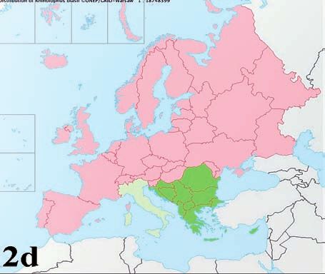
Bats and SARS-like coronaviruses                                       5

nation which, together with high mutation rates,              Nonetheless, the finding of SARS-CoV in mar-
may allow them to adapt to new hosts and eco-                 ketplace animals in China has provided evidence
logical niches. The main targets are epithelial               for interspecies transmission in the genesis of the
cells, causing mainly enteric and respiratory dis-            SARS epidemic (Lau et al., 2005).
eases of varying severity. Transmission occurs                The research of the wild animal reservoir of
through fecal-oral secretions and excretions.                 SARS-CoV subsequently led to the discovery of a
                                                              great variety of coronaviruses belonging to the
Sars and bat coronaviruses                                    first two coronavirus genera in several bat
The severe acute respiratory syndrome-associat-               species, first in Asia (Lau et al., 2005; Li et al.,
ed coronavirus (SARS-CoV or hu-SARS-CoV) is                   2005; Poon et al., 2005; Chu et al., 2006; Tang et
the etiologic agent of the SARS disease (Drosten              al., 2006; Woo et al., 2006; Woo et al., 2007) and
et al., 2003; Ksiazek et al., 2003; Peiris et al., 2003;      then in all the continents in which bats are found
Rota et al., 2003) which caused a global epidem-              (Dominguez et al., 2007; Brandao et al., 2008;
ic in humans in 2002-2003, resulting in more than             Carrington et al., 2008; Gloza-Raush et al., 2008;
8000 infections and 700 deaths (Centers for                   Misra et al., 2009; Pfefferle et al., 2009; Tong et
Disease Control and Prevention (CDC), 2003;                   al., 2009; Donaldson et al., 2010; Drexler et al.,
Peiris et al., 2004; Skowronski et al., 2005).                2010; Quan et al., 2010; Reusken et al., 2010;
From the initial investigations conducted in                  Rihtaric et al., 2010; Watanabe et al., 2010;
China after the outbreak of SARS, it seemed clear             Balboni et al., 2011), whereas none of the bat
that marketplace animals may have been the                    coronaviruses discovered belonged to the third
source of the virus found in humans (Li et al.,               coronavirus genus.
2006; Shi et al., 2008). Evidence of infection was            These discoveries, in association with the ances-
found in Himalayan palm civets (Paguma larva-                 tral nature of bat coronaviruses compared to
ta), Chinese ferret badgers (Melogale moschata)               coronaviruses from other animal species, have
and raccoon dogs (Nyctereutes procyonoides)                   led us to assume that all mammalian coron-
which were found to carry the SARS-CoV.                       aviruses belonging to Alphacoronavirus and
In particular, it was seen that the viruses collect-          Betacoronavirus were derived from ancestral
ed from masked palm civets in 2003 were differ-               viruses residing in bats (Vijaykrishna et al., 2007;
ent from those of 2004 indicating two separate                Woo et al., 2009).
animal-to-human transmissions: the first in the               Currently, there is no evidence of SARS-CoV
main SARS outbreak of 2002-2003 and the sec-                  transmission from bats to humans (Stockman et
ond in a series of sporadic infections in the win-            al., 2008), and the exact natural reservoir host for
ter of 2003-2004 (Song et al., 2005; Shi et al., 2008;        the progenitor virus of SARS-CoV may not have
Graham et al., 2010).                                         been determined as yet. However, a group of
However, subsequent epidemiological studies                   CoVs which are genetically closely related to the
have shown that Palm civets and other market-                 SARS-CoV have been identified in various species
place animals may not be the primary reservoir                of horseshoe bats (Rhinlophus spp.) and in some
hosts of SARS-CoV in nature, but merely a sec-                other bat genera (Lau et al., 2005; Li et al., 2005;
ondary amplifying host which increases the viral              Tang et al., 2006; Woo et al., 2006; Lau et al., 2007;
burden and provides ample contact with humans,                Woo et al., 2007; Pfefferle et al., 2009; Tong et al.,
thereby facilitating animal-to-human and human-               2009; Drexler et al., 2010; Lau et al., 2010; Quan
to-human transmission. This hypothesis is sup-                et al., 2010; Rihtaric et al., 2010; Yuan et al., 2010;
ported by the fact that:                                      Balboni et al., 2011). These viruses were called
1) SARS-CoV was present only in market or                     SARS-like CoVs. These SARS-like CoVs together
    farmed animals but not in those from the wild;            with human and marketplace animal SARS-CoV
2) SARS-CoV genomes evolved rapidly in mar-                   strains are included in the Betacoronavirus genus,
    ketplace animals, suggesting that the virus was           in the severe acute respiratory syndrome-related
    still adapting to these animals rather than per-          coronavirus (SARS-related CoV) species
    sisting in equilibrium, as would be expected              (http://www.ictvonline.org/).
    in a reservoir species (Wang et al., 2006;                There are at least three hypotheses which explain
    Graham et al., 2010).                                     the origin of the SARS-CoV:
6 A. Balboni, M. Battilani, S. Prosperi
1) some unknown intermediate hosts were re- this novel group of coronaviruses is rapidly evolv-
sponsible for the adaptation and transmission ing and may easily cross the species barrier (Lau
of the SARS-CoV from bats to marketplace an- et al., 2010).
imals or humans; Inside this bat genus, the Chinese horseshoe bat
2) there is a SARS-like CoV closely related to the (Rhinolophus sinicus) probably plays an impor-
outbreak SARS-CoV strains in a non-bat ani- tant role in the SARS-like CoV epidemiology.
mal host which is capable of direct transmis- First, the highest virus prevalence detected until
sion from a reservoir host to humans or mar- now was in R. sinicus (Shi et al., 2008) and, in
ketplace animals; addition, two SARS-like-CoVs, Rs672 and Rp3,
3) a bat yet to be identified could be the direct identified in this bat species (Li et al., 2005; Yuan
reservoir of the human or marketplace animal et al., 2010) tightly clustered together and showed
SARS-CoV because it may have an efficient vi- a potential recombinant breakpoint estimated at
ral receptor (ACE2, see below) (Hou et al., the nucleotide immediately after the start codon
2010). of the S gene, with the major parental regions
With regard to the presence of coronaviruses in phylogenetically closer to human-SARS-CoVs
bats, there may be a coevolutionary relationship than to bat-SARS-like-CoVs and the minor
between some bat-CoVs and their hosts. parental region phylogenetically closer to bat-
Therefore, with very few exceptions, most bat SARS-like-CoVs than to human-SARS-CoVs
coronaviruses seem to be species-specific, al- (Yuan et al., 2010). It is possible that the recom-
though one bat genus/species may more than one bination insertion of variant RBDs (Receptor-
type of coronavirus (Wang et al., 2006; Woo et al., Binding Domains) may have mediated the initial
2006; Cui et al., 2007). cross-species transmission event from bats into
Bat-CoV seemed to be more closely associated other mammals (Graham et al., 2010).
with bat species than with sampling location; in-
deed, the same bat species living in different ge- SARS-like-CoV genome
ographical locations can contain the same type All bat-SARS-like CoV full genomes have a gene
of coronavirus and, at the same time, different organization similar to human and civet SARS-
species roosting in the same cave may carry dif- CoVs; the genome is composed of a 5’ non-coding
ferent coronaviruses (Tang et al., 2006; Gloza- leader sequence, polymerase complex ORF1ab,
Raush et al., 2008; Reusken et al., 2010). S, ORF3 (formed to ORF3a,b,c in Rf1 strain), E,
Therefore, virus-host associations could be used M, ORF6, ORF7a,b, ORF8, ORF9a(N),b,c and 3’
to predict geographic distributions of reservoir- terminal non-coding region with a stem loop II-
borne CoVs (Drexler et al., 2010). like motif (s2m) and a polyA tail (Figure 1a).
Moreover, there seems to be a relationship be- Another genome characteristic shared by SARS
tween cornavirus infection and the age of the and SARS-like coronaviruses is the presence of a
bats; in fact, it has been shown that young bats conserved nucleotide sequence (5’-ACGAAC-3’)
provide a susceptible population for amplifying functioning as a transcription regulatory se-
the coronavirus and transmitting it to lactating quence (TRS) located at the 3’ end of the puta-
females in maternity colonies whereas the virus tive 5’ leader sequence of each predicted gene
would replicate less efficiently in adults than in start site (Marra et al., 2003; Rota et al., 2003).
young animals (Gloza-Raush et al., 2008; Rihtaric This motif is indispensable for the production of
et al., 2010). the subgenomic mRNAs and is identical in all
To date, SARS-like coronaviruses have been de- SARS-CoV ORFs and in most bat-SARS-like CoV
tected in China, Africa and Europe. With the ex- ORFs (Li et al., 2005; Woo et al., 2007).
ception of the African strains, all bat-SARS-like Nevertheless, there are considerable diversities
CoVs have been detected in some species of within the genome organisation of SARS-like
horseshoe bats (Rhinolophus spp.) where they CoVs and between these and human and civet
cause acute, self-limiting infection of the gas- SARS-CoVs (Fig.1b) (Shi et al., 2008; Quan et al.,
trointestinal tract (Lau et al., 2010). The detec- 2010). Major differences have been observed in
tion of diverse SARS-like CoV strains in the dif- nsp3 (third non-structural protein codified by
ferent Rhinolophus species have suggested that ORF 1a,b), S, ORF3 and ORF8 regions whichBats and SARS-like coronaviruses 7 showed the lowest nucleotide and amino acidic man and civet SARS-CoVs, especially localized identities in comparison to the rest of the genome in the putative S1 domain. In the bat-SARS-like (Lau et al., 2005; Shi et al., 2008; Lau et al., 2010). CoVs S1 domain, there are several insertions and The principal genome differences between SARS deletions, mainly in the RBD region, which is crit- and SARS-like viruses are located in ORF8 which ical for host-receptor binding and for immunity is a frequent site for deletion. All Bat-SARS-like response (Lau et al., 2005; Li et al., 2005). CoVs (except the HKU3-8 strains, GQ153543), The nsp3 region of ORF1a is relatively variable civet-SARS-CoVs (except one strain) and early- in the genome of bat-SARS-like CoVs and, in par- phase SARS-CoVs harbor a 29-nts sequence ticular, a deletion of 579nt in the nsp3 of the which fuses ORF8a and ORF8b into a single Rs672 strain has been identified, only found in a ORF8. Instead, in successive-phase SARS-CoVs, human-SARS-CoV (AY463060) strain of the late this 29-nts does not exist and ORF 8 is subdivid- phase of the 2003 epidemic. Yuan et al. (2010) ob- ed into two distinct ORF8s (a and b), disrupting served that this deletion probably occurred inde- the functional expression of this open reading pendently in Rs672 and Human-SARS-CoV, and frame (Lau et al., 2005; Oostra et al., 2007; Shi et was not acquired through homologous recombi- al., 2008; Graham et al., 2010; Lau et al., 2010). nation (Ren et al., 2006). The bat-SARS-like CoV strain HKU3-8 is an ex- ception, having a short deletion of 26 bp which Spike protein and its role in host tropism subdivides this ORF into three smaller ORFs (Lau The S protein is the largest structural protein en- et al., 2010). coded by all coronaviruses; it mediates receptor The S gene of bat-SARS-like CoVs presents a low association and fusion of the viral and cellular percentage of sequence identity compared to hu- membranes, and is the major antigenic determi- FIGURE 1A, B - Typical genome organisation of human and civet SARS-CoVs and bat SARS-like-CoVs. 1a (upper panel) is a schematic diagram of SARS-related CoV genome organisation. 1b (lower panel) is a schematic represen- tation of the diversities within the genome organisation of SARS-related CoVs. Light gray: little variable genome tracts, dark gray: highly variable genome tracts. *1: does not include African and European bat-SARS-like CoVs. *2: Rf1 strain presents ORF3a,b,c. *3: HKU3-8 strain presents ORF8a,b,c.
8                                       A. Balboni, M. Battilani, S. Prosperi

nant of coronaviruses. The host range of a spe-              already been shown that Rhinolophus sinicus and
cific coronavirus is largely determined by its S             Myotis daubentonii ACE2s are also capable of
protein which is composed of an N-terminal ex-               supporting SARS-CoV entry, albeit with a differ-
tremity (S1) for receptor binding and a C-termi-             ent efficiency in comparison to that of human
nal extremity (S2) for virus-cell fusion and en-             ACE2 (Hou et al., 2010).
trance. (Li et al., 2006).
The Receptor-Binding Domain (RBD) is a tract                 Possible African origin of the SARS-like CoV
of about 200 amino acids of the S1 extremity with            To date, SARS-like CoVs have been detected in
the function of linking to the cellular host recep-          Asia, Europe and Africa. In Asia and Europe, the
tor, the major immunodominant and potent neu-                SARS-like CoVs are strictly related to some
tralising epitope of the S protein. The RBD is               species of the Rhinolophus bat genus (Lau et al.,
composed of two sub-domains: a core and an ex-               2005; Li et al., 2005; Woo et al., 2006; Lau et al.,
tended loop. In the extended loop, there is the              2007; Woo et al., 2007; Drexler et al., 2010; Lau et
Receptor-Binding Motif (RBM) which directly                  al., 2010; Rihtaric et al., 2010; Yuan et al., 2010;
contacts the cellular host receptor.                         Balboni et al., 2011;) whereas, in Africa, the
The S1 extremity of bat-SARS-like CoVs has a                 SARS-like CoVs have been detected in several
low degree of similarity to that of the SARS-CoV,            species of the Hipposideros and Chaerophon bat
especially in the RBD, whereas the S2 extremity              genera, indicating the lack of a strict species-spe-
has a high degree of similarity between the two vi-          cific host restriction in this geographical area
ral groups. These differences imply that bat-                (Pfefferle et al., 2009; Tong et al., 2009; Quan et
SARS-like CoVs and the SARS-CoV made use of                  al., 2010).
different molecules as entry receptors but have              The absence of a species-specific host restriction,
the same entry mechanism (Ren et al., 2008; Shi              which bat-SARS-like CoVs have shown only in
et al., 2008).                                               the African area, is also supported by the previous
Human and civet angiotensin-converting en-                   detection of reactive antibodies against SARS-
zyme 2 (ACE2) was identified as a functional                 CoV antigens in sera belonging to seven different
cellular receptor for the SARS-CoV (expressed                bat genera (Hypsignathus, Lyssonycteris,
in the lung and the gastrointestinal tract of the            Miniopterus, Mops, Myonycteris, Rhinolophus and
hosts) whereas the SARS-like viruses found in                Rousettus) (Muller et al., 2007)
bats are unable to use any of the human, civet               Of considerable interest is ZBCoV (HQ166910),
and bat ACE2 molecules as their receptors and                the SARS-like CoV strain detected by Quan et al.
utilize another unknown receptor (Li et al., 2006;           (2010) in Nigeria which presents several genome
Ren et al., 2008; Graham et al., 2010; Hou et al.,           peculiarities in comparison to other SARS-like
2010; Yu et al., 2010). This capability of SARS-             CoVs; the principal peculiarities are:
CoVs to bind with human ACE2 is due to a min-                1) the TRS motif is not present in ORF6;
imal variation in the RBD sequence, and it is                2) the putative S protein presents greater se-
therefore possible that bat-SARS-like CoVs may                   quence differences as compared to other
attain this ability to infect human cells through                SARS- and SARS-like CoVs, especially in the
the acquisition of an S sequence capable of bind-                S1 domain involved in receptor binding;
ing to human ACE2. This event could occur by                 3) a short ORF3;
recombination with other CoVs if the same bat                4) a significantly shorter region between the M
cells carry receptors for both types of viruses,                 and N genes, caused by a large deletion, with
one capable and one not capable of binding with                  ORf6 which overlaps with the M gene, ORF7
ACE2 (Ren et al., 2008).                                         which overlaps with ORF6 and is not subdi-
This possibility is favoured by the greater genet-               vided into a and b, ORF8 which overlaps with
ic diversity of ACE2 among bats as opposed to                    ORF7 and the N gene;
that observed among known SARS-CoV-suscep-                   5) the presence of two conserved s2m (instead of
tible mammals (Hou et al., 2010). This diversity                 one) downstream from the N gene.
has also pointed that many uncharacterized bat               The ZBCoV is most closely related to the
species may be able to act as reservoirs of the              GhanaBt-CoVs detected by Pfefferle et al. (2009)
SARS-CoV or its progenitor viruses. Indeed, it has           in Ghana. Phylogenetically, these African strains
Bats and SARS-like coronaviruses                                    9

cluster together and could be considered a sepa-           the positive bats belonged to the Myotis and
rate subgroup within the Betacoronavirus, in an            Pipistrellus bat genera, but no samples from the
ancestral position to the SARS-related CoV sub-            Rhinolophus spp. were included in the study. This
group, which includes SARS-CoVs and the other              study did not identify the coronavirus correlated
Asian and European Rhinolophus SARS-like                   with the SARS-CoV but hypothesised a close as-
CoVs (Pfefferle et al., 2009; Quan et al., 2010).          sociation of bat-CoVs with bat species rather than
These findings could support the possible origin           with sampling locations, and a central role of ju-
of the SARS-like CoVs in Africa rather than in             venile bats in virus amplification and transmis-
Asia, and it is therefore possible to hypothesise          sion to adult females in maternity colonies.
that a subsequent migration occurred from Africa           In 2010, Reusken et al. (2010) carried out a study
to Asia which may have led to the origin of the            on the presence of coronaviruses in the Dutch bat
SARS-related coronavirus subgroup (Quan et al.,            population. More than 200 bats representing 13
2010).                                                     different bat species were tested using RT-PCR
                                                           for the presence of coronaviruses; a high per-
The european scenario                                      centage of infection (16,9%) was detected. This
Despite the large size and mild climate of the             study found no SARS-related CoV but coron-
Mediterranean area, the European bat popula-               aviruses belonging to the Alpha- and
tion is composed of a small number of species              Betacoronavirus were detected in some species of
(Table 2), which contrasts with the high species           Myotis, Nyctalus and Pipistrellus bat genera and
diversity characterising the order Chiroptera. Bats        a closer association of the viruses with bat species
are still present in all European countries, with          than with sample location was observed. The
the Mediterranean area having a major diversity.           16.9% prevalence of coronavirus infection de-
At present, there is still little information on the       tected in the Netherlands was almost double that
presence of coronaviruses in the European bat              found in Northern Germany (9.8%), although
population.                                                some species tested were the same. These data
Not many studies have been carried out and they            might suggest a variation in the geographic dis-
have focused, for the most part, on central-east           tribution of bat-CoVs which could be influenced
Europe, investigating only some of the species             by several factors, such as the latitude and cli-
which make up the diversity of the European bat            mate of the sampling area, the composition and
population.                                                density of the population, and different bat be-
Due to its characteristic climatic conditions and          havior and ecology.
geographical position, the Mediterranean area is           Furthermore, both studies sampled two bat
characterized by remarkable biodiversity and rep-          species belonging to the Myotis genus (M. dauben-
resents a strategic bridge for the viruses from the        tonii and M. dasycneme) more than the other
Middle East and Africa to cross to Europe.                 species and, whereas M. dasycneme had approx-
Nevertheless, with the exception of one prelimi-           imately the same prevalence of infection in both
nary study (Balboni et al., 2011), the Mediterranean       studies, M. daubentonii had a far greater preva-
area has been ignored by most other studies.               lence of infection in the Netherlands than in
The first investigation on the presence of coron-          Northern Germany. Therefore, to confirm what
aviruses in European bats was carried out in 2008          has already been hypothesized, the divergence of
by Gloza-Rausch et al. (2008) in Northern                  the prevalence of infection between different
Germany. Gloza-Rausch et al. tested faeces sam-            European areas may be more related to the sin-
pled from more than 300 bats by reverse tran-              gle species than to general environmental and ge-
scription-polymerase chain reaction (RT-PCR); a            ographical factors.
high conservative tract was amplified for all the          In 2010-2011, three other studies on coron-
coronaviruses of the RNA-dependent RNA poly-               aviruses in bats in different European countries
merase (RdRp, the 12th non structural protein              were published (Drexler et al., 2010; Rihtaric et
codified to ORF1a,b) which is frequently used for          al., 2010; Balboni et al., 2011). In these studies,
the phylogenetic comparison of coronaviruses.              the study sample also included faeces belonging
Bat-CoVs belonging to the Alphacoronavirus were            to bats of the Rhinolophus spp. which, in Europe,
detected with an overall prevalence of 9.8%. All           is represented by five different species having dif-
10 A. Balboni, M. Battilani, S. Prosperi
ferent territorial distributions (Table 2 and Fig. Rihtaric et al. (2010) carried out a study on the
2a-e) (www.faunaeur.org). These studies demon- presence of CoVs in bats in Slovenia, amplifying
strated a strict correlation between SARS-like a highly conservative fragment of the RdRp by
CoVs and Rhinolophus spp. bats in Europe as has RT-PCR. Fourteen of the 106 bats analyzed were
also been shown in Asia. positive and all belonged to the 36 Rhinolophus
TABLE 2 - Bat species present in Europe
European bat species (www.faunaeur.org)
Suborder Family Genus and species IT ES FR GE GB NL BG SL
Macrochiroptera Pteropodidae Rousettus aegyptiacus
Microchiroptera Molossidae Tadarida teniotis X X X X
Rhinolophidae Rhinolophus ferrumequinum X X X X X X X
Rhinolophus hipposideros X X X X X X X
Rhinolophus euryale X X X X X
Rhinolophus blasii X X X X
Rhinolophus mehelyi X X X X
Vespertilionidae Barbastella barbastellus X X X X X X X
Eptesicus botae
Eptesicus nilssonii X X X X X
Eptesicus serotinus X X X X X X X X
Hypsugo savii X X X X X
Miniopterus schreibersii X X X X X
Myotis alcathoe X X
Myotis aurascens X X
Myotis bechsteinii X X X X X X X X
Myotis blythii X X X X X
Myotis brandtii X X X X X X X
Myotis capaccinii X X X X X
Myotis dasycneme X X X X X
Myotis daubentonii X X X X X X X X
Myotis emarginatus X X X X X X X
Myotis myotis X X X X X X X X
Myotis mystacinus X X X X X X X X
Myotis nattereri X X X X X X X X
Myotis punicus X X
Nyctalus azoreum
Nyctalus lasiopterus X X X X
Nyctalus leisleri X X X X X X X X
Nyctalus noctula X X X X X X X X
Pipistrellus kuhlii X X X X X X
Pipistrellus maderensis X
Pipistrellus nathusii X X X X X X X X
Pipistrellus pipistrellus X X X X X X X X
Pipistrellus pygmaeus X X X X X X X
Plecotus auritus X X X X X X X X
Plecotus austriacus X X X X X X X X
Plecotus kolombatovici
Plecotus macrobullaris X X X X
Plecotus sardus X
Plecotus teneriffae X
Vespertilio murinus X X X X X X X
IT=Italy; FR=France; GB=United Kingdom; ES=Spain; GE=Germany; NL=Netherlands; BG=Bulgaria; SL=Slovenia.Bats and SARS-like coronaviruses 11 FIGURE 2 - European distribution of the Rhinolophus bat species (www.faunaeur.org): 2a: R. ferrumequinum; 2b: R. hipposideros; 2c: R. euryale; 2d: R. blasii; 2e: R. mehelyi. GREEN: present. PALE GREEN: doubtful. RED: absent.
12                                      A. Balboni, M. Battilani, S. Prosperi

hipposideros bats included in the population sam-            1) the high divergence of predicted proteins 3b
ple. Only the Rhinolophus spp. was positive to                   and 6;
coronavirus infection, and bioinformatic analysis            2) the absence of ORF8;
showed that the Slovenian bat coronaviruses be-              3) a greater similarity of the Bulgarian SARS-like
longed to the Betacoronaviruses and that the most                virus spikes RBD to SARS-CoVs than to the
closely related virus sequence in GenBank was                    other bat-SARS-like CoVs.
SARS bat Rp3 isolated from Rhinolophus sinicus               From the research carried out to date, it can be
in China in 2004 (Li et al., 2005; Rihtaric et al.,          hypothesized that CoVs are widely diffused in
2010; Yuan et al., 2010). As in the first German             European bats and that SARS-like CoVs are pres-
study, the positive bats were young bats or lactat-          ent in close correlation to bat species belonging
ing females, corroborating the hypothesis that the           to the Rhinolophus genus, as has been noted in
virus replicates more efficiently in young bats than         Asia (Tang et al., 2006; Cui et al., 2007; Lau et al.,
in adults, and that young bats transmit the virus            2007; Shi et al., 2008).
to adult females in maternity colonies.                      The full genome sequence obtained in one
In Rihtaric et al.’s study, 26 Myotis daubentonii            Bulgarian bat-SARS-like CoV (GU190215)
bats were tested and were all negative.                      (Drexler et al., 2010) also has some special fea-
Considering what had already been seen in the                tures which distinguish it from the Asian and the
Netherlands and Northern Germany for this                    African strains.
species, there may be a progressive increase in              The total absence of ORF8 is an unexpected dele-
the prevalence of coronavirus infection proceed-             tion for a bat-SARS-like-CoV genome since al-
ing from southeast to northwest Europe.                      most all SARS-related coronavirus strains de-
The research of Balboni et al. (2011) on bat CoVs            tected in the Rhinolophus spp were character-
involved only one species of the Rhinolophus                 ized by a single ORF8, with the insertion of an
genus found in the Italian peninsula, the R. fer-            29-nts sequence which fuses together the ORF8a
rumequinum.                                                  and 8b distinctive for Human-SARS-CoV. If it
They utilized the same method as that used in                were hypothesized that the 29 nts deletion in the
preceding studies, an RT-PCR amplifying a high-              ORF8 of the Human-SARS-CoV represented a
ly conserved fragment of RdRp, detecting two                 greater functional hallmark in the transition of
positives of the 52 bats sampled, with a virus               the SARS-CoV to humans (Lau et al., 2005;
prevalence of 3.8%. Sequence analysis of one                 Oostra et al., 2007), the absence of this ORF in
strain showed that it was genetically closely re-            the European SARS-like CoV would suggest that
lated to SARS- and other SARS-like coronavirus-              protein 8 may not, in general, be essential for the
es belonging to the Betacoronavirus.                         maintenance of the SARS-like CoV in bats
Furthermore, the Italian strain showed a close               (Drexler et al., 2010).
correlation to the Slovenian strains whereas the             This peculiar deletion in the Bulgarian SARS-like
similarities with the African strain were low.               CoV, in association with the high nucleotide vari-
Drexler et al. (2010) conducted an evaluation of             ability of some genes and the greater similarity
CoVs in rhinolophid and vespertilionid bat                   of spikes RBD with the SARS-CoV than with oth-
species in Bulgaria, in an area in which all five            er bat-SARS-like CoVs, suggests a possible diver-
European Rhinolophidae species coexist. Two                  gence between the Asian bat SARS-like CoVs and
hundred and two of the 499 bats were positive                the European bat SARS-like CoVs.
with RT-PCR or nested RT-PCR when amplifying                 This assumption requires more extensive inves-
a highly conserved fragment of RdRp. The virus-              tigation on the European bat population to ac-
es detected were either Alpha- and Betacorona-               quire more information on the epidemiology of
virus, but all the SARS-like CoVs occurred ex-               bat-SARS-like CoVs on this continent.
clusively in Rhinolophus (101 positives out of 389           The studies should also focus more closely on the
Rhinolophus bats sampled).                                   bat species belonging to the Rhinolophus spp. and
A Bulgarian SARS-like virus was fully sequenced              on the epidemiological situation in the
and its genome showed some special features in               Mediterranean area which could play an impor-
comparison with other known SARS-like CoVs,                  tant role in the evolution of these viruses.
namely:
Bats and SARS-like coronaviruses                                     13

DISCUSSION                                                traced back to a common ancestor with the
                                                          SARS-related CoV subgroup.
In recent decades, with the emergence of several          The African origin may have been followed by a
zoonoses which have had bats as animal reser-             migration which brought the bat-SARS-like CoVs
voirs, numerous studies together on the central           to adapt to bats belonging to the Rhinolophus
role of bats in the dissemination of zoonotic             spp., thus arriving in Asia and Europe.
agents have been carried out. Among these, a              It is unclear whether the arrival of the bat-SARS-
high variability of coronavirus strains has been          like CoVs in Europe followed the viral adaptation
discovered in several species of bats in Asia,            in Asian bats, or whether these viruses were first
North and South America, Africa and Europe af-            colonized in Europe, via the Mediterranean area,
ter the SARS epidemic in the past ten years.              and from there were then passed on to Asia. On
All coronaviruses found in bats belong to two of          the other hand, the arrival of SARS-like CoVs
the three coronavirus genera: Alphacoronavirus            from Africa to European and Asian bats could al-
and Betacoronavirus. Of particular importance,            so have been the result of two independent events,
with regard to the epidemiology of SARS-CoV, is           with the origin of distinct viral populations, rather
a group of coronaviruses found in some species            than two consecutive events.
of bats and belonging to the Betacoronavirus, the         Some special features of the Bulgarian SARS-like
bat-SARS-like coronaviruses. These viruses are            CoV distinguish it from the Asian and African
genetically closely related to the SARS-CoV and           strains (Drexler et al., 2010), but only one com-
are probably the ancestral viruses of the market-         pletely sequenced European strain is not suffi-
place animal- and human- SARS-CoVs which                  cient to clarify the evolution and the migratory
caused the human epidemic in 2002-2003. To                events which could have characterized these
date, bat-SARS-like CoVs have been discovered             viruses in Europe.
in Asia, Africa and Europe. Taking into account           More detailed studies on the European bat pop-
that there have been many more studies carried            ulation with particular reference to the
out on the Asian region than in Africa and                Mediterranean area are required to clarify these
Europe, some differences can be pointed out re-           aspects of the bat-SARS-like CoV evolution and
garding these three continents.                           the ramifications concerning public health as re-
First, the host range of bat-SARS-like CoVs is lim-       gards the potential of coronaviruses to cross
ited to the Rhinolophus bat genus in both Asia            species barriers and thus infect humans, as peo-
and Europe, suggesting a close species-specific           ple come into closer contact with wild animals.
relationship. Instead, bat-SARS-like CoVs were            In this regard, and to better assess the prevalence
found in numerous different bat species belong-           of infection of coronaviruses in bats, the devel-
ing to several genera.                                    opment and use of diagnostic techniques with
Second, numerous genomic features differenti-             high sensitivity is important. Indeed, the princi-
ating the coronaviruses belonging to the three            pal diagnostic method used by various re-
continents have been added to the host differ-            searchers, especially during the initial screening
ences. Thus, if it is reasonable to think that the        phase of the population sample, is a reverse tran-
SARS-CoV causing the human epidemic derived               scription-PCR (RT-PCR) amplifying a fragment
from the SARS-like CoVs found in Asian bats, the          of variable size of the RNA-dependent RNA poly-
theories regarding the area of origin and the phy-        merase gene (RdRp, the 12th non structural pro-
logenetic evolution of these viruses are more con-        tein codified to ORF1a, b), which is a genome
troversial.                                               tract frequently used for the molecular detection
A possible origin of the SARS-like CoVs in Africa         and phylogenetic analysis of bat coronaviruses
was hypothesized by Quan et al. (2010) in the             (Lau et al., 2005; Lau et al., 2007; Woo et al., 2007;
light of the characteristics of some strains, such        Lau et al., 2010; Rihtaric et al., 2010; Yuan et al.,
as the ZBCoV and GhanaBt-CoVs (Pfefferle et al.,          2010; Balboni et al., 2011). However, this method
2009; Quan et al., 2010) identified in Nigeria and        may not have optimal sensitivity, causing an un-
Ghana, respectively, which could form a separate          derestimation of the true prevalence of infection.
subgroup distinct from Asian and European                 This may be due to two main factors: first, bats
Rhinolophus SARS-related CoVs and could be                are reservoirs of coronaviruses and may shed on
14 A. Balboni, M. Battilani, S. Prosperi
a small amount of viruses in faeces, too low to be CHU D.K., POON L.L., CHAN K.H., CHEN H., GUAN Y.,
detected by traditional PCR methods; second, the YUEN K.Y., PEIRIS J.S. (2006). Coronaviruses in bent-
RdRp, although highly conserved in coron- winged bats (Miniopterus spp.). J. Gen. Virol. 87
(Pt 9), 2461-2466.
aviruses, has a moderate degree of nucleotide
CUI J., HAN N., STREICKER D., LI G., TANG X., SHI Z., HU
variability which can result in a sub-optimal Z., ZHAO G., FONTANET A., GUAN Y., WANG L., JONES
match of the primers used. G., FIELD H.E., DASZAK P., ZHANG S. (2007).
In the near future, the tactics used by humans to Evolutionary relationships between bat coron-
prevent the spread of bat-associated zoonosis, aviruses and their hosts. Emerg. Infect. Dis. 13 (10),
should be based on two main lines of action: on 1526-1532.
the one hand systematic surveillance with ap- DOMINGUEZ S.R., O’SHEA T.J., OKO L.M., HOLMES K.V.
(2007). Detection of group 1 coronaviruses in bats
propriate techniques aimed at controlling known
in North America. Emerg. Infect. Dis. 13 (9), 1295-
pathogens, such as the bat-SARS-like CoV, and 1300.
discovering possible new zoonotic agents; on the DONALDSON E.F., HASKEW A.N., GATES J.E., HUYNH J.,
other hand, the conservation of the natural habi- MOORE C.J., FRIEMAN M.B. (2010). Metagenomic
tats of bats, thus avoiding direct contact with hu- analysis of the viromes of three North American
mans and livestock. bat species: viral diversity among different bat
species that share a common habitat. J. Virol. 84
(24), 13004-13018.
REFERENCES DREXLER J.F., GLOZA-RAUSCH F., GLENDE J., CORMAN
V.M., MUTH D., GOETTSCHE M., SEEBENS A., NIEDRIG
ALTRINGHAM J.D. (1996a). Bats, biology and behaviour. M., PFEFFERLE S., YORDANOV S., ZHELYAZKOV L.,
New York: Oxford university press. Introduction, 1-4. HERMANNS U., VALLO P., LUKASHEV A., MÜLLER M.A.,
ALTRINGHAM J.D. (1996b). Bats, biology and behaviour. DENG H., HERRLER G., DROSTEN C. (2010). Genomic
New York: Oxford university press. Chapter 4, 115- characterization of severe acute respiratory syn-
132. drome-related coronavirus in European bats and
ALTRINGHAM J.D. (1996c). Bats, biology and behaviour. classification of coronaviruses based on partial
New York: Oxford university press. Chapter 6, 155- RNA-dependent RNA polymerase gene sequences.
198. J. Virol. 84 (21), 11336-11349.
ALTRINGHAM J.D. (1996d). Bats, biology and behaviour. DROSTEN C., GÜNTHER S., PREISER W., VAN DER WERF S.,
New York: Oxford university press. Chapter 7, 199- BRODT H.R., BECKER S., RABENAU H., PANNING M.,
229. KOLESNIKOVA L., FOUCHIER R.A., BERGER A.,
BALBONI A., PALLADINI A., BOGLIANI G., BATTILANI M. BURGUIÈRE A.M., CINATL J., EICKMANN M., ESCRIOU
(2011). Detection of a virus related to betacoron- N., GRYWNA K., KRAMME S., MANUGUERRA J.C.,
aviruses in Italian greater horseshoe bats. MÜLLER S., RICKERTS V., STÜRMER M., VIETH S.,
Epidemiol. Infect. 139 (2), 216-219. KLENK H.D., OSTERHAUS A.D., SCHMITZ H., DOERR
BRANDÃO P.E., SCHEFFER K., VILLARREAL L.Y., ACHKAR H.W. (2003). Identification of a novel coronavirus
S., OLIVEIRA RDE N., FAHL WDE O., CASTILHO J.G., in patients with severe acute respiratory syndrome.
KOTAIT I., RICHTZENHAIN L.J. (2008). A coronavirus N. Engl. J. Med. 348 (20), 1967-1976.
detected in the vampire bat Desmodus rotundus. FAUQUET C.M., MAYO M.A., MANILOFF J., DESSELBERGER
Braz. J. Infect. Dis. 12 (6), 466-458. U., BALL L.A. (2005). Virus Taxonomy ed.VIII.
CALISHER C.H., CHILDS J.E., FIELD H.E., HOLMES K.V., Amsterdam: Elsevier academic press., 937-955.
SCHOUNTZ T. (2006). Bats: important reservoir hosts GLOZA-RAUSCH F., IPSEN A., SEEBENS A., GÖTTSCHE M.,
of emerging viruses. Clin. Microbiol. Rev. 19 (3), PANNING M., FELIX DREXLER J., PETERSEN N., ANNAN
531-545. A., GRYWNA K., MÜLLER M., PFEFFERLE S., DROSTEN
CARRINGTON C.V., FOSTER J.E., ZHU H.C., ZHANG J.X., C. (2008). Detection and prevalence patterns of
SMITH G.J., THOMPSON N., AUGUSTE A.J., RAMKISSOON group I coronaviruses in bats, northern Germany.
V., ADESIYUN A.A., GUAN Y. (2008). Detection and Emerg. Infect. Dis. 14 (4), 626-631.
phylogenetic analysis of group 1 coronaviruses in GRAHAM R.L., BARIC R.S. (2010). Recombination, reser-
South American bats. Emerg. Infect. Dis. 14 (12), voirs, and the modular spike: mechanisms of coro-
1890-1893. navirus cross-species transmission. J. Virol. 84 (7),
CENTERS FOR DISEASE CONTROL AND PREVENTION (CDC). 3134-3146.
(2003). Outbreak of severe acute respiratory syn- HOU Y., PENG C., YU M., LI Y., HAN Z., LI F., WANG L.F.,
drome-worldwide, 2003. MMWR Morb Mortal Wkly SHI Z. (2010). Angiotensin-converting enzyme 2
Rep. 2003 Mar 21. 52 (11), 226-228. Erratum in: (ACE2) proteins of different bat species confer vari-
MMWR Morb Mortal Wkly Rep. 2003 Apr 4. 52 (13), able susceptibility to SARS-CoV entry. Arch. Virol.
284. 155 (10), 1563-1569.Bats and SARS-like coronaviruses 15 KSIAZEK T.G., ERDMAN D., GOLDSMITH C.S., ZAKI S.R., GARBUTT M., GRAY M., GROLLA A., JONES S., PERET T., EMERY S., TONG S., URBANI C., COMER J.A., FELDMANN H., MEYERS A., KABANI A., LI Y., NORMAND LIM W., ROLLIN P.E., DOWELL S.F., LING A.E., S., STROHER U., TIPPLES G.A., TYLER S., VOGRIG R., HUMPHREY C.D., SHIEH W.J., GUARNER J., PADDOCK WARD D., WATSON B., BRUNHAM R.C., KRAJDEN M., C.D., ROTA P., FIELDS B., DERISI J., YANG J.Y., COX PETRIC M., SKOWRONSKI D.M., UPTON C., ROPER R.L. N., HUGHES J.M., LEDUC J.W., BELLINI W.J., (2003). The Genome sequence of the SARS-associ- ANDERSON L.J.; SARS WORKING GROUP. (2003). A ated coronavirus. Science. 300 (5624), 1399-1404. novel coronavirus associated with severe acute res- MISRA V., DUMONCEAUX T., DUBOIS J., WILLIS C., NADIN- piratory syndrome. N. Engl. J. Med. 348 (20), 1953- DAVIS S., SEVERINI A., WANDELER A., LINDSAY R., 1966. ARTSOB H. (2009). Detection of polyoma and coro- KUNZ T.H., FENTON M.B. (2003a). Bat ecology. Chicago: na viruses in bats of Canada. J. Gen. Virol. 90 (Pt 8), The University of Chicago Press. Chapter 1, 3-89. 2015-2022. KUNZ T.H., FENTON M.B. (2003b). Bat ecology. Chicago: MÜLLER M.A., PAWESKA J.T., LEMAN P.A., DROSTEN C., The University of Chicago Press. 4, 156-208. GRYWNA K., KEMP A., BRAACK L., SONNENBERG K., KUNZ T.H., FENTON M.B. (2003c). Bat ecology. Chicago: NIEDRIG M., SWANEPOEL R. (2007). Coronavirus an- The University of Chicago Press. 14, 622-679. tibodies in African bat species. Emerg. Infect. Dis. LAU S.K., WOO P.C., LI K.S., HUANG Y., TSOI H.W., WONG 13 (9), 1367-1370. B.H., WONG S.S., LEUNG S.Y., CHAN K.H., YUEN K.Y. OOSTRA M., DE HAAN C.A., ROTTIER P.J. (2007). The 29- (2005). Severe acute respiratory syndrome coron- nucleotide deletion present in human but not in avirus-like virus in Chinese horseshoe bats. Proc. animal severe acute respiratory syndrome coron- Natl. Acad. Sci. U S A. 102 (39), 14040-14045. aviruses disrupts the functional expression of open LAU S.K., WOO P.C., LI K.S., HUANG Y., WANG M., LAM reading frame 8. J. Virol. 81 (24), 13876-13888. C.S., XU H., GUO R., CHAN K.H., ZHENG B.J., YUEN PEIRIS J.S., LAI S.T., POON L.L., GUAN Y., YAM L.Y., LIM K.Y. (2007). Complete genome sequence of bat W., NICHOLLS J., YEE W.K., YAN W.W., CHEUNG M.T., coronavirus HKU2 from Chinese horseshoe bats CHENG V.C., CHAN K.H., TSANG D.N., YUNG R.W., NG revealed a much smaller spike gene with a different T.K., YUEN K.Y.; SARS STUDY GROUP. (2003). evolutionary lineage from the rest of the genome. Coronavirus as a possible cause of severe acute res- Virology. 367 (2), 428-439. piratory syndrome. Lancet. 361 (9366), 1319-1325. LAU S.K., LI K.S., HUANG Y., SHEK C.T., TSE H., WANG Peiris J.S., Guan Y., Yuen K.Y. (2004). Severe acute res- M., CHOI G.K., XU H., LAM C.S., GUO R., CHAN K.H., piratory syndrome. Nat. Med. 10, S88-97. ZHENG B.J., WOO P.C., YUEN K.Y. (2010). PFEFFERLE S., OPPONG S., DREXLER J.F., GLOZA-RAUSCH Ecoepidemiology and complete genome compari- F., IPSEN A., SEEBENS A., MÜLLER M.A., ANNAN A., son of different strains of severe acute respiratory VALLO P., ADU-SARKODIE Y., KRUPPA T.F., DROSTEN C. syndrome-related Rhinolophus bat coronavirus in (2009). Distant relatives of severe acute respirato- China reveal bats as a reservoir for acute, self-lim- ry syndrome coronavirus and close relatives of hu- iting infection that allows recombination events. J. man coronavirus 229E in bats, Ghana. Emerg. Virol. 84 (6), 2808-2819. Infect. Dis. 15 (9), 1377-1384. LI W., SHI Z., YU M., REN W., SMITH C., EPSTEIN J.H., POON L.L., CHU D.K., CHAN K.H., WONG O.K., ELLIS WANG H., CRAMERI G., HU Z., ZHANG H., ZHANG J., T.M., LEUNG Y.H., LAU S.K., WOO P.C., SUEN K.Y., MCEACHERN J., FIELD H., DASZAK P., EATON B.T., YUEN K.Y., GUAN Y., PEIRIS J.S. (2005). Identification ZHANG S., WANG L.F. (2005). Bats are natural reser- of a novel coronavirus in bats. J. Virol. 79 (4), 2001- voirs of SARS-like coronaviruses. Science. 310 2009. (5748), 676-679. QUAN P.L., FIRTH C., STREET C., HENRIQUEZ J.A., LI W., WONG S.K., LI F., KUHN J.H., HUANG I.C., CHOE PETROSOV A., TASHMUKHAMEDOVA A., HUTCHISON S.K., H., FARZAN M. (2006). Animal origins of the severe EGHOLM M., OSINUBI M.O., NIEZGODA M., OGUNKOYA acute respiratory syndrome coronavirus: insight A.B., BRIESE T., RUPPRECHT C.E., LIPKIN W.I. (2010). from ACE2-S-protein interactions. J. Virol. 80 (9), Identification of a severe acute respiratory syn- 4211-4219. drome coronavirus-like virus in a leaf-nosed bat in MARRA M.A., JONES S.J., ASTELL C.R., HOLT R.A., Nigeria. MBio. 1 (4), e00208-00210. BROOKS-WILSON A., BUTTERFIELD Y.S., KHATTRA J., REN W., LI W., YU M., HAO P., ZHANG Y., ZHOU P., ZHANG ASANO J.K., BARBER S.A., CHAN S.Y., CLOUTIER A., S., ZHAO G., ZHONG Y., WANG S., WANG L.F., SHI Z. COUGHLIN S.M., FREEMAN D., GIRN N., GRIFFITH O.L., (2006). Full-length genome sequences of two SARS- LEACH S.R., MAYO M., MCDONALD H., MONTGOMERY like coronaviruses in horseshoe bats and genetic S.B., PANDOH P.K., PETRESCU A.S., ROBERTSON A.G., variation analysis. J. Gen. Virol. 87 (Pt 11), 3355- SCHEIN J.E., SIDDIQUI A., SMAILUS D.E., STOTT J.M., 3359. YANG G.S., PLUMMER F., ANDONOV A., ARTSOB H., REN W., QU X., LI W., HAN Z., YU M., ZHOU P., ZHANG BASTIEN N., BERNARD K., BOOTH T.F., BOWNESS D., S.Y., WANG L.F., DENG H., SHI Z. (2008). Difference CZUB M., DREBOT M., FERNANDO L., FLICK R., in receptor usage between severe acute respiratory
You can also read