The structure of DNA: Cooperation and competition
←
→
Page content transcription
If your browser does not render page correctly, please read the page content below
1
The structure of DNA: Cooperation and competition
During the early 1950s, the intellectual journeys of a bird biologist,
an expert on the structure of coal, a designer of underwater mines,
and a nuclear physicist intersected, resulting—not in a submarine
explosion of feathers, as one might expect—but in a discovery that
offered a glimpse of the molecular mechanisms that underlie all life,
paving the way for a revolution in molecular biology. The insight,
innovation, and persistence of James Watson, Rosalind Franklin,
Francis Crick, and Maurice Wilkins led to a detailed understanding
of the structure of DNA, the stuff that genes are made of (Fig. 1).
This discovery brought together information from many disciplines
and many researchers to answer one of the most fundamental ques-
tions in life science: How do living things pass on traits to their
offspring?
This case study highlights the following aspects of the nature of sci-                        Fig. 1. Clockwise from top left, James
                                                                                              Watson, Francis Crick, Maurice Wilkins, and
ence:                                                                                         Rosalind Franklin. Center, Image B 51, a
                                                                                              key piece of evidence in the discovery of the
   • Science can test hypotheses about things that are too small for                          structure of DNA.
   us to observe directly.
   • Science relies on communication within a diverse scientific community.
   • Scientists are expected to give credit where credit is due.
   • Scientific discoveries lead to ongoing research.

The right timing
Scientific discoveries may seem like sudden breakthroughs—the work of a genius who just “sees” the answer—
but new findings don’t come out of nowhere. Each breakthrough is made possible by the work that came be-
fore it. Some scientific discoveries are a bit like putting together the pieces of a puzzle. Many different research-
ers discover important bits of evidence—pieces of the puzzle—and the sudden breakthrough arises when one
group or person sees how the puzzle pieces logically fit together. And sometimes—as in the case of DNA—new
findings and technological advances have made so many new puzzle pieces available that the odds of someone
putting them together seem quite high. Making this final leap often
involves a brilliant insight—but it’s important to recognize all the
clues which made that insight possible.
In the 19th century, the Austrian monk, Gregor Mendel, discovered
basic patterns of inheritance. Traits pass from parent to offspring in
an organized and predictable way. Although the scientists that fol-
lowed in Mendel’s footsteps had no concrete understanding of what
caused these distinct patterns, they knew that the explanation of
inheritance would have to account for them.
By the 1940s and 50s, scientists were getting closer to a physical expla-                     Fig. 2. Human chromosomes magnified
nation of how parents pass on traits to their offspring. New technology                       1000 times.

Crick and Watson photos from an image courtesy of Cold Spring Harbor Laboratory Library and Archive, James D. Watson
Collection; Franklin photo © Henry Grant Collection / Museum of London; Maurice Wilkins in the lab photo © King’s College
Archives; Photo of X-ray diffraction pattern courtesy of Cold Spring Harbor Laboratory Library and Archive, James D. Watson
Collection; chromosomes photo © 2005 The University of New Mexico

© 2007 The University of California Museum of Paleontology, Berkeley, and the Regents of the University of California • www.understandingscience.org
2
had made it possible to observe smaller structures than ever before.
Biologists had found that genetic instructions are carried on parts of
the cell known as chromosomes (Fig. 2), and chemists had discovered
that these chromosomes are made up of two components: proteins and
DNA. Furthermore, experiments looking for the key molecule of life
had zeroed in on DNA, and not protein, as the component that actu-
ally carries genetic information (Fig. 3).
But exactly how could DNA carry all the information needed to Fig. 3.
make a new organism? The answer might be revealed by the mol-
ecule’s three-dimensional structure—and some tantalizing clues regarding this structure were becoming avail-
able. Researchers already knew that DNA was a relatively simple molecule. It seemed to consist of an un-
remarkable chain of phosphates and sugars, in some way attached to a set of ring-shaped molecules called
nitrogenous bases. These bases come in four “flavors”: adenine (A), thymine (T), cytosine (C), and guanine
(G) (Fig. 4). Somehow, these simple components would have to carry all the instructions necessary to make
fruit flies, oak trees, humans, and the rest of life. Some early work suggested that the bases were arranged like
a stack of pancakes in the molecule, each 0.34 nanometers apart from one another,1 but other than that, little
was known about exactly how a DNA molecule was put together.
To further complicate matters, researchers had dis-
covered another intriguing—but perplexing—clue.
DNA’s bases always occur in the same special ratios:
the amount of A is always equal to the amount of
T, and C is always equal to G—though the ratio of
A/T to C/G varies from species to species.2 What this
meant wasn’t clear, but any hypothesis about DNA’s
three-dimensional structure would have to account
for this strange observation.
Around the same time that these three puzzle pieces
were discovered (Fig. 5), more and more physicists
and chemists were becoming interested in applying
their knowledge and skills to learning about the phys-
                                                            Fig. 4. The molecular components of DNA: phosphates,
ical basis for life. To add further fuel to the fire, tech- deoxyribose (a sugar), and the four nitrogenous bases,
nological breakthroughs and refinements had recently adenine, guanine, cytosine, and thymine.
offered scientists new ways to study the positions of
atoms within molecular structures. Together, these factors were providing scientists with the tools and insights
to put the DNA puzzle together. The stage was set for a breakthrough.

1
 Astbury, W.T. 1939. X-ray study of thymonucleic acid. Nature 141(3573):747-748.
2
 Chargaff, E. 1950. Chemical specificity of nucleic acids and mechanism of their enzymatic degradation. Experientia
6(6):201-209.

© 2007 The University of California Museum of Paleontology, Berkeley, and the Regents of the University of California • www.understandingscience.org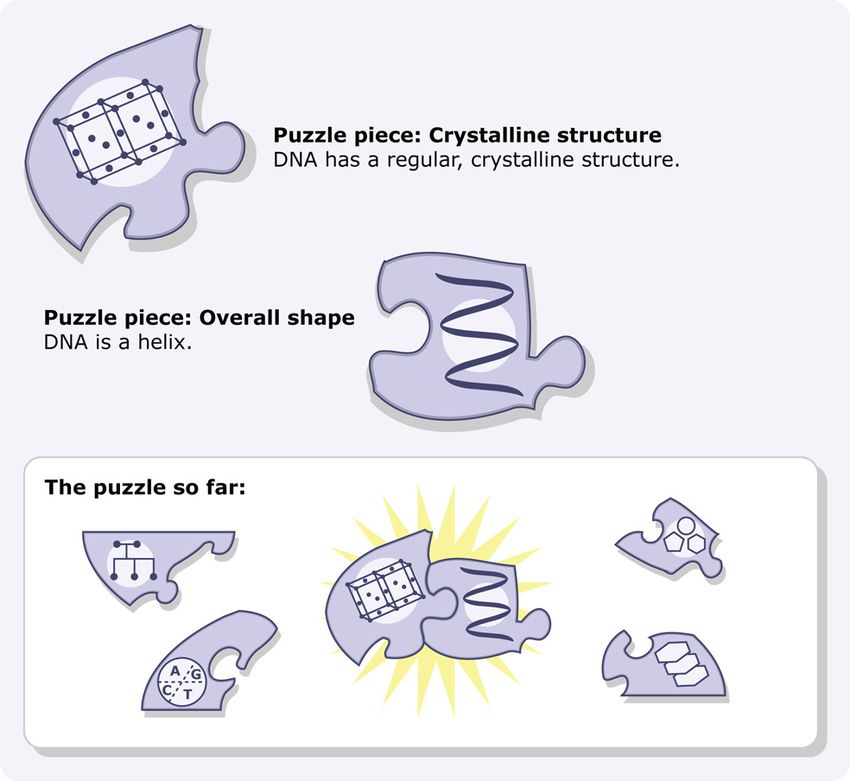
3

                                     Fig. 5.

The great race
Onto the scene, from all different directions, came Wilkins and
Franklin, then Watson and Crick (Fig. 6). Though their scientific
backgrounds were diverse, all four recognized that understanding
how the parts of a DNA molecule fit together would provide impor-
tant information about the way life works. Each hoped to be part of
the team that solved the puzzle first. Wilkins and Franklin worked
together at the University of London, and Crick and Watson col-
laborated at Cambridge University—but they weren’t the only sci-
entists thinking about DNA. Several other groups also recognized
that the three-dimensional structure of DNA was within reach, so
the competition was stiff. Linus Pauling, a soon-to-be Nobel Prize
winner who had already solved a complicated molecular structure
found in proteins, led one of the groups working to identify DNA’s
structure. The number of people investigating the problem made it                             Fig. 6.

a race right from the beginning. The edges of the puzzle were laid
out—but who would be the first to fill in all the pieces?

© 2007 The University of California Museum of Paleontology, Berkeley, and the Regents of the University of California • www.understandingscience.org
4
Atoms and X-rays: Seeing inside a crystal
Maurice Wilkins, the nuclear physicist, entered the race for DNA based on a
stroke of luck. After his work with the Manhattan Project on atomic bombs was
completed, he wanted to switch to a more peaceful line of work and was inspired
to investigate the physical basis for life. He turned to the fast-growing field of bio-
physics, taking up a position at the University of London. Early in his career there,
he happened to attend a conference where a biochemist gave away samples of
high-quality DNA. Wilkins was lucky enough to get a sample—though it might
not have seemed that impressive at the time. It was so slimy and gooey that he
later described it as “just like snot.” Nevertheless, because it contained long, intact
DNA molecules—which were hard to come by at the time—this slippery sample
would turn out to be critical in uncovering clues to DNA’s structure. Raymond
Gosling (Fig. 7), a Ph.D. student in Wilkins’ lab, suggested looking at the DNA                                  Fig. 7. Raymond Gosling, a
with a new observational technique called X-ray diffraction.                                                     Ph.D. student in the lab at
                                                                                                                 the University of London.
                                                             X-ray diffraction, devel-
                                                             oped in the first half of the 20th century, was one of
                                                             the new technologies that made solving the structure
                                                             of DNA possible. The technique works on crystals, a
                                                             kind of molecule with a regular, repeating structure.
                                                             When X-rays are aimed through a sample, they are
                                                             bent or diffracted in different directions depending
                                                             on the locations of the atoms in the sample, and the
                                                             final direction of the X-rays can be recorded on film
                                                             (Fig. 8). Because the X-rays must travel through many
                                                             layers of atoms, it’s important that the atoms always
                                                             occur in the same crystalline arrangement. If they
Fig. 8.
                                                             don’t, the X-rays are bent into overlapping patterns,
leaving the results a fuzzy, indistinct blur. However, if the structure has a repeating arrangement of atoms, they
leave a pattern of sharp, clear spots. Different structures scatter the X-rays into different characteristic patterns.
Even though scientists couldn’t directly observe the atoms within the crystal, they
could work backward from X-ray diffraction patterns to reconstruct the three-di-
mensional structure that produced the scattering. This works a little like trying to fig-
ure out how tall a person is by looking at his or her shadow. Depending on the angle
of the sun, the shadow might be longer or shorter, but if you could compare many
pictures of their shadow at different times of day, you’d eventually be able to figure
out how tall they were. Similarly, scientists compare many “shadows,” or X-ray dif-
fraction patterns, cast by a crystal to determine the arrangement of atoms within it.
Although the DNA didn’t look very crystalline, Gosling wanted to try X-ray diffrac-
tion on the molecule anyway. Wilkins and Gosling knew DNA’s structure might
be too irregular to produce a clear, well-defined X-ray pattern, but as it turned
out, the sample was sticky and stringy because it was made up of lots of long, thin
                                                                                                                 Fig. 9. This X-ray diffraction
molecules of intact, crystalline DNA. Over the summer of 1950, Wilkins and                                       pattern photographed by
Gosling’s patterns showed that DNA did have a regular structure—which meant                                      Gosling and Wilkins in 1950
                                                                                                                 showed that DNA did have a
that X-ray diffraction would be a critical tool in solving the structure (Fig. 9). The                           crystalline structure.

Gosling portrait © King’s College Archives; Wilkins-Gosling 1950 diffraction pattern from a contribution by R.G. Gosling to
Genesis of a Discovery: DNA Structure, edited by S. Chomet, 1993, Newman-Hemisphere, London

© 2007 The University of California Museum of Paleontology, Berkeley, and the Regents of the University of California • www.understandingscience.org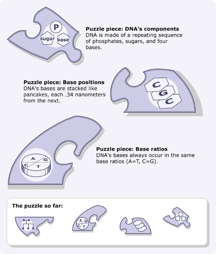
5
patterns even suggested what that basic structure might be. Despite a few confusing blurry spots, the images
hinted that DNA might come in the form of a twisted spiral—better known as a helix—though it was still not
clear how the phosphates, sugars, and bases were arrayed within that helix (Fig. 10).

                                     Fig. 10.

Passing puzzle pieces
                                     Wilkins’ and Gosling’s results were intriguing enough to be communicated to the
                                     scientific community. Researchers regularly share their findings and ideas with one
                                     another so that others can evaluate and build upon them. Scientific conferences
                                     provide a direct way of doing this. In May of 1951, Wilkins set off to present the
                                     results at a conference in Italy. There, these tantalizing clues would inspire another
                                     scientist to join in the race for the structure of DNA.
                             James Watson (Fig. 11) was studying biochemistry at the Naples Marine Station—
                             but he spent every spare moment reading about genes and the molecules they might
                             be made of. Although Watson began his career studying birds, he had switched to
                             genetics as a graduate student. He felt that understanding genes was essential to figur-
                             ing out how life worked—
                             and all the latest evidence
Fig. 11. James Watson, 1949. suggested that genes were
                             made of DNA. So when
Wilkins presented his findings—the most detailed in-
formation then available on the structure of DNA—
Watson was in the audience, watching with interest
(Fig. 12). He could see from Wilkins’ results that there
was a repeating pattern to DNA’s structure. If he could
just figure out what caused that pattern, he thought he
would be able to unravel the structure itself.             Fig. 12.

Watson photo courtesy of Cold Spring Harbor Laboratory Library and Archive, James D. Watson Collection

© 2007 The University of California Museum of Paleontology, Berkeley, and the Regents of the University of California • www.understandingscience.org
6
                                                        Inspired by this clue, Watson decided to devote all his time and
                                                        energy to understanding the structure of DNA. At first, he wanted
                                                        to join Wilkins’ lab—but Wilkins didn’t have any room. Instead,
                                                        in the autumn of 1951, he joined another lab specializing in X-ray
                                                        diffraction, at Cambridge University. There, he shared Wilkins’ and
                                                        Gosling’s clue about DNA with someone who would soon join him
                                                        in the race for the structure of DNA, Francis Crick (Fig. 13).
                                           Like Wilkins, Crick had started out as a physicist. During World
                                           War II, he put his scientific training to work designing underwater
                                           mines. After the war, he got interested in studying the physical basis
Fig. 13. Francis Crick and James Watson in
the 1950s.                                 of life and joined a Cambridge biology lab. There, Crick launched
                                           into an investigation of protein structure—but his concentration on
this project was soon to be interrupted by Watson’s arrival in the lab.
Watson’s enthusiasm for DNA was contagious. He
was convinced by the published results suggesting that
genes were made of DNA. And though he did not yet
know that a helical structure had been suggested for
DNA, he had seen the evidence from Wilkins’ pre-
sentation indicating that the structure of DNA was
simple enough to solve. Watson shared this evidence
with Crick—who eventually decided to join the race
himself (Fig. 14).

                                                                           Fig. 14.

Franklin joins the fray
                                     While Crick and Watson were joining forces at Cambridge, things were changing
                                     back in Wilkins’ lab at the University of London too. The preliminary findings
                                     were exciting—they knew that DNA had a regular structure—but they still had
                                     to figure out what that structure was. Expert help was needed to improve and
                                     interpret the X-ray results. Luckily, Rosalind Franklin, a scientist who specialized
                                     in X-ray diffraction, had just joined the lab (Fig. 15). Franklin was used to work-
                                     ing with messy materials that came from living things—she had just finished an
                                     important study applying X-ray diffraction to coal, the compressed remains of
                                     ancient swamp plants. She was asked to lend her expertise to the DNA project,
                                     and it soon caught her imagination.
Fig. 15. Rosalind Franklin at
                            Franklin began working with Raymond Gosling, the graduate student who had
work in 1954.
                            encouraged Wilkins to try X-ray diffraction on his DNA sample. Over the sum-
mer of 1951, she taught Gosling the exacting X-ray diffraction techniques she’d developed. They exposed the
special high-quality DNA sample to a range of different humidities, from wet to dry. In the dry atmosphere,
the strands appeared to thicken, and the X-ray patterns turned into a sharp scatter with many distinct spots.
As they added moisture to the atmosphere, the strands stretched, and the X-ray pattern changed to a clear x
shape (Fig. 16).

Crick and Watson photo adapted from an image courtesy of Cold Spring Harbor Laboratory Library and Archive, James D.
Watson Collection; Franklin photo © Henry Grant Collection / Museum of London

© 2007 The University of California Museum of Paleontology, Berkeley, and the Regents of the University of California • www.understandingscience.org
7
The two different patterns demonstrated that DNA
existed in two forms: the dry A form, which held less
water, and the wet B form, in which water molecules
cling to the DNA, causing it to stretch out (Fig. 17).
The first X-ray images of DNA taken by Wilkins and
Gosling (Fig. 9) had been sharp, but they had con-
tained a few confusing blurry spots. Franklin and Gos-
ling’s new images explained why: the previous images
were based on a blend of the two forms mixed together.
Fig. 16. X-ray diffraction patterns for the two forms of DNA;
at left, form A, at right, form B. The Univer
sity of Lon-
don group had now uncovered several important clues to DNA’s
structure: it was crystalline, at least one of its forms took the shape
of a helix, and many water molecules could cling to it. Franklin took
things one step further, fitting together a few of the existing puzzle
pieces. Based on the ease with which DNA took up water, she rea-
soned that the phosphates (which attract water) must be on the out-
side of the helix (Fig. 18). The London crew was off to a good start— Fig. 17.
but they would soon face stiff competition from another approach.
Fig. 18.
Diffraction patterns of Forms A and B of DNA courtesy of Cold Spring Harbor Laboratory Library and Archive, James D.
Watson Collection
© 2007 The University of California Museum of Paleontology, Berkeley, and the Regents of the University of California • www.understandingscience.org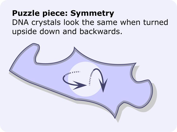
8
A model approach
Crick and Watson wanted to work on DNA’s structure, but they couldn’t ap-
proach it as Wilkins and Franklin were—through X-ray diffraction. First, Crick
was a friend of Wilkins and didn’t want to step on his toes. Second, Watson and
Crick didn’t have the high-quality DNA samples necessary for X-ray diffraction.
But Watson and Crick had another way of working—they could form hypotheses
about DNA’s structure by building a physical model of how its atoms fit together
(Fig. 19).
                                                                                                                 Fig. 19. One type of
                                     Today, ball-and-stick models like the ones Watson molecular modeling kit that
                                     and Crick used are available in most chemistry class- is widely available today.
                                                                                             The original Watson and
                                     es—but in 1951, they were found in only the best- Crick model was made using
                                     equipped labs. In the first half of the 20th century, wire and pieces of flat metal,
                                                                                             before good molecular
                                     painstaking chemical work established the approxi- model components became
                                     mate sizes of atoms, the number of bonds they form affordable.
                                     with other atoms, and the angles at which these bonds
                                     form. The models Watson and Crick worked with incorporated all of this infor-
                                     mation. The flexibility and accuracy of the models allowed them to try out many
                                     different structures and quickly see whether they agreed with what was known
                                     about chemical bonding. This made the models a good way to form new hypoth-
                                     eses about the shape of a molecule—something too small to observe directly.
                             Watson and Crick were also encouraged by the fact that Linus Pauling (Fig. 20),
Fig. 20. Linus Pauling
with a model of the helical
                             a chemist who studied bond formation, had just used models to figure out the
structure exhibited in some  helices that are part of the structures of many proteins. Pauling came up with the
segments of proteins.
                             solution by starting with X-ray diffraction data, then using ball-and-stick model-
building as a shortcut. This approach had allowed him to find the solution much more quickly than he could
have by using X-ray data alone. The success of his approach inspired Watson and Crick to try the same thing.

A false start
In order to try model building, Crick and Watson still needed data on DNA as a starting point. Molecular
model-building works because it lets researchers explore different hypotheses about molecular structures and
see which hypotheses fit well both with our knowledge about how atoms bond together and with evidence
regarding the structure of a particular molecule like DNA. But evidence of DNA’s structure came mainly from
X-ray diffraction, Wilkins and Franklin’s domain.
Fortunately for Crick and Watson, communicating evidence and results is a standard part of the process of
science. They kept an eye out for any talks or papers related to DNA’s structure, and as soon as they heard that
Franklin was going to share her findings in a talk at the University of London, Watson made plans to go. At the
presentation, Franklin showed X-ray diffraction patterns produced by DNA A and B, and discussed how the
two forms seemed to be produced by surrounding the DNA molecules with different amounts of water. She
also described the spacing between the atoms in DNA, based on the patterns in her diffraction images. Watson
listened with interest (Fig. 21). Yet the next day, his memory failed him when he met up with Crick to discuss
the evidence Franklin had shared. In particular, he couldn’t seem to remember how much water Franklin had

Molecular model image from photos courtesy of Dave Barnes, Arbor Scientific (www.arborscientific.com); photo of Pauling
with model courtesy of the Ava Helen and Linus Pauling Papers, Oregon State University Special Collections

© 2007 The University of California Museum of Paleontology, Berkeley, and the Regents of the University of California • www.understandingscience.org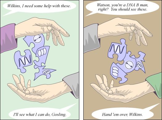
9
said surrounded the molecule. Nonetheless, Crick had
experience in X-ray diffraction and thought he could
put the pieces together. They decided that they had
enough evidence to build a model of DNA’s structure.
In their model, three long twists of the sugar-phos-
phate chain were held together by magnesium ions,
and the bases flopped outward from this central
backbone (Fig. 22). Watson and Crick excitedly in-
vited Wilkins, Franklin, and Gosling to come see the
model. When Franklin arrived, she quickly saw that
Watson had remembered several things incorrectly—
in particular, he had forgotten the amount of water                        Fig. 21.

that surrounded each strand. DNA crystals contained
at least ten times as much water as their model al-
lowed for, and there was no evidence that DNA con-
tained any magnesium at all. If it did, all that water
would cling to the magnesium ions, tearing the mol-
ecule apart. It was clear that the hypothesis Watson
and Crick had formulated using their metal-and-wire
models didn’t fit the available evidence on DNA. It
would have to be rejected.                                                 Fig. 22. Watson and Crick’s model erroneously placed
                                                                           the bases on the outside of the DNA molecule with the
                                                                           phosphates, bound by magnesium or calcium ions, inside.

The accidental image
While Watson and Crick went back to their model building, Franklin
continued to work on DNA by making X-ray diffraction images and
analyzing these results. She and Gosling focused on DNA A, produc-
ing many clear images and uncovering more clues to its structure: the
size of the repeating units that made up the molecule and the symme-
try of these units. DNA crystals, it turned out, look the same when
they are turned upside down and backwards (Figs. 23, 24).
                                                        Each image took many
                                                        hours of X-ray exposure to
                                                        develop—sometimes up to
                                                        100 hours—so Franklin
                                                        and Gosling occasionally Fig. 23.
                                                        exposed them overnight.
                                                        On the morning of May 2nd, 1952, they returned to the lab to dis-
                                                        cover that the DNA had hydrated during the night and the image
                                                        they had taken was actually of DNA B. It was unusually sharp—and
                                                        illuminating. It showed an obvious x shape, a pattern that previous
Fig. 24.
                                                        work associated with helical structures (Fig. 25). The image also

© 2007 The University of California Museum of Paleontology, Berkeley, and the Regents of the University of California • www.understandingscience.org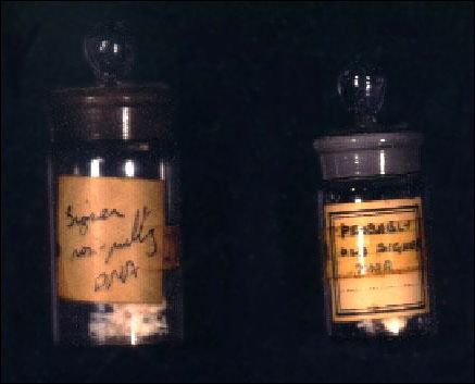
10
confirmed the idea that DNA’s bases were stacked pancake-style, 0.34 nanometers
apart, and suggested that 10 of these layers occurred in every twist of the helix.
It even delineated the width of the diameter of the helix: 2 nanometers (Fig. 26).
Since it was the 51st image taken, they called it image B 51. They set it aside and
decided to come back to it once they’d solved the structure of DNA A.

                                                                                                                 Fig. 25. X-ray diffraction
                                                                                                                 image B 51 taken by
                                                                                                                 Franklin and Gosling.

                                     Fig. 26.

Photo of X-ray diffraction pattern courtesy of Cold Spring Harbor Laboratory Library and Archive, James D. Watson Collection

© 2007 The University of California Museum of Paleontology, Berkeley, and the Regents of the University of California • www.understandingscience.org
11
Personal personnel problems
With Franklin and Gosling gathering additional evidence, and Crick and Watson
concentrating on generating new hypotheses, the puzzle of DNA seemed close to
being solved. But a personal conflict would soon change the course of this discov-
ery. From the time that Franklin started working in the lab, she and Wilkins had
argued about which of them would get to work on DNA. Initially, their boss had
asked Wilkins to hand the project over to Franklin—so Wilkins gave her all of the
high-quality DNA sample (Fig. 27). Later, he decided he wanted to keep working
on the problem anyway, but Franklin had already gotten started and didn’t want Fig. 27. Bottles containing
the high-quality DNA
to be pushed out. The resulting tension made both of them unhappy, and shortly samples that Franklin
after image B 51 was taken, Franklin notified her boss that she wanted to leave obtained from Wilkins.
the lab. This left Gosling, her student, upset and without a Ph.D. supervisor. He decided to seek advice from
Wilkins—and when he did, he took a critical piece of evidence with him: image B 51.
Wilkins had always been more interested in DNA B
anyway, and he took special notice of the clear, in-
formative image. Later that month, Watson came to
London for another lab colloquium. After the talk,
Wilkins had dinner with Watson and showed him the
beautiful image of DNA B produced by Franklin (Fig.
28). Because Crick had helped Watson learn how to
interpret the X-ray patterns produced by helices, Wat-
son immediately recognized the tell-tale evidence of a
helix—which he had suspected all along—as well as
other clues that would help Watson and Crick put all
the puzzle pieces together. Determined not to make
the same mistake as before, Watson asked Wilkins for
more details, and this time, he wrote everything down. Fig. 28.
The race to discovery
When he returned to Cambridge, Watson shared the new results
with Crick and they applied the information to their ball-and-stick
models. Watson wanted to try making a model in which just two
phosphate-sugar-base chains were linked together. He thought it
made sense for genes to come in pairs, partly because most organ-
isms have two parents. Watson and Crick also decided to try orient-
ing the bases towards the center of the pair. Watson later recounted
that they tried this approach simply because it was something they
hadn’t yet tried, though Franklin had previously given them good
reason to think that the bases should be on the inside and phosphates
on the outside of the molecule where they could attract water. Both Fig. 29.
of them were surprised by how well the new two-strand, bases-in
model (Fig. 29) fit the clues Watson had scribbled down during his dinner with Wilkins. But Watson and
Photo of the bottles containing DNA samples © King’s College Archives
© 2007 The University of California Museum of Paleontology, Berkeley, and the Regents of the University of California • www.understandingscience.org12 Crick weren’t the only ones thinking about a double helix—Rosalind Franklin’s notes from February 10th show that she started wondering if DNA B might be a two-chain helix around the same time. Of course, because she had produced the results, Franklin was the only one with all the data—and Watson and Crick needed more information to keep working. In science, researchers regularly share their findings with other scientists through journal publi- cations, but Franklin’s results were so new that they Fig. 30. hadn’t been thoroughly peer-reviewed and published. However, Watson and Crick were able to find out more about Franklin’s work from another source. Her lab was funded by the Medical Research Council, which required grant recipients to report on their progress at the end of each year. All of the clues that Franklin had uncovered were summarized in that report. Such reports are supposed to be confidential, but Watson and Crick happened to know someone on the Medical Research Council who had a copy of the report and was willing to show it to them. When Crick saw the evidence in the report (Fig. 30), he recognized the type of crystal symmetry Franklin described, and realized something that she hadn’t. If DNA crystals could be flipped upside down and backwards, and still look the same, the strands of the backbone must be identical, and they must run in opposite directions (Fig. 31). By this time, Franklin had also concluded that DNA was a two-chain helix, composed of two intertwined sugar-phosphate backbones. Fig- uring out the shape of the backbones, though, still left the bases an open question. She knew from details in her X-ray images that the phosphates were on the outside of the helix, which meant that the bases must point toward the center. But how did they fit together? Each base is a slightly different size, but the smooth twists of the sugar-phosphate chain never varied. How could the bases fit inside the chains without touching and repelling one another? She was sure there was a clue in DNA’s unique base ratios—one of the puzzle piec- es discovered before Franklin had even begun to study DNA—but she still wasn’t sure exactly what that clue meant. By February 23rd, her notes show that she realized that if A were physically interchange- able with G, and C with T, then the amount of A would have to equal T, and likewise for C and G. She was getting close—but she had yet to put the pieces together into a complete hypothesis. Meanwhile, back in Cambridge, Watson and Crick were working on the same problem … Fig. 31. © 2007 The University of California Museum of Paleontology, Berkeley, and the Regents of the University of California • www.understandingscience.org
13
The finish line
Watson and Crick were also stuck on what to do with
the bases. At first, Watson thought they paired togeth-
er A-A, C-C, T-T, G-G—but because of the different
sizes of the bases, the hypothesis had to be discarded.
It would have required a sugar-phosphate backbone
that wiggled in and out, rather than winding around
in smooth twists. Then, Watson and Crick got a key
piece of evidence about the shapes of the bases from
a visiting American chemist, Jerry Donohue. At that
time, most chemistry textbooks reported a particular
placement of hydrogen on the bases. That placement
made it impossible to match A to T, or G to C—they
just didn’t fit. Donohue told Watson that the text- Fig.       32. The visiting American chemist, Jerry Donohue,
                                                          provided a key piece of evidence when he revealed that
books were outdated. More was now known about the forms given for thymine and guanine in most textbooks
the shapes these bases might take: one of the hydro- were       wrong. Note the changes, indicated by the glowing
                                                          hydrogens.
gen atoms could be attached to the base in another
location (Fig. 32). In fact, based on a few different lines of evidence, Donohue thought that the bases likely
took shapes that Watson had not yet tried (Figs. 33, 34).
                                                        Watson tried to fit the new shapes into the two-chain model he and
                                                        Crick had developed. On February 28th, he was playing with paper
                                                        cutouts of each base when he suddenly saw the answer. The A fit with
                                                        T, and G fit with C. Plus, the A-T pair had the exact same molecular
                                                        length as the G-C pair! Bonded together like this, the bases wouldn’t
                                                        bump and repel one another. Crick realized that if the bases paired up
                                                        like this, it would explain the mysterious
                                                        base ratios: A=T, G=C (Fig. 35). Sud-
                                                        denly, it made perfect sense that the base
Fig. 33.
                                                        pairs must be in the center of the mol-
                                                        ecule, and that the two sugar-phosphate
                                                                        strands wound around
                                                                        them. It even suggested
                                                                        how one strand could be
                                                                        used to copy the other.
                                                                        Because each base always
                                                                        matches up with the same
                                                                        partner, the order of bases Fig. 35. Given the correct
                                                                        on one strand could de- forms for the bases, Watson
                                                                        termine the exact order was      able to figure out
                                                                                                    how adenine-thymine and
Fig. 34.
                                                                        of bases on a new strand. guanine-cytosine pairs
                                                                        Within a week, Watson matched         up, and formed
                                                                                                    weak hydrogen bonds with
                                                                        and Crick had worked one another. Watson and
                                                                        out the details of their Crick    originally suggested
                                                                                                    that there were two bonds
                                                                        hypothesis about the mo- between guanine and
                                                                        lecular structure of DNA. cytosine    but later it was
                                                                                                    found that a third existed.

© 2007 The University of California Museum of Paleontology, Berkeley, and the Regents of the University of California • www.understandingscience.org
14
Credit and debt
Watson and Crick published their proposed structure for DNA in April 1953 in
the journal Nature 3 (Fig. 36).In the same issue, Wilkins, Franklin, Gosling, and
their colleagues presented the evidence they’d collected, which supported Watson
and Crick’s two-chain helix hypothesis.4 In this way, the evidence and hypothesis
relating to the structure of DNA entered the scientific literature and became avail-
able for other researchers to build on.
But not everything that went into these papers came from freely available sources.
Scientists often use others’ data and ideas (Fig. 37), but they are expected to give
credit to their sources. This allows science to grow by building on existing ideas,
while rewarding individual scientists for their contributions. Crick and Watson’s
paper did give credit for much of the evidence they’d collected during their investi-
gation of the structure of DNA. However, data inspiring some of their key insights
came from Franklin’s 1952 report to the Medical Research Council—which was
supposed to be confidential information. Franklin never gave Watson and Crick
permission to use that work, and in their paper—the scientific record of this dis-
covery—they do not credit Franklin for supplying this evidence or for image B 51,
which was so critical to their discovery. Retrospectively, both Crick and Watson
Fig. 36. acknowledged their debt. According to Crick, “all the really relevant experimental
work on the X-ray diffraction patterns of DNA” came from Franklin’s lab, and
Watson later claimed that their discovery would not have been possible without the data collected by Franklin.
The failure to give full credit to important evidence
is considered a serious infringement of scientific eth-
ics. Crick and Watson have both had highly success-
ful scientific careers, but the issue of whether or not
they acted fairly has continued to follow them. In
interviews and public appearances, they were—and
are—frequently questioned about their choices and
about Franklin’s role in their most famous discovery,
and have had to endure the scrutiny and judgment of
the scientific community.
Fig. 37.
It’s also worth noting that Franklin was a pioneer in
terms of women’s presence in the sciences. At the time Franklin was working on DNA, less than five percent
of Ph.D.s in the physical sciences were awarded to women.5 Franklin never reported specific examples of dis-
crimination (aside from not being allowed to eat with her male colleagues in the senior common room), but
she did worry that her work might not be taken seriously because of her gender. Though we can never know
for sure, it’s certainly possible that the discovery of DNA’s structure—and the credit given for it—would have
played out differently, had the social environment for women scientists been fairer.
3
Watson, J.D., and F.H.C. Crick. 1953. A structure for deoxyribose nucleic acid. Nature 171:737-738.
4
Franklin, R., and R.G. Gosling. 1953. Molecular configuration in sodium thymonucleate. Nature 171:740-741.
5
In 2005, that number was closer to 30%. Ivie, R., and K.N. Ray. Feb, 2005. Women in physics and astronomy, 2005.
American Institute of Physics. Retrieved July 3, 2008 from http://www.aip.org/statistics/trends/reports/women05.pdf
© 2007 The University of California Museum of Paleontology, Berkeley, and the Regents of the University of California • www.understandingscience.org15
DNA then and now
After unraveling the structure of DNA, all four re-
searchers continued to study genetics and molecular
biology, although along their separate paths (Fig. 38).
Wilkins, Watson, and Crick went on to collect ad-
ditional evidence on DNA’s structure, examine how
DNA copies itself, and investigate the genetic code Fig. 38. From left, Rosalind Franklin in 1956, James Watson
inherent in the DNA molecule. Sadly, Franklin’s re- in the 1980s, Francis Crick in the 1980s, and Maurice Wilkins
search was cut short when she died of cancer—just in the early 1990s. Franklin died in 1958. Both Crick and
Wilkins died in 2004.
five years after the landmark Nature publication. This
also meant that Franklin missed out on many of the honors awarded for their discovery, including the possibil-
ity of a Nobel Prize—which cannot be awarded posthumously.
Despite her early death, Franklin’s work, along with
that of the others, has earned a permanent place in
our accumulated scientific knowledge (Fig. 39). Ge-
netic researchers today still build on the foundation
laid by these half-century old ideas and findings. If
we trace the roots of today’s cutting-edge technolo-
gies like DNA fingerprinting, genetic engineering,
and genome sequencing back in time, we will find
ourselves once again in the X-ray diffraction lab at
the University of London and tinkering with models Fig. 39. The discovery of DNA’s structure opened the door
at Cambridge. And continuing even further back in to an entire field of genetic research and application.
time, we’ll encounter the community of researchers
who set the stage for this discovery by developing X-ray diffraction techniques and by uncovering those first
puzzle pieces that inspired Wilkins, Franklin, Watson, and Crick to join the race and chase down the double
helix. With many open questions involving DNA, its structure will continue to be a key piece of evidence in
many new discoveries yet to come.
Though the discovery of the structure of DNA is
frequently attributed to Watson and Crick, the story
behind this discovery highlights just how indebted
to other researchers they were (Fig. 40). Reliance on
the clues discovered by others is a key theme, not just
of this story, but of the process of science in general.
Science is too big a job and involves too many com-
plex ideas for any one person to tackle a problem in
complete isolation. Even the few scientists who work
alone on a day-to-day basis rely on the cumulative
knowledge of the scientific community as a starting
point and contribute their findings to this knowledge
base so that others can build upon them. Because of Fig. 40.
science’s collaborative nature, communication—shar-
ing pieces of the puzzle—has played a critical role in many scientific discoveries. As we saw in the race for the
structure of DNA, science works not solely through the brilliance and good fortune of a few individuals, but
through the work of a diverse community.
Photos of James Watson, Francis Crick, and Rosalind Franklin courtesy of Cold Spring Harbor Laboratory Library and Archive,
James D. Watson Collection; Wilkins photo courtesy of TVNZ; grain genetics photo provided by USDA; researcher and PCR
photos provided by NIH
© 2007 The University of California Museum of Paleontology, Berkeley, and the Regents of the University of California • www.understandingscience.org16 Want to learn more? Check out these references Popular and historical accounts: Maddox, B. 2003. The Dark Lady of DNA. London: HarperCollins. Watson, J.D. 1969. The Double Helix: A Personal Account of the Discovery of the Structure of DNA. New York: Mentar Books. A few scientific articles: Avery, O.T., C.M. MacLeod, and M. McCarty. 1944. Studies on the chemical nature of the substance in- ducing transformation of Pneumococcal types. Journal of Experimental Medicine 79:137–159. Franklin, R., and R.G. Gosling. 1953. Molecular configuration in sodium thymonucleate. Nature 171:740–741. Watson, J.D., and F.H.C. Crick. 1953. A structure for deoxyribose nucleic acid. Nature 171:737–738. © 2007 The University of California Museum of Paleontology, Berkeley, and the Regents of the University of California • www.understandingscience.org
You can also read