Transmission of Human Respiratory Syncytial Virus in the Immunocompromised Ferret Model - Viroclinics
←
→
Page content transcription
If your browser does not render page correctly, please read the page content below
© Published by Viruses 2018

      Transmission of Human Respiratory Syncytial Virus
                     in the Immunocompromised Ferret Model
Leon de Waal 1, Saskia L. Smits 1, Edwin J. B. Veldhuis Kroeze 1,2, Geert van Amerongen 1, Marie O. Pohl 1, Albert D. M. E. Osterhaus 1,3 and Koert J. Stittelaar 1*

            1
                Viroclinics Biosciences BV, Rotterdam 3029 AK, The Netherlands; dewaal@viroclinics.com (L.d.W.); smits@viroclinics.com (S.L.S.);
  edwinvk@gmail.com (E.J.B.V.K.); amerongen@viroclinics.com (G.v.A.); pohl@viroclinics.com (M.O.P.); albert.osterhaus@tiho-hannover.de (A.D.M.E.O.)
                                       2
                                           Department of Viroscience, Erasmus MC, Rotterdam 3015 CN, The Netherlands
            3
                Research Centre for Emerging Infections and Zoonoses, University of Veterinary Medicine Hannover, 30559 Hannover, Germany

           Human respiratory syncytial virus (HRSV) causes substantial morbidity and mortality in vulnerable
           patients, such as the very young, the elderly, and immunocompromised individuals of any age. Nosocomial
           transmission of HRSV remains a serious challenge in hospital settings, with intervention strategies largely
           limited to infection control measures, including isolation of cases, high standards of hand hygiene, cohort
           nursing, and use of personal protective equipment.
           No vaccines against HRSV are currently available, and treatment options are largely supportive care and
           expensive monoclonal antibody or antiviral therapy. The limitations of current animal models for HRSV
           infection impede the development of new preventive and therapeutic agents, and the assessment of
           their potential for limiting HRSV transmission, in particular in nosocomial settings. Here, we demonstrate
           the efficient transmission of HRSV from immunocompromised ferrets to both immunocompromised and
           immunocompetent contact ferrets, with pathological findings reproducing HRSV pathology in humans.
           The immunocompromised ferret-HRSV model represents a novel tool for the evaluation of intervention
           strategies against nosocomial transmission of HRSV.

           © 2018 by the authors. Licensee MDPI, Basel, Switzerland. This article is an open access article distributed under the terms and conditions
           of the Creative Commons Attribution (CC BY) license

        * Corresponding author at:
          Viroclinics Biosciences B.V.
          Marconistraat 16
          3029 AK Rotterdam
          The Netherlands.

          E-mail address:
          stittelaar@viroclinics.com (K.J. Stittelaar)

                                                                            knows your target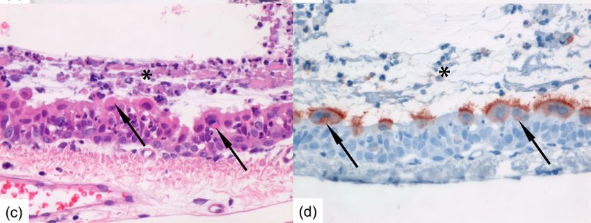
viruses
Article
Transmission of Human Respiratory Syncytial Virus
in the Immunocompromised Ferret Model
Leon de Waal 1 , Saskia L. Smits 1 , Edwin J. B. Veldhuis Kroeze 1,2 , Geert van Amerongen 1 ,
Marie O. Pohl 1 , Albert D. M. E. Osterhaus 1,3 and Koert J. Stittelaar 1, *
 1    Viroclinics Biosciences BV, Rotterdam 3029 AK, The Netherlands; dewaal@viroclinics.com (L.d.W.);
      smits@viroclinics.com (S.L.S.); edwinvk@gmail.com (E.J.B.V.K.); amerongen@viroclinics.com (G.v.A.);
      pohl@viroclinics.com (M.O.P.); albert.osterhaus@tiho-hannover.de (A.D.M.E.O.)
 2    Department of Viroscience, Erasmus MC, Rotterdam 3015 CN, The Netherlands
 3    Research Centre for Emerging Infections and Zoonoses, University of Veterinary Medicine Hannover,
      30559 Hannover, Germany
 *    Correspondence: stittelaar@viroclinics.com; Tel.: +31-88-668-4727

 Received: 1 December 2017; Accepted: 30 December 2017; Published: 2 January 2018

 Abstract: Human respiratory syncytial virus (HRSV) causes substantial morbidity and mortality
 in vulnerable patients, such as the very young, the elderly, and immunocompromised individuals
 of any age. Nosocomial transmission of HRSV remains a serious challenge in hospital settings,
 with intervention strategies largely limited to infection control measures, including isolation of
 cases, high standards of hand hygiene, cohort nursing, and use of personal protective equipment.
 No vaccines against HRSV are currently available, and treatment options are largely supportive care
 and expensive monoclonal antibody or antiviral therapy. The limitations of current animal models
 for HRSV infection impede the development of new preventive and therapeutic agents, and the
 assessment of their potential for limiting HRSV transmission, in particular in nosocomial settings.
 Here, we demonstrate the efficient transmission of HRSV from immunocompromised ferrets to both
 immunocompromised and immunocompetent contact ferrets, with pathological findings reproducing
 HRSV pathology in humans. The immunocompromised ferret-HRSV model represents a novel tool
 for the evaluation of intervention strategies against nosocomial transmission of HRSV.

 Keywords:      respiratory syncytial virus;          animal model;        ferret;   immunocompromised;
 transmission; contact

1. Introduction
     Human respiratory syncytial virus (HRSV) is the leading cause of acute lower respiratory tract
infection in children less than 5 years of age, with high hospitalization rates in infants younger
than 6 months [1]. It also causes significant morbidity in adults, and contributes to excess mortality in
older individuals and immunocompromised individuals of any age [2,3]. In the latter, infection may
be prolonged and often complicated by bacterial co-infection, leading to pneumonia and severe
respiratory distress [4]. Outbreaks among vulnerable patients in hospital settings are of particular
concern [5,6]. Nosocomial transmission poses a substantial risk and remains a serious challenge
for hospital infection control. In the absence of an effective vaccine, HRSV treatment options are
largely limited to supportive care and expensive monoclonal antibody or antiviral therapy [7,8].
Infection control measures, including isolation of cases, high standards of hand hygiene, cohort nursing,
and use of personal protective equipment have been reported as relatively effective in the prevention
and control of HRSV outbreaks in nosocomial settings [5,9,10]. The prophylaxis use of monoclonal
antibody therapy also can reduce nosocomial transmission [11]. There is nonetheless a need for a

Viruses 2018, 10, 18; doi:10.3390/v10010018                                      www.mdpi.com/journal/viruses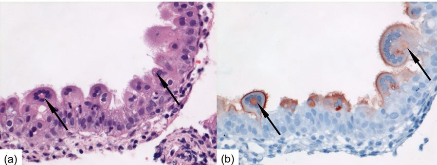
Viruses 2018, 10, 18                                                                                  2 of 11

larger armamentarium of HRSV-specific treatments and evidence-based control measures for HRSV
infection prevention and control.
      The lack of an appropriate animal model fully recapitulating HRSV pathogenesis of infection and
disease has long represented a hurdle for the development of new intervention strategies against HRSV,
including its spread in nosocomial settings [12]. In humans, HRSV is transmitted by direct contact
and large aerosol droplets, and via contaminated surfaces, initiating viral replication in the upper
respiratory tract after an incubation period of 4–5 days [2]. In severe cases, HRSV further spreads to
and replicates in the lower respiratory tract. The virus replicates in ciliated epithelial cells along the
respiratory tract, as well as in type I and type II alveolar pneumocytes, resulting in inflammation and
necrosis. Animal models for HRSV infection are mostly semi-permissive, requiring a large inoculum
of HRSV to result in a productive infection [12]. They include non-human primate (NHP) models,
which, apart from chimpanzees and African green monkeys, generally develop no or little clinical
signs or pathological changes. The most used non-primate models of HRSV infection are cotton rats,
mice, and the neonatal lamb. These models have contributed to improving our understanding of
HRSV pathogenesis and host immune responses [13,14]. Furthermore, NHPs and cotton rats may also
develop atypical enhanced disease following vaccination with formalin-inactivated HRSV, as seen in
humans. However, they all have their limitations [12]. In none of these models HRSV is transmitted to
contact individuals, or they are highly impractical to study transmission.
      The ferret model of HRSV infection had initially yielded disappointing results, with poor,
age-dependent replication of the virus in the lower respiratory tract [12,15]. However, we reported
recently that a low-passage clinical isolate of HRSV efficiently replicates in both the upper and lower
respiratory tracts of adult ferrets [16]. In addition, we described the use of immunocompromised ferrets
as a relevant model for HRSV infection in immunocompromised individuals [16]. It is characterized by
high replication levels in the lower respiratory tract, with viral antigen detected in tracheal, bronchial,
and bronchiolar ciliated epithelial cells, and by prolonged viral replication and shedding, reflecting the
increased viral loads and delayed viral clearance observed in immunocompromised individuals.
Prolonged infection is thought to be an important risk factor for nosocomial transmission [17].
      Prince et al. reported that infant ferrets generate higher virus loads than adult ferrets, resulting in
successful contact transmission of HRSV from infant ferrets to their mother [15]. Recently, Chan et al.
showed limited horizontal transmission in a preliminary study between immunocompetent ferrets
without proof of replicating virus in the contact ferrets [18]. The immunocompromised ferret-HRSV
model [16] offers a new tool to study HRSV transmission and its prevention, which may be used
to address the potential of intervention strategies, including vaccines and antibody or antiviral
treatments, to block or limit HRSV transmission. Here we report the effective transmission of a
low-passage clinical isolate of HRSV from immunocompromised ferrets to both immunocompetent
and immunocompromised contact ferrets with the detection of replicating virus in both donor and
contact ferrets. Both the intranasal and intratracheal route of infection were explored to determine
which site of virus replication would be responsible for transmission of the virus.

2. Materials and Methods

2.1. Virus
     A clinical isolate of HRSV subgroup A strain was isolated in human respiratory epithelium
carcinoma (HEp-2) cells from a nasal lavage of an infant hospitalized at Erasmus Medical Centre,
Rotterdam, The Netherlands, in 2011. The virus was passaged exclusively in HEp-2 cells, cultured as
described previously [16]. The challenge stock used in the animal experiments was passage 3,
and was prepared as described previously [16]. Virus titers were determined by end-point titration on
monolayers of HEp-2 cells and cytopathic effect (CPE) screening six days after infection. Viral titers
were calculated using the method of Spearman and Karber. The challenge stock tested negative for a
large panel of respiratory human viruses and mycoplasma by RT-PCR.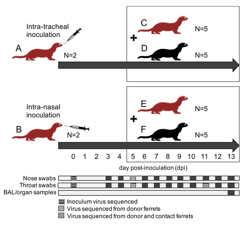
Viruses 2018, 10, 18 3 of 11
Viruses 2018, 10, 18 3 of 11
were calculated using the method of Spearman and Karber. The challenge stock tested negative for a
2.2. Ferrets
large panel of respiratory human viruses and mycoplasma by RT-PCR.
Ferrets (Mustela putorius furo) were 16- to 18-month-old purpose-bred males, which were
2.2. Ferrets for Aleutian disease virus, and RSV subgroup A and B. They were housed in
seronegative
groups Ferrets
of 2 to(Mustela
12, as described previously
putorius furo) were 16- [16]. Male ferretspurpose-bred
to 18-month-old were selected irrespective
males, of any
which were
HRSV-related
seronegativeparameter,
for Aleutianbecause maleand
disease virus, ferrets would beA more
RSV subgroup and B. robust to endure
They were housed inthe treatment
groups of
procedures.
2 to 12, as The present
described experiment
previously [16]. complied with
Male ferrets wereallselected
national and international
irrespective guidelines for
of any HRSV-related
careparameter,
and use of because male
animals ferrets
and was would be approved
ethically more robustonto15endure the treatment
December 2015 by procedures. The
the Dutch Central
present experiment
Committee complied with
Animal Experiments (CCD),all protocol
national number
and international guidelines for care and use of
AVD277002015161.
animals and was ethically approved
Immunocompromised on 15 December
ferrets received 2015daily
orally twice by thethe
Dutch Central Committeemedication
immunosuppressive Animal
Experiments (CCD), protocol number AVD277002015161.
of 20 mg/kg of body weight (bw) Mycophenolate mofetil (MMF) (CellCept , Roche, ®
Woerden, Immunocompromised
The Netherlands), 0.5 ferrets
mg/kgreceived orally twice (Prograft
bw tacrolimus daily the immunosuppressive
® , Astellas Pharmamedication of
BV, Leiderdorp,
20 mg/kg of body weight (bw) Mycophenolate mofetil (MMF) (CellCept®, Roche, Woerden, The
The Netherlands), 8 mg/kg bw prednisolone (Hospital Pharmacy, UMCN St Radboud,
Netherlands), 0.5 mg/kg bw tacrolimus (Prograft®, Astellas Pharma BV, Leiderdorp, The
Nijmegen, The Netherlands), together with antibiotic prophylaxis of 10 mg/kg bw amoxicillin
Netherlands), 8 mg/kg bw prednisolone (Hospital Pharmacy, UMCN St Radboud, Nijmegen, The
and 2.5 mg/kg bw clavulanic acid (Pharmachemie BV, Haarlem, The Netherlands), as described
Netherlands), together with antibiotic prophylaxis of 10 mg/kg bw amoxicillin and 2.5 mg/kg bw
previously
clavulanic[16,19].
acid (Pharmachemie BV, Haarlem, The Netherlands), as described previously [16,19].
2.3. HRSV Transmission Experiment
2.3. HRSV Transmission Experiment
A total
A total of of
twenty-four
twenty-fourferrets
ferrets were
were randomly assignedtotothe
randomly assigned thefollowing
following treatment
treatment groups
groups
(Figure 1): (A)
(Figure 2 immunocompromised
1): (A) 2 immunocompromised donor
donorferrets
ferretsinfected
infectedwith
with HRSV
HRSV via the intra-tracheal
via the intra-trachealroute
route of
inoculation; (B) 2 immunocompromised donor ferrets infected with HRSV via
of inoculation; (B) 2 immunocompromised donor ferrets infected with HRSV via the intra-nasal the intra-nasal route
of inoculation; (C) 5 immunocompromised
route of inoculation; (C) 5 immunocompromised contact ferrets
contact placed
ferrets on day
placed 5 post-inoculation
on day 5 post-inoculation (dpi)
with group
(dpi) withAgroup
ferrets;
A (D) 5 immunocompetent
ferrets; (D) 5 immunocompetent contact ferretsferrets
contact placed on 5 on
placed dpi5 with group
dpi with A ferrets;
group A
(E) 5ferrets; (E) 5 immunocompromised
immunocompromised contact
contact ferrets placed ferrets placed
on 5 dpi withon 5 dpi
group with group
B ferrets; B ferrets; (F) 5
(F) 5 immunocompetent
immunocompetent contact ferrets placed on
contact ferrets placed on 5 dpi with group B ferrets. 5 dpi with group B ferrets.
Figure 1. 1.Schematics
Figure overviewofofthe
Schematics overview theexperimental
experimental design;
design; ferretferret
groups groups are lettered
are lettered (A–F);
(A–F); red
red ferrets
ferretsare
areimmunocompromised
immunocompromisedferrets; ferrets; black
black ferrets
ferrets areare immunocompetent
immunocompetent ferrets;
ferrets; contact
contact ferrets
ferrets
(group
(group C–F)C–F)
are are placed
placed in the
in the samesame cages
cages as donor
as donor ferrets
ferrets (group
(group A,B)
A,B) on on
dayday 5 post-inoculation
5 post-inoculation (dpi);
(dpi); all
all ferrets are ferrets are euthanized
euthanized at 13ofdpi.
at 13 dpi. Days Days of
collection collection
of nose swabs,of throat
nose swabs,
swabs, broncho-alveolar
throat swabs,
broncho-alveolar
lavages lavages (BAL),
(BAL), and respiratory tractand respiratory
samples tract samples
are indicated are indicated
with dark with dark
squares; samples squares;
used for viral
samples used
sequencing for viral sequencing
are indicated are indicated with patterns.
with patterns.
After a one-week acclimatization period, the ferrets assigned to group A, B, C, and E (Figure 1)
were started on the immunosuppression protocol, with daily oral prophylactic antibiotics startingViruses 2018, 10, 18 4 of 11
four days before HRSV infection, and twice-daily oral immunosuppressants starting three days before
HRSV infection. The ferrets assigned to group A and B were inoculated on day 0. Donor ferrets
sedated with a mixture of ketamine and medetomidine (IM doses of 0.2 and 0.01 mL/kg body weight,
respectively) were inoculated with 105 median tissue-culture infective dose (TCID50 ) of low-passage
clinical isolate HRSV subgroup A by intra-tracheal (IT; group A) or intra-nasal (IN; group B) inoculation
with a volume of 3 or 0.3 mL, respectively.
The ferrets assigned to group C to F were introduced in the cages of group A and B ferrets on 5 dpi
to enable direct contact. Throat (Copan; rayon tipped) and nose (Copan; polyester tipped) swabs were
collected from all ferrets daily from 3 dpi onwards in a 3mL virus transport medium [16]. Donor ferrets
were euthanized by exsanguination under anesthesia with ketamine-medetomidine at 13 dpi and
contact ferrets at day 8 post-exposure (dpe; corresponding to 13 dpi). Broncho-alveolar lavages (BAL)
and samples of the nasal turbinates, trachea, primary bronchus, and lung parenchyma were collected
upon autopsy.
All personnel involved in the collection of study data on a day-to-day basis and all personnel
performing the laboratory analysis in which interpretation of the data is required were not aware of the
so-called Random Treatment Allocation Key at any time prior to completion of the study. All samples
were labeled with a unique sample number.
2.4. Samples and Assays
All samples were processed within four hours of sample collection. Infectious virus titers and
concentrations of viral RNA were measured by virus isolation and reverse transcription-PCR (RT-PCR),
respectively, as previously described [16]. Samples of the nasal turbinates, trachea, primary bronchus,
and right lung were weighed and subsequently homogenized with a FastPrep-24 (MP Biomedicals,
Eindhoven, The Netherlands) in medium and centrifuged briefly before viral load assessment by virus
isolation and quantitative RT-PCR.
RNA extraction was performed using a MagNA Pure 96 (Roche Diagnostics Nederland B.V.,
Almere, The Netherlands) and MagNA Pure 96 DNA and Viral NA Small Volume Kit with an
input volume of 190 µL and output volume of 100 µL according to the manufacturer’s instructions
(Roche Diagnostics Nederland B.V.). The extraction was internally controlled by the addition of a
known concentration of phocine distemper virus (PDV) [20]. 20 µL extracted RNA was amplified in
a 50 µL final volume, containing 12.5 µL 4× TaqMan Fast Virus 1-Step Master Mix (Life Technologies,
Bleiswijk, The Netherlands), and 1 µL of primers and probe mixture for detection of RSV A, RSV B,
and PDV in a triplex reaction (Table S1). The RT-PCR temperature profile was 5 min at 50 ◦ C, 20 sec
at 95 ◦ C, 45 cycles of 3 s at 95 ◦ C, and 32 s at 60 ◦ C.
Dilutions of an electron microscopic-counted HRSV stock (ABIOnline.com Columbia) were used
for conversion of RT-PCR threshold cycle (Ct) values into a quantitative measurement of viral particles.
Concentrations of viral RNA are expressed as log10 vp/mL. Infectious virus titers in tissue are
expressed as log10 TCID50 per gram tissue, and infectious virus titer in throat and nose swabs are
expressed as log10 TCID50 /mL. Significant differences in shedding loads were assessed based on the
areas under the curve and using the Student t-test.
Formalin-fixed tissue sections of nasal turbinates, trachea, and left lung were routinely processed,
paraffin embedded, and sectioned at 3–4 µm, deparaffinized with xylene and rehydrated using graded
alcohols, and stained with hematoxylin and eosin (H&E) for histopathological examination by light
microscopy. For immunohistochemistry (IHC), additional serial slides were sectioned simultaneously
and incubated for 1 h with a goat anti-HRSV-peroxidase (PO) (Virostat, Portland, ME, USA)
polyclonal antibody following antigen retrieval using citric acid buffer. Endogenous PO was
blocked with 3% hydrogen peroxide. The bound PO was visualized by incubating slides with
3-amino-9-ethylcarbazole for 10 min as substrate, resulting in a reddish brown finely-granular staining
of HRSV-infected epithelial cells, followed by hematoxylin counterstain. Negative controls were
performed in the absence of the antibody.Viruses 2018, 10, 18 5 of 11
2.5. Nested PCR Amplification and Sanger Sequencing
Viruses 2018, 10, 18 5 of 11
Full-length F and G gene sequencing was performed on the inoculum virus and RT-PCR
positive2.5.nose
Nested PCR Amplification
and/or and Sanger
throat swabs Sequencing
collected from the donor ferrets at 5 and 11 dpi and from
the contactFull-length
ferrets atF6and dpeG(corresponding
gene sequencing to was11performed
dpi). RNAon theextraction
inoculum was
virusperformed
and RT-PCRusing a
MagNA positive
Pure 96nose and/or Diagnostics)
(Roche throat swabs collected
and MagNA from the donor
Pure 96 ferrets
DNA at and5 and 11 NA
Viral dpi and
Smallfrom the
Volume Kit
contact ferrets at 6 dpe (corresponding to 11 dpi). RNA extraction was performed
with an input volume of 200 µL and output volume of 100 µL according to the manufacturer’s using a MagNA
Pure 96 (Roche Diagnostics) and MagNA Pure 96 DNA and Viral NA Small Volume Kit with an
instructions (Roche Applied Science). cDNA synthesis was performed using Superscript III Reverse
input volume of 200 µL and output volume of 100 µL according to the manufacturer’s instructions
Transcriptase
(Roche (Invitrogen,
Applied Science).Carlsbad,
cDNACA,synthesis
USA). Semi-nested PCR using
was performed amplification wasIII
Superscript performed
Reverse using
HotStarTaq
Transcriptase (Invitrogen, Carlsbad, CA, USA). Semi-nested PCR amplification was performed with
DNA Polymerase (Qiagen, Hilden, Germany). Sequence reactions were performed
using® Terminator
the BigDye HotStarTaq v3.1 DNAcycle sequencing
Polymerase kit (Applied
(Qiagen, Hilden, Biosystems, Foster City,
Germany). Sequence CA, USA)
reactions were and an
performed
ABI Prism with theanalyzer.
3730 genetic BigDye®Terminator v3.1indicated
Primers are cycle sequencing kitS1.
in Table (Applied Biosystems, Foster City,
CA, USA) and an ABI Prism 3730 genetic analyzer. Primers are indicated in Table S1.
3. Results
3. Results
3.1. Donor Ferrets
3.1. Donor Ferrets
All donor ferrets inoculated with HRSV either intra-tracheally (group A) or intra-nasally (group B)
All donor ferrets inoculated with HRSV either intra-tracheally (group A) or intra-nasally (group
developed a productive infection.infection.
B) developed a productive Both HRSVBothRNA
HRSVandRNAreplication-competent virus virus
and replication-competent were were
detected in
throat and nose swabs of group A and group B ferrets (Figure 2).
detected in throat and nose swabs of group A and group B ferrets (Figure 2).
Figure Figure
2. HRSV2. HRSV
RNA RNA concentrationsand
concentrations and viral
viraltiters in in
titers thethe
throat and nose
throat andswabs
nose of donorof
swabs ferrets;
donor (a) ferrets;
HRSV concentration in the throat swabs (plain line) and nose swabs (dashed line) of
(a) HRSV concentration in the throat swabs (plain line) and nose swabs (dashed line) of group A ferrets group A ferrets
inoculated intra-tracheally (I.T); (b) HRSV viral titers in the throat swabs (plain line) and nose swabs
inoculated intra-tracheally (I.T); (b) HRSV viral titers in the throat swabs (plain line) and nose swabs
(dashed line) of group A ferrets inoculated intra-tracheally (I.T); (c) HRSV concentration in the throat
(dashedswabs
line)(plain
of group A ferrets inoculated intra-tracheally (I.T); (c) HRSV concentration in the throat
line) and nose swabs (dashed line) of group B ferrets inoculated intra-nasally (I.N); (d)
swabs HRSV
(plainviral
line)titers
andinnose swabs
the throat (dashed
swabs (plainline)
line) of
andgroup B ferrets
nose swabs inoculated
(dashed intra-nasally
line) of group B ferrets (I.N);
(d) HRSV viral titers
inoculated in the(I.N);
intra-nasally throat
eachswabs (plain line)
line represents andferret;
a single noseforswabs (dashed line)limit
(a,c): quantification of ofgroup B
ferrets inoculated
qPCR data isintra-nasally
indicated by a (I.N);
dottedeach linenon-quantifiable
line, and represents a single ferret;
Ct values forassigned
were (a,c): quantification
the value 1.3 limit
of qPCR log10 vp/mL.
data is indicated by a dotted line, and non-quantifiable Ct values were assigned the
value 1.3 log10 vp/mL.
Detectable RNA concentrations and viral titers in throat and nose swabs were typically recorded
from 3–4 dpi to 10–13 dpi, except in the nose swabs of group A ferrets, with quantifiable RNAViruses 2018, 10, 18 6 of 11
Viruses 2018, 10, 18 6 of 11
Detectable RNA concentrations and viral titers in throat and nose swabs were typically
concentrations
recorded andfrom viral titers
3–4 dpi appearing
to 10–13 later,
dpi, except in between 6 andof10
the nose swabs dpi.AIn
group group
ferrets, A ferrets,
with viral RNA
quantifiable
concentrations and viral titers
RNA concentrations weretiters
and viral found to be significantly
appearing later, betweenhigher in dpi.
6 and 10 the throat
In group swabs thanviral
A ferrets, in the nose
RNA concentrations
swabs, based on the areasand viral the
under titerscurve
were found
(3.17x, topbe=significantly higherpin=the
0.04 and 3.21x, throat
0.04, swabs than in
respectively; Figure 3).
Viral RNA concentrations and viral titers in the nose swabs of group A ferrets were respectively;
the nose swabs, based on the areas under the curve (3.17x, p = 0.04 and 3.21x, p = 0.04, significantly lower
Figure 3). Viral RNA concentrations and viral titers in the nose swabs of group A ferrets were
than in the nose swabs of group B ferrets, based on the areas under the curve (−4.05x, p = 0.03 and 4.8x,
significantly lower than in the nose swabs of group B ferrets, based on the areas under the curve
p = 0.02,(−4.05x,
respectively; Figure
p = 0.03 and 4.8x,3).
p =HRSV RNA concentrations
0.02, respectively; andRNA
Figure 3). HRSV viralconcentrations
titers in organ andsamples and BAL
viral titers
are given in Table 1.
in organ samples and BAL are given in Table 1.
Figure 3. Average RNA concentrations and viral titers in the throat and nose swabs of group A to F
Figure 3. Average RNA concentrations and viral titers in the throat and nose swabs of group A to
ferrets, expressed as areas under the curve (AUC); (a) AUC RNA concentration; (b) AUC viral titers;
F ferrets, expressed as areas under the curve (AUC); (a) AUC RNA concentration; (b) AUC viral
significant differences are marked with asterisks (* p ≤ 0.05 and ** p ≤ 0.01); error bars indicate
titers; significant differences are marked with asterisks (* p ≤ 0.05 and ** p ≤ 0.01); error bars indicate
standard deviations.
standard deviations.
Table 1. HRSV viral titers (log10 TCID50/g) and RNA concentrations (log10 virus particles (vp)/mL)
Table 1.inHRSV
sampled tissues
viral of the
titers respiratory
(log10 TCID50 tract
/g)ofand
donor
RNA and concentrations
contact ferrets. (log10 virus particles (vp)/mL)
in sampled tissues of the respiratory tract Viral
Ferret
of donor and contact ferrets.
Titers (log10 TCID50/g)
RT-PCR
(log10 vp/mL)
Nasal Primary
Ferret Group Number Viral Titers (log10 TCID50 /g) Lung
Trachea BAL Lung RT-PCR (log10 vp/mL)
BAL
Turbinates Bronchus
Group Number Nasal Turbinates
1 Trachea
4.9 Primary
2.1 Bronchus
- 2.9Lung 5 BAL
8.8 7.3 Lung BAL
A.
1 2
4.9 - 2.1 - - - - 2.9 - 6.9
5 5.2 8.8 7.3
A.
2 B.
-3 6.1 - 1.7 - 2.3 4.2 - 6.8 7.5
- 7.1 6.9 5.2
4 - - - - - 5.8 3.2
3 6.1 1.7 2.3 4.2 6.8 7.5 7.1
B. 5 5.3 1.4 - - 2.8 5.6 2.9
4 - - - - - 5.8 3.2
6 - 3.3 - - 4.3 6.5 5.8
5 C. 5.3
7 4.3 1.4 3 - - 2.9 - 4.5 2.8
6 5.5 5.6 2.9
6 -8 - 3.3 - - - - - - 4.3
- - 6.5 5.8
C. 7 4.3
9 6.1 3 1.7 - - - 2.9 - 4.5
NQ NQ 6 5.5
8 D.
-
15 -
- 1.5
- - 2.3
- -
-
6.4 4.3
- -
9 6.1 1.7 - - - NQ NQ
15 - 1.5 - 2.3 - 6.4 4.3
16 5 - - - 1.8 5.3 4.3
D. 17 - - - - - - -
18 - - - - - NQ 3.9
19 3.4 - - - - - -Viruses 2018, 10, 18 7 of 11
Table 1. Cont.
Viruses 2018, 10, 18 7 of 11
Ferret Viral Titers (log10 TCID50 /g) RT-PCR (log10 vp/mL)
Group Number 16
Nasal Turbinates 5
Trachea -
Primary -
Bronchus - Lung 1.8 5.3
BAL 4.3Lung BAL
17 - - - - - - -
10 6.7
18 - 3.5 - - - - 2.4 - -
NQ 3.9 6.5 4.4
11 6.5
19 3.4 5.2 - - - - 3.2 - 3.5- - 6.6 5.5
E. 12 6.2 5.3 - 3.1 5.8 6.5 6.4
10 6.7 3.5 - 2.4 - 6.5 4.4
13 4.6 2.3 - - 1.8 5.6 NQ
11 6.5 5.2 - 3.2 3.5 6.6 5.5
14 - - - - - 5.7 NQ
E. 12 6.2 5.3 - 3.1 5.8 6.5 6.4
20 -13 4.6 - 2.3 - - - - 1.8 -
5.6 NQ NQ -
21 1.4
14 - - - - - - - - -
5.7 NQ - -
F. 22 3.7
20 - - - - - - - - -
NQ - NQ -
23 -21 1.4 - - - - - - - -- - NQ NQ
24 F. -22 3.7 - - - - - 2.2 - -
NQ - 5.3 NQ
23 -
NQ: -
non quantifiable - values. -
Ct - NQ NQ
24 - - - 2.2 - 5.3 NQ
Upon microscopic examination, pathological NQ: non quantifiable Ct values.
changes were observed in some of the respiratory
tissues sampled from group A and group B ferrets (Figure 4). In each group, cells positive for
Upon microscopic examination, pathological changes were observed in some of the respiratory
HRSV-antigen were detected
tissues sampled by immunohistochemistry
from group in one 4).
A and group B ferrets (Figure of the inoculated
In each ferrets.
group, cells In the
positive forgroup A
positiveHRSV-antigen
ferret, positivewere detected by immunohistochemistry in one of the inoculated ferrets. In the group of the
cells were found in small to moderate number in ciliated epithelial cells
nasal turbinates,
A positive trachea, primary
ferret, positive cells bronchus,
were found and bronchioles.
in small to moderate Positive
number syncytial cells were
in ciliated epithelial observed
cells of in
the nasaltheturbinates.
nasal turbinates,
In thetrachea,
group B primary bronchus,
positive and bronchioles.
ferret, positive cells werePositive
found syncytial cells were
in moderate number in
ciliated observed
epithelialin the nasal turbinates. In the group B positive ferret, positive cells were found in moderate
cells of the primary bronchus and in large number in ciliated epithelial cells of the
number in ciliated epithelial cells of the primary bronchus and in large number in ciliated epithelial
nasal turbinates and trachea, including positive syncytial cells (Figure 4).
cells of the nasal turbinates and trachea, including positive syncytial cells (Figure 4).
Figure 4. Photomicrographs of intranasally HRSV-inoculated immunocompromised donor ferret
Figure 4. Photomicrographs
(animal #3) inflamed mucosa of intranasally HRSV-inoculated
of the nasal turbinates immunocompromised
at 13 dpi (a,b) and of immunocompromised donor ferret
(animalcontact
#3) inflamed mucosa
ferret (animal #10)of the nasal
tracheal mucosaturbinates
followingatproductive
13 dpi (a,b)HRSV and of immunocompromised
transmission at 8 days
contact post-exposure
ferret (animal (c,d).#10)
(a) RSV-infected epithelialfollowing
tracheal mucosa cells may develop bright HRSV
productive eosinophilic viral inclusion
transmission at 8 days
bodies (arrows)
post-exposure (c,d). (a)and may form syncytial
RSV-infected multinucleated
epithelial cells (H&E-stain);
cells may develop (b) Serial slideviral
bright eosinophilic showsinclusion
HRSV-antigen expression faintly of the cytoplasmic inclusion bodies (arrows) enclosed by a corona
bodies (arrows) and may form syncytial multinucleated cells (H&E-stain); (b) Serial slide shows
of multiple nuclei, and markedly of the outer cellular membranes and cilia of RSV-infected nasal
HRSV-antigen expression faintly of the cytoplasmic inclusion bodies (arrows) enclosed by a corona
epithelial cells as red-brown staining (Immunoperoxidase, hematoxylin-counterstain); (c) The
of multiple
mucosanuclei, and markedly
is markedly inflamed of theinfiltrated
with outer cellular membranes
neutrophils and with
and covered cilia layer
of RSV-infected
of exudate nasal
epithelial cells asmultinucleated
(asterisk); red-brown staining (Immunoperoxidase,
epithelial hematoxylin-counterstain);
syncytial cells containing (c) The mucosa
bright eosinophilic viral inclusion
is markedly
bodies inflamed
(arrows) arewith infiltrated
present neutrophils
within RSV-infected andepithelial
tracheal coveredcellswith layer of(d)
(H&E-stain); exudate (asterisk);
Serial slide
shows HRSV-antigen
multinucleated epithelial expression
syncytialfaintly
cellsofcontaining
the cytoplasmic inclusion
bright bodies (arrows)
eosinophilic viralandinclusion
markedly bodies
(arrows)of are
the apical side and cilia of RSV-infected tracheal epithelial cells as red-brown staining
present within RSV-infected tracheal epithelial cells (H&E-stain); (d) Serial slide
(Immunoperoxidase, hematoxylin-counterstain). Original magnifications 400×.
shows HRSV-antigen expression faintly of the cytoplasmic inclusion bodies (arrows) and markedly
of the apical side and cilia of RSV-infected tracheal epithelial cells as red-brown staining
(Immunoperoxidase, hematoxylin-counterstain). Original magnifications 400×.Viruses 2018, 10, 18 8 of 11
3.2. Contact Ferrets
HRSV was detected by RT-PCR in the throat swabs of four to five contact ferrets, and in
the nose swabs of two to four contact ferrets in each of the four groups C–F (Table 2).
HRSV replication-competent virus was detected in the throat swabs of three to four contact ferrets,
and in the nose swabs of one to three contact ferrets in each of these four groups (Table 2). HRSV was
detected from day 4–6 to day 8 post-exposure (dpe) in the positive contact ferrets. HRSV RNA
concentrations and viral titers were similar in these four groups based on the areas under the curve.
In general, viral titers were higher in the throat swabs than in the nose swabs of the contact ferrets
(Figure S1). They tended to reach similar peak values in the throat swabs, yet lower peak values in the
nose swabs than those in the donor ferrets (Figure S1). HRSV RNA and replication-competent virus
was detected in the lung and or BAL of at least one contact ferret in each of the four groups of contact
ferrets. RNA concentrations and viral titers in organ samples and BAL of the contact ferrets are given
in Table 1.
Table 2. Number of ferrets with throat and nose swabs positive for HRSV RNA and for
replication-competent HRSV.
Ferret Group Throat Swab RT-PCR Nose Swab RT-PCR Throat Swab VI Nose Swab VI
A (N = 2) 2 2 2 2
B (N = 2) 2 2 2 2
C (N = 5) 4 3 4 2
D (N = 5) 4 4 4 3
E (N = 5) 4 2 4 2
F (N = 5) 5 4 3 1
Upon microscopic examination, pathological changes were observed in several of the respiratory
tissues sampled from contact ferrets. The infected mucosae of, especially, the tracheas and nasal
turbinates showed mild inflammation characterized by infiltration of mainly neutrophils within
the epithelial lining accompanied by exfoliation of degenerated and/or infected epithelial cells and
intraluminal exudation of fibrinous material. Large, bright eosinophilic inclusion bodies composed of
viral protein may be present within the cytoplasm of infected epithelial cells (Figure 4). In each group,
cells positive for HRSV-antigen were detected by immunohistochemistry in one to three contact ferrets.
In group C, positive ciliated epithelial cells were found in the trachea and primary bronchus of one
contact ferret. In group D, positive ciliated epithelial cells were found in the nasal turbinates or trachea
of three contact ferrets. In group E, positive ciliated epithelial cells were found in the trachea and/or
primary bronchus of three contact ferrets, including positive syncytial cells (Figure 4). In group F,
positive ciliated epithelial cells were found in the nasal turbinates of one contact ferret.
3.3. Virus
A low-passage clinical isolate of HRSV was used to inoculate the donor ferrets. The F and G
genes of the inoculum virus, as well as of shed virus sampled from the throat or nose swabs of donor
and contact ferrets, were sequenced to evaluate any genetic changes potentially marking adaptation
to the ferret model (Table S2). Three substitutions in the HRSV G protein and one substitution in
HRSV F protein were detected in four of the contact ferrets. Point mutations resulted in amino acid
changes T226I in the G protein in animal #5 (group C), Q81Q/L in the G protein in animal #7 (group C),
N20N/D in the G protein in animal #14 (group D), and T244T/A in the F protein in animal #23
(group F). No consistent changes in amino acid sequences were observed, suggesting that transmission
of virus is independent of virus adaptation to ferrets.Viruses 2018, 10, 18 9 of 11
4. Discussion
Immunocompromised ferrets inoculated with HRSV can transmit the virus to contact ferrets that
are either immunocompromised or immunocompetent. In general, HRSV viral loads were found similar
in immunocompromised and immunocompetent contact ferrets. Although immunocompromised
ferrets inoculated intra-nasally tended to have higher RNA or viral loads in the nose swabs than
immunocompromised ferrets inoculated intra-tracheally, HRSV was transmitted as efficiently from
either group to contact ferrets.
The transmission rate of HRSV in our ferret model using a low-passage HRSV isolate is
high, reaching 80 to 100%, based on RT-PCR results. In contrast to the study by Chan et al. [18],
we chose to use the immunocompromised ferret model as donors for transmission studies, as it
is more relevant for the clinical situation, and in addition more and longer HRSV shedding was
observed [16]. It demonstrates the potential of these models to be used for the quantitative assessment
of HRSV transmission in the context of intervention studies. The immunocompromised donor ferrets
reproduced a prolonged infection with high RNA and viral loads in nose and throat swabs as described
previously, confirming the robustness of the model [16]. Ferrets inoculated intra-nasally developed
higher RNA and viral loads in the nose than ferrets inoculated intra-tracheally. Ferrets inoculated
intra-nasally also tended to have more cells positive for HRSV-antigen in the upper regions of the
respiratory tract, i.e., in the nasal turbinates and trachea than ferrets inoculated intra-tracheally.
Conversely, ferrets inoculated intra-tracheally tended to have more positive cells for HRSV-antigen
deeper down the respiratory tract, in the bronchi and bronchioles, than ferrets inoculated intra-nasally.
This supports the fact that the route of transmission can have an impact on HRSV kinetics upon
infection, as previously shown for influenza A virus in ferrets [21,22].
Both immunocompromised and immunocompetent contact ferrets effectively developed
a productive infection following contact transmission of HRSV. In both donor and contact
immunocompromised and immunocompetent ferrets, ciliated epithelial cells along the respiratory
tract, from the nasal turbinates to the bronchi and bronchioles, were found infected, in association
with inflammatory lesions. Only a few substitutions were sporadically detected in the F and G
proteins of HRSV shed by contact ferrets, suggesting that little adaptation to the ferret model had
occurred during the prolonged infection in donor immunocompromised ferrets and upon contact
transmission. These observations further support the appropriateness of the ferret model to reproduce
HRSV infection and pathogenesis as seen in humans, including after contact transmission of the
virus. The use of a contact transmission model nonetheless precludes the assessment of the respective
role of large droplets and aerosols in the transmission of HRSV. However, and despite differences in
RNA and viral loads in the nose, donor ferrets inoculated intra-nasally and donor ferrets inoculated
intra-tracheally transmitted HRSV to contact ferrets with similar efficiency. This suggests that HRSV
transmission in these ferret models does not correlate with viral loads in the upper respiratory tract.
Future studies using molecularly cloned HRSV constructs expressing a reporter gene, like EGFP,
may facilitate studies of transmission mechanisms of HRSV.
In conclusion, the ferret models presented in this paper can be used to mimic nosocomial
transmission of HRSV in vulnerable patients, as well as population-based transmission in humans,
and also can be used to evaluate intervention strategies for the prevention of HRSV transmission.
Supplementary Materials: The following are available online at www.mdpi.com/1999-4915/10/1/18/s1,
Figure S1: HRSV virus titer in throat and nose swabs of donor and contact ferrets; Group A: donor ferrets
inoculated intra-tracheally; Group B: donor ferrets inoculated intra-nasally; Group C: immunocompromised
ferrets in contact with group A ferrets; Group D: immunocompetent ferrets in contact with group A ferrets;
Group E: immunocompromised ferrets in contact with group B ferrets; Group F: immunocompetent ferrets in
contact with group B ferrets; each line represents a single ferret; Table S1: RT-PCR and sequencing primers and
probes; Table S2: Sequence mutations in the F and G genes of virus shed from donor and contact ferrets compared
to the inoculum virus.Viruses 2018, 10, 18 10 of 11
Acknowledgments: The authors wish to thank Saskia Berkhof and her colleagues of the preclinical department
of Viroclinics Biosciences B.V. and Peter van Run of the department of Viroscience for their excellent technical
support. The authors thank Pikado B.V. for editorial support during the preparation of this manuscript.
Author Contributions: A.D.M.E.O. and K.J.S. conceived and designed the experiments; L.d.W. and G.v.A.
performed the experiments; S.L.S., E.J.B.V.K., M.O.P., and K.J.S. analyzed the data; all authors wrote the paper.
Conflicts of Interest: A.D.M.E.O. is CSO at Viroclinics-Biosciences BV, and SAB member/ad hoc consultant for
public and private entities. The other authors declare no conflict of interest.
References
1. Shi, T.; McAllister, D.A.; O’Brien, K.L.; Simoes, E.A.F.; Madhi, S.A.; Gessner, B.D.; Polack, F.P.; Balsells, E.;
Acacio, S.; Aguayo, C.; et al. Global, regional, and national disease burden estimates of acute lower
respiratory infections due to respiratory syncytial virus in young children in 2015: A systematic review and
modelling study. Lancet 2017, 390, 946–958. [CrossRef]
2. Hall, C.B.; Simoes, E.A.; Anderson, L.J. Clinical and epidemiological features of respiratory syncytial virus.
Curr. Top. Microbiol. Immunol. 2013, 372, 39–58. [PubMed]
3. Dominguez-Pinilla, N.; Belda Hofheinz, S.; Vivanco Martinez, J.L.; Baro-Fernandez, M.; Ruiz-Contreras, J.;
Gonzales-Granado, L.I. Respiratory syncytial virus in immunocompromised patients in a paediatric hospital:
5 years’ experience. Anal. Pediatr. 2014, 82, 35–40. [CrossRef]
4. Hall, C.B.; Powell, K.R.; MacDonald, N.E.; Gala, C.L.; Menegus, M.E.; Suffin, S.C.; Cohen, H.J.
Respiratory syncytial viral infection in children with compromised immune function. N. Engl. J. Med.
1986, 315, 77–81. [CrossRef] [PubMed]
5. French, C.E.; McKenzie, B.C.; Coope, C.; Rajanaidu, S.; Paranthaman, K.; Pebody, R.; Nguyen-Van-Tam, J.S.;
Noso-RSV, S.G.; Higgins, J.P.; Beck, C.R. Risk of nosocomial respiratory syncytial virus infection and
effectiveness of control measures to prevent transmission events: A systematic review. Influ. Other
Respir. Viruses 2016, 10, 268–290. [CrossRef] [PubMed]
6. Englund, J.A.; Anderson, L.J.; Rhame, F.S. Nosocomial transmission of respiratory syncytial virus in
immunocompromised adults. J. Clin. Microbiol. 1991, 29, 115–119. [PubMed]
7. Santos, R.P.; Chao, J.; Nepo, A.G.; Butt, S.; Stellrecht, K.A.; Pearce, J.M.; Lepow, M.L. The use of intravenous
palivizumab for treatment of persistent RSV infection in children with leukemia. Pediatrics 2012, 130,
e1695–e1699. [CrossRef] [PubMed]
8. Shah, J.N.; Chemaly, R.F. Management of RSV infections in adult recipients of hematopoietic stem cell
transplantation. Blood 2011, 117, 2755–2763. [CrossRef] [PubMed]
9. Inkster, T.; Ferguson, K.; Edwardson, A.; Gunson, R.; Soutar, R. Consecutive yearly outbreaks of respiratory
syncytial virus in a haemato-oncology ward and efficacy of infection control measures. J. Hosp. Infect. 2017,
96, 353–359. [CrossRef] [PubMed]
10. Lavergne, V.; Ghannoum, M.; Weiss, K.; Roy, J.; Beliveau, C. Successful prevention of respiratory
syncytial virus nosocomial transmission following an enhanced seasonal infection control program.
Bone Marrow Transpl. 2011, 46, 137–142. [CrossRef] [PubMed]
11. Kassis, C.; Champlin, R.E.; Hachem, R.Y.; Hosing, C.; Tarrand, J.J.; Perego, C.A.; Neumann, J.L.; Raad, II;
Chemaly, R.F. Detection and control of a nosocomial respiratory syncytial virus outbreak in a stem cell
transplantation unit: The role of palivizumab. Biol. Blood Marrow Transpl. 2010, 16, 1265–1271. [CrossRef]
[PubMed]
12. Taylor, G. Animal models of respiratory syncytial virus infection. Vaccine 2017, 35, 469–480. [CrossRef]
[PubMed]
13. Boukhvalova, M.S.; Blanco, J. The Cotton Rat Sigmodon Hispidus Model of Respiratory Syncytial Virus
Infection. Curr. Top. Microbiol. Immunol. 2013, 372, 347–358. [PubMed]
14. Openshaw, P.J. The Mouse Model of Respiratory Syncytial Virus Disease. Curr. Top. Microbiol. Immunol. 2013,
372, 359–370. [PubMed]
15. Prince, G.A.; Porter, D.D. The pathogenesis of respiratory syncytial virus infection in infant ferrets.
Am. J. Pathol. 1976, 82, 339–352. [PubMed]Viruses 2018, 10, 18 11 of 11
16. Stittelaar, K.J.; de Waal, L.; van Amerongen, G.; Veldhuis Kroeze, E.J.; Fraaij, P.L.; van Baalen, C.A.;
van Kampen, J.J.; van der Vries, E.; Osterhaus, A.D.; de Swart, R.L. Ferrets as a Novel Animal Model for
Studying Human Respiratory Syncytial Virus Infections in Immunocompetent and Immunocompromised
Hosts. Viruses 2016, 8, 168. [CrossRef] [PubMed]
17. Geis, S.; Prifert, C.; Weissbrich, B.; Lehners, N.; Egerer, G.; Eisenbach, C.; Buchholz, U.; Aichinger, E.;
Dreger, P.; Neben, K.; et al. Molecular characterization of a respiratory syncytial virus outbreak in a
hematology unit in Heidelberg, Germany. J. Clin. Microbiol. 2013, 51, 155–162. [CrossRef] [PubMed]
18. Chan, K.F.; Carolan, L.A.; Druce, J.; Chappell, K.; Watterson, D.; Young, P.; Korenkov, D.; Subbarao, K.;
Barr, I.G.; Laurie, K.L.; et al. Pathogenesis, humoral immune responses and transmission between co-housed
animals in a ferret model of human RSV infection. J. Virol. 2017. [CrossRef] [PubMed]
19. Van der Vries, E.; Stittelaar, K.J.; van Amerongen, G.; Veldhuis Kroeze, E.J.; de Waal, L.; Fraaij, P.L.;
Meesters, R.J.; Luider, T.M.; van der Nagel, B.; Koch, B.; et al. Prolonged influenza virus shedding and
emergence of antiviral resistance in immunocompromised patients and ferrets. PLoS Pathog. 2013, 9, e1003343.
[CrossRef] [PubMed]
20. Hoek, R.A.; Paats, M.S.; Pas, S.D.; Bakker, M.; Hoogsteden, H.C.; Boucher, C.A.; van der Eerden, M.M.
Incidence of viral respiratory pathogens causing exacerbations in adult cystic fibrosis patients. Scand. J.
Infect. Dis. 2013, 45, 65–69. [CrossRef] [PubMed]
21. Bodewes, R.; Kreijtz, J.H.; van Amerongen, G.; Fouchier, R.A.; Osterhaus, A.D.; Rimmelzwaan, G.F.; Kuiken, T.
Pathogenesis of Influenza A/H5N1 Virus Infection in Ferrets Differs between Intranasal and Intratracheal
Routes of Inoculation. Am. J. Pathol. 2011, 179, 30–36. [CrossRef] [PubMed]
22. Van den Brand, J.M.; Stittelaar, K.J.; Leijten, L.M.; van Amerongen, G.; Simon, J.H.; Osterhaus, A.D.; Kuiken, T.
Modification of the ferret model for pneumonia from seasonal human influenza A virus infection. Vet. Pathol.
2012, 49, 562–568. [CrossRef] [PubMed]
© 2018 by the authors. Licensee MDPI, Basel, Switzerland. This article is an open access
article distributed under the terms and conditions of the Creative Commons Attribution
(CC BY) license (http://creativecommons.org/licenses/by/4.0/).Transmission of Human
Respiratory Syncytial Virus in the
Immunocompromised Ferret Model
Learn more about Viroclinics Biosciences contributions to
vaccine and antiviral development:
www.viroclinics.com/wv2018
Viroclinics Biosciences
Rotterdam Science Tower
www.viroclinics.com/wv2018
info@viroclinics.com
Tel. + 31 88 668 4787
Marconistraat 16
3029 AK Rotterdam
The Netherlands
knows your targetYou can also read