UC Irvine UC Irvine Previously Published Works - eScholarship
←
→
Page content transcription
If your browser does not render page correctly, please read the page content below
UC Irvine
UC Irvine Previously Published Works

Title
Finger strength, individuation, and their interaction: Relationship to hand function and
corticospinal tract injury after stroke.

Permalink
https://escholarship.org/uc/item/9c77d729

Journal
Clinical neurophysiology : official journal of the International Federation of Clinical
Neurophysiology, 129(4)

ISSN
1872-8952

Authors
Wolbrecht, Eric T
Rowe, Justin B
Chan, Vicky
et al.

Publication Date
2018-04-03

Peer reviewed

 eScholarship.org                                     Powered by the California Digital Library
                                                                      University of California
HHS Public Access
                                Author manuscript
                                Clin Neurophysiol. Author manuscript; available in PMC 2019 April 01.
Author Manuscript

                    Published in final edited form as:
                     Clin Neurophysiol. 2018 April ; 129(4): 797–808. doi:10.1016/j.clinph.2018.01.057.

                    Finger Strength, Individuation, and their Interaction:
                    Relationship to Hand Function and Corticospinal Tract Injury
                    after Stroke
                    Eric T. Wolbrecht1, Justin B. Rowe2, Vicky Chan3, Morgan L. Ingemanson4, Steven C.
                    Cramer3,4,6, and David J. Reinkensmeyer2,4,5,6
                    1Department       of Mech. Engineering, University of Idaho
Author Manuscript

                    2Department       of Biomedical Engineering, University of California at Irvine
                    3Department       of Neurology, University of California at Irvine
                    4Department       of Anatomy and Neurobiology, University of California at Irvine
                    5Department       of Mechanical and Aerospace Engineering, University of California at Irvine
                    6Department       of Physical Medicine and Rehabilitation, University of California at Irvine

                    Abstract
                         Objective—The goal of this study was to determine the relative contributions of finger weakness
                         and reduced finger individuation to reduced hand function after stroke, and their association with
Author Manuscript

                         corticospinal tract (CST) injury.

                         Methods—We measured individuated and synergistic maximum voluntary contractions (MVCs)
                         of the index and middle fingers, in both flexion and extension, of 26 individuals with a chronic
                         stroke using a robotic exoskeleton. We quantified finger strength and individuation, and defined a
                         novel metric that combines them – “multifinger capacity”. We used stepwise linear regression to
                         identify which measure best predicted hand function (Box and Blocks Test, Nine Hole Peg Test)
                         and arm impairment (the Upper Extremity Fugl-Meyer Test).

                         Results—Compared to metrics of strength or individuation, capacity survived the stepwise
                         regression as the strongest predictor of hand function and arm impairment. Capacity was also most
                         strongly related to presence or absence of lesion overlap with the CST.

                         Conclusions—Reduced strength and individuation combine to shrink the space of achievable
Author Manuscript

                         finger torques, and it is the resulting size of this space – the multifinger capacity – that is of
                         elevated importance for predicting loss of hand function.

                    corresponding author, ewolbrec@uidaho.edu.
                    Publisher's Disclaimer: This is a PDF file of an unedited manuscript that has been accepted for publication. As a service to our
                    customers we are providing this early version of the manuscript. The manuscript will undergo copyediting, typesetting, and review of
                    the resulting proof before it is published in its final citable form. Please note that during the production process errors may be
                    discovered which could affect the content, and all legal disclaimers that apply to the journal pertain.
                    Conflict of Interest Statement
                    David J. Reinkensmeyer has a financial interest in Hocoma A.G. and Flint Rehabilitation Devices, LLC, companies that develop and
                    sell rehabilitation devices. The terms of these arrangements have been reviewed and approved by the University of California, Irvine,
                    in accordance with its conflict of interest policies. The remaining authors declare that they have no competing interests.
Wolbrecht et al.                                                                                             Page 2

                             Significance—Multi-finger capacity may be an important target for rehabilitative hand training.
Author Manuscript

                       Keywords
                             Finger individuation; finger strength; multi-finger strength; stroke; neurorehabilitation; hand
                             function; corticospinal tract (CST) injury

                       1. Introduction
                                       Many activities of daily living require dexterous use of the fingers, such as opening a door,
                                       buttoning a shirt, and holding a fork. Such activities often become more effortful and slower
                                       after a stroke, and sometimes impossible to achieve with the hemiparetic hand. Thus,
                                       approximately 50% of the 700,000 individuals who survive a stroke each year in the U.S.
                                       have persistent upper extremity impairment (Dobkin, 1996; Heller et al., 1987; Ma et al.,
Author Manuscript

                                       2014; Warabi et al., 1990). Understanding the pathophysiological mechanisms that cause
                                       reduced hand function is essential for targeting stroke therapies.

                                       Two of the most common and prominent deficits after a stroke are weakness and loss of
                                       independent control of the fingers. Weakness is usually more severe distally (Gandevia,
                                       1993), and grip strength is one of the best predictors of functional deficit after stroke
                                       (Bohannon et al., 1991; Canning et al., 2004; Dobkin, 1996; Harris and Eng, 2007; Heller et
                                       al., 1987; Lang and Beebe, 2007; Ma et al., 2014; Warabi et al., 1990). Weakness appears to
                                       arise primarily due to an inability to activate motoneuronal pools (Kamper et al., 2003;
                                       Kamper et al., 2006), although other factors, including abnormal muscle coactivation
                                       (Kamper and Rymer, 2001) and muscle atrophy may also play a role (Triandafilou and
                                       Kamper, 2012). Grip strength has been reported to depend on the integrity of the
                                       corticospinal tract, as are other aspects of hand impairment and hand function (Cho et al.,
Author Manuscript

                                       2007; Lindenberg et al., 2010; Nouri and Cramer, 2011; Riley et al., 2011; Rosso et al.,
                                       2013; Xu et al., 2015; Zhu et al., 2010). Amplitude of the motor evoked potential (measured
                                       from hand muscle activity arising from transcranial, magnetic brain stimulation) is also
                                       significantly correlated with grip strength following stroke (Brouwer and Schryburt-Brown,
                                       2006; Lindberg et al., 2012; Thickbroom et al., 2002).

                                       Less is known about the loss of independent control of the fingers. Studies to date have
                                       typically focused on developing quantitative measures of synergy, i.e., loss of individuation
                                       in people with stroke. For example, one metric of finger individuation, the individuation
                                       index (Lang and Schieber, 2003), quantifies how much non-intended fingers move during an
                                       attempted, isolated, movement with another finger. People with a stroke demonstrate
                                       reduced finger individuation measured with this index (Lang and Schieber, 2003; Schieber et
Author Manuscript

                                       al., 2009) or similar metrics (Raghavan et al., 2006). Some studies have measured isometric
                                       force generation instead of free movement of the fingers after stroke, and likewise found
                                       “finger enslaving” (Li et al., 2003), reduced force independence (Kim et al., 2014), or
                                       increased frequency of generation of unwanted finger movements (Térémetz et al., 2015), all
                                       indicative of reduced finger individuation.

                                       It has been hypothesized based on primate lesion/inactivation studies that the CST is a
                                       primary mediator of finger individuation (Lawrence and Kuypers, 1968; Schieber and

                                           Clin Neurophysiol. Author manuscript; available in PMC 2019 April 01.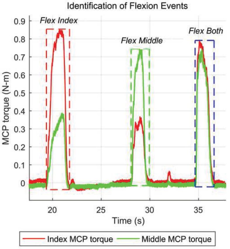
Wolbrecht et al.                                                                                            Page 3

                                       Poliakov, 1998). In humans, however, this hypothesis is not well tested. Some studies have
Author Manuscript

                                       limited subject populations to those with damage to the CST or primary motor cortex and
                                       demonstrated reduced finger individuation (Lang and Schieber, 2003; Lang and Schieber,
                                       2004). But, to our knowledge few attempts have been made to determine if the presence or
                                       absence of CST damage correlates with loss of finger individuation.

                                       Critically, in contrast to the well-known contribution of hand strength, the contribution of
                                       reduced finger individuation to hand function also remains unclear at present. One study did
                                       not find correlation between finger individuation and clinical tests of hand function
                                       (Raghavan et al., 2006). Another found that measures of finger individuation based on
                                       unwanted extra finger movements correlated with the Action Research Arm Test (ARAT)
                                       and the Moberg Pick-Up Test scores (Térémetz et al., 2015).

                                       Understanding the relative contributions of finger strength and individuation to hand
Author Manuscript

                                       function is essential for optimal development and application of stroke therapies, and for
                                       clarifying which of these factors should be targeted during rehabilitation. For example,
                                       robotic and sensor-based devices are being proposed to target training of finger
                                       independence (Adamovich et al., 2009; Dovat et al., 2010; Friedman et al., 2014; Thielbar et
                                       al., 2014), but this approach makes most sense if finger individuation plays a leading role in
                                       causing loss of hand function. In this paper, we investigated the impact of stroke on both
                                       finger strength and individuation, as well as on a novel metric of their combined effect on
                                       multifinger force production. We also estimated lesion load of the CST to determine if it
                                       correlates with these impairments, and used stepwise linear regression to identify which of
                                       these measures best predicted hand function.

                       2. Methods
Author Manuscript

                       2.1. Subjects
                                       Twenty-six individuals with a history of a single stroke were recruited to participate in a
                                       parent study (Rowe et al., 2017) that used the FINGER robotic exoskeleton device (Figure 1,
                                       (Taheri et al., 2014)) for robot-assisted hand therapy. Four additional subjects completed the
                                       parent study, but their data were not analyzed because the torques they generated were very
                                       small and noisy, due to the severity of their impairment. All of the participants provided
                                       informed consent following a protocol approved by the local Institutional Review Board.
                                       The study was registered on Clinical-Trials.gov (NCT02048826).

                       2.2. Clinical Tests of Hand Function
                                       Besides the Box and Blocks Test, subjects completed the Nine-Hole Peg Test to measure
Author Manuscript

                                       finger function. The tests were administered by a single, blinded, and experienced physical
                                       therapist two-weeks prior to a three-week training period with the FINGER robot. The
                                       therapist also assessed the Fugl-Meyer Test of the Upper Extremity (See et al., 2013), an
                                       impairment-based measure that judges the ability to control arm joints independently and
                                       ranges from 0 (no arm movement) to 66 (normal arm movement function). All three clinical
                                       assessments were repeated at the conclusion of the three-week training period; results
                                       reported here are averages from the two separate assessments. Scores from the Box and

                                          Clin Neurophysiol. Author manuscript; available in PMC 2019 April 01.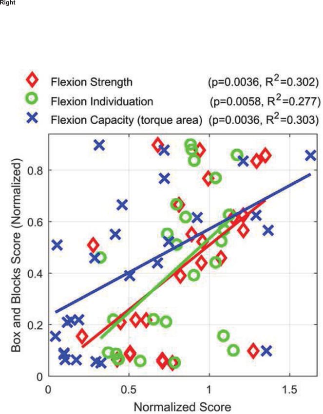
Wolbrecht et al.                                                                                            Page 4

                                       Blocks and Nine-Hole Peg tests were normalized to the performance of the unimpaired arms
Author Manuscript

                                       of the subjects, giving a range of 0–1 for those tests.

                       2.3. Quantification of Corticospinal Tract Injury
                                       High-resolution T1-weighted and T2 FLAIR (fluid- attenuated inversion recovery) images
                                       were acquired using a 3.0T Philips (Best, the Netherlands) Achieva system. Using MRIcron
                                       software (http://www.mccauslandcenter.sc.edu/mricro/mricron), each patient's infarct was
                                       outlined by hand on the T1-weighted MRI image, informed by the T2 FLAIR image as
                                       described previously (Burke et al., 2014). Stroke masks were transformed into MNI
                                       (Montreal Neurological Institute) standard stereotaxic space using FSL (Functional
                                       Magnetic Resonance Imaging of the Brain Software Library). Stroke masks for patients with
                                       right-sided lesions were flipped about the midsagittal plane onto the left hemisphere, to
                                       allow direct comparison between patients.
Author Manuscript

                                       Because fiber tracking with diffusion tensor imaging can be problematic in brain regions
                                       affected by stroke, injury to the CST for each patient was quantified by examining the extent
                                       to which the stroke infarct overlapped with a template CST generated from healthy controls
                                       (Burke et al., 2014; Burke Quinlan et al., 2015; Nouri and Cramer, 2011; Riley et al., 2011).
                                       To simulate damage to groups of axons, the template CST was divided into 16 separate
                                       longitudinal subsections using a standard canonical template. The number of subsections
                                       was determined from a prior report (Riley et al., 2011) which found that dividing the CST
                                       into 16 subsections provided the strongest correlations with behavioral status in patients with
                                       stroke. For each subject, the binary stroke mask was overlapped onto each CST subsection.
                                       The percentage of CST injury was calculated from the summed number of subsections
                                       damaged more than 5% (percentage also based on Riley et al. (2011)) divided by the total
                                       number of subsections, which was then converted to a percentage. To determine percent
Author Manuscript

                                       damage, the lesions were manually outlined and binarized using validated methods
                                       previously reported (Burke et al., 2014). CST injury for subjects with infarcts below the
                                       level of the thalamus were omitted from relevant statistical analyses.

                       2.4. The FINGER Robotic Exoskeleton
                                       The FINGER robot consists of two stacked single-degree-of-freedom 8-bar mechanisms for
                                       guiding the index and middle fingers through a naturalistic grasping motion (Taheri et al.,
                                       2014; Taheri et al., 2012). FINGER can assist in flexion and extension of index and middle
                                       fingers independently or together. During the grasping motion, the metacarpophalangeal
                                       (MCP) joint can move from full extension to 60 degrees of flexion. Each mechanism
                                       connects to the middle and proximal phalanx of the finger through small load cells (Futek
                                       LSB200 miniature s-beam load cells) located on the dorsal surface of the finger (see Figure
Author Manuscript

                                       1). FINGER is actuated by two brushless linear actuators (Dunkermotoren Servotube
                                       STA116-168-S-S03C). The combination of these low-friction actuators and precision low-
                                       friction mechanisms makes FINGER highly backdriveable and directly force controllable.

                       2.5. Robot Tests of Finger Strength and Individuation
                                       The parent study (Rowe et al., 2017) included three weeks of robot-assisted finger training,
                                       with three, one-hour sessions per week. During movement training with FINGER the

                                          Clin Neurophysiol. Author manuscript; available in PMC 2019 April 01.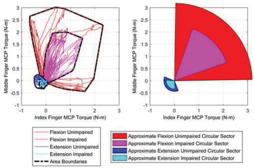
Wolbrecht et al.                                                                                             Page 5

                                       subjects played a musical computer game similar to Guitar Hero (Taheri et al., 2012). While
Author Manuscript

                                       playing the game, they attempted to move their index and middle fingers (both individually
                                       or together) to meet streaming musical notes at a specific place and time on the computer
                                       screen. Varying levels of robotic assistance were provided to the subjects during gameplay,
                                       based on the rate at which they successfully met the notes, following the algorithm described
                                       in (Taheri et al., 2014). Further details of the musical computer game and parent study can
                                       be found in (Rowe et al., 2017).

                                       In addition to playing the musical computer game, subjects also completed a weekly robotic
                                       test designed to quantify finger movement ability. Here, we analyzed a maximum voluntary
                                       contraction (MVC) test for measuring finger flexion and extension strength. This test was
                                       administered two weeks prior to the three-week training period, at the start of each week of
                                       training, and at the conclusion of the training period, for a total of five tests. Each subject
                                       also completed these tests once with their unimpaired hand, in order to provide data for
Author Manuscript

                                       normalizing the impaired hand performance.

                                       During the MVC test, the FINGER robot held the index and middle fingers fixed (at an MCP
                                       angle of approximately 30 degrees) by simulating a stiff, damped spring. Participants were
                                       instructed to “Flex as hard as you can” against the robot: first their index finger alone (“flex
                                       index”), followed by their middle finger alone (“flex middle”), and finally both middle and
                                       index fingers at the same time (“flex both”). They then repeated this sequence for finger
                                       extension. They typically held the flexion or extension for 2–5 seconds, and typically there
                                       were 5–10 seconds between the events, which were always performed in the same sequence.
                                       An example showing MVC tests in flexion is shown in Figure 2.

                       2.6. Data Collection and Analysis
Author Manuscript

                                       Data from all four load cells (two per finger) and the FINGER position sensors were
                                       recorded at 1000 Hz during all experiments. Based on the kinematic and mechanical design
                                       of FINGER, the measured force on the proximal phalanx (fp) and middle phalanx (fm) of
                                       each finger were mapped to the equivalent proximal interphalangeal (PIP) torque (τPIP) and
                                       MCP torque (τMCP), according to

                                                                                                                    (1)

                                       where rp is distance from the MCP joint to the proximal phalanx force sensor, rm is the
                                       distance from the PIP joint to the middle phalanx force sensor, and lp is the length of the
Author Manuscript

                                       proximal phalanx. The angle of the middle phalanx force, fp, is always normal to the dorsal
                                       surface. This is not the case with middle phalanx force sensor, where the angle relative to the
                                       dorsal surface normal, θA, changes during motion. The angle θB is the angle of fm relative to
                                       fp. These two angles, θA and θB, are determined as a function of actuator position according
                                       to the kinematics of the 8-bar mechanism (Taheri et al., 2014).

                                       The calculated MCP torque includes contributions from the forces measured at both the
                                       middle and proximal phalanx and thus provides a more complete measure of finger strength

                                          Clin Neurophysiol. Author manuscript; available in PMC 2019 April 01.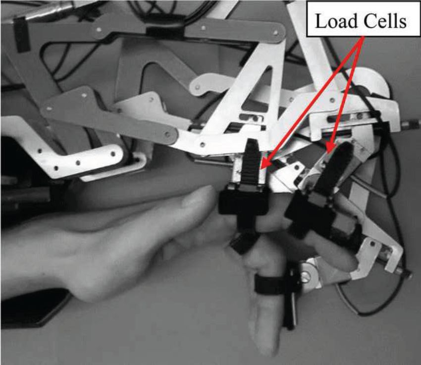
Wolbrecht et al.                                                                                              Page 6

                                       during finger flexion than the PIP torque and is our focus here. Unfiltered time-series MCP
Author Manuscript

                                       torque data (Figure 2) were used to identify each flexion event (flex index, flex middle, flex
                                       both). Bias torques from tone, determined from the intervals between the events, were
                                       subtracted from the MCP torque data. Extension torques were much smaller and difficult to
                                       identify, and thus not parsed out of the time series. Kinematic and kinetic data were
                                       smoothed using a 50th-order windowed linear-phase finite-impulse-response digital filter
                                       with cut-off frequency fc = 100 Hz. Time series with clear outlier data were either trimmed
                                       or omitted; for example when flexion events were either missing or could not be clearly
                                       identified or when there was clear confusion on the part of the subjects regarding when and
                                       how to move their middle and index fingers.

                       2.7. Quantification of Finger Strength and Individuation from Multifinger Capacity Plots
                                       For the primary analyses below, MCP torque data were combined from all MVC tests across
Author Manuscript

                                       all evaluations from each of the 26 subjects. Plotting index and middle finger torques on the
                                       x and y axes, respectively, provided a means to visualize and quantify finger strength and
                                       individuation (left side of Figure 3). We term such a plot a “multifinger capacity plot” (or
                                       “capacity plot”). A convex hull can be fit such that it circumscribes the torque data (right
                                       side of Figure 3). Analysis of the convex hull boundary provides metrics of both strength
                                       and individuation, including the total area, which combines both.

                                       To define individuation and strength, a pie-wedge shape (named a “circular sector” in plane
                                       geometry) was fit to the convex hull boundary. The radius of the circular sector, which
                                       represents an average of strength across the two fingers, was defined as the average of the
                                       maximum distances from the origin along three directions: the x -axis, the y -axis, and the y
                                       = x direction. To define individuation from a multifinger capacity plot, first note that full
                                       individuation ability (I = 1) would result in a 90° sector (i.e. a quarter pie wedge), and zero
Author Manuscript

                                       individuation would result in a 0° sector. Thus, individuation can be found from the convex
                                       hull area A and the radius r :

                                                                                                    (2)

                                       Examples of circular sectors fit to flexion and extension hulls for both their unimpaired and
                                       impaired sides are shown in the right side of Figure 3. Here, we define flexion capacity and
                                       extension capacity as the area of the circular sectors in flexion and extension, respectively, as
                                       they represent a combined capacity of finger individuation and strength. Below we often
                                       report strength and capacity of the impaired hand normalized to the values of unimpaired
Author Manuscript

                                       hand, in which case we identify the measure as “normalized” and report the measure without
                                       units. We did not normalize individuation, since individuation scores (e.g. 0 or 1) have
                                       interpretations that normalization would obscure. It should be noted that individuation
                                       values greater than 1 are possible when MCP torques of opposite signs are recorded from the
                                       two fingers (resulting in torque vectors in quadrants II and IV of the multi-finger capacity
                                       plots). This appears to be a “leveraging” strategy, in which the subject “pushes off” with one
                                       finger to maximize the torque produced by the other finger.

                                          Clin Neurophysiol. Author manuscript; available in PMC 2019 April 01.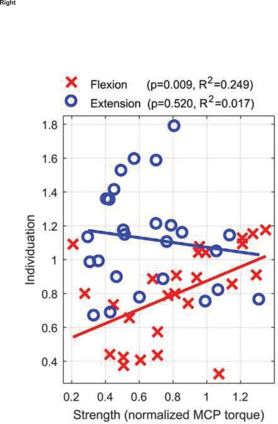
Wolbrecht et al.                                                                                            Page 7

                       2.8. Statistics and Stepwise Linear Regression
Author Manuscript

                                       To compare measures of strength, individuation, or capacity, we used paired t-tests across
                                       the 26 subjects. To evaluate data normality, we used normal probability plots and the
                                       Anderson-Darling test. The %CST injury data showed significant departure from normality,
                                       so we used Spearman’s method for correlation analysis. For our three response variables
                                       (Fugl-Meyer score, Box and Blocks score, and Nine Hole Peg Test score), only the Nine
                                       Hole Peg Test score data displayed non-normality due to a floor effect for 9 of 26 subjects
                                       who scored zero; when we removed those subjects, Nine Hole Peg Test score data passed
                                       normality, and the regression values did not change substantially; capacity was still the
                                       strongest predictor (see below). For simplicity and consistency, we report the values for all
                                       subjects, as for the other measures, and we used Pearson’s method for all correlation
                                       analysis. For each clinical outcome, we used stepwise linear regression to identify the
                                       simplest model with the strongest predictive power. Table 1 lists the six predictor variables
Author Manuscript

                                       (strength, individuation, and capacity in both flexion and extension) and the three response
                                       variables (Fugl-Meyer score, Box and Blocks score, and Nine Hole Peg Test score)
                                       considered. All predictor variables were normalized to the unimpaired hand. Linear,
                                       quadratic, and cross terms were considered during the stepwise linear regression, which was
                                       performed in the forward direction using five different criteria for each (sum-of-squared
                                       error, Akaike information, Bayesian information, R2, and Adj. R2). The forward direction
                                       was chosen because no clear model hypothesis existed and the simplest model was
                                       preferred.

                       3. Results
                                       All subjects had experienced a single, unilateral stroke at least six months before the study,
                                       were between 18–73 years old, and were able to score a minimum of three blocks on the
Author Manuscript

                                       Box and Blocks test (see Table 2, (Mathiowetz et al., 1985)). Of the 26 subjects, 54% had
                                       hypertension, 54% had hypercholesterolemia, 19% had diabetes mellitus, and 15% had atrial
                                       fibrillation. The stroke affected the right arm in 11 and the left arm in 15. Subjects were a
                                       median of 19.4 [interquartile range (IQR) 11 – 49] months post-stroke at time of study
                                       enrollment. Across subjects, infarct locations varied, including hemispheric and brainstem,
                                       affected the cerebral cortex in 15, and had median volume of 5.3 cc [IQR 1.5 – 38.5]. Table
                                       2 shows the demographics and baseline Box and Blocks and Fugl-Meyer scores of the 26
                                       individuals with a stroke who participated in the study. All participants had at least some
                                       minimal hand function, defined as ability to lift at least three blocks in the Box and Blocks
                                       Test.

                       3.1. Multifinger Capacity Plots: Visualizing Finger Strength, Individuation, and Capacity
Author Manuscript

                                       We first present a new way to visualize hand impairment after stroke, “Multifinger capacity
                                       plots”, which graph index and middle finger torques generated during MVC tests against
                                       each other, overlaying tests of each finger in isolation and together (Figure 4). These plots
                                       provide a way to simultaneously visualize finger strength (radius of the approximating
                                       convex hull) and individuation ability (angular width of the hull). As can be seen, for the 26
                                       individuals with a stroke studied here, flexion torques (hulls in the upper right quadrants)
                                       were in general much larger than extension torques (hulls in the lower left quadrants), both

                                          Clin Neurophysiol. Author manuscript; available in PMC 2019 April 01.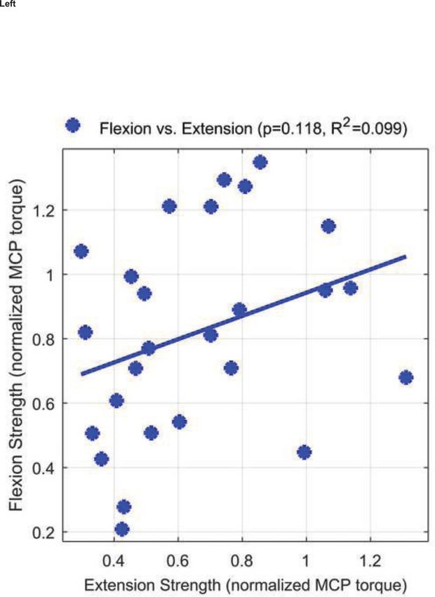
Wolbrecht et al.                                                                                            Page 8

                                       before and after stroke (Figure 4). The hulls were more shrunken for fingers that were
Author Manuscript

                                       weaker, and were also often more narrowed so that they looked like cigars, rather than round
                                       balloons. The narrowing reflected the inability of these fingers to independently generate
                                       torques; the fingers operated in tandem, synergistically generating torque together. In sum,
                                       multifinger capacity plots depict the torque generation resource the hands have to work with
                                       to achieve manipulative function. The convex hull area is a combined metric of strength and
                                       individuation that quantifies this resource, and we will refer to this measure as “capacity”.

                                       To validate analyses based on multifinger capacity plots, we tested if the measures of finger
                                       strength and individuation derived from these plots correlated with previously used metrics
                                       for strength and individuation. Finger flexion strength correlated with pinch grip strength (3-
                                       jaw chuck, p = 0.001, R2 = 0.353) and lateral pinch strength (lateral/key pinch, p = 0.004, R2
                                       = 0.295) obtained with a dynamometer. Finger individuation strongly correlated with the
                                       Individuation Index used in previous studies (p < 0.001, R2 = 0.81) (Lang and Schieber,
Author Manuscript

                                       2003; Schieber, 1991), which we calculated from torque path distances of the flex index and
                                       flex middle events of the MVC tests, rather than angular path distances, since the
                                       measurements were isometric.

                       3.2. Finger Strength, Finger Individuation, and their Relationship
                                       Based on analysis of the multifinger capacity plots, finger flexion strength was significantly
                                       greater than finger extension strength (MCP torque) both for the impaired hand (2.17 ± 0.93
                                       N-m compared to 0.46 ± 0.18 N-m, p < 0.001) and the unimpaired hand (2.69 ± 0.64 N-m
                                       compared to 0.74 ± 0.27 N-m, p < 0.001). The ratio of flexion to extension strength was 5.06
                                       ± 2.23 for the paretic hand, compared to 3.97 ± 1.46 for the non-paretic, a significant
                                       difference (p = 0.042). Relative to the unimpaired hand of each participant, the average
                                       finger flexion strength was 0.82 ± 0.32, while the average finger extension strength was 0.66
Author Manuscript

                                       ± 0.28, a significant difference (p = 0.029). Normalized finger flexion and extension torques
                                       were nearly correlated (Figure 5 left, p = 0.118, R2 = 0.099).

                                       Average flexion individuation for the impaired hand was 0.80 ± 0.27 and extension
                                       individuation was 1.12 ± 0.30, a significant difference (p < 0.001). The two were weakly
                                       correlated (p = 0.028, R2 = 0.186).

                                       To test the hypothesis that finger weakness and loss of individuation arise from a common
                                       mechanism after stroke, we evaluated their correlation for this population of participants
                                       with a wide variety of lesion locations and sizes. Flexion strength, normalized to the
                                       unimpaired hand, was positively correlated with flexion individuation (Figure 5 right, p =
                                       0.009, R2 = 0.249). Normalized extension strength was not, however, correlated with finger
Author Manuscript

                                       extension individuation (Figure 5 right, p = 0.52, R2 = 0.017). Thus, finger strength and
                                       individuation were related, but only for flexion.

                       3.3. Relationship to Lesion Overlap with the CST
                                       We quantified CST injury for each participant as the percentage of overlap between the
                                       infarct and a healthy control template CST. Six subjects had infarcts below the level of the
                                       thalamus, for which the template tract was not available. Of the remaining subjects, three
                                       had no lesion overlap with the CST and 17 had an overlap of the CST ranging from 6.25% to

                                          Clin Neurophysiol. Author manuscript; available in PMC 2019 April 01.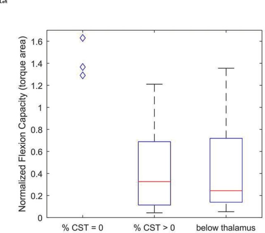
Wolbrecht et al.                                                                                              Page 9

                                       100%. As a first analysis, we divided the participants into three groups: % CST = 0, % CST
Author Manuscript

                                       > 0, and, for completeness, lesion below thalamus (i.e. CST overlap unknown). For the
                                       flexion capacity metric, the % CST = 0 injury group was significantly different from the %
                                       CST > 0 group (p < 0.001), as well as the below thalamus group (p = 0.003) (see Figure 6
                                       Left). Thus, for injuries above the level of the thalamus, lesions that overlapped the CST
                                       impaired finger capacity compared to those that did not. For the strength and individuation
                                       metrics, repeating the same group comparisons did not always produce significant results,
                                       although the comparison trended toward significance (flexion strength: p = 0.040 for % CST
                                       > 0 and p = 0.064 for below thalamus, individuation: p = 0.072 for % CST > 0 and p = 0.146
                                       for below thalamus). In sum, CST injury best corresponded to reduced capacity.

                                       For all three flexion metrics, the below Thalamus and % CST > 0 groups were not
                                       significantly different (p = 1). None of the groups were significantly different for the
                                       extension metrics.
Author Manuscript

                                       Next, we assessed whether the percentage of CST injury (rather than the presence or absence
                                       of CST injury, as in the previous paragraph) correlated with strength, individuation, and
                                       capacity in both flexion and extension (for 6 total correlations). Because the % CST injury
                                       data was not normally distributed, we used Spearman’s rank correlation (Spearman’s ρ) for
                                       significance analysis. When all injuries above the thalamus were included (% CST ≥ 0), all
                                       three flexion metrics (strength, individuation, and capacity) correlated with % CST injury
                                       (Figure 6 Right, and Table 3). We also repeated this analysis for the 17 subjects in the %
                                       CST > 0 group due to the clear separation between this group and the % CST = 0 group. In
                                       this case, none of the three metrics correlated with % CST injury in flexion or extension
                                       (Table 3). Thus, while presence or absence of CST injury predicted reduced finger flexion
                                       capacity, percentage of CST injury did not correlate with amount of reduction of finger
Author Manuscript

                                       capacity, strength, or individuation (when at least some CST injury was present). CST injury
                                       was thus best considered as a binary, rather than graded, predictor of flexion capacity.

                       3.4. Relationship to Clinical Measures of Hand Impairment and Function
                                       We first assessed to what extent strength (normalized to the unimpaired hand for these
                                       analyses), individuation, and capacity (normalized to the unimpaired hand for these
                                       analyses) correlated with the Upper Extremity Fugl-Meyer score. We again individually
                                       tested these three measures for flexion and extension, for six total correlations (Table 3). For
                                       finger flexion, all three measures were positively correlated with the Fugl-Meyer score, with
                                       flexion capacity correlated the most strongly (Figure 7). Extension capacity correlated more
                                       strongly than strength with the Fugl-Meyer score; extension individuation did not correlate
                                       significantly (Table 3).
Author Manuscript

                                       Next, we analyzed relationship of strength, individuation, and capacity to hand function
                                       measured using well-established clinical tests that required rapidly manipulating small
                                       objects. Flexion capacity positively correlated the most strongly to both the Box and Blocks
                                       score and Nine Hole Peg Test score (Figure 7, Table 3). The other two metrics (individuation
                                       and strength) were also positively correlated in the flexion direction. In the extension
                                       direction, only capacity correlated significantly with hand function, measured with the Box
                                       and Blocks score. In general, the capacity metric accounted for higher variance in the

                                          Clin Neurophysiol. Author manuscript; available in PMC 2019 April 01.
Wolbrecht et al. Page 10
correlations with the impairment and function measures (average R2 = 0.27 across six
functional correlations in Table 3), followed by strength (average R2 = 0.23) then
Author Manuscript
individuation (average R2 = 0.15). In addition, the capacity metric correlations were more
significant, in terms of p-values, for all six functional correlations (Table 3).
In 10 of the total 15 applications of stepwise linear regression, the resulting model was 1 +
FC (normalized flexion capacity, Table 4). The remaining models (some of which are also
shown in Table 4) were either minimally more predictive, although the model 1 + (FC)(EC)
that added additional complexity through a cross-term combining flexion and extension
capacity was more predictive than 1 + FC in terms of effect size R2. Each resulting model
included flexion capacity as a term in the model.
4. Discussion
Author Manuscript
Loss of finger strength and loss of finger individuation are two motor impairments thought
to diminish hand function after stroke. The results of this study suggest that multifinger
capacity, which represents the combined effect of strength and individuation, explains more
of the inter-subject variance in hand function than either alone. Further validation of the
capacity metric came from the finding that it was also significantly related to presence or
absence of lesion overlap with the corticospinal tract. We discuss now the implications of
this work, as well as limitations and directions for future research.
Why is finger capacity more related to hand function than strength or individuation alone?
Previous studies of the determinants of hand function after stroke have often focused on
measuring force production of all fingers working together. One key such study found that it
was the inability to activate motor neuronal pools, rather than muscle atrophy, increased
Author Manuscript
passive stiffness, or spasticity, that limited force production (Kamper et al., 2006). Many
studies have found that this activation inability, manifested as power grip weakness, is one of
the best predictors of functional upper extremity deficit after stroke (Bohannon et al., 1991;
Canning et al., 2004; Dobkin, 1996; Harris and Eng, 2007; Heller et al., 1987; Lang and
Beebe, 2007; Ma et al., 2014; Warabi et al., 1990).
In contrast, studies that have focused on the role of individuation in hand function are
limited and equivocal. One study did not find correlation between finger individuation and
clinical tests of hand function (Raghavan et al., 2006). Another found that measures of finger
individuation based on unwanted extra finger movements correlated with the Action
Research Arm Test and the Moberg Pick-Up Test scores (Térémetz et al., 2015). Another
recent report found a strong correlation between both strength and individuation with the
Author Manuscript
Fugl-Meyer score as well as a functional hand score, the ARAT, with similar strengths of
correlation for strength and individuation (Xu et al., 2017).
Consistent with these findings that individuation affects hand function, recent studies
examining the ability to coordinate hand muscles after stroke support the concept that
increased multiple-muscle coupling contributes significantly to hand impairment after
stroke. For example, greater hand impairment was associated with greater muscle
coactivation in a recent study of hand muscle EMG synergies after stroke (Lee et al., 2013).
Clin Neurophysiol. Author manuscript; available in PMC 2019 April 01.Wolbrecht et al. Page 11
Flexor coupling between the thumb and fingers was also found to contribute to undesired
Author Manuscript
thumb movement after stroke, likely impacting hand function (Kamper et al., 2014).
To our knowledge, multifinger capacity, calculated as the area of torque space traced out
during individuated and synergistic multi-finger MVCs, does not have a direct comparison
in existing literature. This measure assesses the overall finger capacity for generating torques
individually and independently, in flexion or extension. We hypothesize that capacity
mattered particularly for the type of hand function we studied here – gripping blocks and
pegs. One must have enough individuation ability to position the fingers in a way that allows
one to grip such small objects, and then be able to exert enough force to maintain the grip on
the object.
It is interesting to note that finger capacity is the mathematical product of strength and
individuation. The stepwise linear regression procedure considered models that contained
Author Manuscript
the sum of strength and individuation, as well, but it is their product that survived the
stepwise selection process. Their product has a geometric interpretation, which is the area of
the multifinger capacity plot, which can be understood as the area of the space of achievable
flexor finger torques, a key resource for hand function. From the capacity point of view,
strength and individuation are very similar. Both require the motor system to generate a
balanced pattern of multiple-muscle activity such that a desired force outcome is met. Both
work to expand the workspace of achievable finger torques.
Do impairments of finger strength and individuation arise from a common anatomical
injury?
In the flexion direction, finger strength correlated strongly with finger individuation,
confirming the conjecture that stroke affects both with at least some commonality. In fact,
Author Manuscript
strength and individuation showed comparable correlations to Fugl-Meyer, Box and Blocks,
and Nine Hole Peg scores. This is somewhat unexpected, given some previous research
suggesting that CST injury affects fine motor control more than strength (Jang, 2009).
Analysis of the extension strength and individuation data did not typically provide
significant correlations.
Xu et al. (2017) recently reported a nonlinear relationship between strength and
individuation after stroke. Initially after a stroke, strength and individuation were strongly
correlated for most patients – i.e. the relationship was fairly linear. Then, a year later,
patients whose hand strength exceeded 60% of the ipsilesional side (now over half of the
individuals) exhibited individuation near maximum that was uncorrelated with strength. This
saturation of individuation without full recovery of strength produced a nonlinear
Author Manuscript
relationship. We did not observe a saturation in individuation for the chronic stroke
participants studied here, even though they had a wide range of strength recovery. In
particular, in the flexion direction, individuation positively and linearly correlated with
strength (Fig. 5 right). In the extension direction, strength and individuation were not
correlated. The difference with Xu may be due to the apparatus, visual feedback provided, or
data analysis techniques for estimating individuation and strength being different.
Differences in stroke populations studied can affect results, too, and so this difference may
also be due to relatively fewer participants in the present study (N = 26) versus theirs (N =
Clin Neurophysiol. Author manuscript; available in PMC 2019 April 01.Wolbrecht et al. Page 12
54) obscuring the relationship. For now, the most robust result, observed in both studies, is
Author Manuscript
that strength and individuation are strongly correlated, at least until individuation is nearly
fully recovered. This suggests they arise from a common source.
Is the source injury to the CST? Xu et al. (2017) found different time courses and patterns of
variability in strength and individuation. They also found that individuation correlated more
with CST injury than strength. These observations led them to speculate that strength
recovery, along with some individuation, can be attributed to descending systems other than
the CST, whereas individuation relies more on the CST. This interpretation is difficult to test
with our data, since we measured at only the chronic stage of recovery. We also found
comparable correlations between strength, individuation, and capacity with CST injury,
when considering all above-thalamus injuries together (% CST ≥ 0). That is, the three
correlations had comparable strength, making it difficult to associate CST injury with one
individual metric. Again, the most robust conclusion at this point is that CST injury is
Author Manuscript
involved in both strength and individuation deficits.
In the cases where the injury was above the level of the thalamus, subjects whose injuries
overlapped the CST (% CST > 0) exhibited significantly greater impairment compared to
those whose injuries did not (% CST = 0). We note that it is this difference that drove the
correlation found in the % CST ≥ 0 cases, rather than a strong correlation among only the
CST > 0 cases. Therefore, while the results from this study support previous studies showing
that amount of injury CST overlap predicts motor impairment and functional ability (Pineiro
et al., 2000; Sterr et al., 2014; Sterr et al., 2010; Zhu et al., 2010), they suggest that this
correlation may be driven by a binary pattern, in which % CST = 0 spares function, and, in
contrast, % CST > 0 produces relatively lower but variable and non-graded levels of
impairment. It is possible this binary pattern is an artifact of the subsection technique we
Author Manuscript
used to quantify CST injury, which, among other possible shortcomings, does not account
for CST somatopy.
The lack of independent control of the fingers, manifested as the “cigar-shaped” torque
traces for the most impaired participants, can be understood as a form of abnormal muscle
synergy or abnormal muscular coupling. A flexion synergy has been quantified for the upper
extremity, manifesting as unwanted activation coupling between shoulder abductor elbow
flexor muscles (Ellis et al., 2016). The flexion synergy has been suggested to arise as a result
of dependence on more diffusely innervating motor pathways, such as the reticulospinal
tract, following CST damage (Ellis et al., 2012). The present data are consistent with the
idea that a reliance on more diffusely innervating motor pathways could cause the abnormal
coupling between the index and middle fingers.
Author Manuscript
Relative impairment in finger extension versus flexion and relationship to hand function
Previous studies have found that stroke affects finger extension strength more than flexion
strength on average (Conrad and Kamper, 2012). The results of this study support this
observation, finding that normalized finger flexion strength after stroke was larger (0.82)
than normalized finger extension strength (0.66). Looked at another way, the ratio of flexion
to extension strength in the paretic hand (5.1) was significantly larger than the non-paretic
hand (4.0), again consistent with the concept that extension was relatively more affected. It
Clin Neurophysiol. Author manuscript; available in PMC 2019 April 01.Wolbrecht et al. Page 13
is worthwhile to note, however, that extension will always appear weak in an absolute sense
Author Manuscript
because, even before a stroke, finger flexion strength is four times stronger than extension
strength.
To our knowledge, we measured individuation ability for both finger flexion and extension
after stroke for the first time. Average individuation was significantly less in the flexion
direction (0.8) than extension direction (1.1). Thus, in contrast to strength, stroke appears to
affect finger flexion individuation more than finger extension individuation. The reason is
unknown, but this finding suggests that the neural subsystems that control flexion and
extension are somewhat separable. A caveat is that the signal-to-noise ratio of the extensors
was low compared to flexors, and this may have affected the extension individuation
measure.
Flexion capacity survived the stepwise linear regression analysis most frequently when
Author Manuscript
testing for a parsimonious predictor of hand function (Table 3), explaining 30–40% of the
variance in the hand function assessments. However, adding extension capacity to the
modeled explained an additional ~15% of the variance. In light of the specific population
studied here (i.e. people who could open their hand a small amount), it may be that if
individuals preserve some finger extension ability, finger flexion metrics are relatively more
important for predicting function, although finger extension still plays a role.
Limitations and future directions
Limitations of this study are as follows. The study had a limited sample size and should be
verified with a larger population. We only included individuals with at least a small amount
of hand function, and caution should be exerted in comparing this study to other studies that
included participants with more serve loss of hand function. We relied on the participants to
Author Manuscript
accurately interpret the instructions to move one or the other finger. If, in practice, they
never actually attempted to isolate their finger movements, it could have made the capacity
appear smaller than actual; we consider this possibility unlikely as the instructions were
simple and clear. Another caveat already mentioned was that extension torques were small
and variable, and thus caution should be applied in interpreting lack of significant
correlation of function or anatomical injury with any of the measures of extension ability.
Another aspect of the study is that we averaged measurements of finger force production
taken over a three week study of robotic therapy, and this may have increased measurement
variance if the measures changed significantly over time. Preliminary analysis suggests they
did not change, suggesting, for example, that individuation is difficult to improve after
stroke, but future work will examine this issue. Other possible sources of variance were: we
always measured flexion before extension (and isolated movement before synergistic
Author Manuscript
movement); possible fatigue encountered during the multiple MVCs at each weekly test; and
variability in level of preserved finger sensation (Rowe et al., 2017). Finally, we only studied
one type of hand function, which involved picking and placing small objects (i.e. blocks and
pegs). However, the fact that the capacity metric also best predicted a more general upper
extremity impairment measure (the Fugl-Meyer test), affords some confidence that it reflects
more general aspects of hand function.
Clin Neurophysiol. Author manuscript; available in PMC 2019 April 01.Wolbrecht et al. Page 14
As for future directions, this study focused on MCP torques generated by the middle and
Author Manuscript
index fingers, both individually and together, during maximum voluntary contraction tests.
Multifinger capacity plots could be created not only for isometric force production, but for
dynamic movement of the fingers, when force production deficits become even more
exaggerated (Conrad and Kamper, 2012). Further, although the current study was conducted
using the FINGER robotic device, which is a capable of evaluating the performance of the
index and middle fingers, this approach may be applied to data collected from other devices
and may be extended to include other digits of the hand, albeit without the ease of
visualization provided by the two degree-of-freedom case.
The previous point highlights another limitation, which is lack of strength, individuation,
and capacity metrics for the thumb. The thumb has been shown to be a strong predictor of
arm function (Lang et al., 2003) even though thumb individuation ability after stroke
remains unclear (Raghaven et al., 2006). The participants used the thumb for the functional
Author Manuscript
tests measured here, and adding measures of thumb movement into models may increase
their predictive power.
Finally, in considering therapeutic targets, this study suggests that rehabilitative hand
training should seek to improve both strength and individuation. If one could simultaneously
improve both, one theoretically would have a multiplicative effect on improving multifinger
capacity and thus hand function. An interesting question is, since strength and individuation
are correlated, at least in flexion as we found here, will training one improve the other, or
should they be independently targeted? Further, extensor individuation may not be as
important to target as extension strength.
Acknowledgments
Author Manuscript
Steven C. Cramer serves or has served as a consultant for GlaxoSmithKline, RAND Corporation, Dart
Neuroscience, and MicroTransponder, and is a cofounder of personalRN. He acknowledges support from K24
HD074722 and UL1 TR000153.
This project was supported by NIH-R01HD062744 from the National Center for Medical Rehabilitation Research
at the National Institute of Child Health and Human Development, and the National Center for Research Resources
and the National Center for Advancing Translational Sciences, National Institutes of Health, through Grant UL1
TR000153.
References
Adamovich SV, Fluet GG, Mathai A, Qiu Q, Lewis J, Merians AS. Design of a complex virtual reality
simulation to train finger motion for persons with hemiparesis: a proof of concept study. J Neuroeng
and Rehabil. 2009; 6(1):28. [PubMed: 19615045]
Bohannon RW, Warren ME, Cogman KA. Motor variables correlated with the hand-to-mouth
Author Manuscript
maneuver in stroke patients. Arch Phys Med Rehabil. 1991; 72(9):682–684. [PubMed: 1859265]
Brouwer BJ, Schryburt-Brown K. Hand function and motor cortical output poststroke: are they
related? Arch Phys Med Rehabil. 2006; 87(5):627–634. [PubMed: 16635624]
Burke E, Dodakian L, See J, McKenzie A, Riley JD, Le V, Cramer SC. A multimodal approach to
understanding motor impairment and disability after stroke. J Neurol. 2014; 261(6):1178–1186.
[PubMed: 24728337]
Burke Quinlan E, Dodakian L, See J, McKenzie A, Le V, Wojnowicz M, Shahbaba B, Cramer SC.
Neural function, injury, and stroke subtype predict treatment gains after stroke. Ann Neurol. 2015;
77(1):132–145. [PubMed: 25382315]
Clin Neurophysiol. Author manuscript; available in PMC 2019 April 01.Wolbrecht et al. Page 15
Canning CG, Ada L, Adams R, O'Dwyer NJ. Loss of strength contributes more to physical disability
after stroke than loss of dexterity. Clin Rehabil. 2004; 18(3):300–308. [PubMed: 15137561]
Author Manuscript
Cho S-H, Kim DG, Kim D-S, Kim Y-H, Lee C-H, Jang SH. Motor outcome according to the integrity
of the corticospinal tract determined by diffusion tensor tractography in the early stage of corona
radiata infarct. Neurosci Lett. 2007; 426(2):123–127. [PubMed: 17897782]
Conrad MO, Kamper DG. Isokinetic strength and power deficits in the hand following stroke. Clin
Neurophysiol. 2012; 123(6):1200–1206. [PubMed: 22055766]
Dobkin, BH. Neurologic Rehabilitation. Vol. 47. F.A. Davis Company; Philadelphia: 1996.
Dovat L, Lambercy O, Salman B, Johnson V, Milner T, Gassert R, Burdet E, Leong TC. A technique to
train finger coordination and independence after stroke. Disabil Rehabil Assist Technol. 2010;
5(4):279–287. [PubMed: 20370489]
Ellis MD, Drogos J, Carmona C, Keller T, Dewald JP. Neck rotation modulates flexion synergy
torques, indicating an ipsilateral reticulospinal source for impairment in stroke. J Neurophysiol.
2012; 108(11):3096–3104. [PubMed: 22956793]
Ellis MD, Lan Y, Yao J, Dewald JPA. Robotic quantification of upper extremity loss of independent
joint control or flexion synergy in individuals with hemiparetic stroke: a review of paradigms
Author Manuscript
addressing the effects of shoulder abduction loading. J Neuroeng Rehabil. 2016; 13:95. [PubMed:
27794362]
Friedman N, Chan V, Reinkensmeyer AN, Beroukhim A, Zambrano GJ, Bachman M, Reinkensmeyer
DJ. Retraining and assessing hand movement after stroke using the MusicGlove: comparison with
conventional hand therapy and isometric grip training. J Neuroeng Rehabil. 2014; 11:76.
[PubMed: 24885076]
Gandevia, SC. Spasticity: Mechanisms and Management. Springer-Verlag; Berlin: 1993. Strength
changes in hemiparesis: Measurements and mechanisms; p. 111-122.
Harris JE, Eng JJ. Paretic upper-limb strength best explains arm activity in people with stroke. Phys
Ther. 2007; 87(11):88–97. [PubMed: 17179441]
Heller A, Wade D, Wood VA, Sunderland A, Hewer RL, Ward E. Arm function after stroke:
measurement and recovery over the first three months. J Neurol Neurosurg Psychiatry. 1987;
50(6):714–719. [PubMed: 3612152]
Jang SH. The role of the corticospinal tract in motor recovery in patients with a stroke: a review.
Author Manuscript
NeuroRehabilitation. 2009; 24(3):285–290. [PubMed: 19458437]
Kamper D, Harvey R, Suresh S, Rymer W. Relative contributions of neural mechanisms versus muscle
mechanics in promoting finger extension deficits following stroke. Muscle Nerve. 2003; 28(3):
309–318. [PubMed: 12929190]
Kamper D, Rymer W. Impairment of voluntary control of finger motion following stroke: role of
inappropriate muscle coactivation. Muscle Nerve. 2001; 24(5):673–681. [PubMed: 11317278]
Kamper DG, Fischer HC, Conrad MO, Towles JD, Rymer WZ, Triandafilou KM. Finger-thumb
coupling contributes to exaggerated thumb flexion in stroke survivors. J Neurophysiol. 2014;
111(12):2665–2674. [PubMed: 24671534]
Kamper DG, Fischer HC, Cruz EG, Rymer WZ. Weakness is the primary contributor to finger
impairment in chronic stroke. Arch Phys Med and Rehabil. 2006; 87(9):1262–1269. [PubMed:
16935065]
Kim Y, Kim W-S, Yoon B. The effect of stroke on motor selectivity for force control in single-and
multi-finger force production tasks. NeuroRehabilitation. 2014; 34(3):429–435. [PubMed:
24473243]
Author Manuscript
Lang CE, Beebe JA. Relating movement control at 9 upper extremity segments to loss of hand function
in people with chronic hemiparesis. Neurorehabil Neural Repair. 2007; 21(3):279–91. [PubMed:
17353458]
Lang CE, Schieber MH. Differential impairment of individuated finger movements in humans after
damage to the motor cortex or the corticospinal tract. J Neurophysiol. 2003; 90(2):1160–1170.
[PubMed: 12660350]
Lang CE, Schieber MH. Reduced muscle selectivity during individuated finger movements in humans
after damage to the motor cortex or corticospinal tract. J Neurophysiol. 2004; 91(4):1722–34.
[PubMed: 14668295]
Clin Neurophysiol. Author manuscript; available in PMC 2019 April 01.Wolbrecht et al. Page 16
Lawrence DG, Kuypers HGJM. The functional organization of the motor cortex in monkeys. I. The
effects of bilateral pyramidal lesions. Brain. 1968; 91(1):1–14. [PubMed: 4966862]
Author Manuscript
Lee SW, Triandafilou K, Lock BA, Kamper DG. Impairment in task-specific modulation of muscle
coordination correlates with the severity of hand impairment following stroke. PloS One. 2013;
8(7):e68745.doi: 10.1371/journal.pone.0068745 [PubMed: 23874745]
Li S, Latash ML, Yue GH, Siemionow V, Sahgal V. The effects of stroke and age on finger interaction
in multi-finger force production tasks. Clin Neurophysiol. 2003; 114(9):1646–1655. [PubMed:
12948793]
Lindberg PG, Roche N, Robertson J, Roby-Brami A, Bussel B, Maier MA. Affected and unaffected
quantitative aspects of grip force control in hemiparetic patients after stroke. Brain Res. 2012;
1452:96–107. [PubMed: 22464180]
Lindenberg R, Renga V, Zhu L, Betzler F, Alsop D, Schlaug G. Structural integrity of corticospinal
motor fibers predicts motor impairment in chronic stroke. Neurology. 2010; 74(4):280–287.
[PubMed: 20101033]
Ma VY, Chan L, Carruthers KJ. Incidence, prevalence, costs, and impact on disability of common
conditions requiring rehabilitation in the United States: stroke, spinal cord injury, traumatic brain
Author Manuscript
injury, multiple sclerosis, osteoarthritis, rheumatoid arthritis, limb loss, and back pain. Arch Phys
Med and Rehabil. 2014; 95(5):986–995. [PubMed: 24462839]
Mathiowetz V, Volland G, Kashman N, Weber K. Adult norms for the Box and Block Test of manual
dexterity. Am J Occup Ther. 1985; 39(6):386–391. [PubMed: 3160243]
Nouri S, Cramer SC. Anatomy and physiology predict response to motor cortex stimulation after
stroke. Neurology. 2011; 77(11):1076–1083. [PubMed: 21880996]
Pineiro R, Pendlebury S, Smith S, Flitney D, Blamire A, Styles P, Matthews P. Relating MRI changes
to motor deficit after ischemic stroke by segmentation of functional motor pathways. Stroke. 2000;
31(3):672–679. [PubMed: 10700503]
Raghavan P, Petra E, Krakauer JW, Gordon AM. Patterns of impairment in digit independence after
subcortical stroke. J Neurophysiol. 2006; 95(1):369–78. [PubMed: 16207778]
Riley JD, Le V, Der-Yeghiaian L, See J, Newton JM, Ward NS, Cramer SC. Anatomy of stroke injury
predicts gains from therapy. Stroke. 2011; 42(2):421–426. [PubMed: 21164128]
Rosso C, Valabregue R, Attal Y, Vargas P, Gaudron M, Baronnet F, Bertasi E, Humbert F, Peskine A,
Author Manuscript
Perlbarg V. Contribution of corticospinal tract and functional connectivity in hand motor
impairment after stroke. PloS One. 2013; 8(9)
Rowe JB, Chan V, Ingemanson ML, Cramer SC, Wolbrecht ET, Reinkensmeyer DJ. Robotic assistance
for training finger movement using a Hebbian model: A randomized controlled trial. Neurorehabil
Neural Repair. 2017; 31(8):769–780. [PubMed: 28803535]
Schieber MH. Individuated finger movements of rhesus monkeys: a means of quantifying the
independence of the digits. J Neurophysiol. 1991; 65(6):1381–1391. [PubMed: 1875247]
Schieber, MH., Lang, C., Reilly, K., McNulty, P., Sirigu, A. Progress in Motor Control. Springer; 2009.
Selective activation of human finger muscles after stroke or amputation; p. 559-575.
Schieber MH, Poliakov AV. Partial inactivation of the primary motor cortex hand area: effects on
individuated finger movements. J Neurosci. 1998; 18(21):9038–9054. [PubMed: 9787008]
See J, Dodakian L, Chou C, Chan V, McKenzie A, Reinkensmeyer DJ, Cramer SC. A standardized
approach to the Fugl-Meyer assessment and its implications for clinical trials. Neurorehabil Neural
Repair. 2013; 27(8):732–741. [PubMed: 23774125]
Sterr A, Dean PJ, Szameitat AJ, Conforto AB, Shen S. Corticospinal tract integrity and lesion volume
Author Manuscript
play different roles in chronic hemiparesis and its improvement through motor practice.
Neurorehabil Neural Repair. 2014; 28(4):335–343. [PubMed: 24334657]
Sterr A, Shen S, Szameitat AJ, Herron KA. The role of corticospinal tract damage in chronic motor
recovery and neurorehabilitation: a pilot study. Neurorehabil Neural Repair. 2010; 24(5):413–419.
[PubMed: 20516488]
Taheri H, Rowe JB, Gardner D, Chan V, Gray K, Bower C, Reinkensmeyer DJ, Wolbrecht ET. Design
and preliminary evaluation of the FINGER rehabilitation robot: controlling challenge and
quantifying finger individuation during musical computer game play. J Neuroeng Rehabil. 2014;
11:10.doi: 10.1186/1743-0003-11-10 [PubMed: 24495432]
Clin Neurophysiol. Author manuscript; available in PMC 2019 April 01.You can also read