Ultrasound Features of the First Gout Attack and the Association with Duration of Hyperuricemia
←
→
Page content transcription
If your browser does not render page correctly, please read the page content below
Uncorrected Proof

Iran J Radiol. 2019 July; 16(3):e85140.                                                                                            doi: 10.5812/iranjradiol.85140.

Published online 2019 July 30.                                                                                                                      Research Article

Ultrasound Features of the First Gout Attack and the Association with
Duration of Hyperuricemia
Wenting Fan 1 , Jiaan Zhu 1, * , Zheng Chen 1 and Wenxue Li 1
1
    Department of Ultrasound, Peking University People’s Hospital, Beijing, China
*
 Corresponding author: Department of Ultrasound, Peking University People’s Hospital, 11 Xizhimen South St., Beijing 100044, China. Tel: +86-1088325379, Fax: +86-1068318386,
Email: zhujiaan@pkuph.edu.cn

Received 2018 October 08; Revised 2019 May 29; Accepted 2019 June 22.

     Abstract

     Background: Gout is the most common form of inflammatory arthritis. Unfortunately, the burden of gout is increasing and treat-
     ment is still suboptimal. Nowadays, ultrasound is increasingly used to evaluate gout, especially in the early stage. However, little is
     known about the prevalence of the ultrasound signs in the first gout attack.
     Objectives: The aim of this study was to evaluate the prevalence of ultrasound features in the first gout attack and to correlate those
     features with the duration of hyperuricemia.
     Patients and Methods: We analyzed the ultrasound features of the knees, ankles and the first metatarsal-phalangeal joints (1st
     MTP) of patients with first gout attack compared to individuals with asymptomatic hyperuricemia (AHU). The findings were also
     compared with clinical, laboratory parameters and hyperuricemia duration.
     Results: Forty-eight patients with first gout attack gout were studied. The first attack affected the first MTP in 61%, the ankle in 33%,
     and the knee in 6% of the instances. The prevalence of snow-storm sign, double contour (DC) sign, tophi, bone erosion and abnormal
     blood flow was 92%, 29%, 6%, 13% and 83%, respectively. The prevalence of snow-storm sign and abnormal blood flow was significantly
     higher in the first attack of gout compared to AHU (P < 0.001). The hyperuricemia duration of patients with tophi and bone erosion
     was significantly longer than those with snow-storm sign and DC sign (7.5y and 6.5y vs 4.0y and 2y) (P = 0.004).
     Conclusion: Ultrasound features are associated with hyperuricemia duration. Furthermore, tophi and bone erosion can be de-
     tected in first gout attack. These data suggest that low-grade inflammation induced by uric acid may also occur in individuals with
     AHU.

     Keywords: Ultrasound, Gout, First Attack, Hyperuricemia, Duration

1. Background                                                                            78.9%, 42.3%, 28.2% and 39.4%, respectively. In previous stud-
                                                                                         ies, ultrasound demonstrated good sensitivity and speci-
     Gout is one of the most common joint diseases (1).
                                                                                         ficity for diagnosing gout (85.9% and 86.7%), especially with
Unfortunately, former epidemiology study showed an ele-
                                                                                         regard for the specificity of DC sign and tophi (96.7% and
vated risk of gout and suboptimal treatment (2). A proper
                                                                                         100%) (7). Currently, ultrasound is increasingly used to eval-
diagnosis is needed to achieve a successful treatment out-
                                                                                         uate gout, especially in its early stage. However, little is
come. Monosodium urate (MSU) identification by aspira-
                                                                                         known about the prevalence of ultrasound signs in first
tion is considered the gold standard for a diagnosis of gout
                                                                                         gout attack.
(3). However, multiple clinical factors may lead to MSU de-
tection failure, and a false-negative result can significantly
delay gout treatment (4, 5). In 2015, the American College                               2. Objectives
of Rheumatology (ACR) developed new gout classification
criteria that included imaging as one of the domains and                                     The aim of the present study was to evaluate the preva-
increased sensitivity and specificity to 92% and 89%, respec-                            lence of ultrasound features in first attack gout. By demon-
tively (6).                                                                              strating that some patients with first gout attack have bone
     For diagnosing gout, ultrasound is a useful tool with                               erosion and tophi, we hope to show that gout involves
great potential. Snowstorm sign, double contour (DC)                                     chronic inflammation of articular tissues. We also corre-
sign, tophi and bone erosion are established ultrasound                                  lated these ultrasound features with hyperuricemia dura-
features of gout. The prevalences of these signs in gout are                             tion to improve our understanding of the relationship be-

Copyright © 2019, Author(s). This is an open-access article distributed under the terms of the Creative Commons Attribution-NonCommercial 4.0 International License
(http://creativecommons.org/licenses/by-nc/4.0/) which permits copy and redistribute the material just in noncommercial usages, provided the original work is properly
cited.
Uncorrected Proof
Fan W et al.
tween gout and hyperuricemia and what factors trigger and synovium (9). Abnormal blood flow was defined as the
the onset of acute gout. presence of a power Doppler signal in the synovium.
3.2. Statistical Analysis
3. Patients and Methods
All analyses were performed using SPSS V. 19.0 (IBM
Subjects were recruited from January 2015 to Novem- Corp. Released 2010. IBM SPSS Statistics for Windows, Ver-
ber 2016 from the Department of Rheumatology of Peking sion 19.0. Armonk, NY: IBM Corp). The distributions of the
University People’s Hospital. Subjects with suspected first variables were checked with the Kolmogorov-Smirnov test.
gout attack underwent ultrasound examination the same Continuous variables are expressed as the mean ± stan-
day they saw a doctor. Gout was defined according to dard deviation (SD) if they had a normal distribution and
the 2015 ACR gout classification criteria (6). Subjects with as the median and interquartile range if they had a skewed
asymptomatic hyperuricemia were selected through a re- distribution. Categorical variables are expressed as per-
view of the medical records of our hospital. Then, tele- centages and numbers. Differences between means were
phone interviews were performed to exclude those with compared by unpaired t-tests when the variables showed a
gout. Hyperuricemia was defined as a serum urate acid normal distribution or by the Mann-Whitney U test when
(SUA) level ≥ 360 µmol/L in women or ≥ 420 µmol/L in they did not. Differences were compared by the chi-square
men. The exclusion criteria applied in all subjects were age test for binomial variables. A P value < 0.05 was consid-
< 18 years old, a prior history of arthritis (including gout, ered to be significant. Intra- and interobserver reliability
rheumatoid arthritis, systemic lupus erythematosus and was estimated based on the Cohen κ coefficient. κ values
similar conditions), or a prior history of joint trauma. Af- of 0 - 0.20 were considered poor, 0.20 - 0.40 fair, 0.40 - 0.60
ter informed consent was provided, all subjects who were moderate, 0.60 - 0.80 good, and 0.80 - 1 excellent.
included in our study underwent a detailed clinical ex-
amination, laboratory tests and ultrasound examination 4. Results
within 3 days. All subjects completed a questionnaire that
reported information on their demographic characteris- 4.1. Subjects’ Characteristics
tics (age and race), duration of hyperuricemia, medication A total of 48 patients with first gout attack and 43 pa-
history and individual knowledge about nutrition. tients with asymptomatic hyperuricemia (AHU) were re-
cruited for this study in our department. In terms of na-
3.1. Ultrasound Examination tionality, all of the included individuals were Han. No pa-
Ultrasonography was performed with an 18 MHz trans- tients in our study were receiving regular treatment for hy-
ducer using a Toshiba Aplio 500 system (Toshiba Medi- peruricemia, and no patients took any uric acid-lowering
cal Systems Corporation, Tochigi, Japan). Standardized ex- drugs within 1 week before their ultrasound examination.
aminations were completed on six joints three bilateral All of the patients with first gout attack were men, and 72%
joints) in each patient beginning with the first metatar- of them had a high purine diet level before experiencing
sophalangeal (1st MTP) joint followed by the ankle and a gout attack (28% seafood, 27% beer and 17% bean prod-
knee joints. All scans were performed in two dimensions: ucts). The clinical and laboratory parameters are shown
from side-to-side in the longitudinal plane and distally to in Table 1. The patients with first gout attack were signifi-
proximally in the transverse plane. Each site was scanned cantly older than the patients with AHU. There was no sig-
in both grayscale mode and with the power Doppler tech- nificant difference in laboratory parameters between the
nique during the same examination by the same sonogra- two groups except for the fasting plasma glucose (FPG) lev-
pher. Sonographic images were stored for all patients. All els.
subjects were scanned by two experienced sonographers
who were blinded to patient histories. Intra- and interob- 4.2. Ultrasound Findings
server reliability was also calculated. According to the out- A total of 288 joints associated with first gout attack
come measures in rheumatology (OMERACT) definition, and 258 associated with AHU were examined, and their
an abnormal hyperechoic band over the superficial mar- ultrasound features are shown in Figure 1. Among the
gin of the articular hyaline cartilage was regarded as a DC first gout attack patients, the 1st MTPs were affected in 61%
sign. Circumscribed, inhomogeneous, hyperechoic and/or (29/48) of the patients, the ankles in 33% (16/48) and the
hypoechoic aggregation with or without acoustic shadow knees in 6% (3/48). The patient-based and joint-based ultra-
was defined as indicating the presence of tophi. Bone ero- sound features of the included individuals are presented
sion was defined as cortical discontinuity (8). Snowstorm in Tables 2 and 3. Among the patients with AHU, 60% (26/43)
sign was defined as hyperechoic spots in the joint fluid had normal ultrasound findings. Abnormal blood flow
2 Iran J Radiol. 2019; 16(3):e85140.Uncorrected Proof
Fan W et al.
Table 1. Clinical Features of Patients with First Gout Attack and Individuals with AHUa
AHU (n = 43) First gout attack (n = 48) P value
Age (year) 41.67 ± 1.42 49.39 ± 2.34 0.005
Sex (female) 9 0
BMI (kg/m2 ) 26.21 ± 0.43 25.73 ± 0.38 0.417
SUA (µmol/L) 495.70 ± 12.89 496.22 ± 19.62 0.982
FPG (mmol/L) 5.77 ± 0.20 5.28 ± 0.09 0.039
LDL (mmol/L) 3.20 ± 0.15 3.30 ± 0.12 0.630
HDL (mmol/L) 1.09 ± 0.03 1.02 ± 0.03 0.102
TC (mmol/L) 5.19 ± 0.20 5.01 ± 0.16 0.492
TG (mmol/L) 2.79 ± 0.43 2.09 ± 0.18 0.164
GFR (mL/min × 1.73 m2 ) 92.04 ± 3.22 86.24 ± 3.22 0.211
HU duration (year) (mean range) 3 (1, 5) 4.5 (1, 6.75) 0.358
Abbreviations: AHU, asymptomatic hyperuricemia; BMI, body mass index; FPG, fast plasma glucose; GFR, glomerular filtration rate; HDL, high density lipoprotein; HU,
hyperuricemia; LDL, low density lipoprotein; SD, standard deviation; SUA, serum urate acid; TC, total cholesterol; TG, triglyceride.
a
Values are expressed as mean ± SD, unless it was mentioned.
was also found in the unaffected joints of 10 first gout at- shown in previous studies (7, 10). Naredo et al. performed
tack patients. ultrasound in 91 male gout patients, and the prevalences
In the patient-based evaluation, the prevalence of of hyperechoic cloudy area and DC sign were 87.5% and
snowstorm signs and abnormal blood flow was signifi- 74.7%, respectively, both of which were significantly higher
cantly higher in first gout attack than in AHU (P < 0.001). than the results obtained in the healthy control group
In the joint-based evaluation, the snowstorm sign in the 1st (10). Another study also reported that the prevalences of
MTPs was the most frequent finding in both patients with snowstorm sign, DC sign, tophi and bone erosion in pa-
AHU and patients with first gout attack. The prevalences of tients with gout were 78.9%, 42.3%, 28.2% and 39.4%, re-
both snowstorm signs and abnormal blood flow were sig- spectively (7). This may be because these previous stud-
nificantly higher in both 1st MTPs and ankles in first gout ies included patients with recurrent attacks of gout. A
attack than in AHU (P < 0.001). Intraobserver reliability long disease duration and flares of acute gout attack cause
(mean κ 0.74) and interobserver reliability (mean κ 0.63) damage to the joints. Furthermore, we also studied the
were both good. prevalences of the four evaluated ultrasound features in
The median duration of hyperuricemia was 7.5 years in the joints of AHU patients, and our results were similar to
patients with a first gout attack with tophi was 7.5 years those presented in previous studies (11). While uric acid
and 6.5, 4 and 2 years in those with bone erosion, DC sign becomes inflammatory only when it crystallizes to form
and snowstorm sign, respectively. The hyperuricemia du- monosodium urate crystals, the factors that lead to the for-
ration was significantly longer in patients with tophi and mation of monosodium urate crystals and the stimulation
bone erosion than in those with snowstorm and DC signs of innate immune inflammation remain unclear. In recent
(P = 0.004). However, there was no significant difference years, multiple factors have been reported to be involved in
between patients with tophi and bone erosion (P = 0.366) the development of gout; these include genetics, diet and
or between patients with snowstorm signs and DC signs metabolic disease (12, 13). Interestingly, we detected more
(P = 0.455). The hyperuricemia duration was 7 years in pa- urate crystal deposition in patients with first gout attack
tients with AHU with tophi and bone erosion. There was than we found in patients with AHU, even though there was
no significant difference in the median hyperuricemia du- no significant difference in serum urate acid levels or hype-
ration of AHU patients with DC signs and snowstorm signs ruricemia duration between the two groups. Our results
(4 years vs. 2 years, P = 0.194). provide evidence showing that hyperuricemia is not suffi-
cient to initiate the onset of acute gout attacks. A combina-
tion of genetic factors and environmental exposure leads
5. Discussion to the development of gout.
Very few studies have addressed the ultrasound fea- According to the 2012 ACR guidelines, it is important to
tures of first gout attack. In our study, the prevalences start the treatment as soon as possible during an acute at-
of the four evaluated ultrasound features were lower in tack of gout (14). During an acute gout attack, patients have
the joints of patients with first gout attack than has been severe swelling and pain in the affected joint, which con-
Iran J Radiol. 2019; 16(3):e85140. 3Uncorrected Proof
Fan W et al.
Table 2. Ultrasound Features of Patients with First Gout Attack and Individual with AHUa
AHU (n = 43) First gout attack (n = 48) P value
Snow storm sign 12 (28) 44 (92) < 0.001
DC sign 10 (21) 14 (29) 0.523
Tophi 1 (2) 3 (6) 0.619
Bone erosion 1 (2) 6 (13) 0.074
Abnormal blood flow 2 (5) 40 (83) < 0.001
Abbreviations: AHU, asymptomatic hyperuricemia; DC, double contour.
a
Values are expressed as No. (%).
Table 3. Ultrasound Features of Joints with First Gout Attack and Individual with AHUa
AHU (n = 258) First gout attack (n = 288) P value
Snow storm sign
1st MTP 19 (7.4) 58 (20.1) < 0.001
Ankle 1 (0.3) 28 (10.8) < 0.001
Knee 1 (0.3) 6 (3.4) 0.079
DC sign
1st MTP 15 (5.8) 14 (4.9) 0.620
Ankle 3 (1.2) 4 (1.4) 0.815
Knee 2 (0.8) 5 (1.7) 0.326
Tophi
1st MTP 1 (0.3) 2 (0.7) 0.628
Ankle - 2 (0.7) 0.501
Knee - 2 (0.7) 0.501
Bone erosion
1st MTP 2 (0.8) 7 (2.4) 0.182
Ankle - 5 (1.7) 0.063
Knee - 2 (0.7) 0.501
Abnormal blood flow
1st MTP 2 (0.8) 30 (10.4) < 0.001
Ankle - 19 (6.6) < 0.001
Knee - 5 (1.7) 0.063
Abbreviations: AHU, asymptomatic hyperuricemia; 1st MTP, first metatarsal-phalangeal joints.
a
Values are expressed as No. (%).
tribute to disability and loss of productivity (15, 16). Biopsy trasound examinations should be performed of these two
studies have shown that blood flow detected by ultrasound joints in patients with gout and AHU to identify joint in-
was associated with the overall pathology of the synovium flammation. To our knowledge, this is the first report to
and can be used to evaluate synovial inflammation (17, 18). show the ultrasound features of first gout attack and its as-
In our study, we detected abnormal blood flow in both af- sociation with hyperuricemia duration. Our results show
fected and unaffected joints in first attack patients. We that hyperuricemia duration decreased in the following
also detected abnormal blood flow in the joints of patients order: tophi > bone erosion > DC sign > snowstorm sign.
with AHU. Our findings suggest that low-grade inflamma- This finding is similar to those presented in a study by El-
tion may present without clinical symptoms. It may there- saman, in which the authors found a significant correla-
fore be more appropriate to divide asymptomatic hyper- tion between ultrasound features and gout duration (7).
uricemia into two categories: patients with and without in- The presence of bone erosion is a severe complication of
flammation. Ultrasound is a more sensitive indicator than gout that eventually leads to joint damage and disability,
clinical manifestations of local synovial inflammation ac- and tophi are strongly associated with bone erosion (19).
tivity. Because the most frequently affected joints were However, in our study, tophi and bone erosion were also de-
the 1st MTPs and ankles, we recommend that regular ul- tected in patients with first gout attack who had a relatively
4 Iran J Radiol. 2019; 16(3):e85140.Uncorrected Proof
Fan W et al.
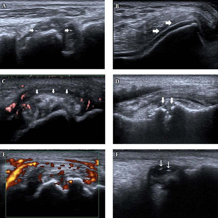
Figure 1. Ultrasound features of joints in first gout attack and asymptomatic hyperuricemia (AHU). A, First metatarso-phalangeal joints (1st MTP) longitudinal view, B-mode
ultrasound shows synovial thickening and hyper echoic foci (white arrows) in patients with first gout attack. B, Knee transverse view, B-mode ultrasound shows double contour
(DC) sign (white arrows) on the surface of the cartilage in patients with first gout attack. C, 1st MTP longitudinal view, tophi (white arrows) with surrounding power Doppler
signal in patients with first gout attack. D, 1st MTP longitudinal view, bone erosion (white arrows) in patients with first gout attack. E, 1st MTP longitudinal view, synovial
thickening and power Doppler signals in patients with first gout attack. F, Ankle longitudinal view, hyperechoic foci (white arrows) inside the synovial fluid.
longer hyperuricemia duration. Furthermore, the forma- this may have led to errors and underestimation. To make
tion of uric acid crystallization begins within two years of our results more reliable, we excluded subjects who were
hyperuricemia onset. Routine ultrasound should be per- not clear about their disease duration. Moreover, the rel-
formed as soon as possible in affected patients to prevent atively small sample size may have limited the statistical
joint damage even when there are no clinical symptoms. power of our analysis, and further studies that include
A limitation of our study is that the hyperuricemia du- larger sample sizes are needed to confirm our conclusions.
ration was obtained through a questionnaire survey, and In conclusion, ultrasound features are associated with
Iran J Radiol. 2019; 16(3):e85140. 5Uncorrected Proof
Fan W et al.
hyperuricemia duration. Furthermore, tophi and bone 6. Neogi T, Jansen TL, Dalbeth N, Fransen J, Schumacher HR, Berend-
erosion can be detected in first gout attack. These data sen D, et al. 2015 Gout classification criteria: An American Col-
lege of Rheumatology/European League Against Rheumatism
suggest that the low-grade inflammation induced by urate
collaborative initiative. Ann Rheum Dis. 2015;74(10):1789–98. doi:
acid may also be present in individuals with AHU. 10.1136/annrheumdis-2015-208237. [PubMed: 26359487]. [PubMed
Central: PMC4602275].
7. Elsaman AM, Muhammad EM, Pessler F. Sonographic find-
Footnotes ings in gouty arthritis: Diagnostic value and association with
disease duration. Ultrasound Med Biol. 2016;42(6):1330–6. doi:
Authors’ Contributions: Study concept and design: 10.1016/j.ultrasmedbio.2016.01.014. [PubMed: 26995154].
Wenting Fan and Jiaan Zhu; analysis and interpretation of 8. Wakefield RJ, Balint PV, Szkudlarek M, Filippucci E, Backhaus M,
D’Agostino MA, et al. Musculoskeletal ultrasound including defini-
data: Wenting Fan, Zheng Chen and Wenxue Li; drafting
tions for ultrasonographic pathology. J Rheumatol. 2005;32(12):2485–
of the manuscript: Wenting Fan; critical revision of the 7. [PubMed: 16331793].
manuscript for important intellectual content: Jiaan Zhu; 9. Lamers-Karnebeek FB, Van Riel PL, Jansen TL. Additive value for ul-
statistical analysis: Wenting Fan, Zheng Chen and Wenxue trasonographic signal in a screening algorithm for patients pre-
senting with acute mono-/oligoarthritis in whom gout is sus-
Li. pected. Clin Rheumatol. 2014;33(4):555–9. doi: 10.1007/s10067-014-
Conflict of Interests: The authors declare that they have 2505-6. [PubMed: 24510062].
no conflicts of interest for this study. 10. Naredo E, Uson J, Jimenez-Palop M, Martinez A, Vicente E, Brito E,
et al. Ultrasound-detected musculoskeletal urate crystal deposition:
Ethical Approval: Ethical approval for the study was Which joints and what findings should be assessed for diagnosing
granted by the Ethics Committee of Peking University Peo- gout? Ann Rheum Dis. 2014;73(8):1522–8. doi: 10.1136/annrheumdis-
ple’s Hospital (FWA00001384). 2013-203487. [PubMed: 23709244].
11. De Miguel E, Puig JG, Castillo C, Peiteado D, Torres RJ, Martin-
Financial Disclosure: The listed authors have no finan- Mola E. Diagnosis of gout in patients with asymptomatic hyperuri-
cial disclosures to report. caemia: A pilot ultrasound study. Ann Rheum Dis. 2012;71(1):157–8. doi:
10.1136/ard.2011.154997. [PubMed: 21953340].
Funding/Support: This work was supported by National
12. Miao Z, Li C, Chen Y, Zhao S, Wang Y, Wang Z, et al. Dietary and
Natural Science Foundation of China (No. 81571684 to Jiaan lifestyle changes associated with high prevalence of hyperuricemia
Zhu), Peking University People’s Hospital Research and De- and gout in the Shandong coastal cities of Eastern China. J Rheumatol.
velopment Funds (RDC2014-02 to Wenting Fan). 2008;35(9):1859–64. [PubMed: 18634142].
13. Beydoun MA, Canas JA, Fanelli-Kuczmarski MT, Tajuddin SM, Evans
Patient Consent: All subjects completed the informed MK, Zonderman AB. Genetic risk scores, sex and dietary factors inter-
consents. act to alter serum uric acid trajectory among African-American urban
adults. Br J Nutr. 2017;117(5):686–97. doi: 10.1017/S0007114517000411.
[PubMed: 28345493]. [PubMed Central: PMC5679207].
References 14. Khanna D, Fitzgerald JD, Khanna PP, Bae S, Singh MK, Neogi T, et al.
2012 American College of Rheumatology guidelines for management
1. Liu R, Han C, Wu D, Xia X, Gu J, Guan H, et al. Prevalence of hype- of gout. Part 1: Systematic nonpharmacologic and pharmacologic
ruricemia and gout in mainland China from 2000 to 2014: A sys- therapeutic approaches to hyperuricemia. Arthritis Care Res (Hobo-
tematic review and meta-analysis. Biomed Res Int. 2015;2015:762820. ken). 2012;64(10):1431–46. doi: 10.1002/acr.21772. [PubMed: 23024028].
doi: 10.1155/2015/762820. [PubMed: 26640795]. [PubMed Central: [PubMed Central: PMC3683400].
PMC4657091]. 15. Edwards NL, Sundy JS, Forsythe A, Blume S, Pan F, Becker MA. Work
2. Kuo CF, Grainge MJ, Mallen C, Zhang W, Doherty M. Rising bur- productivity loss due to flares in patients with chronic gout re-
den of gout in the UK but continuing suboptimal management: fractory to conventional therapy. J Med Econ. 2011;14(1):10–5. doi:
A nationwide population study. Ann Rheum Dis. 2015;74(4):661–7. 10.3111/13696998.2010.540874. [PubMed: 21138339].
doi: 10.1136/annrheumdis-2013-204463. [PubMed: 24431399]. [PubMed 16. Dalbeth N, Merriman TR, Stamp LK. Gout. Lancet.
Central: PMC4392307]. 2016;388(10055):2039–52. doi: 10.1016/S0140-6736(16)00346-9.
3. Zhang W, Doherty M, Pascual E, Bardin T, Barskova V, Conaghan P, [PubMed: 27112094].
et al. EULAR evidence based recommendations for gout. Part I: Di- 17. Andersen M, Ellegaard K, Hebsgaard JB, Christensen R, Torp-Pedersen
agnosis. Report of a task force of the Standing Committee for In- S, Kvist PH, et al. Ultrasound colour Doppler is associated with syn-
ternational Clinical Studies Including Therapeutics (ESCISIT). Ann ovial pathology in biopsies from hand joints in rheumatoid arthri-
Rheum Dis. 2006;65(10):1301–11. doi: 10.1136/ard.2006.055251. [PubMed: tis patients: A cross-sectional study. Ann Rheum Dis. 2014;73(4):678–83.
16707533]. [PubMed Central: PMC1798330]. doi: 10.1136/annrheumdis-2012-202669. [PubMed: 23475981].
4. Swan A, Amer H, Dieppe P. The value of synovial fluid assays in 18. Wittoek R, Carron P, Verbruggen G. Structural and inflammatory
the diagnosis of joint disease: A literature survey. Ann Rheum sonographic findings in erosive and non-erosive osteoarthritis of the
Dis. 2002;61(6):493–8. doi: 10.1136/ard.61.6.493. [PubMed: 12006320]. interphalangeal finger joints. Ann Rheum Dis. 2010;69(12):2173–6. doi:
[PubMed Central: PMC1754135]. 10.1136/ard.2010.128504. [PubMed: 20693271].
5. Park JW, Ko DJ, Yoo JJ, Chang SH, Cho HJ, Kang EH, et al. Clinical factors 19. Sapsford M, Gamble GD, Aati O, Knight J, Horne A, Doyle AJ, et al.
and treatment outcomes associated with failure in the detection of Relationship of bone erosion with the urate and soft tissue compo-
urate crystal in patients with acute gouty arthritis. Korean J Intern Med. nents of the tophus in gout: A dual energy computed tomography
2014;29(3):361–9. doi: 10.3904/kjim.2014.29.3.361. [PubMed: 24851071]. study. Rheumatology (Oxford). 2017;56(1):129–33. doi: 10.1093/rheuma-
[PubMed Central: PMC4028526]. tology/kew383. [PubMed: 27803304].
6 Iran J Radiol. 2019; 16(3):e85140.You can also read