Uncovering evolutionary changes in the mitochondrial genomes of lichenized fungi within the order Arthoniales - Semantic Scholar
←
→
Page content transcription
If your browser does not render page correctly, please read the page content below
University of Colorado, Boulder
CU Scholar
Undergraduate Honors Theses                                                                                                      Honors Program

Spring 2019

Uncovering evolutionary changes in the
mitochondrial genomes of lichenized fungi within
the order Arthoniales
Dustin Bailey
Dustin.Bailey@Colorado.EDU

Follow this and additional works at: https://scholar.colorado.edu/honr_theses
     Part of the Evolution Commons, Genetics Commons, and the Genomics Commons

Recommended Citation
Bailey, Dustin, "Uncovering evolutionary changes in the mitochondrial genomes of lichenized fungi within the order Arthoniales"
(2019). Undergraduate Honors Theses. 1874.
https://scholar.colorado.edu/honr_theses/1874

This Thesis is brought to you for free and open access by Honors Program at CU Scholar. It has been accepted for inclusion in Undergraduate Honors
Theses by an authorized administrator of CU Scholar. For more information, please contact cuscholaradmin@colorado.edu.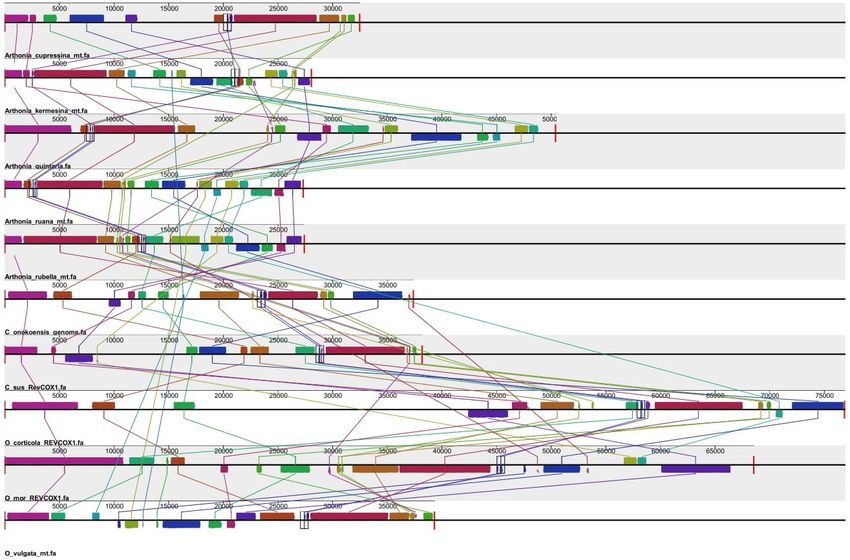
Uncovering evolutionary changes in the mitochondrial
genomes of lichenized fungi within the order Arthoniales

                                Dustin W. Bailey

                            Defense Date: 04/08/2019

         Thesis Advisor: Prof. Nolan Kane, Ecology & Evolutionary Biology

                               Defense Committee:
                Prof. Nolan Kane, Ecology & Evolutionary Biology
  Dr. Brent Hulke, USDA-ARS & Ecology & Evolutionary Biology Adjunct Faculty
 Prof. Barbara Demming-Adams, Ecology & Evolutionary Biology and Honors Council
                                  Representative
Keywords: Lichen, Symbiosis, Mitochondria, Genomics
Abstract
Lichens are symbiotic organisms that are understudied despite their usefulness to a wide
range of disciplines. Studying the genetics of lichen, will contribute to a better understanding of
the genetic mechanisms behind symbiosis and fill in grey areas of taxonomic classification of
lichens. The lichen order of Arthoniales is even less studied compared to its sister order
Lecanorales. This study expands the genomic resources for the former order of lichen and
reveals some novel genetic trends found among species within Arthoniales. Some genetic trends
have yet to be identified in other lichen species such as the trans-splicing of nad4 among
Arthonia species and lack of atp9 in select species in Opegrapha and Chrysothrix.
Trans-splicing of nad4 in Arthonia species is characterized by two exons of nad4 being
split by thousands of base pairs. In some instances, the second is also on the opposite strand as
the first exon. This was thought to be unique to the mitochondrial genomes of plants (Knoop,
1991) until trans-splicing was also identified in fungi in 2012 (Pelin, 2012). In plants,
trans-splicing allows their extremely flexible genomes to rearrange without losing the function of
essential genes.
The lack of atp9 in the mitochondrial mycobiont genome has proven to be a significant
characteristic among certain species of lichen (Pogoda, 2018). In obligate symbiotic organisms,
the exchange of genes between species allows core genomic processes to be streamlined. In the
past study of genomic obligate symbiosis of lichen, the loss of atp9 occured in all species of a
related genus. Our findings show the loss of atp9 among single species within a genus.
This research fills a gaping hole in the genetic research of lichenized fungi within the
order Arthoniales and reveals unique genetic features yet to be seen among lichenized fungi.
Introduction
Lichens are obligate symbioses between a fungal species (mycobiont) and one or more
photosynthetic organisms (photobiont), usually a green alga or cyanobacterium. Lichens areecologically important, successful in many different terrestrial habitats, inhabit many different
niche environments and come in a variety of colors and growth forms (Fryday et al., 2007). The
Arthoniales are the second largest order of lichenized fungi after Lecanorales. Despite the large
number of extant species within Arthoniales, very little genomic information is available,
especially whole organellar genomes. Of the 299 mitochondrial sequences associated with
Arthoniales in NCBI’s GenBank, complete annotated mitochondrial resources are available only
for Arthonia susa and Opegrapha vulgata. The ten mitochondrial mycobiont genomes we
assembled and annotated during this study will comprise the majority of genomic resources
available for species within this order. Using these annotations, we can determine variation
between species’ genome length, conservation of gene order, and other unique features of these
lichen species. With this information, we identify unusual trends and patterns among Arthoniales
and their relevance to the orders evolutionary history.
Mitochondria in plant, animal, and fungal cells are the powerhouses that drive energy
production needed for all biological processes. Containing protein-coding genes required for the
electron transport chain as well as ATP synthase, mitochondria serve an essential role for
complex eukaryotic life (Gray et al., 1999). The endosymbiotic theory states that mitochondria
were once free-living alpha-proteobacteria, and their presence in eukaryotic cells today is due to
an early symbioses event that caused a bacterium to be engulfed, but not digested by a eukaryotic
cell (Zimorski, 2014). This view is supported by the similarity of the mitochondrial genome to
that of Rickettsia prowazekii, an obligate intracellular parasite that causes typhus (Gray et al.,
1999). By studying mitochondrial genomes, the evolutionary history of these species can be
elucidated, including genetic changes that are defining characteristics for this order of lichenized
fungi.
The mitochondrial genome of most lichenized fungi, contains 15, sometimes 14,
protein-coding genes along with the small ribosomal subunit, large ribosomal subunit, and a
number of tRNAs. One of the protein-coding genes, atp9, is absent in two of our ten Arthoniales
mitochondrial genomes. The absence of atp9 has been seen in a previous paper with the absence
being consistent with an entire genus (Pogoda, 2018). The present report may be the first to
demonstrate atp9 missing among specific species within a genus. Prior studies show that theabsence of atp9 generally occurs within an entire genus. Of the ten species characterized here,
Opegrapha corticola and Chrysothrix susquehannensis of the order Arthoniales are the only
species missing atp9.
In addition, retrotransposons were identified in the genera Opegrapha and Chrysothrix
but none in Arthonia species. Retrotransposable elements have been useful in distinguishing
between large evolutionary lineages (Pogoda, 2019). They are parasitic homing endonucleases
which find their way into the genomes introns and intein segments. Previous research
hypothesized that lack of gene synteny could be linked to the presence of retrotransposons in the
genome (Bennetzen, 2005). However, as mentioned before, only half of the ten Arthoniales
species had retrotransposons present but all the species showed a lack of gene synteny.
Lastly, trans-splicing of nad4 in Arthonia was found with the exons for nad4 thousands of
base pairs apart and occasionally on different strands depending on the species. Trans-splicing
has been identified in metazoans, arbuscular mycorrhizal fungi, and a number of plant species
but this is the first time trans-splicing has been identified in lichenized fungi. In plants,
trans-splicing is thought to play a role in the mediating the impact of gene rearrangement on gene
function (Knoop, 1991). Plant mitochondrial genomes are flexible and Arthoniales genomes also
seem to be flexible based on gene synteny figures. Further research is needed to confidently say
trans-splicing is a result of highly variable gene order.
Methods
Sample Collection
To study major lineage differences between Arthoniales and Lecanorales, ten species
representing three genera were compared to ten previously annotated Lecanorales genomes. All
the Arthoniales and Lecanorales species are native to the southern Appalachian Mountain
biodiversity hotspot of eastern United States area and were collected in the wild during fieldwork
between 2016 and 2018. Collected specimens were deposited in both the herbaria of the New
York Botanical Garden (NY) and University of Colorado, Boulder (COLO). Tissue collection
efforts were as follows: for macrolichens (lichens with larger structures/fruiting bodies), ca. 1 x 1cm of tissue was removed, targeting the thallus margins and lobes. For microlichens (difficult to
see structures without close examination), tissue was scraped from rock or tree substrates using a
sterile razor blade. Tissue samples were then air dried in a laminar flow hood for 24 hours then
kept frozen at -20°C until transport to the University of Colorado for subsequent DNA extraction
and sequencing.
DNA Extraction and Sequencing
Genomic DNA was extracted from tissue samples of A. rubella, A. ruana, A. quintaria,
A. kermesina, A. cupressina, O. vulgata, O. moroziana, O. corticola, C. susquehannensis, a nd C.
onokoensis using a Qiagen DNeasy 96 plant kit (Qiagen, Germany) with a modified protocol by
Cloe Pogoda and Kyle Keepers. This modified protocol included a 10 minute 65°C incubation
step during the lysis phase. A 100% ethanol wash was included before finally drying the
membrane before elution. Genomic libraries were prepared using Nextera® XT DNA library
prep kits (Illumina®, California) and each sample was barcoded by the adapters Nextera® i5 and
i7.
Samples that passed QC were processed for paired end 150 base pair reads on the
Illumina NextSeq® sequencer at the University of Colorado’s BioFrontiers Institute
Next-Generation Sequencing Facility in Boulder, Colorado.
Assembly and Annotation
The mitochondrial genomes for each species were assembled de novo using SPAdes v3.9
(Saint Petersburg State University, Russia). To identify the mitochondrial contigs, a BLAST
search was conducted of the assemblies against known mycobiont mitochondrial protein coding
genes. The contigs containing mitochondrial genes were then web BLASTed to identify if the
contig belonged to the mycobiont or photobiont partner. Once enough contigs were identified to
contain all the protein coding genes for a species, they were tiled together by referencing the
reverse complement for each contig and the trimmed fastq files to find overlapping sequences
and ultimately form a full circularized sequence. Once circularized, the mycobiont mitochondrial
sequences were BLASTed against previously annotated sequences to determine the proper
orientation and to standardize the sequences to start at cox1. Standardized sequences were errorcorrected utilizing the SAMTOOLS suite to determine SNPs between the trimmed fastq files and
the circularized mitochondrial sequence. Add in info about orientation, circularization and error
correction (tview).
Identification of genomic content
Gene features of each assembly were annotated using DOGMA , NCBI BLAST (National Center
for Biotechnology Information, U.S. National Library of Medicine 8600 Rockville Pike,
Bethesda MD, 20894 USA), CHLOROBOX, and Sequin v15.1. DOGMA was used to
approximately assign the gene order for each species. We modified the DOGMA parameters to
mitochondrial genome type, gapped alignment, mold mitochondrial for genetic code for Blastx,
and percent identity cutoff for both proteins and rRNA was set to 40. After using DOGMA to get
a general idea of the gene order, the mitochondrial genomes were circularized and oriented to
cytochrome c oxidase subunit I (cox1). By circulzarizing and orienting to cox1, all the species
analyzed were standardized for further comparison.
For NCBI BLAST, either BLASTn or BLASTx were used depending on whether
nucleotide FASTA sequences or the protein FASTA sequences were available. To locate a
specific gene feature, the function “align two or more sequences” was used with the Genetic
Code option “Mold Mitochondrial;...4” .
Parameters were modified for use of Chlorobox GeSeq. For our circular, mitochondrial
genomes, BLAST searches were set to an identity of 40, and tRNAscan-SE v2.0 output was
included in the analysis. Parameters were set to include organellar tRNAs, a genetic code for
Mold/Protozoan/Coelenterate Mitochondrial, with a cut-off score of 40. Reference species used
included Peltigera dolichorhiza, Peltigera malacea, Imshaugia aleurites, a nd Usnea ceratina
from the order, Lecanorales.
Identification of “missing” atp9For Opegrapha corticola and Chrysothrix susquehannensis, a command line tBLASTn
was performed to locate matches of atp9 within each species meta-assembly file. Contig matches
were either associated with the mycobionts nuclear genome or a photobiont partner’s
mitochondrion. Benchmarking Universal Single-Copy Orthologs (BUSCO) for fungi were
utilized to determine which contigs were associated with the mycobionts nuclear genome. An
online BLASTn confirmed each contigs similarity to algal species, allowing us to assume the
contig that matched belonged to a photobiont partner.
Homing Endonucleases
Parasitic elements such as LAGLIDAGs and GIY-YIGs were identified using ORFfinder
and NCBI SMART BLAST. After all of the features were annotated in Sequin, the mitogenome
was submitted to NCBI GenBank and we were given temporary accession numbers which can be
found near the conclusion of this paper.
Phylogenetic analyses
The phylogeny was created using ribosomal DNA sequences from our assemblies as well as
large ribosomal subunit sequences from 17 other species within Arthoniales available on
genbank (Ertz et al., 2009). Gene alignments created very discordant trees, while the highly
conserved ribosomal sequences produced highly supported phylogenetic relationships.
Sequences were aligned in MEGA7 utilizing the MUSCLE algorithm and curated in PhyDE
(Kumar et al., 2015; Edgar, 2004). The tree was created utilizing the Maximum-likelihood
method with 500 bootstraps and rooted with Cladonia grayi and Cladonia uncialis.
Results
All but two of the mitogenomes contained 15 protein-coding genes and genes for two
subunit rRNAs: three cytochrome c oxidase subunits (cox1-3), seven subunits of NAD
dehydrogenase (nad1-6, nad4L), three ATP synthases (atp6,8,9), one ribosomal protein (rps3) ,
one cytochrome oxidase b (cob) , and the large and small rRNA subunits (LSU and SSU) . Theother two species, Opegrapha corticola a nd Chrysothrix susquehannensis, contained all of these
features with the exception of ATP synthase subunit 9 (atp9) . [See Figure 2]
The absence of atp9 i n the mitogenomes of Opegrapha corticola and Chrysothrix
susquehannensis was associated with evidence of its presence elsewhere. A tBLASTn revealed
matches for atp9 in each of the mycobiont’s nuclear genome as well as within the mitogenome of
their photobiont partners. For O. corticola, the contig associated with the mycobionts nuclear
ith an E-value of 1e-08, and a contig with a similarity to algae matched
genome matched atp9 w
with an E-value of 1e-04. Although the BLAST results only identified parts of atp9, the low
E-value offers significant evidence that a copy of atp9 may be present in either the mycobionts
nuclear genome or the mitochondrial genome of its algal counterpart. For C. susquehannensis,
ith an E-value of
the contig associated with the mycobionts nuclear genome matched atp9 w
2e-16, and a contig with a similarity to algae matched with an E-value of 3e-06.
Each annotated species within Arthonia was observed to contain a trans-spliced nad4
gene. This gene was found in two exons, the location of which differed among species. In
addition, there was no conserved pattern of which strand an exon was found on. Across the five
Arthonia species, each exon was found on the plus and minus strands, and of the four
combinations possible (plus/plus, minus/minus, plus/minus, minus/plus) all were observed
except for both exons on the minus strand. The exons for Arthonia were separated by thousands
of base pairs or found on different strands. This trans-splicing of nad4 is present in all Arthonia
species even through translocation. Trans-splicing had not been observed in any other lichenized
fungi but trans-splicing has been observed in plant mitochondrial genomes, and associated with
group II introns (Bonen, 2008).
A triple cotranscription was observed in Arthonia kermesina between cob-cox1-cox2, in
that order (Table 1). Cotranscription is common within mitochondrial genomes, and has been
frequently observed especially in plants (Gualberto et al., 1988; Hoffman et al., 1999; Itani et al.,
1998). While cotranscription of cob and cox2 is a common characterization of species within
Lecornorales, this is the first evidence, to our knowledge, of a triple cotranscription event
recorded for the mitochondrion of a lichenized fungus. Evolutionarily, cotranscription iseconomically beneficial to an organism as it requires less energy to transcribe three
protein-coding genes at once rather than having intergenic regions where errors could occur
(Sneppen, 2010).
Arthonia is distinguished by a lack of introns and retrotransposable elements, although
many are identified in Opegrapha and Chrysothrix. Of the species annotated within Arthonia,
only A. quintaria contained a significant number of introns and retrotransposable elements
compared to the other four which contained almost all of their protein coding genes as a single
exon (with the exception of nad4). A. quintaria contained at least a one intron in cob, cox1, cox3,
nad5, and rps3 while A. kermesina, A. cupressina, A. rubella, a nd A. ruana only contained an
intron in rps3.
Discussion
Comparison of the genomes of the ten Arthoniales species to the genomes of previously
annotated Lecanorales species demonstrated the order of their genes to be highly variable.
Phylogenetic Analysis
Previous studies produced similar trees to the one developed with the data obtained for
this project. Opegrapha was shown to be paraphyletic and Arthonia monophyletic (Ertz et al.,
2009; Nelsen et al., 2009). In addition, placement of Arthonia cupressina with Chrysothrix
xanthina is supported by a high bootstrap value (81), as well as its close relationship to Arthonia
caesia t hat was previously shown to group with C. xanthina (Nelsen et al., 2009). This finding
presents evidence that A. cupressina may truly belong to the genus Chrysothrix, and further
research may uncover other species of lichen that need to be reassigned. Development of
phylogenetic trees based on the morphology of lichen using analysis of structural components,
mode of reproduction, etc have limitations, but genomic resources are helpful in clarifying the
grey areas of lichen phylogenetics (Nelsen et al., 2009).
Evolutionary History in relation to gene syntenyA number of hypotheses have been considered by lichenologists when trying to explain
the observed difference between Lecanorales and Arthoniales. The most promising hypothesis
considers the evolutionary history of the order, Arthoniales. Since within the Ascomycota tree,
Arthoniales diverged much earlier than Lecanorales, it is inferred that there is a large number of
extinct individuals in the Arthoniales evolutionary history (Grube, 1998; Ertz et al., 2009). As a
number of species became extinct, gaps began to emerge in the phylogenetic tree for the
Arthoniales. The extant Arthoniales species present today represent a very small percentage of
the entire Arthoniales tree. Therefore, it is likely that the ten species we annotated are more
divergent from one another than originally thought. This could explain why gene order is so
variable from species to species among Arthonia, Chrysothrix, and Opegrapha.
Gene synteny in relation to retrotransposable elements.
Another hypothesis attempts to link inconsistent gene order to the presence of
retrotransposable elements (Bennetzen et al., 2005; Beaudet et al., 2013) . In fungi, high
variability in the mitochondrial genome has been observed even within members of the same
phylum (Aguileta et al., 2014). Changes within the mitochondrial genome have been associated
with DNA polymerases and selfish retrotransposable elements such as homing endonucleases
(Kanzi et al., 2016). These elements can cause shifts in gene order and changes in genome size
through the movement of genetic elements (Beaudet et al., 2013; Bennetzen, 2005; Nadimi et al.,
2015).
In the case of lichen species, a few common retrotransposable elements found are
LAGLIDADG’s and GIY-YIG’s. Activation of these retrotransposons have been associated with
environmental stressors and have been linked to variability in genome size (Grandbastien, 1998;
Joardar et al., 2012). While this link may be true among Lecanorales, there are almost no
retrotransposons in Arthonia s pecies that exhibit clear gene disorder. However, these
retrotransposons can be found in most Chrysothrix and Opegrapha species, which could
potentially explain their gene disorder. The causes of gene reorganization within Arthonia are
unclear. Introns and retrotransposable are widely discussed (Grandbastien, 1998; Joardar et al.,2012; Aguileta et al., 2014; Bennetzen et al., 2005; Beaudet et al., 2013) as major contributors to
rearrangement; a lack thereof may indicate another force driving divergence within Arthonia.
Absence of atp9
The absence of atp9 in the mycobiont mitochondrial genome is not a new finding and has
been identified in a number of genera in the order Lecanorales (Pogoda et al., 2018). The latter
authors explain how relocation of atp9 to the photobiont’s mitochondrial genome suggests that
obligate symbiosis involves consolidating genome space among asexually reproducing lichens.
Although energy efficiency and consolidation are possible factors, another important factor could
be the symbiotic relationship itself. To better coordinate and regulate the symbiotic relationship,
the partitioning of gene copies is a common attribute of symbiotic species (Khachane et al.,
2007; Tsaousis et al., 2008; Corradi et al., 2010; Baumgarten et al., 2015). In lichens however,
the partitioning of specific gene copies to the photobiont is completely dependent on the mode of
sexual reproduction.
The life cycle of a sexually reproducing lichen exemplifies why the loss of atp9 is so
unusual. Generally, a fungal spore is released from a mature lichen, but this spore does not
include the photosynthetic symbiont. For the first stage of development, a sexually reproducing
lichen thus develops as a fungus without it’s photosynthetic partner (Honegger, 1998). The
young lichen requires ATP synthase in order to create energy but, without ATP synthase subunit
9 in the mycobiont mitochondrial genome, it is not clear how this species would develop
independent from the photobiont partner.
Asexually reproducing lichen are more likely to partition specific genes from the
mycobiont to the photobiont because they are never separated (Pogoda et al., 2018). Many
asexually reproducing lichen multiply as propagules containing both the photosynthetic partner
and the mycobiont. This allows the mycobiont to allocate a copy of atp9 to the photobiont
because they are associated with the photobiont throughout their life history.
Chrysothrix susquehannensis and Opegrapha corticola are unique in having a portion of
atp9 residing in the mycobiont nuclear genome and a copy present in the photobiontmitochondrial genome. Further analysis is needed to confirm whether the “portion” of atp9 is a
functional piece of the protein-coding gene or whether it is a remnant copy. The transfer of atp9
from the mitochondria to the nucleus has been revealed to impact the functionality of atp9 but
serves a regulatory function rather than a being lethal (Dequard-Chablat et al., 2011; Sellem et al.
2016; Bietenhader et al., 2012)
While the members from the genus Usnea, which belongs to the order Lecanorales, were
missing atp9, only one species in the genera Chrysothrix a nd Opegrapha w
ere missing atp9 a nd
each had a copy of atp9 in its nuclear genome for both species. This finding suggests that a
functional atp9- subunit may be produced within the nucleus and imported into the mitochondria
(Bietenhader et al., 2012), which may reflect an evolutionary reduction within the mitochondrial
genomes of these species of lichen through gene transfer to the nucleus. The majority of
mitochondrial genes are encoded within the nuclear genomes, with the exceptions of cytochrome
b, cox1, and most notably, atp9 ( Bietenhader et al., 2012). When atp9 is transferred to the
nucleus however, a heat shock response is activated (Bietenhader et al., 2012). Further research
is necessary to examine whether this heat shock response might be beneficial to some lichens in
specific regions of the world. Perhaps there may be an underlying ecological pressure for the
transfer of atp9 from the mitochondria to the nucleus in specific species.
This finding suggests that C. susquehannensis a nd O. corticola are evolving to further
reduce their mitochondrial genomes through transfer of atp9 into their nuclear genomes.
Previous studies demonstrated loss of atp9 was isolated to Lecanorales, Teloschistales and
Ostropales (Pogoda 2018). The loss of atp9 in Chrysothrix susquehannensis and Opegrapha
corticola extends this feature to members of the order Arthoniales.
Conclusions
Lichens of the order Arthoniales are widespread and constitute a large portion of known
lichenized fungi. The data presented provide genetic information on this order for which little
such information had been available. Unique differences in the mitochondrial genomes of
Arthoniales and Lecornorales raise new questions about the evolutionary history of these lichen.
What drives the grand gene disorder in Arthonia? Does the transfer of atp9 in O. corticola and C.susquehannensis f rom the mycobiont mitochondrial genome to the mycobiont nucleus or
photobiont mitochondria serve an ecological function? Arthoniales do appear to have a much
different evolutionary history from previously studied orders of lichenized fungi. Future research
should address additional methods of genome rearrangement, ecological impacts on lichen
genomic trends, as well as processes acting towards obligate symbiosis.
Data Accessibility
Species Accession
Number
Arthonia rubella MH308714
Arthonia ruana MH308713
Arthonia quintaria MH308712
Arthonia cupresina MH308710
Arthonia kermesina MH308711
Opegrapha corticola MH746206
Opegrapha moroziana Awaiting Accesion
Opegrapha vulgata MH845230
Chrysothrix susquehannensis Awaiting Accesion
Chrysothrix onokoensis MH998153
Chrysothrix and Opegrapha species assembled and annotated by Dustin Bailey. Arthonia species assembled and
annotated by Arif Nadiadi
ReferencesAguileta, G., Vienne, D., M, D., Ross, O. N., Hood, M. E., Giraud, T., … Gabaldón, T. (2014). High Variability of Mitochondrial Gene Order among Fungi. Genome Biology and Evolution, 6(2), 451–465. https://doi.org/10.1093/gbe/evu028 Baumgarten, S., Simakov, O., Esherick, L.Y., Liew, Y.J., Lehnert, E.M., Michell, C.T., ... & Gough, J. (2015). The genome of Aiptasia, a sea anemone model for coral symbiosis. Proceedings of the National Academy of Sciences of the United States of America, 112(38), 11893-11898. Beaudet, D., Nadimi, M., Iffis, B., & Hijri, M. (2013). Rapid Mitochondrial Genome Evolution through Invasion of Mobile Elements in Two Closely Related Species of Arbuscular Mycorrhizal Fungi. PLOS ONE, 8(4), e60768. https://doi.org/10.1371/journal.pone.0060768 Bennetzen, J. L. (2005). Transposable elements, gene creation and genome rearrangement in flowering plants. Current Opinion in Genetics & Development, 15(6), 621–627. https://doi.org/10.1016/j.gde.2005.09.010 Bietenhader, M., Martos, A., Tetaud, E., Aiyar, R. S., Sellem, C. H., Kucharczyk, R., … Rago, J.-P. di. (2012). Experimental Relocation of the Mitochondrial ATP9 Gene to the Nucleus Reveals Forces Underlying Mitochondrial Genome Evolution. PLOS Genetics, 8(8), e1002876. https://doi.org/10.1371/journal.pgen.1002876 Bonen, L. (2008). Cis- and trans-splicing of group II introns in plant mitochondria. Mitochondrion, 8(1), 26–34. https://doi.org/10.1016/j.mito.2007.09.005 Corradi, N., Pombert, J. F., Farinelli, L., Didier, E. S., & Keeling, P. J. (2010). The complete sequence of the smallest known nuclear genome from the microsporidian Encephalitozoon intestinalis. Nature communications, 1, 77.
Edgar, Robert C. (2004), MUSCLE: multiple sequence alignment with high accuracy and high throughput, Nucleic Acids Research 32(5), 1792-97 Ertz, D., Miadlikowska, J., Lutzoni, F., Dessein, S., Raspé, O., Vigneron, N., … Diederich, P. (2009). Towards a new classification of the Arthoniales (Ascomycota) based on a three-gene phylogeny focussing on the genus Opegrapha. Mycological Research, 113(1), 141–152. https://doi.org/10.1016/j.mycres.2008.09.002 Frisch, A., Thor, G., Ertz, D., Grube, M. (2014). The Arthonialen challenge: Restructuring Arthoniaceae. Systematics and Phylogeny, 727-744 https://doi.org/10.12705/634.20 Grandbastien, M.-A. (1998). Activation of plant retrotransposons under stress conditions. Trends in Plant Science, 3(5), 181–187. https://doi.org/10.1016/S1360-1385(98)01232-1 Gray, M. W., Burger, G., & Lang, B. F. (1999). Mitochondrial Evolution. Science, 283(5407), 1476–1481. https://doi.org/10.1126/science.283.5407.1476 Grube, Martin. “Classification and Phylogeny in the Arthoniales (Lichenized Ascomycetes).” The Bryologist, vol. 101, no. 3, 1998, p. 377., doi:10.1639/0007-2745(1998)101[377:capita]2.0.co;2. Gualberto, J.M., Wintz, H., Weil, JH. et al. Mol Gen Genet (1988) 215: 118. https://doi.org/10.1007/BF00331312 Hoffmann, M., Dombrowski, S., Guha, C. et al. Mol Gen Genet (1999) 261: 537. https://doi.org/10.1007/s004380050998
Honegger, R. (1998). THE LICHEN SYMBIOSIS—WHAT IS SO SPECTACULAR ABOUT IT? The Lichenologist, 30(3), 193–212. https://doi.org/10.1006/lich.1998.0140 Itani, K. & Handa, H. Curr Genet (1998) 34: 318. https://doi.org/10.1007/s002940050402 Joardar, V., Abrams, N. F., Hostetler, J., Paukstelis, P. J., Pakala, S., Pakala, S. B., … Nierman, W. C. (2012). Sequencing of mitochondrial genomes of nine Aspergillus and Penicillium species identifies mobile introns and accessory genes as main sources of genome size variability. BMC Genomics, 13, 698. https://doi.org/10.1186/1471-2164-13-698 Kanzi, A.M., Wingfield, B.D., Steenkamp, E.T., Naidoo, S., & van der Merwe, N.A. (2016). Intron Derived Size Polymorphism in the Mitochondrial Genomes of Closely Related Chrysoporthe Species. PloS one, 11(6), https://doi.org/10.1371/journal.pone.0156104 Khachane, A.N., Timmis, K.N., & dos Santos, V.A.M. (2007). Dynamics of reductive genome evolution in mitochondria and obligate intracellular microbes. Molecular biology and evolution, 24(2), 449-456. Knoop, V., et al. “Trans Splicing Integrates an Exon of 22 Nucleotides into the nad5 MRNA in Higher Plant Mitochondria.” The EMBO Journal, vol. 10, no. 11, 1991, pp. 3483–3493., doi:10.1002/j.1460-2075.1991.tb04912.x. Lohse, M., Drechsel, O., & Bock, R. (2007). OrganellarGenomeDRAW (OGDRAW): a tool for the easy generation of high-quality custom graphical maps of plastid and mitochondrial genomes. Current Genetics, 52(5–6), 267–274. https://doi.org/10.1007/s00294-007-0161-y Nadimi, M., Stefani, F. O. P., & Hijri, M. (2015). The Mitochondrial Genome of the
Glomeromycete Rhizophagus sp. DAOM 213198 Reveals an Unusual Organization Consisting of Two Circular Chromosomes. Genome Biology and Evolution, 7(1), 96–105. https://doi.org/10.1093/gbe/evu268 Nelsen, M. P., Lücking, R., Grube, M., Mbatchou, J. S., Muggia, L., Plata, E. R., & Lumbsch, H. T. (2009). Unravelling the phylogenetic relationships of lichenised fungi in Dothideomyceta. Studies in Mycology, 64, 135–144. https://doi.org/10.3114/sim.2009.64.07 Pelin, A. , Pombert, J. , Salvioli, A. , Bonen, L. , Bonfante, P. and Corradi, N. (2012), The mitochondrial genome of the arbuscular mycorrhizal fungus Gigaspora margarita reveals two unsuspected trans-splicing events of group I introns. New Phytologist, 194: 836-845. doi:10.1111/j.1469-8137.2012.04072.x Pogoda, C. S., Keepers, K. G., Lendemer, J. C., Kane, N. C., & Tripp, E. A. (2018). Reductions in complexity of mitochondrial genomes in lichen-forming fungi shed light on genome architecture of obligate symbioses. Molecular ecology, 27(5), 1155-1169. Sneppen, Kim, et al. “Economy of Operon Formation: Cotranscription Minimizes Shortfall in Protein Complexes.” MBio, vol. 1, no. 4, 2010, doi:10.1128/mbio.00177-10. Tsaousis, A. D., Kunji, E. R., Goldberg, A. V., Lucocq, J. M., Hirt, R. P., & Embley, T. M. (2008). A novel route for ATP acquisition by the remnant mitochondria of Encephalitozoon cuniculi. Nature, 453(7194), 553. Zimorski, Verena, et al. “Endosymbiotic Theory for Organelle Origins.” Current Opinion in Microbiology, vol. 22, 2014, pp. 38–48., doi:10.1016/j.mib.2014.09.008.
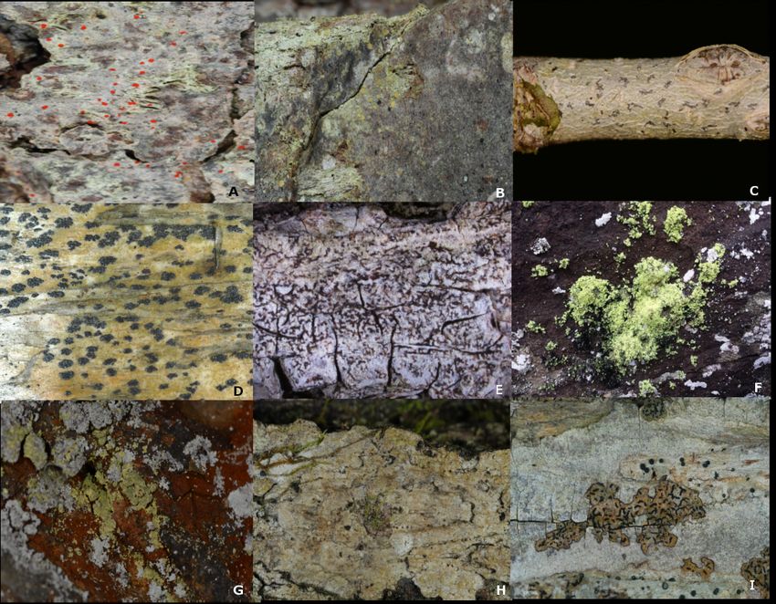
Kumar, S., Stecher, G., & Tamura, K. (2016). MEGA7: Molecular Evolutionary Genetics Analysis Version 7.0 for Bigger Datasets. Molecular Biology and Evolution, 33(7), 1870–1874. https://doi.org/10.1093/molbev/msw054 Automatic annotation of organellar genomes with DOGMA. Wyman SK, Jansen RK, Boore JL, Bioinformatics 2004 20(17):3252-3255 Figures and Tables Figure 1: Arthonia kermesina a ka “Hot dots” (A), Arthonia cupressina (B), Arthonia quintaria (C), Arthonia ruana (D), Arthonia rubella ( E), Chrysothrix onokoensis ( F), Chrysothrix susquehannensis ( G), Opegrapha corticola (H), Opegrapha vulgata ( I). All pictures taken by Erin A. Tripp and James C. Lendemer.
Arthonia rubella cox1 rps3 cox3 nad6 atp9 nad4 atp8 atp6 nad3 nad2 nad5 nad4L nad1 cox2 cob
Arthonia ruana cox1 rps3 atp9 nad1 nad4L nad5 nad4 atp8 atp6 nad3 nad2 nad6 cox3 cox2 cob
Arthonia quintaria cox1 rps3 cob cox2 cox3 nad6 nad4 atp8 nad5 nad4L nad1 atp6 nad3 nad2 atp9
Arthonia kermesina cox1 cox2 rps3 atp6 nad1 atp9 nad4 atp8 nad5 nad4L nad6 cox3 nad3 nad2 cob
Arthonia cupressina cox1 cox2 nad1 cox3 nad6 nad4L nad5 cob atp6 atp8 nad4 atp9 nad3 nad2 rps3
Opegrapha vulgata cox1 cox3 atp6 nad6 atp8 nad4 atp9 nad5 nad4L nad1 cox2 cob nad2 nad3 rps3
Opegrapha moroziana cox1 cox3 nad6 nad4 atp6 atp8 cox2 nad1 atp9 rps3 nad5 nad4L nad3 nad2 cob
Opegrapha corticola cox1 nad4 rps3 nad1 atp6 atp8 cob cox2 nad6 cox3 nad2 nad3 nad4L nad5
Chrysothrix
susquehannensis cox1 cox2 cob atp6 atp8 nad2 nad3 nad1 nad4L nad5 nad4 nad6 cox3 rps3
Chrysothrix onokoensis cox1 nad4 rps3 cob cox2 cox3 nad1 atp6 atp8 atp9 nad6 nad2 nad3 nad4L nad5
Cladonia apodocarpa cox1 nad1 nad4 rps3 cob cox2 atp9 atp6 atp8 nad6 cox3 nad2 nad3 nad4L nad5
Cladonia caroliniana cox1 nad1 nad4 rps3 cob cox2 atp9 atp6 atp8 nad6 cox3 nad2 nad3 nad4L nad5
Cladonia furcata cox1 nad1 nad4 rps3 cob cox2 atp9 atp6 atp8 nad6 cox3 nad2 nad3 nad4L nad5
Usnea ceratina cox1 nad4 rps3 nad1 atp6 atp8 cob cox2 nad6 cox3 nad2 nad3 nad4L nad5
Usnea cornuta cox1 nad4 rps3 nad1 atp6 atp8 cob cox2 nad6 cox3 nad2 nad3 nad4L nad5
Usnea halei cox1 nad4 rps3 nad1 atp6 atp8 cob cox2 nad6 cox3 nad2 nad3 nad4L nad5
Table 1: Gene order of 15 protein-coding sequences oriented to cox1 across species annotated in this study as well
as six species from the order Lecanorales. Within the order Arthoniales, high levels of genomic rearrangement is
observed. However, certain gene orders were conserved such as nad2-nad3, nad5-nad4L, cox1-cox2, and cox2-cob.
In contrast, the genera Cladonia and Usnea (belonging to the order Lecanorales) both show very conserved gene
order across all species annotated (Pogoda et al., 2018). For trans-splicing in the case of Arthonia, only the first exon
of nad4 is indicated. Created by Dustin Bailey and Arif Nadiadi.Figure 3: This cladogram shows an estimation of the relationships between 33 species of lichen including the ten
species annotated in this study. Utilizing rDNA from each species, this cladogram was created using the
urther
Maximum-likelihood method with 500 bootstraps and rooted with Cladonia grayi and Cladonia uncialis. Fprograms are being tested to produce a better phylogenetic representation of Arthoniales. Figure produced by Arif Nadiadi. Figure 4: Mauve alignment of all ten species produced by Arif Nadiadi and Dustin Bailey
You can also read