Uncovering Local Endemism in the Kimberley, Western Australia: Description of New Species of the Genus Amplirhagada Iredale, 1933 Pulmonata: ...
←
→
Page content transcription
If your browser does not render page correctly, please read the page content below
© The Author, 2010. Journal compilation © Australian Museum, Sydney, 2010
Records of the Australian Museum (2010) Vol. 62: 217–284. ISSN 0067-1975
doi:10.3853/j.0067-1975.62.2010.1554

                 Uncovering Local Endemism in the Kimberley,
               Western Australia: Description of New Species of the
                       Genus Amplirhagada Iredale, 1933
                           (Pulmonata: Camaenidae)

                                                       Frank Köhler

                       Department of Environment and Conservation of Western Australia, Science Division,
                                       Wildlife Place, Woodvale WA 6026, Australia; and

                                Australian Museum, 6 College Street, Sydney NSW 2010, Australia
                                                frank.koehler@austmus.gov.au

         Abstract. In this work twenty-six species of the camaenid land snail Amplirhagada, which is endemic to
         the Kimberley region in Western Australia, are newly described. In addition, the anatomy of two further
         species, A. katerana and A. combeana, is described and a further, yet undescribed species is reported from
         Boongaree Island based on dry shell material. Most of these species occur on islands of the Bonaparte
         Archipelago off the mainland coast. The patterns of distribution and differentiation of these island species
         are comparable, however, with those found on the mainland. Mainland species are usually restricted to
         single rainforest patches. Frequently, single patches harbour only one or two congeneric species. Similarly,
         smaller islands usually support one endemic Amplirhagada species whereas on larger islands up to four
         species are found to occur in sympatry. Species are distinguishable particularly by the characteristic
         anatomy of the inner penial wall. Sympatric species generally exhibit marked morphological differences
         in shells and genital anatomy. A molecular phylogeny based on partial sequences of the mitochondrial
         cytochrome c oxidase unit 1 gene (COI) reveals a basal polytomy among species of the genus, which
         are generally genetically well differentiated. Relationships among species in the molecular tree mainly
         reflect geographical patterns.

         Köhler, Frank, 2010. Uncovering local endemism in the Kimberley, Western Australia: description of new species of
         the genus Amplirhagada Iredale, 1933 (Pulmonata: Camaenidae). Records of the Australian Museum 62(2): 217–284.

Camaenid land snails of the genus Amplirhagada Iredale,            River Reserve, Mt Elizabeth Station and the region south of
1933, are endemic to the Kimberley region of Western               Wyndham (Solem, 1981a, 1988). Many of these species are
Australia, where they have radiated extensively. Thirty            restricted to single localities, such as rainforest patches or
species are currently considered valid, most of which usually      more open woodlands (Solem, 1991). Supposed exceptions,
occupy small distributional ranges along the Kimberley             with species occupying larger ranges, are likely to result
coast from the Buccaneer Archipelago in the south to               from the inadequate delimitation of morphologically cryptic
Kalumburu in the north as well as in inland areas of the           species (Solem, 1981a). Earlier accounts were predominantly
Napier, Harding, and King Leopold Ranges, the Drysdale             based on dry shells (e.g., Smith, 1894; Iredale, 1933,
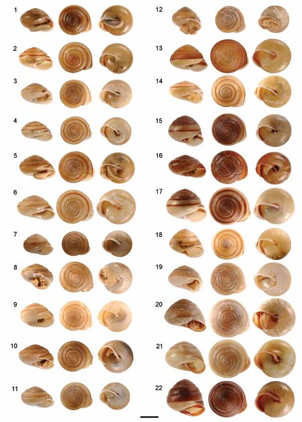
218       Records of the Australian Museum (2010) Vol. 62

Figure 1. Distribution of Amplirhagada species in the Kimberley region, northwestern Australia. (1) A. euroa n.sp., Adolphus Island. (2)
A. solemiana n.sp., Middle Osborn Island. (3) A. indistincta n.sp., Southwest Osborn Island, Kidney Island. (4) A. combeana, Cassini
Island. (5) A. mckenziei n.sp., Oliver Island. (6) A. ponderi n.sp., Kingsmill Island. (7) A. montesquieuana n.sp., Fenelon Island. (8) A.
descartesana n.sp., Descartes Island. (9) A. katerana, Katers Island. (10) A. puescheli n.sp., unnamed island near Prudhoe Island. (11) A.
decora n.sp., A. kessneri n.sp., Bigge Island. (12) A. berthierana n.sp., Berthier Island. (13) A. lamarckiana n.sp., Lamarck Island. (14) A.
anderdonensis n.sp., unnamed island, Anderdon Islands. (15) A. tricenaria n.sp., Prince Frederick Harbour, north of Hunter River mouth.
(16) A. regia n.sp., A. boongareensis n.sp., A. gibsoni n.sp., and Amplirhagada sp., Boongaree Island. (17) A. yorkensis n.sp., Coronation
Island. (18) A. buffonensis n.sp., Buffon Island. (19) A. uwinsensis n.sp., Uwins Island. (20) A. sphaeroidea n.sp., St. Andrews Island.
(21) A. basilica n.sp., and A. camdenensis n.sp., Augustus Island. (22) A. kimberleyana n.sp., NW of Wilson Point. (23) A. gemina n.sp.,
1.5 km SE of Hall Point. (24) A. dubitabile n.sp., Steep Island. (25) A. napierana, north Napier ranges. (26) A. percita, Napier ranges.
(27) A. castra, A. mitchelliana, and A. varia, Mitchell Plateau.

1938, 1939). However, Solem (1981a, 1988) demonstrated                  as a convenient source of key morphological characters for
that Amplirhagada species frequently show only subtle                   the recognition of species not only in this genus but also in
differences in shell features, such as colour, shape, and               other camaenid genera (Solem, 1979, 1981a, 1981b, 1984,
sculpture. In addition, shells are prone to homoplasy, which            1985; Willan et al., 2009; Köhler, 2010). In Amplirhagada
renders the delimitation of species by shell features alone             structure of the inner penial wall and development of the
difficult or impossible. In fact, proper species delimitation           main stimulatory pilaster provide particularly valuable
requires the study of genital anatomy, which was identified             diagnostic characters for delimiting species (Solem, 1988).
Köhler: New Amplirhagada species from the Kimberley         219

   A comparative study, mainly of the genital anatomy,                            Material and methods
enabled Solem (1981a) to identify a large number of species
that were unrecognized by previous authors, who relied              This study is primarily based on ethanol preserved specimens
exclusively on shell features. Solem showed that relatively         and supplementary dry shell material collected during
restricted areas in the Kimberley, such as the Mitchell             the Kimberley Island Survey (KIS) of the Department of
Plateau, may support a surprisingly large number of species         Environment and Conservation, Western Australia (DEC)
that can be differentiated on morphological criteria only by        in 2007–2009, the Kimberley Rainforest Survey of the then
their genital anatomy. Solem’s milestone publications on            Department of Conservation and Land Management, Western
the Camaenidae in Western Australia (Solem, 1979, 1981a,            Australia (RFS) in 1987–1988 and additional field work
1981b, 1984, 1985, 1988, 1991; Solem & Christensen, 1984)           conducted between 1988 and 2002 by various parties. Types
highlighted the remarkable diversity and patterns of local          and other voucher material are deposited in the Western
endemism within this group of land snails. Even though he           Australian Museum, Perth (WAM), the Field Museum
published extensively on Western Australian Camaenidae,             of Natural History, Chicago (FMNH), and the Australian
Solem was not able to approach a complete documentation             Museum, Sydney (AMS).
of their extant diversity (Cameron et al., 2005).                      Morphological descriptions focus on characters of the
   In addition to thirty named species of Amplirhagada,             shell, the mantle cavity, the genital organs, and the radula.
Solem (1991) listed 25 undescribed species from rainforest          Morphometric shell parameters, such as height of shell (H),
patches and woodlands throughout the Kimberley region.              diameter of shell (D), height of last whorl (LW), and width of
When Alan Solem died in 1990 he left numerous examples of           umbilicus (U) were measured with callipers precise to 0.1 mm.
undescribed Amplirhagada species that are currently housed in       The numbers of whorls (N), including the protoconch, were
the Field Museum in Chicago. Given the poor documentation           counted as described Barker (1999: Fig. 6). The parameter
of camaenid land snails from vast areas, such as most of            “angle of aperture” describes the angle formed between aperture
the islands off the Kimberley coast, and with documented            and the horizontal in degrees when the shell is in an upright
but undescribed material being kept in the collections of           position. The morphometric shell parameters H, D, LW, U, N,
various museums, many more species undoubtedly remain               H/D were subjected to morphometric analyses when considered
to be discovered (Solem, 1988). Taking this high proportion         necessary for the delimitation of species with similar shells.
of undiscovered diversity into consideration; Amplirhagada             Anatomy of mantle and genital organs was studied
is surely the most species diverse camaenid genus in                using a binocular microscope with drawing mirror. If not
northwestern Australia, with only a small fraction of its actual    stated otherwise, the final inking of drawings was done by
species diversity having been described.                            Martin Püschel (Sydney). Radulae and jaws were extracted
   The aim of the present work is to enhance the                    manually, cleaned by soaking in 10% KOH solution for about
documentation of diversity of this endemic genus in the             six hours followed by rinsing in water and ethanol. They
Western Australian Kimberley region. Special attention              were mounted on carbon specimen tabs for electron scanning
is paid to islands off the Kimberley coast, which have              microscopy. Radular tooth formula gives the numbers of
previously been neglected. Twenty-seven Amplirhagada                teeth as follows: C (central row of teeth)+number of lateral
species are newly described herein based on a comparative           rows of teeth+number of transitional rows of teeth+number
study of samples from various sources. The core material            of marginal rows of teeth. The anatomy was studied in two
was collected in 2007–2008 during the Kimberley                     (in small series) to five specimens per sample in order to
Island Survey of the Department of Environment and                  confirm that morphological observations are consistently
Conservation, Western Australia. This material is                   found among conspecific specimens.
supplemented by samples that were collected as early as                DNA was extracted from small pieces of foot muscle
1987 and are housed in museum collections.                          by use of a QIAGEN DNA extraction kit for animal
   In addition to the study of morphological differentiation,       tissue following the standard procedure of the manual.
molecular phylogenies have been reconstructed based                 A fragment of the mitochondrial cytochrome c oxidase
on sequences of a partial fragment of the mitochondrial             gene (COI) was amplified by PCR using the standard
cytochrome c oxidase gene (COI) in order to compare                 primers LCO1490 and HCO2198 of Folmer et al. (1994).
rates of genetic and morphological differentiation and to           Reactions were performed under standard conditions with
assess the diagnostic value of anatomical characters in a           an annealing temperature of 50°C. Phylogenetic trees were
phylogenetic context.                                               reconstructed by application of Maximum Likelihood (ML)
                                                                    using the software Treefinder version October 2008 (Jobb
                                                                    et al., 2004), Bayesian Inference (BI) using the software
                                                                    MrBayes 3.1.2 (Ronquist & Huelsenbeck, 2003), and
                                                                    Maximum Parsimony using the Ratchet implemented in
                                                                    Winclada (Nixon, 1999) with 1,000 iterations, 10 trees
                                                                    kept at each step. Nodal support of topologies was inferred
                                                                    by applying MP bootstrapping (Felsenstein, 1985) or by
                                                                    calculating Bayesian posterior clade probabilities (Larget
                                                                    & Simon, 1999) and Expected-Likelihood Weights of Local
                                                                    Rearrangements of tree topology (LR-ELW) (Strimmer
                                                                    & Rambaut, 2002). Pair-wise genetic distances were
                                                                    calculated with the software DAMBE (Xia & Xie, 2001).
                                                                    Sequences have been deposited with GenBank under the
                                                                    accessions GU302247 to GU302299.
220 Records of the Australian Museum (2010) Vol. 62
Systematic descriptions preserved specimens, as holotype), WAM S37390 (3 shells, 15°06'18"S
128°09'04"E), WAM S37391 (shell, 15°06'35"S 128°09'57"E), AMS
Gastropoda C463723 (6 shells, 15°06'32"S 128°09'08"E), WAM S37392 (11 shells,
15°06'32"S 128°09'08"E), WAM S37393 (3 shells, north of Adolphus
Heterobranchia Island, 15°04'19"S 128°08'18"E).
Stylommatophora Etymology. From euroa (Latin = eastern), for this species
occurring at the easternmost limits of the known range of
Camaenidae Pilsbry, 1895 the genus.
Sealing strategy. Rock sealer.
Amplirhagada Iredale, 1933
Shell (Fig. 2A–B, Pl. 1.1–2). Broadly conical with low to
Amplirhagada Iredale, 1933: 52; Solem, 1981a: 147–320; medium high spire, thin to translucent. Periphery evenly
Solem, 1988: 28–32; Solem, 1991: 187–202. Type species
Helix (Hadra) sykesi Smith, 1894 by original designation.
rounded to slightly angulate; upper and basal sectors of
Tenuigada Iredale, 1939: 68. Type species Tenuigada percita whorls rounded. Umbilicus open, narrowly winding, 50–90
Iredale, 1939 by original designation. percent concealed by columellar reflection. Background
colour yellowish brown with chestnut brown, thin to
Diagnosis moderately broad, diffuse to well defined sub-sutural and
mid-whorl bands, clearly visible on last whorls only; ventral
Rock or free sealer with medium sized (15–30 mm in colour brownish horn to whitish; outer lip colour tends
diameter), thin and translucent to moderately thick, broadly to be lighter than shell till whitish; inner lip translucent.
conical to dome-shaped shell with moderately to strongly Protoconch c. 3 mm in diameter, comprising about 1.7
elevated spire. Umbilicus concealed by columellar reflection whorls, with faint, indistinct axial riblets. Teleoconch with
or narrowly open. Protoconch with weak to strongly fine axial lirae, curved if viewed from above, pointed in
developed radially elongated pustulations; transition to cross-section, irregularly spaced, spaces as wide as thickness
teleoconch inconspicuous. Teleoconch with very weak axial of lirae. Lirae evenly distributed across whorl diameter, with
growth lines that may become almost invisible on last whorl reduced height underneath suture; present on all whorls.
or sculptured by well-developed, regular axial lirae. Last Angle of aperture 45°. Outer lip simple rounded, sharp,
whorl moderate to wide in cross-section; periphery well slightly to well expanded, slightly reflected; basal node
rounded to slightly angulate (often transitions are found absent or very weak; palatal node absent. Parietal wall of
within single populations). Shell colour usually variable inner lip absent or inconspicuous.
even within populations, background often yellowish brown
to horn, frequently with darker, brown bands located below Pallial morphology. Pallial cavity deep, extending one
suture and on periphery of whorl; some species are uniform whorl; mottled or spotted black mantle pigmentation. Kidney
in colour. Genitalia typically; development depends on extending about half of pallial cavity.
seasonal activity and maturity. Penis with well-developed
sheath, extending entire length, thin proximally, thick Genital morphology (Figs. 3–4). Penis straight, more or
distally. No well-developed epiphallus present. Penial less of same length as anterior part of oviduct. Vas deferens
retractor muscle attached at apex of penial complex. Vas coils before entering penis. Penial retractor muscle shorter
deferens entering sheath from halfway up to almost apically; than penis complex. Penial verge short, less than 1⁄5 of
entering penial chamber through verge. Inner penial wall length of penial chamber, slender with rounded tip. Penial
supports characteristic pustulation and basal pilasters; wall pustules normal to elongated, arranged in rows over
a main stimulatory pilaster varies in development from entire length of inner penial wall. Main stimulatory pilaster
undifferentiated to very large, cone-shaped. Spermatheca large, cone-shaped, comprising entire length of inner penial
relatively simple, short reaching base of spermoviduct; duct wall; sculptured by ridges with smooth, flattened pustules.
and head usually well differentiated; inner spermathecal Vas deferens entering penial sheath in upper third. Vagina
and vaginal wall with longitudinal pilasters that vary in of medium length, posteriorly inflated; inner vaginal wall
development and finer structure. Albumen gland elongate. supports undulating longitudinal pilasters. Spermatheca
Hermaphroditic duct tightly undulating. Radula rectangular short, reaching base of spermoviduct; duct wide, inner
in shape, usually between 3.5 and 5.5 mm long with 120–170 wall with smooth longitudinal pilasters; head globular to
rows of teeth. Tooth formula variable, C+12–20+3–4+15–22. elongately inflated, connected with oviduct by connective
tissue, wall of head delicate, smooth. Free oviduct rather
Amplirhagada euroa n.sp. straight comprising about half of length of anterior part of
oviduct. Spermoviduct longer than anterior part of oviduct.
Type locality (Fig. 1). Western Australia, eastern Kimberley, Talon embedded in albumen gland close to anterior end of
Cambridge Gulf, eastern section of Adolphus Island, albumen gland.
15°06'32"S 128°09'08"E; KIS 3–113. East-facing gully with
ephemeral stream and patches of vine thicket, fig trees. Scree Radular morphology (Fig. 2C–E). Rectangular. Tooth
on very steep slopes. In loose soil under large boulders (leg. formula C+12–13+3–4+20–21. Average number of rows
V. Kessner, 7 August 2008). of teeth 146±7 with 36.2±0.7 rows of teeth per mm (n = 2).
Central teeth with sharply pointed triangular mesocones,
Type material. Holotype WAM S34601 (Pl. 1.1). Paratypes AMS shorter than base of tooth; ectocones vestigial. Lateral teeth
C463680 (6 preserved specimens, as holotype), WAM S37083 (12 with bluntly pointed triangular mesocones, length equal toKöhler: New Amplirhagada species from the Kimberley 221
Figure 2. SEM photographs of Amplirhagada euroa n.sp., paratype AMS C463680: (A) apical whorl viewed from above (scale 200 µm);
(B) penultimate whorls viewed from above (scale 200 µm); (C) central and lateral radular teeth (scale 20 µm); (D) details of central and
lateral teeth (scale 10 µm); (E) details of outer lateral and inner marginal teeth (scale 10 µm); (F) jaw (scale 100 µm).
base of tooth; small ectocones; endocones absent. Marginal Island) occur in closer proximity. The shell of A. cambridg
teeth multicuspic, mesocone and endocone similar in length, ensis is very similar but this species differs in the morphology
ectocone smaller than endocone, occasionally subdivided. of the inner penial wall with pustules being arranged to form
corrugated longitudinal pilasters. Amplirhagada euroa differs
Comparative remarks. Amplirhagada euroa is geographic from A. questronana by umbilicus forming a chink instead
ally well separated from most other congeneric species. Ampli of being open and by its long main stimulatory pilaster that
rhagada cambridgensis Solem, 1988 (from the western bank supports flattened pustules (A. questronana has a short pilaster
of Cambridge Sound) and A. questronana Solem, 1981a (from with corrugations). Otherwise, both species have rather similar
El Questro Station near Wyndham, c. 100 km S of Adolphus shells with regard to shape and size.222 Records of the Australian Museum (2010) Vol. 62
Figure 3. Genitalia of Amplirhagada euroa n.sp., paratype AMS
C463680 (7 August, scale 10 mm). Labelling of structures: ag,
albumen gland; hd, hermaphroditic duct; p, penial complex (penis
and penis sheath); ov, oviduct (free anterior part); rm, penial
retractor muscle; sd, spermathecal duct; sh, spermathecal head; sp,
spermoviduct (uterus, prostate); t, talon; va, vagina; vd, vas deferens.
Amplirhagada solemiana n.sp.
Type locality (Fig. 1). Western Australia, northwestern
Kimberley, Bonaparte Archipelago, Admiralty Gulf, west
coast of Middle Osborn Island, 14°18'18"S 125°59'35"E;
KIS 2–030. Small vine thicket at base of hill, west facing
slopes. Common on trees and bushes (leg. V. Kessner, 13
February 2008).
Type material. Holotype WAM S34602 (Pl. 1.3). Paratypes AMS
C463681 (8 preserved specimens, as holotype), WAM S36860 (20
preserved specimens, as holotype), AMS C463683 (4 shells, 14°18'37.4"S
125°59'18.4"E), WAM S36572 (10 shells, 14°18'37.4"S 125°59'18.4"E),
AMS C463682 (2 preserved specimens, east coast, 14°18'53.5"S
126°02'06.7"E), WAM S36859 (6 preserved specimens, east coast, Figure 4. Interior of penial chamber of Amplirhagada euroa n.sp.,
14°18'53.5"S 126°02'06.7"E). paratype AMS C463680 (7 August, scale 5 mm). Labelling of
structures: iw, inner penial wall; lp, longitudinal (main) pilaster;
Additional, non-type material. WAM S36471, WAM S36566–71, WAM rm, penial retractor muscle; sh, penial sheath; vd, vas deferens;
S36573, WAM S36585–99, WAM S36858, WAM S36861, WAM S36971, vg, penial verge.
AMS C463684–5 (Middle Osborn Island).
Etymology. Named in honour of Alan Solem, in recognition
of his achievements in camaenid systematics. mid-whorl bands, visible on entire shell; ventral and outer
lip colour horn; inner lip translucent, pale. Protoconch c. 1.8
Sealing strategy. Rock sealer. mm in diameter, comprising about 1.5 whorls, sculptured by
comparatively strong axial ribs. Teleoconch sculptured by
Shell (Fig. 5A–E, Pl. 1.3–4). Semi-globose with moderately coarse, regular lirae, rounded in cross-section; spaces equal
high spire. Thin to solid, translucent. Periphery evenly to thickness of lirae; sculpture evenly distributed across
rounded to slightly angulate; upper and basal sectors of whorls of shell and across whorl diameter, height of lirae
whorls well rounded. Umbilicus open, forming a chink, reduced underneath suture. Angle of aperture 45°; outer
to 80–90 percent concealed by columellar reflection. lip sharp to moderately thick, rounded, slightly expanded,
Background colour horn to yellowish brown, with brown, slightly reflected; basal and palatal node absent. Parietal wall
well defined to diffuse, moderately broad, sub-sutural and of inner lip inconspicuous.
Figure 5 (facing page). SEM photographs of Amplirhagada solemiana n.sp. (A–E). Shell, paratype AMS C463683: (A) apical whorl viewed
from above (scale 200 µm); (B) first four whorls viewed from above (scale 1 mm); (C) shell, lateral view (scale 1 mm); (D) details of axial
sculpture on last whorl, lateral view (scale 1 mm); (E) close-up of axial lirae on last whorl (scale 100 µm). (F–I) Radula, paratype AMS
C463681: (F) rows of central and lateral teeth (Scale 20 µm); (G) details if central and lateral teeth (scale 20 µm); (H) outer lateral and
inner marginal teeth (scale 10 µm); (I) middle and outer marginal teeth (scale 10 µm). (J) jaw, paratype AMS C463681 (scale 100 µm).Köhler: New Amplirhagada species from the Kimberley 223
224      Records of the Australian Museum (2010) Vol. 62

Figure 6. Genitalia of Amplirhagada solemiana n.sp., paratype
WAM S36581 (13 February, scale 10 mm). Compare with Fig. 3
for labelling of structures.

Pallial morphology. Pallial cavity deep, extending one
whorl. Pigmentation on mantle consists of sparsely spaced
dark grey-black spots. Kidney extending about half of pallial
cavity or slightly more.

Genital morphology (Figs. 6–7). Penis straight to slightly
curved; same length as anterior part of oviduct or slightly
longer. Vas deferens forms simple loop before entering penis.
Penial retractor muscle about as long as penis. Penial verge
long to very long (c. 1⁄3 to ½ of penial chamber), slender to
spatulate with pointed tip. Pustulation comprising entire
length of inner penial wall; pustules small to moderate in
size, densely arranged in rows, some of which form four to
five corrugated longitudinal pilasters along entire length of
penial chamber; main stimulatory pilaster not differentiated.
Vas deferens entering penial sheath apically. Vagina of
medium length, tubular or posteriorly inflated. Inner vaginal
wall densely ciliated, ciliae arranged to form smooth
longitudinal pilasters, pilasters may be weakly developed.
Spermathecal duct of medium thickness; internally with
smooth longitudinal pilasters. Spermathecal head elongately      Figure 7. Interior of penial chamber of Amplirhagada solemiana
inflated, connected with oviduct by connective tissue; inside    n.sp., paratype AMS C463681 (13 February, scale 3 mm). Compare
entirely smooth; wall moderately thick. Length of free           with Fig. 4 for labelling of structures.
oviduct equivalent to about half of anterior part of oviduct;
coiled underneath entrance to spermoviduct. Spermoviduct
longer than anterior part of oviduct. Talon embedded in          Baudin Island (A. imitata) and the two Osborn Islands (A.
albumen gland close to anterior end.                             solemiana and A. indistincta) there are numerous other islands
                                                                 that are occupied by Amplirhagada species (e.g., Kingsmill,
Radular morphology (Fig. 5F–I). Rectangular. Tooth               Corneille, Fenelon, and Descartes Islands) (Fig. 1). Given the
formula C+9–12+3–4+19–21. Average number of rows of              usually very restricted occurrence of Amplirhagada species
teeth 131±22 with 29.9±0.6 rows per mm (n = 3).                  and the marked genetic differentiation of A. solemiana and
   Central teeth with bluntly pointed, ovate mesocones,          A. indistincta, it is considered very unlikely that either of
shorter than base of tooth; ectocones vestigial. Lateral teeth   them could be conspecific with A. imitata from Baudin
with bluntly pointed ovate mesocones, length equal to base       Island, which is located in a considerable distance (c. 50 km
of tooth; small ectocones; endocones absent. Marginal teeth      NW of Osborn Islands). Based on shell features only, Solem
multicuspic, mesocone and endocone similar in length,            (1981a) also synonymized A. burrowsena Iredale, 1938 from
ectocone smaller than endocone, occasionally subdivided.         Vansittart Bay (c. 80 km E of Baudin Is) with A. imitata and
                                                                 reported this species to further occur in rainforest patches on
Comparative remarks. Shells are close to Amplirhagada            the mainland (RFS 03/3, 11/1). These reports, however, almost
imitata (E. A. Smith, 1894) in overall shape, colouration, and   certainly refer to extralimital populations. Species such as A.
sculpture, but see Fig. 8. The type locality of A. imitata was   imitata, A. solemiana, A. indistincta, A. burrowsena and likely
restricted to Baudin Island by Solem (1981a). Amplirhagada       an undescribed species from the rainforest patches on the
imitata and A. solemiana exhibit the most sharply defined        mainland may possibly form a group of sibling species that are
and prominent axial sculpture among all congeners except         not clearly differentiated by means of their shell morphology
for A. indistincta. The anatomy of A. imitata is unknown for     as is also known from the species group of A. mitchelliana
only dry shells were available to Solem (1981a). In between      Solem, 1981 from the Mitchell Plateau.
Köhler: New Amplirhagada species from the Kimberley 225
Figure 9. Genitalia of Amplirhagada indistincta n.sp., paratype
AMS C463687 (11 February, scale 10 mm). Compare with Fig. 3
for labelling of structures.
Figure 8. Comparison of A. solemiana and A. indistincta by means
of shell parameters. Scatter-plot showing the ratio of shell height
(H) versus diameter (D).
Amplirhagada indistincta n.sp.
Type locality (Fig. 1). Western Australia, northwestern
Kimberley, Bonaparte Archipelago, Admiralty Gulf, West
coast of South West Osborn Island, 14°22'26"S 125°56'13"E;
KIS 3-7. Isolated vine thicket patch on dune behind sand
beach, on trees, under logs (leg. V. Kessner, 29 July 2007).
Type material. Holotype WAM S34603 (Pl. 1.5). Paratypes AMS
C463686 (5 preserved specimens, as holotype), WAM S36617 (10
preserved specimens, as holotype), AMS C463688 (5 shells, 14°22'26"S
125°56'13"E),WAM S36575 (15 shells, 14°22'26"S 125°56'13"E), AMS
C463687 (20 preserved specimens, 14°22'26.5"S 125°56'17.8"E), WAM
S36865 (40 preserved specimens, 14°22'26.5"S 125°56'17.8"E), AMS
C463689 (11 shells, east coast, 14°22'47.3"S 125°56'00.6"E), WAM S36962
(25 shells, east coast, 14°22'47.3"S 125°56'00.6"E).
Additional, non-type material. WAM S28521, WAM S36491–501, WAM
S36574, WAM S36577, WAM S36612–16, WAM S36618–25, WAM
S36863–4, WAM S36866, AMS C463690–1 (South West Osborn Island);
WAM S41454, AMS C463724 (Kidney Island; 14.329°S 125.985°E).
Etymology. From indistincta (Latin = indistinct) referring
to the close morphological resemblance with A. solemiana.
Sealing strategy. Rock sealer.
Shell (Fig. 11A–D, Pl. 1.5–6). Semi-globose to broadly
conical with moderate to high spire; solid to thick, not
translucent. Periphery well rounded to slightly angulate.
Umbilicus completely concealed by columellar reflection.
Background colour yellowish to brownish white; with thin
to moderately broad brown to yellowish brown sub-sutural
and mid-whorls bands that are visible on most whorls, sub-
sutural band diffuse, mid-whorl band well marked; ventral Figure 10. Interior of penial chamber of Amplirhagada indistincta
colour whitish to greyish white; outer lip colour differs n.sp., paratype AMS C463687 (11 February, scale 5 mm). Compare
from shell, whitish; inner lip translucent, white. Protoconch with Fig. 4 for labelling of structures.226 Records of the Australian Museum (2010) Vol. 62 Figure 11. SEM photographs of Amplirhagada indistincta n.sp. (A–D) Shell: (A) apical whorl viewed from above, paratype AMS C463688 (scale 200 µm); (B) first four whorls viewed from above, paratype AMS C463689 (scale 1 mm); (C) details of axial sculpture on last whorl, lateral view, paratype AMS C463689 (scale 1 mm); (D) close-up of sculpture on last whorl, paratype AMS C463689 (scale 100 µm); (E) jaw, paratype AMS C463687 (scale 100 µm). (F–G) Radula, paratype AMS C463687: (F) rows of central and lateral teeth (Scale 20 µm); (G) outer lateral and inner marginal teeth (scale 10 µm). c. 2 mm in diameter, comprising about 1.5 whorls, with Pallial morphology. Pallial cavity deep, extending one strong axial sculpture. Teleoconch sculptured by coarse, whorl. Pigmentation on mantle consists of sparsely regular, curved, in cross-section rounded lirae; sculpture distributed dark brown to black spots. Kidney extending evenly distributed across shell and whorl diameter; spaces more than half of pallial cavity. between lirae equal to thickness of lirae, height of lirae reduced underneath suture. Angle of aperture 30°; outer Genital morphology (Figs. 9–10). Penis straight to slightly lip rounded, sharp to moderately thick, expanded, slightly curved; same length as anterior part of oviduct or slightly reflected; basal node of lip weak, palatal node absent. longer. Vas deferens forms simple loop before entering penis. Parietal wall of inner lip inconspicuous. Penial retractor muscle about as long as penis. Penial verge
Köhler: New Amplirhagada species from the Kimberley              227

              Figure 12. Genitalia of Amplirhagada combeana               Amplirhagada combeana Iredale, 1938
              AMS C463726 (25 August, scale 10 mm).
              Compare with Fig. 3 for labelling of structures.       Helix (Hadra) imitata var. cassiniensis Smith, 1894: 92, pl. 7,
                                                                       fig. 16 (Cassini Island). Amplirhagada combeana Iredale,
                                                                       1938: 113 (nomen novum), Solem, 1981a: 310–312, pl.
                                                                       12b, figs. 71h–i).

                                                                   Nomenclatural remarks. The original specific epithet
                                                                   “cassiniensis” employed by Smith (1894) is preoccupied
                                                                   and has been replaced with “combeana” by Iredale (1938),
                                                                   who elevated the taxon to the rank of an independent species.
                                                                   Solem (1981a: 310–312) described details of the shell but
                                                                   was not able to describe the anatomy because no preserved
                                                                   material was available.
                                                                   Material examined. WAM S41450, AMS C463725 (Western Australia,
                                                                   northwestern Kimberley, Bonaparte Archipelago, Cassini Island,
                                                                   13°57'04"S 125°38'39"), WAM S41455, AMS C463726 (13°57'22"S
                                                                   125°37'53"E) (Fig. 1).

                                                                   Sealing strategy. Rock sealer.

long to very long (c. 1⁄3 to ½ of penial chamber), slender to      Shell (Fig. 14A–C, Pl. 1.7). Broadly conical with low to
spatulate with pointed tip. Pustulation comprising entire          medium spire; solid (not translucent). Periphery well rounded
length of inner penial wall; penial wall pustules small to         to slightly angulate. Umbilicus completely concealed by
moderate in size, densely arranged in rows, some of which          columellar reflection or forming a chink. Background
form four to five longitudinal pilasters along entire length of    colour horn to yellowish brown; with diffuse to well defined,
penial chamber; main stimulatory pilaster not differentiated.      thin to moderately broad, chestnut brown sub-sutural and
Vas deferens entering penial sheath apically. Vagina of
medium length, tubular or posteriorly inflated. Inner vaginal
wall densely ciliated, ciliae arranged to form smooth
longitudinal pilasters, pilasters may be weakly developed.
Spermathecal duct of medium thickness; internally with
smooth longitudinal pilasters. Spermathecal head elongately
                                                                                     Figure 13. Interior of penial chamber of
inflated, connected with oviduct by connective tissue; inside                        Amplirhagada combeana AMS C463726 (25
entirely smooth; wall moderately thick. Length of free                               August, scale 3 mm). Compare with Fig. 4 for
oviduct equivalent to about half of anterior part of oviduct;                        labelling of structures.
coiled underneath entrance to spermoviduct. Spermoviduct
longer than anterior part of oviduct. Talon embedded in
albumen gland close to anterior end.

Radular morphology (Fig. 11F–G). Rectangular. Tooth
formula C+12+3–4+20–21. Average number of rows of teeth
126±14 with 26.3±1.5 rows per mm (n = 2). Central teeth
with bluntly pointed, ovate mesocones, shorter than base of
tooth; ectocones vestigial. Lateral teeth with bluntly pointed,
ovate mesocones, shorter than base of tooth; ectocones small,
endocones absent. Marginal teeth multicuspic; ectocones
shorter and narrower than mesocones, split into two denticles;
endocones of approximately same size as ectocones.

Comparative remarks. Most similar to A. solemiana in
shell and genital anatomy. Shells of A. indistincta are larger
than those of A. solemiana (Table 1); a one-way ANOVA
revealed that both species differ significantly in the shell
parameters H, D, and H/D (Fig. 8). Specimens from Kidney
Island, which is located in between Middle and Southwest
Osborn Island, tend to have slightly larger and more robust
shells than specimens from the type locality. In the molecular
phylogeny they cluster closely together with A. indistincta and
are therefore considered conspecific because their anatomy
corresponds with the specimens from Southwest Osborn
Island. Amplirhagada solemiana and A. indistincta exhibit
a virtually identical genital anatomy but cluster as clearly
distinct lineages in the mitochondrial phylogeny (see below).
For relationship with A. imitata see under A. solemiana.
228 Records of the Australian Museum (2010) Vol. 62
Figure 14. SEM photographs of Amplirhagada combeana, AMS C463725. (A–C) Shell: (A) apical whorl viewed from above (scale 200
µm); (B) first four whorls viewed from above (scale 1 mm); (C) details of axial sculpture on last whorl (scale 100 µm). (D–E) Radula:
(D) close-up of central and lateral radular teeth (Scale 10 µm); (E) outer lateral and inner marginal teeth (scale 10 µm).
mid-whorl bands that are most conspicuous on last whorl; moderately thick, inside with smooth longitudinal pilasters.
ventral colour horn; outer lip colour same as shell; inner Spermathecal head globular, connected with oviduct by
lip translucent. Protoconch c. 1.7 mm in diameter with 1.5 connective tissue, inside entirely smooth with delicate
whorls, comparatively strong axial sculpture. Teleoconch wall. Free oviduct comprising about half of anterior part
sculptured by coarse, curved, in cross-section rounded, of oviduct; coiled underneath entrance to spermoviduct.
irregularly spaced lirae; spaces equal to thickness of lirae; Spermoviduct longer than anterior part of oviduct. Talon
lirae evenly distributed across shell and whorl diameter, embedded in albumen gland at junction with spermoviduct.
height reduced underneath suture. Angle of aperture 30°;
outer lip rounded, moderately thick, expanded, slightly Radular morphology (Fig. 14D–E). Rectangular. Tooth
reflected; basal node of lip weak, palatal node absent. formula C+12+2–3+18–20 with 162.5±3.5 rows of teeth,
33.5±1.9 rows per mm (n = 2). Central teeth with bluntly
Pallial morphology. Pallial cavity moderately deep, pointed, triangular to ovate mesocones, shorter than base of
extending 3⁄4 whorl. Pigmentation on mantle consists of tooth; ectocones well developed. Lateral teeth with bluntly
sparse greyish patches or spots. Kidney extending not more pointed, triangular to ovate mesocones; length equal to base
than half of pallial cavity. of tooth; ectocones well developed, endocones vestigial.
Marginal multicuspic; ectocones shorter and narrower
Genital morphology (Figs. 12–13). Penis straight; more than mesocones; endocones approximately same size as
or less of same length as anterior part of oviduct or longer. ectocones; divided into two denticles.
Vas deferens forms simple loop before entering penis.
Penial retractor muscle shorter than penis. Penial verge Comparative remarks. This species differs from all previous
short (Köhler: New Amplirhagada species from the Kimberley 229
Amplirhagada mckenziei n.sp. Genital morphology (Figs. 15–16). Penis straight, slightly
longer than anterior part of oviduct. Vas deferens forms
Type locality (Fig. 1). Western Australia, northwestern simple loop before entering penis. Penial retractor muscle
Kimberley, Bonaparte Archipelago, Admiralty Gulf, Mont shorter than penis. Penial verge very short (barely visible) to
esquieu Islands, Oliver Island, 14°05'42"S 125°44'30"E; short (230 Records of the Australian Museum (2010) Vol. 62 Figure 17. SEM photographs of Amplirhagada mckenziei n.sp., paratypes WAM S41488. (A–C) Shell: (A) apical whorl viewed from above (scale 200 µm); (B) first four whorls viewed from above (scale 200 µm); (C) details of axial sculpture on last whorl (scale 100 µm). (D) Jaw (scale 100 µm). (E–F) Radula: (E) close-up of central and inner lateral radular teeth (Scale 10 µm); (E) outer lateral and inner marginal teeth (scale 10 µm). Radular morphology (Fig. 17E–F). Rectangular. Tooth Comparative remarks. The studied museum material was formula C+12–18+2–3+18–20. With in average 135±12.5 labelled as “Amplirhagada sp. 72” by Solem. This species rows of teeth, 31.2±1.2 rows per mm (n = 2). Central teeth is closely related to A. montesquieuana (see below) and A. with sharply pointed, triangular mesocones, shorter than combeana, both inhabiting islands in relative proximity. base of tooth; ectocones vestigial. Lateral teeth with sharply Amplirhagada combeana is similar in exhibiting a relatively pointed, triangular mesocones, length equal to base of pronounced but irregular axial sculpture but differs in having tooth; ectocones tiny, endocones vestigial. Marginal teeth smaller main stimulatory pilaster, penial verge and no penial with triangular to ovate mesocones; ectocones shorter and wall pustules. narrower than mesocones; endocones reduced in size.
Köhler: New Amplirhagada species from the Kimberley        231

              Amplirhagada ponderi n.sp.                              penial sheath in upper third. Vagina relatively long, tubular,
                                                                      posteriorly slightly inflated. Inner vaginal wall with smooth
Type locality (Fig. 1). Western Australia, northwestern               longitudinal pilasters. Spermathecal duct moderately thick
Kimberley, Bonaparte Archipelago, Admiralty Gulf,                     with smooth longitudinal pilasters inside. Spermathecal head
Kingsmill Island, 14°09'24"S 125°46'16"E; KC-067 (leg.                elongately inflated, connected with oviduct by connective
V. Kessner & A. Longbottom, 19 July 1988).                            tissue, entirely smooth inside; wall delicate. Length of free
                                                                      oviduct less than half of anterior part of oviduct, zig-zag-
Type material. Holotype WAM S34605 (Pl. 1.10). Paratype WAM S41489    folded underneath entrance to spermoviduct. Posterior part
(1 preserved specimen), FMNH 219268 (1 preserved specimen).           of genitalia unknown.
Etymology. Named in honour of Winston F. Ponder, senior               Radular morphology (Fig. 20A–C). Rectangular. Tooth
fellow of the Australian Museum, in recognition of his                formula C+16+3+18. Average number of rows of teeth 165 (n
achievements in malacological research.                               = 1) with 35.6 rows per mm (n = 1). Central teeth with sharply
                                                                      pointed, elongate to triangular mesocones, shorter than base
Shell (Pl. 1.10). Broadly conical, with low spire; thin               of tooth; ectocones vestigial. Lateral teeth with sharply
(translucent) to solid. Periphery slightly angulate; upper            pointed, elongate to triangular mesocones, not exceeding
and basal sectors of whorls rounded. Umbilicus open,                  base of tooth; ectocones well developed, endocones vestigial.
narrowly winding, c. 40–60 percent concealed by columellar            Marginal teeth with elongate to triangular mesocones;
reflection. Background colour yellowish brown to horn; with           ectocones shorter and narrower than mesocones; endocones
diffuse, thin, light brown sub-sutural and mid-whorl bands            reduced in size.
being most conspicuous on last whorl; ventral colour whitish
horn; outer lip colour same as shell; inner lip colour horn to        Comparative remarks. The studied museum material
translucent. Protoconch and teleoconch smooth except for              was labelled as “Amplirhagada sp. 73” by Solem. The
growth lines. Angle of aperture 45°; outer lip rounded, sharp         shell of this species is similar to those of other species
to moderately thick, expanded, slightly reflected; basal node         from the Montesquieu Islands, such as A. mckenziei, A.
of lip weak to moderately developed; palatal node absent.             montesquieuana or A. puescheli (for comparison with the
Parietal wall of inner lip inconspicuous.                             latter two see below), and is not readily differentiated from
                                                                      the former in shell characters alone. The inner penial wall
Pallial morphology. Pallial cavity moderately deep,
extending 3⁄4 whorl. Blackish mottled pigmentation on
mantle. Kidney extending half of pallial cavity.

Genital morphology (Figs. 18–19). Penis bent; much
longer than anterior part of oviduct. Vas deferens forms
complex coiling before entering penis. Penial retractor
muscle stubby, much shorter than penis. Penial verge
medium sized to long (1⁄8–1⁄3 of penial chamber), slender
to spatulate with pointed tip. Penial wall pustules very
small, arranged in rows over entire length of inner penial
wall. Main stimulatory pilaster well-differentiated, narrow
and quite long comprising apical to median portion of
penial chamber; corrugated by ridges of undifferentiated
pustules that support little hooks. Vas deferens entering

Figure 18. Genitalia of Amplirhagada ponderi n.sp., paratype          Figure 19. Interior of penial chamber of Amplirhagada ponderi
FMNH 219268 (19 July, scale 10 mm). Compare with Fig. 3 for           n.sp., paratype FMNH 219268 (19 July, scale 5 mm). Compare
labelling of structures.                                              with Fig. 4 for labelling of structures.
232 Records of the Australian Museum (2010) Vol. 62
Figure 20. SEM photographs of the radula of Amplirhagada ponderi n.sp., paratype FMNH 219268: (A) central and inner lateral teeth (Scale
20 µm); (B) close-up of inner lateral teeth (scale 10 µm); (C) outer lateral and inner marginal teeth (scale 10 µm); (D) jaw (scale 100 µm).
of A. ponderi, however, is very characteristic. It differs opening, 80–95 percent concealed by columellar reflection.
from A. mckenziei most markedly by its much longer and Background colour whitish or yellowish to golden brown;
narrow main pilaster, which extends almost to the anterior with diffuse, thin, brown sub-sutural and mid-whorl bands
end of the penial chamber. The penis of A. ponderi is most conspicuous on last whorl; ventral colour whitish;
proportionally longer. For comparison with other species outer lip colour same as shell or lighter; inner lip whitish
from the Montesquieu Islands see below. Phylogenetically, and translucent. Protoconch c. 2 mm in diameter with 1.5
A. ponderi is more closely related to A. varia, A. solemiana, whorls, sculptured by fine but distinct axial lirae. Teleoconch
and A. indistincta. From all these it differs by possessing a with coarse axial lirae, rounded in cross-section with regular
well-developed, corrugated main pilaster. spacing, spaces equal to thickness of lirae. Sculpture evenly
distributed across shell and whorl diameter, height reduced
underneath suture. Angle of aperture 30°, outer lip rounded,
Amplirhagada montesquieuana n.sp. sharp to moderately thick, slightly expanded and reflected;
basal node of lip weak, palatal node absent. Parietal wall of
Type locality (Fig. 1). Western Australia, northwestern inner lip inconspicuous.
Kimberley, Bonaparte Archipelago, Admiralty Gulf, Fenelon
Island, 14°08'16"S 125°41'55"E; FERT 04 (leg. Harvey, Pallial morphology. Pallial cavity moderately deep,
28.08.2002). extending 3⁄4 whorl. Mantle with mottled, black pigmentation.
Type material. Holotype WAM S34606 (Pl. 1.11). Paratypes AMS Kidney extending about half of pallial cavity.
C463748 (9 preserved specimens), WAM S41453 (15 preserved
specimens). Genital morphology (Figs. 22–23). Penis straight, of more
or less same length as anterior part of oviduct to slightly
Etymology. In reference to Montesquieu Islands, to which
longer. Vas deferens coils once before entering penis. Penial
Fenelon Island belongs.
retractor muscle clearly shorter than penis. Penial verge
Sealing strategy. Rock sealer. very short, slender to spatulate, with pointed tip. Penial
wall pustules of normal size, slightly elongated, arranged in
Shell (Fig. 21A–B, Pl. 1.11). Broadly conical with low spire; sparsely distributed, distinct rows over entire length of inner
thin (translucent). Whorls evenly rounded in cross-section. penial wall. Towards base of penial chamber, pustules fuse to
Umbilicus open, forming a chink to narrowly winding form corrugated, narrow pilasters. Main stimulatory pilasterKöhler: New Amplirhagada species from the Kimberley 233 Figure 21. SEM photographs of Amplirhagada montesquieuana n.sp., paratypes AMS C463748. (A–B) Shell: (A) apical whorl viewed from above (scale 200 µm); (B) sculpture on last two whorls viewed from above (scale 200 µm). (C–F) Radula: (C) central and lateral teeth (Scale 20 µm); (D) close-up of central and inner lateral teeth (scale 10 µm); (E) outer lateral and inner marginal teeth (scale 10 µm); (F) close-up of middle marginal teeth (scale 10 µm). (G) Jaw (scale 100 µm). differentiated, forming cone-shaped, prominent ridge covered embedded in albumen gland at junction with spermoviduct. by enlarged pustules at apical to median portion of penial chamber. Pilaster ridges carry little toe-shaped extensions Radular morphology (Fig. 21C–F). Rectangular. Tooth that support hooks. Vas deferens entering penial sheath in formula C+11+4+14. Average number of rows of teeth upper third. Vagina moderately long, tubular. Inner vaginal 150±32.5 with 38.1±0.3 rows per mm (n = 2). Central teeth wall with smooth longitudinal pilasters. Spermathecal duct with sharply pointed, triangular mesocones, shorter than base wide; inner wall with transversely structured, longitudinal of tooth; ectocones small. Lateral teeth with bluntly pointed, pilasters. Spermathecal head globular to elongate, connected triangular mesocones, length equal to base of tooth; ectocones with oviduct by connective tissue; wall delicate. Free oviduct well developed, endocones vestigial. Marginal teeth with comprising±half of anterior part of oviduct, rather straight. elongate mesocones; ectocones shorter and narrower than Spermoviduct longer than anterior part of oviduct. Talon mesocones, split into two denticles; endocones reduced in size.
234       Records of the Australian Museum (2010) Vol. 62

                                                               flatter shell; a one-way ANOVA revealed that both species
                                                               differ significantly in the shell parameters H, D, FW and
                                                               H/D. The inner penial wall is characteristic by its rather
                                                               sparsely distributed rows of pustules that give rise to
                                                               corrugated, narrow, longitudinal pilasters towards the base
                                                               of the penial chamber, and the shape and development of the
                                                               main stimulatory pilaster being shorter and broader than in
                                                               A. ponderi and larger than in A. mckenziei. The possession
                                                               of well-developed “toe-shaped” extensions of the pilaster
                                                               ridges is unique amongst species from Montesquieu Islands.

                                                                          Amplirhagada descartesana n.sp.

                                                               Type locality (Fig. 1). Western Australia, northwestern
                                                               Kimberley, Admiralty Gulf, Institute Islands, Descartes
                                                               Island, 14°10'26"S 125°40'38"E; KC-071 (leg. V. Kessner
Figure 22. Genitalia of Amplirhagada montesquieuana n.sp.,     & A. Longbottom, 19 July 1988).
paratype AMS C463748 (28 August, scale 10 mm). Compare with
Fig. 3 for labelling of structures.                            Type material. Holotype WAM S34607 (Pl. 1.12). Paratypes WAM
                                                               S41491 (4 preserved specimens, as holotype), FMNH 219276 (5 preserved
                                                               specimens, as holotype), FMNH 219272 (16 preserved specimens, unnamed
                                                               island SW of Descartes Island, 14°10'45"S 125°40'00"E, KC/070), WAM
                                                               S41490 (10 preserved specimens, same as FMNH 219272), AMS C463749
                                                               (7 preserved specimens, same as FMNH 219272).

                                                               Additional, non-type material. WAM S41452 (Descartes Island,
                                                               125°40'47"E 14°09'50"S).

                                                               Etymology. In reference to Descartes Island, where this
                                                               species occurs.

                                                               Shell (Fig. 24A–D, Pl. 1.12). Semi-globose to broadly conical
                                                               with medium high spire; thin (translucent) to solid. Periphery
                                                               evenly rounded to slightly angulate; upper and basal sectors
                                                               of whorls rounded. Umbilicus forming a chink to narrowly
                                                               winding opening, 30–100 percent concealed by columellar
                                                               reflection. Background colour brownish horn; uniform or
                                                               banded; if present sub-sutural and mid-whorl bands diffuse,
                                                               thin, brown, on last whorl(s) only; ventral colour, outer and
                                                               inner lip colour whitish to cream. Protoconch c. 2.8 mm in
                                                               diameter with 1.7 whorls, almost smooth, sculptured by faint
                                                               axial lirae. Teleoconch sculptured by coarse lirae, rounded
                                                               in cross-section, regularly spaced, spaces equal to thickness
                                                               of lirae, evenly distributed across shell and whorl diameter
                                                               even, reduced underneath suture. Angle of aperture 45°, outer
                                                               lip rounded, moderately thick, slightly expanded, slightly
                                                               reflected; basal node absent or weak, palatal node absent.
                                                               Parietal wall of inner lip inconspicuous.

                                                               Pallial morphology. Pallial cavity moderately deep,
                                                               extending 3⁄4 whorl. Mottled pigmentation on mantle dark
Figure 23. Interior of penial chamber                          grey. Kidney extending about half of pallial cavity.
of Amplirhagada montesquieuana
n.sp., paratype AMS C463748 (28                                Genital morphology (Figs. 25–26). Penis straight to
August, scale 5 mm). Compare with                              slightly curved, longer than anterior part of oviduct. Vas
Fig. 4 for labelling of structures.                            deferens forms simple loop before entering penis. Penial
                                                               retractor muscle shorter than penis. Penial verge short
                                                               (
Köhler: New Amplirhagada species from the Kimberley 235 Figure 24. SEM photographs of Amplirhagada descartesana n.sp., paratype AMS C463749. (A–D) Shell: (A) apical whorl viewed from above (scale 200 µm); (B) sculpture on first four whorls viewed from above (scale 1 mm); (C) sculpture, lateral view (scale 100 µm); (D) close-up of sculpture, obliquely from above (scale 100 µm). (E–G) Radula: (E) central and lateral teeth (Scale 20 µm); (F) close-up of central and inner lateral teeth (scale 10 µm); (G) outer lateral and inner marginal teeth (scale 10 µm). (H) Jaw (scale 100 µm).
236 Records of the Australian Museum (2010) Vol. 62
Comparative remarks. The present material has been
labelled as “Amplirhagada sp. 74” by Solem. The species
is most readily distinguished from other species from the
Montesquieu Islands by its more turreted shell. Similar to A.
montesquieuana, its axial sculpture is more regular than that
of A. combeana and A. mckenziei. From A. montesquieuana
it differs by its larger size and more conical shape. The main
stimulatory pilaster is similar to that in A. montesquieuana
in shape but lacks “hooked toes”; broader than pilaster in
A. ponderi and A. mckenziei; penial wall pustulation differs
markedly from that in A. montesquieuana.
Figure 25. Genitalia of Amplirhagada descartesana n.sp., paratype
WAM S41491 (20 July, scale 10 mm). Compare with Fig. 3 for Amplirhagada katerana Solem, 1981
labelling of structures.
Amplirhagada katerana Solem, 1981a: 198–201, figs. 37d,
41a, 43e–f.
short to moderately long, posteriorly inflated. Inner vaginal
wall with smooth longitudinal pilasters. Spermathecal duct Material examined. AMS C463692, WAM S36601 (preserved specimens,
comparatively wide, internally with smooth longitudinal Bonaparte Archipelago, Montague Sound, Kater’s Island, 14°26'51.6"S
pilasters. Spermathecal head elongately inflated, connected 125°31'07.1"E), WAM S36878 (preserved specimens, 14°26'52"S
with oviduct by connective tissue, internally smooth, with 125°31'13"E), AMS C463695, WAM S36879 (preserved specimens,
delicate wall. Free oviduct comprising about half of anterior 14°26'49"S 125°31'15"E), AMS C463696, WAM S36880 (preserved
specimens, 14°26'56.8"S 125°31'11.0"E) (Fig. 1).
part of oviduct, rather straight. Spermoviduct clearly longer
than anterior part of oviduct. Talon embedded in albumen
Sealing strategy. Free sealer.
gland close to anterior end.
Shell (Fig. 27A–C, Pl. 1.13). Semi-globose with medium
Radular morphology (Fig. 24E–G). Rectangular. Tooth
spire; solid. Periphery evenly rounded to angulate. Umbilicus
formula C+16–17+3–4+18–20. In average with 139±5.5
forming a chink or narrowly winding opening, 80–90 percent
rows of teeth, 31.4±0.2 rows per mm (n = 2). Central teeth
concealed by columellar reflection. Background colour horn;
with bluntly pointed, triangular mesocones, shorter than
with diffuse to well marked, dark brown, moderately to very
base of tooth; ectocones small. Lateral teeth with bluntly
thick sub-sutural and mod-whorl bands; bands may fuse with
pointed, triangular mesocones, length equal to base of tooth;
each other concealing background colour completely; ventral
ectocones small, endocones vestigial. Marginal teeth with
colour, outer lip and inner lip horn. Protoconch c. 2.3 mm in
elongate mesocones; ectocones shorter and narrower than
diameter, comprising 2 whorls, sculptured by fine, indistinct
mesocones, divided into two denticles; endocones smaller
axial lirae. Teleoconch smooth except for axial growth lines.
than ectocones.
Angle of aperture 30°; outer lip rounded, sharp to moderately
thick, slightly expanded, not or slightly reflected, basal and
palatal node absent. Parietal wall of inner lip inconspicuous.
Pallial morphology. Pallial cavity deep, comprising one
whorl. Mottled pigmentation on mantle dark greyish brown.
Kidney extending about half of pallial cavity.
Genital morphology (Figs. 28–29). Penis straight, of
about same length as anterior part of oviduct. Vas deferens
forms simple loop or coils before entering penis. Penial
retractor muscle shorter than penis of same length. Penial
sheath evenly thick. Penial verge short ( 1⁄8–¼ of penial
chamber), broad, with pointed tip. Penial wall covered by
small pustules, arranged in rows over entire length of inner
penial wall. Main stimulatory pilaster not differentiated.
Three pilasters are formed by rows of thickened pustules
comprising entire length of inner penial wall. Vas deferens
slightly undulated, entering penial sheath close to apical
portion. Vagina elongated, tubular. Inner vaginal wall with
smooth longitudinal pilasters. Spermathecal duct and head
not well-differentiated; duct moderately thick, internally
Figure 26. Interior of penial chamber of Amplirhagada with smooth longitudinal pilasters; head globular to
descartesana n.sp., paratype FMNH 219276 (20 July, scale 5 elongately inflated, internally smooth, with delicate wall;
mm). Compare with Fig. 4 for labelling of structures. only tip of head connected with oviduct by connectiveYou can also read