Understanding the glioblastoma immune microenvironment as basis for the development of new immunotherapeutic strategies - eLife
←
→
Page content transcription
If your browser does not render page correctly, please read the page content below
REVIEW ARTICLE
Understanding the glioblastoma immune
microenvironment as basis for the
development of new immunotherapeutic
strategies
Ana Rita Pombo Antunes1,2, Isabelle Scheyltjens1,2, Johnny Duerinck3,
Bart Neyns4, Kiavash Movahedi1,2*, Jo A Van Ginderachter1,2*
1
Myeloid Cell Immunology Lab, VIB Center for Inflammation Research, Brussels,
Belgium; 2Lab of Cellular and Molecular Immunology, Vrije Universiteit Brussel,
Brussels, Belgium; 3Department of Neurosurgery, UZ Brussels, Brussels, Belgium;
4
Department of Medical Oncology, UZ Brussels, Brussels, Belgium
Abstract Cancer immunotherapy by immune checkpoint blockade has proven its great potential
by saving the lives of a proportion of late stage patients with immunogenic tumor types. However,
even in these sensitive tumor types, the majority of patients do not sufficiently respond to the
therapy. Furthermore, other tumor types, including glioblastoma, remain largely refractory. The
glioblastoma immune microenvironment is recognized as highly immunosuppressive, posing a
major hurdle for inducing immune-mediated destruction of cancer cells. Scattered information is
available about the presence and activity of immunosuppressive or immunostimulatory cell types in
glioblastoma tumors, including tumor-associated macrophages, tumor-infiltrating dendritic cells
and regulatory T cells. These cell types are heterogeneous at the level of ontogeny, spatial
distribution and functionality within the tumor immune compartment, providing insight in the
complex cellular and molecular interplay that determines the immune refractory state in
*For correspondence:
glioblastoma. This knowledge may also yield next generation molecular targets for therapeutic
Kiavash.Movahedi@vub.be (KM);
jo.van.ginderachter@vub.be intervention.
(JAVG)
Competing interests: The
authors declare that no
competing interests exist.
Introduction
During the past decade, immunotherapy of cancer has reached the status of being one of the most
Funding: See page 9 effective cancer therapies for defined tumor types. The main progress came from immune check-
Received: 25 September 2019 point blockers (ICB), monoclonal antibodies that inhibit the function of molecules involved in down-
Accepted: 30 January 2020 regulating T-cell activation such as CTLA-4 or PD-1. ICB has shown the spectacular potential of
Published: 04 February 2020 curing late stage metastatic patients with highly immunogenic tumors such as melanoma, Merkel cell
carcinoma or microsatellite instability (MSI)-high cancers, largely explaining its success. However, the
Reviewing editor: Caetano Reis
e Sousa, The Francis Crick
majority of patients, even in responsive tumor types such as melanoma, do not benefit from ICB.
Institute, United Kingdom Even more troublesome, some tumor types have shown nearly complete refractoriness to ICB, for as
yet not fully defined reasons.
Copyright Pombo Antunes et
Glioblastoma (GBM), the highest-grade, most prevalent and most aggressive glial tumor, is one
al. This article is distributed under
of the cancers in which ICB has met little success so far. Several underlying mechanisms could be
the terms of the Creative
Commons Attribution License, responsible for this failure, including the inherently heterogenous nature of this tumor type within
which permits unrestricted use individuals and the establishment of an immunosuppressive tumor microenvironment.
and redistribution provided that Growth of GBM tumors, but also resistance to radiotherapy and chemotherapies, is mediated by
the original author and source are stem-like cells, whose tumor-propagating nature is fully regulated by a core set of neurodevelop-
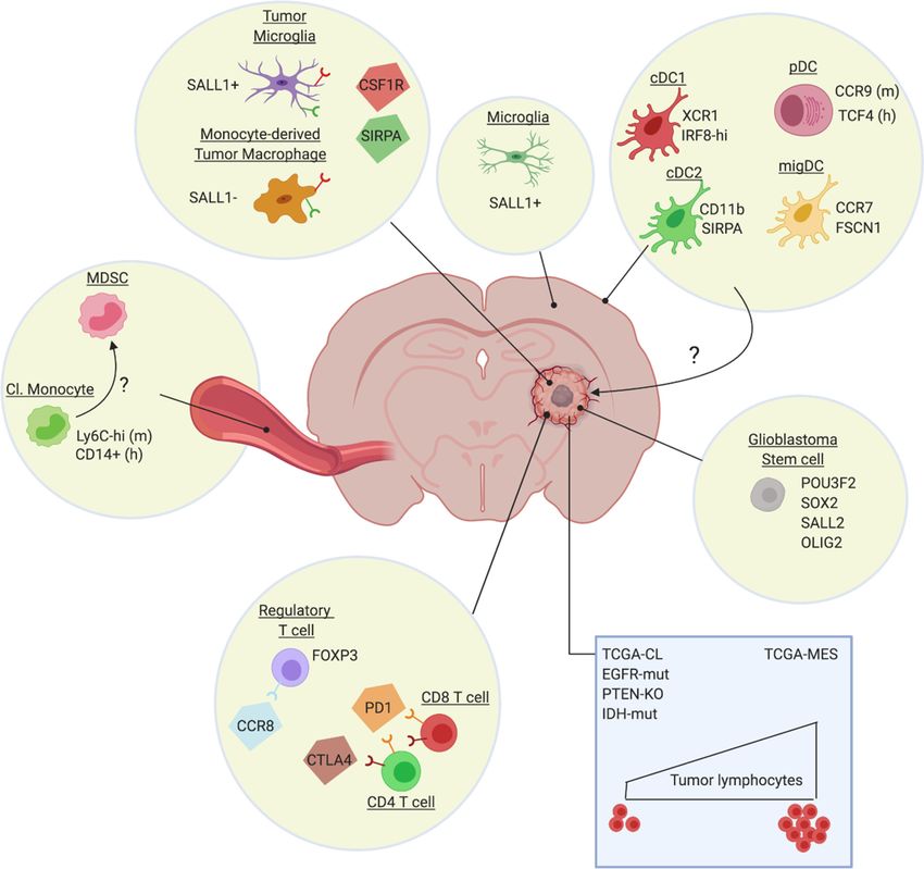
credited. mental transcription factors such as POU3F2, SOX2, SALL2, and OLIG2 (Suvà et al., 2014) (Figure 1).
Pombo Antunes et al. eLife 2020;9:e52176. DOI: https://doi.org/10.7554/eLife.52176 1 of 16Review Article Cancer Biology
Various markers have been suggested for glioblastoma stem cells (Lathia et al., 2015), but it is
unclear at present whether different subpopulations of GBM stem cells exist and whether these give
rise to tumors with a different cellular composition. In any case, expression profiling of GBM tumors
identified at least three GBM subtypes: proneural (TCGA-PN), classical (TCGA-CL) and mesenchymal
(TCGA-MES) (Verhaak et al., 2010; Wang et al., 2017), which tend to differentially associate with
abnormalities in PDGFRA, IDH1, EGFR and NF1 (Verhaak et al., 2010). This level of heterogeneity
is dramatically increased by the notion that different GBM subtypes can be found within the same
tumor and are dynamic in function of time or in response to therapy (Sottoriva et al., 2013;
Patel et al., 2014; Wang et al., 2017). More recent high-resolution single-cell RNA sequencing pro-
vided even more granularity to the concept of intra-tumoral heterogeneity by identifying four cellular
states for glioblastoma cells: mesenchymal-like (MES-like), astrocyte-like (AC-like), oligodendrocytic
Figure 1. Heterogeneity of the glioblastoma immune microenvironment and potential therapeutic targets. Within glioblastoma tumors reside
ontogenically distinct, immunoregulatory macrophages (Sall1+ tumor microglia, Sall1- monocyte-derived macrophages), immunosuppressive Treg (eg
CCR8+) and dysfunctional T-cell populations (CTLA-4/PD-1hi). Not much is known about intratumoral DC subsets, although distinct DC populations are
found in other brain regions, such as the dura mater (Van Hove et al., 2019). Glioblastoma also affects the phenotype of classical monocytes (Cl.
Monocyte) in the periphery, which acquire an immunosuppressive (MDSC-like?) phenotype. Notably, the genetic make-up of the cancer cells (blue
rectangle) and potentially also of the glioblastoma stem cells, affect the immune composition of the tumor, with for example a higher presence of
lymphocytes in TCGA-MES tumors. Several potential therapeutic targets (CSF1R, SIRPa, CCR8, PD-1, CTLA-4), either already tested in the clinic or
promising for the future, are highlighted.
Pombo Antunes et al. eLife 2020;9:e52176. DOI: https://doi.org/10.7554/eLife.52176 2 of 16Review Article Cancer Biology
precursor cell-like (OPC-like) and neural progenitor cell-like (NPC-like) (Neftel et al., 2019). There is
a preponderance of particular states in each TCGA tumor type, with TCGA-CL and TCGA-MES
being enriched in AC-like and MES-like states, respectively, and TCGA-PN encompassing both OPC-
like and NPC-like states. Notably, some genetic alterations favor specific cellular states, with for
example EGFR overexpression driving an AC-like program (Neftel et al., 2019). Finally, non-genetic
heterogeneity within GBM tumors is determined by the relative proximity of cancer cells to blood
vessels, with mTOR activity being upregulated in the few cell layers closest to the vessels
(Kumar et al., 2019). In these cells, mTOR conveys superior invasive and migratory capabilities and
resistance to therapy. Together, this highly heterogeneous nature of GBM strongly undermines the
efficacy of therapy, considering the likely presence of cancer cell clones which are able to escape.
In addition, defects in anti-tumor T-cell responses are commonly observed in GBM, suggesting
the active induction of immunosuppression. In this respect, intracranial tumors of various histological
origins (not only GBM) cause an entrapment of T cells in the bone marrow due to a loss of surface
spingosine-1-phosphate receptor 1 (S1P1) (Chongsathidkiet et al., 2018). Why only intracranial
tumors induce this defect and via which mechanism is currently unknown, but a reversal of this defi-
ciency enables an enhanced migration of T cells into GBM tumors. Along the same line, thymic invo-
lution in GBM tumor-bearers results in diminished T-cell production and, hence, reduced availability
of T cells for anti-GBM immunity (Andaloussi et al., 2008). Moreover, once T cells do get inside the
GBM tumor, they are likely to become dysfunctional via various mechanisms (Woroniecka et al.,
2018a). Recent studies provide evidence for the enriched presence of exhausted T cells within GBM
tumors, with tumor-infiltrating lymphocytes (TILs) more often co-expressing the immune checkpoint
molecules PD-1, LAG3 and TIM-3 than peripheral blood cells of patients or healthy controls
(Woroniecka et al., 2018b; Davidson et al., 2019). Interestingly, PD-1+ LAG3+ TIM-3+ T cells
showed impaired T-cell functions, while single positive cells for any of these markers did not. A sec-
ond prominent mechanism of T-cell dysfunctionality is tolerance, mediated by the presence of sup-
pressive cells such as regulatory T cells (Treg) and tumor-associated macrophages (TAM). These
aspects will be discussed in detail in this review.
Immunotherapeutic approaches in glioblastoma
Previous reviews have described the attempts to incorporate ICB in the treatment of GBM
(Romani et al., 2018; Simonelli et al., 2018). In summary, preclinical models have provided a proof-
of-concept that ICB with anti-CTLA-4 and/or anti-PD-1, either as monotherapy or in conjunction with
standard-of-care treatments, improves tumor outcome. This has set the stage for multiple clinical tri-
als in glioblastoma patients, the results of which are up to this date mostly unavailable. Here, we will
focus on the most recent progress in this field. One of the most noticeable advances comes from
the realization that the timing of ICB administration could majorly impact its effect. Classically, ICB is
given in an adjuvant setting, improving recurrence-free survival and overall survival after surgery in
cancer types such as melanoma. Neoadjuvant (pre-operative) therapy has proven to be advanta-
geous in breast, bladder and other types of cancer, but this finding remained restricted to chemo-
and radiation therapy with very little data available on ICB neoadjuvant therapy until recently
(O’Donnell et al., 2019). Liu et al. (2016) demonstrated increased anti-tumor immune responses
and survival upon neoadjuvant immunotherapy, in two mouse models of triple-negative breast can-
cer. This finding has now been reproduced in several other preclinical models (O’Donnell et al.,
2019), and importantly, also in the clinical setting of resectable melanoma (Blank et al., 2018;
Amaria et al., 2018; Rozeman et al., 2019) and glioblastoma (Cloughesy et al., 2019;
Schalper et al., 2019). In resectable glioblastoma, either as primary or recurrent tumor, one neoad-
juvant injection of anti-PD-1 monoclonal antibodies, followed by surgical resection of the tumor and
repeated adjuvant anti-PD-1 treatment, resulted in a significant increase in overall survival and pro-
gression-free survival compared to adjuvant treatment alone (Cloughesy et al., 2019;
Schalper et al., 2019). Remarkably, the same conclusions were reached by two independent
research groups that used a different monoclonal antibody, either pembrolizumab or nivolumab.
Both studies also demonstrated an intratumoral immunomodulatory effect of the neoadjuvant treat-
ment, with an increased interferon response, production of chemokines, and signs of increased TIL
activity. However, larger-scale randomized trials should be performed to fully prove the benefit of
this neoadjuvant approach.
Pombo Antunes et al. eLife 2020;9:e52176. DOI: https://doi.org/10.7554/eLife.52176 3 of 16Review Article Cancer Biology
A second level of innovation comes from the assessment of novel combination therapies in pre-
clinical models, whereby anti-PD-1 efficacy is boosted by a concurrent administration of chemother-
apy (Park et al., 2019), radiation therapy (Rajani et al., 2018), other immune checkpoint blocking
monoclonal antibodies (Wu et al., 2019), cancer cell-directed immunotoxins (Chandramohan et al.,
2019), and nanocarriers that improve the shuttling of ICB across the blood-brain barrier
(Galstyan et al., 2019). Most of these studies show a clear benefit of combining different
approaches to increase anti-GBM immunity. Indeed, monotherapies usually have little impact,
highlighting the strongly immunosuppressive nature of the GBM tumor microenvironment (TME). A
novel avenue to increase the immunogenicity of the GBM TME comes from virotherapy, whereby
oncolytic viruses not only cause a direct killing of highly proliferative cancer cells but also trigger an
(innate) immune response (Iorgulescu et al., 2018). For example, a recent clinical trial applied intra-
tumoral injections of a polio-rhinovirus chimera and could achieve a remarkable 21% of longer term
survivors (at least 2 to 3 years), while 3 years from treatment, and three patients had
a 95% reduction of the tumor, which was probably due to direct oncolytic effects of the virus fol-
lowed by elicitation of an immune-mediated antiglioma response (Lang et al., 2018). A passive
transfer of anti-tumor T-effector cells could be an alternative strategy to attack GBM tumors,
although such T cells should then be able to withstand the suppressive TME. Novel generations of
CAR T-cells are being generated that should show improved functionality in the TME, including the
TME from GBM (Petersen and Krenciute, 2019). Finally, the adoptive transfer of antigen-presenting
dendritic cells (i.e. DC vaccination) has yielded some efficacy in improving the median overall survival
in clinical trials (Srivastava et al., 2019) and further research is warranted to improve the treatment
regimens and the choice of the most optimal DC type and the most optimal tumor antigens. Neoan-
tigens are the most tumor-specific and arguably the most promising antigen candidates, that show
efficacy as vaccine candidates in glioblastoma patients (Keskin et al., 2019; Hilf et al., 2019).
Heterogeneity of the tumor immune microenvironment in glioblastoma
The immune contexture of human tumors, referring to the exact composition of distinct immune cell
types within tumors, changes at each tumor stage and is known to have an impact on clinical out-
come (Fridman et al., 2012; Bindea et al., 2013). Hence, understanding the tumor (immune) micro-
environment has recently been recognized as one of the future challenges to improve therapy of
brain tumors (Aldape et al., 2019). Glioblastoma tumors are known to contain a variety of immune
cell types, but with a dominance of immunosuppressive cells. Recent findings further finetune this
concept through the identification of distinct subpopulations within the immunoregulatory macro-
phage compartment of tumors, but also distinct Treg and dendritic cell subsets have been identified
and will be discussed in this review.
Correlation between tumor genotype and the glioblastoma immune
microenvironment
As mentioned earlier, innovative techniques such as extensive exome and transcriptome sequencing
have revealed the existence of molecularly distinct proneural, mesenchymal and classical subtypes of
glioblastoma (Verhaak et al., 2010; Wang et al., 2017). In addition, a diverse array of recurrent
genomic mutations were found in these tumors, including TP53, IDH1, NF1, PTEN, PDGFRA, EGFR
and MAPK pathway mutations, with particular mutations being enriched in particular glioblastoma
subtypes.
Of importance in the context of ICB immunotherapy, is the observation that the number of
immune cells in the human glioblastoma microenvironment appears to be associated with the pre-
dominant genetic alteration (Figure 1). TILs are enriched in TCGA-MES glioblastomas and strongly
associated with mutations in NF1 and RB1. Conversely, TILs are depleted in the TCGA-CL class,
EGFR-amplified, and homozygous PTEN-deleted tumors and rare in histologies characterized by
these alterations (Rutledge et al., 2013). In addition, the IDH mutant status in glioblastoma tumors
significantly associates with TIL infiltration. The IDH-wt status is associated with significantly higher
TIL infiltration and PD-L1 expression (Berghoff et al., 2017), while IDH mutations result in the oppo-
site phenotype, that is a reduced IFNg signature and reduced presence of CD8+ and CD4+ cells
Pombo Antunes et al. eLife 2020;9:e52176. DOI: https://doi.org/10.7554/eLife.52176 4 of 16Review Article Cancer Biology
(Kohanbash et al., 2017; Luoto et al., 2018). Nevertheless, patients bearing IDH-mutated tumors
have a more favourable outcome (Ceccarelli et al., 2016), suggesting that other cancer cell-intrinsic
or -extrinsic mechanisms are at play beyond the presence of TILs.
One of those mechanisms could be the presence of immunoregulatory macrophages and neutro-
phils. In a study comparing a small number of treatment-naive IDH-mutated and IDH-wt glioblas-
toma tumors, IDH-mutants were shown to contain less macrophages which displayed a more pro-
inflammatory M1-like activation state, possibly contributing to the longer survival of these patients
(Poon et al., 2019). The reduced presence of macrophages in IDH-mutant, but also CDK4-amplified
tumors, was also demonstrated via a computational analysis of a large number of TCGA RNAseq
samples (Luoto et al., 2018). Hence, although IDH-mutated tumors contain a lower number of T
lymphocytes, these cells may be more optimally activated due to the relative paucity of immunosup-
pressive macrophages and neutrophils. The frequency of macrophages and neutrophils is generally
increased in TCGA-MES tumors versus the other TCGA types (Wang et al., 2017). Remarkably,
genes for both tumor-promoting M2-like as well as potentially anti-tumoral M1-like macrophages
were enriched in TCGA-MES tumors, in particular in the NF1-deficient cases (Wang et al., 2017;
Luoto et al., 2018). The absence of NF1 appears to result in chemoattraction of macrophages via
an as yet undefined mechanism. In addition, high B-cell and CD4+ T-cell components were com-
monly observed in TCGA-MES samples, while immune cell components tend to be low in TCGA-PN
samples (Luoto et al., 2018). In general terms, computational analysis grouped glioblastoma tumors
into three immune response-related subgroups, termed negative (defined by a relative paucity of
immune cells; enriched by TCGA-PN and CDK4-MARCH9 amplification), humoral (defined by a high
B-cell and CD4+ T-cell compartment; enriched by TCGA-MES) and cellular-like (defined by a higher
‘negative regulation of T-cell activation’ and ‘gamma delta T-cell’ cluster; enriched by TCGA-CL and
samples with a high macrophage content) (Luoto et al., 2018).
The question remains how these findings relate to the responsiveness of patients to ICB. Surpris-
ingly, in spite of the immunologically different tumor microenvironment in TCGA subtypes, no asso-
ciation was found with ICB responsiveness (Zhao et al., 2019). Rather, non-responding patients bear
significantly more PTEN mutations, which are associated with immunosuppressive gene expression
signatures, while responders are enriched in MAPK pathway alterations (PTPN11, BRAF). These find-
ings illustrate that responsiveness to ICB is of a greater complexity than the mere presence of
immune cell types within the microenvironment.
Impact of standard-of-care treatments on the Glioblastoma immune
microenvironment
Long-standing treatment options for glioblastoma include surgery, radiotherapy and temozolomide
chemotherapy, all of which may affect the immune composition of glioblastoma tumors. In addition,
brain oedema is most often treated with dexamethasone, a steroid with known immunoregulatory
capacities.
Radiation therapy obviously aims to eliminate the proliferating cancer cells, but one of its side
effects is the induction of hypoxia. In turn, hypoxia initiates a lethal cascade of events consisting of
the increased production of CXCL12, the subsequent recruitment of CXCR4 and CXCR7-positive
bone marrow-derived monocytes and hematopoietic progenitor cells that differentiate into tumor-
promoting macrophages and mediate vasculogenesis and tumor recurrence in mice
(Tabatabai et al., 2006; Kioi et al., 2010; Tseng et al., 2011). Hence, blockade of CXCL12
(Liu et al., 2014) and CXCR7 (Walters et al., 2014) delays recurrence of tumors upon irradiation in
preclinical models. Most importantly, a recent Phase I/II clinical trial with the reversible CXCR4 inhibi-
tor plerixafor – a strategy named Macrophage Exclusion after Radiation Therapy or MERT – in con-
junction with chemoirradiation improved local control of tumor recurrence in newly diagnosed
glioblastoma patients (Thomas et al., 2019). Another effect of radiation is the upregulation of PD-L1
at the surface of tumor-infiltrating myeloid cells. This effect has been exploited to target anti-PD-L1-
functionalized lipid nanoparticles, carrying toxic compounds, to these myeloid cells in conjunction
with irradiation, leading to an improved anti-tumor response in mice (Zhang et al., 2019).
Temozolomide is the most widely used chemotherapy for patients with glioblastoma (GBM),
whose action not only directly affects cancer cells but also depends on its immunomodulatory prop-
erties (Karachi et al., 2018). Temozolomide induces lymphopenia, which, counterintuitively, can be
harnessed to improve immunotherapy. Indeed, lymphoablative doses of temozolomide were shown
Pombo Antunes et al. eLife 2020;9:e52176. DOI: https://doi.org/10.7554/eLife.52176 5 of 16Review Article Cancer Biology
to increase tumor antigen-specific immune responses in GBM patients (Sampson et al., 2011;
Batich et al., 2017) and GBM-bearing mice (Sanchez-Perez et al., 2013). Mechanistically, it was
suggested that compensatory homeostatic cytokines after temozolomide therapy cause enhanced
immune responses by reduction of the T-cell activation threshold and proliferation induction.
In recent years, it became clear that dexamethasone treatment of patients may strongly affect
outcome, with a lower overall survival in recurrent glioblastoma patients (Wong et al., 2015). This
adverse effect is most likely due to the well-known immunomodulatory function of steroids. Indeed,
glioblastoma patients treated with dexamethasone contained significantly less lymphocytes in their
blood, but more myeloid cells with potentially suppressive functions (Chitadze et al., 2017).
Heterogeneity of macrophages within the Glioblastoma immune
microenvironment
Macrophages are a large component of many tumor types and have been an attractive target for
glioblastoma therapy (Pyonteck et al., 2013; Quail et al., 2016). Originally, TAMs were often
thought to exclusively originate from tumor-infiltrating monocytes, but both in CNS and peripheral
tumor types a fraction can be derived from the tissue-resident macrophage pool (Movahedi and
Van Ginderachter, 2016; Laviron and Boissonnas, 2019). Recent work that has relied on single-cell
RNA sequencing combined with fate mapping approaches has highlighted the rich diversity of
murine tissue-resident brain macrophages. (Van Hove et al., 2019) (Figure 1). In this respect, paren-
chymal microglia differ from macrophages located in the brain’s border regions, such as the menin-
ges and the choroid plexus. Whether border-associated macrophages contribute to glioblastoma or
metastatic brain tumors is not known. However, there is now firm evidence that TAMs in GBM
tumors are partly derived from embryonic microglia (Figure 1). Tumor-associated microglia and
monocyte-derived macrophages can be distinguished via genetic lineage tracing, which was used to
sort these populations from transplantable and spontaneous mouse models of glioblastoma fol-
lowed by RNA-sequencing (Bowman et al., 2016; Chen et al., 2017). These data revealed that the
glioblastoma microenvironment instructs the transcriptional landscape of these cells, with prominent
differences between microglia and monocyte-derived TAMs at multiple levels, including differential
expression of genes involved in IL-1 signaling, chemokine and inflammatory cytokine production and
antigen presentation. Whether this transcriptional divergence is reflected at the functional level and
whether these subsets differentially affect glioblastoma progression is not known, but seems likely.
Indeed, tissue-resident interstitial macrophages and their monocyte-derived counterparts are also
found in mouse lung tumors, where resident TAMs are suggested to support cancer cell expansion,
while monocyte-derived cells might contribute to cancer cell dissemination (Loyher et al., 2018).
Also in mouse mammary carcinoma, a distinction was made between monocyte-derived TAMs and
resident mammary tissue macrophages, with only the former contributing to a suppression of anti-
tumor cytotoxic T-cell responses (Franklin et al., 2014).
In glioblastomas, monocyte-derived macrophages tend to be more enriched in the tumor core,
while microglia-derived TAM are typically found at the tumor periphery (Chen et al., 2017). Live in
vivo 2-photon microscopy confirmed the distinction between the two TAM populations in the mouse
and demonstrated that monocyte-derived cells are small and motile, while microglia are large sessile
cells whose processes are continuously extending and retracting within tumors (Chen et al., 2019a).
Importantly, the existence of microglia and monocyte-derived TAM populations has now also been
shown in human GBM (Müller et al., 2017; Darmanis et al., 2017). However, it will be interesting to
evaluate whether microglia and monocyte-derived TAM further consist of specialized subpopula-
tions, for example instructed by their precise intratumoral localization, as was demonstrated for
TAM in other tumor models (Movahedi et al., 2010; Casazza et al., 2013). For this reason, future
single-cell RNA-sequencing efforts will provide crucial new insights, and will undoubtedly allow the
identification of multiple TAM subsets, as was already shown in mouse and human lung tumors
(Zilionis et al., 2019). An interesting possibility is that microglia- and monocyte-derived TAMs may
exhibit a differential ability to infiltrate or colonize specific tumor microenvironments or tumor mac-
rophage niches.
Interesting novel findings in preclinical models provide further insights in the molecular mecha-
nisms that govern the attraction of monocyte-derived macrophages to the glioblastoma microenvi-
ronment. Cancer cell- and host cell-derived osteopontin mediates macrophage infiltration through
the interaction with integrin avb5 and instructs a pro-tumoral phenotype in these cells (Wei et al.,
Pombo Antunes et al. eLife 2020;9:e52176. DOI: https://doi.org/10.7554/eLife.52176 6 of 16Review Article Cancer Biology
2019). Also the Aryl Hydrocarbon Receptor (AHR) is indirectly implicated in recruiting monocytes to
the tumor environment by driving CCR2 expression, the receptor for the major monocyte chemoat-
tractants CCL2 and CCL7 (Takenaka et al., 2019). In PTEN-deficient tumor models, enhanced LOX
production is responsible for the enhanced attraction of macrophages via the b1 integrin-PYK2 path-
way, which subsequently promote tumor growth via SPP1 (Chen et al., 2019b). However, how this
finding correlates with the enhanced responsiveness of PTEN-mutant tumors to ICB therapy in
patients is not clear and requires further investigation, including the potential differences between
the PTEN mutations found in patients and the PTEN-null phenotype in mouse models.
Another point of interest is the systemic influence of brain tumors on peripheral myeloid cells.
GBM-secreted IL-6 induces immunosuppressive myeloid cells in the periphery that express high lev-
els of PD-L1 in orthotopic mouse models (Lamano et al., 2019). This finding is in line with CyTOF
fingerprinting of patients’ peripheral blood, indicating a significant elevation of myeloid-derived sup-
pressor cells (MDSC), but not Treg in the circulation (Alban et al., 2018) (Figure 1). Hence, the mye-
loid cells that infiltrate GBM tumors may have already been primed into a tumor-promoting role.
The importance of MDSC, both the monocytic and granulocytic subtype (Movahedi et al., 2008), is
further confirmed by the finding that their presence in mouse GBM tumors is associated with a
reduction in the number of tumor-infiltrating lymphocytes (Raychaudhuri et al., 2015). Moreover,
within the murine glioblastoma environment, MDSC were shown to induce regulatory B cells (Lee-
Chang et al., 2019). Interestingly, CCL2 produced by TAM mediates the recruitment of suppressive
CCR2+ monocytic MDSC and CCR4+ Treg, explaining the clinical observation that elevated intratu-
moral CCL2 expression levels correlate with reduced overall survival (Chang et al., 2016). Of note,
recruited monocytic MDSC may in turn differentiate into TAM. In a preclinical setting, this notion has
now been taken further by showing that a small molecule CCR2 antagonist sensitizes otherwise resis-
tant murine gliomas to ICB therapy (Flores-Toro et al., 2020).
Heterogeneity of regulatory T cells (Treg) within the glioblastoma
immune microenvironment
The presence of Treg has been amply described in multiple cancer types, but their value as predic-
tors of disease outcome is debatable in glioblastoma. Independent researchers described an
increased presence of FOXP3+ Treg in higher tumor grades of various brain tumor types, including
glioblastoma (El Andaloussi and Lesniak, 2007; Jacobs et al., 2010). However, the correlation of
Treg with decreased survival, which was anticipated based on the T-cell suppressive capacity of
these cells, has been very moderate at best (Jacobs et al., 2010; Yue et al., 2014; Thomas et al.,
2015). Nevertheless, Treg-directed therapies, such as agonist anti-GITR or anti-LAP (Latency-associ-
ated Peptide) mAb treatment, have shown some promise in mouse glioblastoma models
(Miska et al., 2016; Patel et al., 2016; Gabriely et al., 2017).
A potential reason for the unclear association of Treg with disease outcome could be the exis-
tence of distinct Treg subsets which may differ in their suppressive capacity and whose presence in
tumors may vary between individual patients. Early preclinical work suggested that the majority of
Tregs infiltrating glioblastoma tumors are thymus-derived (Wainwright et al., 2011), although it
seems likely that the local de novo induction of Treg in the tumor microenvironment is a possibility
as well. As a matter of fact, indirect evidence based on correlative studies proposed glioblastoma-
induced PD-L1 expression as a mechanism of Treg induction and maintenance in patients
(DiDomenico et al., 2018). The attraction of these Treg to the tumor microenvironment may be
mediated by various mechanisms, including IDO activity (Wainwright et al., 2012) and CCL2 pro-
duction, which interacts with CCR4 at the surface of Treg (Chang et al., 2016). Interestingly, another
chemokine receptor, CCR8, has recently been identified as a marker that is specifically expressed in
at least a subset of tumor-infiltrating Treg, but not Treg in the periphery, as part of a tumor-infiltrat-
ing Treg transcriptional signature that is conserved across species and tumor types (Plitas et al.,
2016; De Simone et al., 2016; Zheng et al., 2017; Magnuson et al., 2018) (Figure 1). CCR8+ Treg
were reported as very potent suppressors, suggesting that this Treg subset is likely important in the
creation of an immunosuppressive tumor microenvironment (Barsheshet et al., 2017). However, to
what extent the tumor-infiltrating Treg signature and CCR8+ Treg are present within glioblastoma
tumors is currently unknown.
Another marker that is functionally important on Treg within tumors is Neuropilin-1 (Nrp1). The
interaction of Nrp1 with its ligand Semaphorin 4a was shown to stabilize the Treg phenotype in the
Pombo Antunes et al. eLife 2020;9:e52176. DOI: https://doi.org/10.7554/eLife.52176 7 of 16Review Article Cancer Biology
mouse, which turns out to be especially important for the suppression of anti-tumor immune
responses (Delgoffe et al., 2013). The absence of Nrp1 in Treg leads to their loss of suppressive
potential but also to their production of IFNg, which subsequently annihilates the suppressive capac-
ity of neighbouring Nrp1+ Treg in murine tumors (Overacre-Delgoffe et al., 2017). This mechanism
may be stimulated by enhanced HIF-1a expression in Nrp1-deficient Treg. Interestingly, HIF-1a
expression in hypoxic tumor-infiltrating Treg was indeed shown to decrease the intrinsic suppressive
activity of these Treg, but was at the same time required for the migration of Treg into mouse glio-
blastoma tumors (Miska et al., 2019). Consequently, the absence of HIF1a in Treg leads to the
reduced growth of glioblastoma tumors.
Altogether, suppressive Treg subsets are induced in the microenvironment of multiple tumor
types. As these cells could be prime targets for therapeutic intervention, it will be important to
assess their presence and relevance in glioblastoma.
Heterogeneity of dendritic cells within the Glioblastoma immune
microenvironment
Recent findings, in other tumor types than glioblastoma, have uncovered the existence of distinct
DC populations in solid tumors. Indeed, at least three types of conventional DCs (cDCs), in addition
to plasmacytoid DCs (pDCs), are present in the tumor microenvironment of multiple mouse and
human tumors (Laoui et al., 2016; Roberts et al., 2016; Zilionis et al., 2019). The functionality of
these cells is being explored in mouse tumor models. cDC1 are rare cells within tumors that were
repeatedly reported to migrate to the tumor-draining lymph nodes where they cross-present tumor
antigens to CD8+ T cells (Broz et al., 2014; Roberts et al., 2016; Laoui et al., 2016). Interestingly,
NK cells attract cDC1 to the tumor environment via the secretion of various growth factors and che-
mokines, such as Flt3L, CCL5 and XCL1 (Böttcher et al., 2018; Barry et al., 2018). cDC2, which are
typically more abundant within tumors than cDC1, also have the capacity to drive T-cell responses,
but are more directed towards the activation of CD4+ T cells. As such, these cells were shown to ini-
tiate a Th17 response (Laoui et al., 2016), but their full T-cell activating potential may only be
unleashed in the absence of Treg (Binnewies et al., 2019). Both mouse and human lung tumors also
harbor an additional DC subset that exhibits a migratory gene signature (Zilionis et al., 2019),
although this cannot be taken as proof for actual migration.
Translating these findings to glioblastoma is not trivial, as a systematic analysis of DC subpopula-
tions in these tumors is currently lacking. Recent work has shown that various dendritic cell subsets
are present in the murine homeostatic brain (Mrdjen et al., 2018; Van Hove et al., 2019), where
they are thought be critical for initiating neuroinflammation (Mundt et al., 2019; Jordão et al.,
2019). In the context of glioblastoma, one study reported that CCR2+ Lin- hematopoietic stem and
progenitor cells (HSC) can differentiate into cross-presenting DC within mouse glioblastoma tumors
(Flores et al., 2018). These HSC-derived DC can be isolated from glioblastoma tumors to success-
fully vaccinate mice and induce anti-tumor immunity in the vaccine recipients. In addition, their pres-
ence potentiates the effect of immune checkpoint blockade immunotherapy. Along the same line,
increasing the number of intratumoral CD103+ cDC1 through the administration of Flt3L
(Miao et al., 2018), or activating them via TLR3 agonists (Garzon-Muvdi et al., 2018), magnifies the
response of mouse glioblastomas to immune checkpoint blockade, suggesting the importance of
this DC type to augment immunity against glioblastoma, similar to other tumor types. However,
Fibrinogen-like Protein 2 (FGL2), that is predominantly secreted by the cancer cells, may interfere
with the induction of these immunostimulatory DCs (Yan et al., 2019). Indeed, FGL2 interferes with
GM-CSF signalling, blunting the differentiation of CD103+ cDC1 and consequently lowering the
CD8+ T-cell response. Overall, it can be concluded that interventions which increase the number of
CD103+ cDC1 in glioblastoma tumors or augment their activation state may be beneficial. Whether
cDC2 can also contribute to anti-glioblastoma immunity remains to be determined.
Perspective on novel immunotherapies directed against cells within the
tumor immune microenvironment
Finally, how can the recently acquired knowledge on the heterogeneity of the glioblastoma immune
microenvironment be turned into novel therapeutic avenues? An important caveat in this respect is
the capacity of drugs to cross the blood-brain barrier (BBB) and to reach sufficiently high
Pombo Antunes et al. eLife 2020;9:e52176. DOI: https://doi.org/10.7554/eLife.52176 8 of 16Review Article Cancer Biology
concentrations in the brain. A number of very recent studies have highlighted the potential of nano-
particles to deliver therapeutic cargo to the mouse brain. Solid lipid nanoparticles can deliver small
interfering RNAs to the mouse glioblastoma microenvironment, following a low-dose irradiation to
prime brain uptake (Erel-Akbaba et al., 2019). A novel tumor penetrating peptide that targets cell
surface p32, LinTT1 (AKRGARSTA), has also been reported as a GBM targeting ligand for systemi-
cally-administered nanoparticles (Säälik et al., 2019). Moreover, nanoscale immunoconjugates
(NICs) on a natural biopolymer scaffold, poly(b-L-malic acid), were produced with covalently
attached anti-CTLA-4 or anti-PD-1 for systemic delivery across the BBB and activation of local brain
anti-tumor immune responses (Galstyan et al., 2019). Notably, nanoparticles often end up being
phagocytosed by tumor-associated macrophages and are, hence, interesting tools to modulate the
activity of these cells. The fact that modulating the macrophage compartment is indeed a promising
approach was shown a couple of years ago by the administration of a brain penetrating CSF1R inhib-
itor in mice, which repolarized TAM into a more anti-tumoral phenotype resulting in a reduced GBM
progression (Pyonteck et al., 2013). Likewise, a blocking anti-CD47 antibody mobilizes the phago-
cytic capacity of both monocyte-derived and microglial TAMs in mouse glioblastoma models
(Hutter et al., 2019). As a matter of fact, this type of approaches could now be combined with strat-
egies to increase the presence of antigen-presenting cells, by the targeted delivery of growth factors
or chemokines for cDCs.
Targeting tumor-infiltrating Treg, without affecting their peripheral counterparts to avoid auto-
immune complications, could be another interesting approach. The recent identification of mole-
cules, such as CCR8, that are specifically upregulated on these cells within tumors may provide novel
therapeutic anchor points in multiple tumor types. It needs to be evaluated whether these tiTreg-
specific molecules are implicated in Treg functionality as this will determine the type of compound
we need for therapy (agonists, antagonists,...).
Overall, increasing knowledge about subpopulations of immune cells that either promote or
inhibit glioblastoma tumor progression will allow more specific therapeutic approaches against this
aggressive type of brain tumor.
Additional information
Funding
Funder Author
Kom op tegen Kanker Bart Neyns
Kiavash Movahedi
Jo A Van Ginderachter
Stichting Tegen Kanker Jo A Van Ginderachter
Fonds Wetenschappelijk On- Isabelle Scheyltjens
derzoek Jo A Van Ginderachter
Innoviris Kiavash Movahedi
The funders had no role in study design, data collection and
interpretation, or the decision to submit the work for publication.
Author ORCIDs
Jo A Van Ginderachter https://orcid.org/0000-0002-4442-7474
References
Alban TJ, Alvarado AG, Sorensen MD, Bayik D, Volovetz J, Serbinowski E, Mulkearns-Hubert EE, Sinyuk M, Hale
JS, Onzi GR, McGraw M, Huang P, Grabowski MM, Wathen CA, Ahluwalia MS, Radivoyevitch T, Kornblum HI,
Kristensen BW, Vogelbaum MA, Lathia JD. 2018. Global immune fingerprinting in glioblastoma patient
peripheral blood reveals immune-suppression signatures associated with prognosis. JCI Insight 3:122264.
DOI: https://doi.org/10.1172/jci.insight.122264, PMID: 30385717
Aldape K, Brindle KM, Chesler L, Chopra R, Gajjar A, Gilbert MR, Gottardo N, Gutmann DH, Hargrave D,
Holland EC, Jones DTW, Joyce JA, Kearns P, Kieran MW, Mellinghoff IK, Merchant M, Pfister SM, Pollard SM,
Pombo Antunes et al. eLife 2020;9:e52176. DOI: https://doi.org/10.7554/eLife.52176 9 of 16Review Article Cancer Biology
Ramaswamy V, Rich JN, et al. 2019. Challenges to curing primary brain tumours. Nature Reviews Clinical
Oncology 16:509–520. DOI: https://doi.org/10.1038/s41571-019-0177-5, PMID: 30733593
Amaria RN, Reddy SM, Tawbi HA, Davies MA, Ross MI, Glitza IC, Cormier JN, Lewis C, Hwu WJ, Hanna E, Diab
A, Wong MK, Royal R, Gross N, Weber R, Lai SY, Ehlers R, Blando J, Milton DR, Woodman S, et al. 2018.
Neoadjuvant immune checkpoint blockade in high-risk resectable melanoma. Nature Medicine 24:1649–1654.
DOI: https://doi.org/10.1038/s41591-018-0197-1, PMID: 30297909
Andaloussi AE, Han Y, Lesniak MS. 2008. Progression of intracranial glioma disrupts thymic homeostasis and
induces T-cell apoptosis in vivo. Cancer Immunology, Immunotherapy 57:1807–1816. DOI: https://doi.org/10.
1007/s00262-008-0508-3
Barry KC, Hsu J, Broz ML, Cueto FJ, Binnewies M, Combes AJ, Nelson AE, Loo K, Kumar R, Rosenblum MD,
Alvarado MD, Wolf DM, Bogunovic D, Bhardwaj N, Daud AI, Ha PK, Ryan WR, Pollack JL, Samad B, Asthana S,
et al. 2018. A natural killer-dendritic cell Axis defines checkpoint therapy-responsive tumor microenvironments.
Nature Medicine 24:1178–1191. DOI: https://doi.org/10.1038/s41591-018-0085-8, PMID: 29942093
Barsheshet Y, Wildbaum G, Levy E, Vitenshtein A, Akinseye C, Griggs J, Lira SA, Karin N. 2017. CCR8+FOXp3+
Treg cells as master drivers of immune regulation. PNAS 114:6086–6091. DOI: https://doi.org/10.1073/pnas.
1621280114
Batich KA, Reap EA, Archer GE, Sanchez-Perez L, Nair SK, Schmittling RJ, Norberg P, Xie W, Herndon JE, Healy
P, McLendon RE, Friedman AH, Friedman HS, Bigner D, Vlahovic G, Mitchell DA, Sampson JH. 2017. Long-
term survival in glioblastoma with Cytomegalovirus pp65-Targeted vaccination. Clinical Cancer Research 23:
1898–1909. DOI: https://doi.org/10.1158/1078-0432.CCR-16-2057, PMID: 28411277
Berghoff AS, Kiesel B, Widhalm G, Wilhelm D, Rajky O, Kurscheid S, Kresl P, Wöhrer A, Marosi C, Hegi ME,
Preusser M. 2017. Correlation of immune phenotype with IDH mutation in diffuse glioma. Neuro-Oncology 19:
1460–1468. DOI: https://doi.org/10.1093/neuonc/nox054, PMID: 28531337
Bindea G, Mlecnik B, Tosolini M, Kirilovsky A, Waldner M, Obenauf AC, Angell H, Fredriksen T, Lafontaine L,
Berger A, Bruneval P, Fridman WH, Becker C, Pagès F, Speicher MR, Trajanoski Z, Galon J. 2013.
Spatiotemporal dynamics of intratumoral immune cells reveal the immune landscape in human Cancer.
Immunity 39:782–795. DOI: https://doi.org/10.1016/j.immuni.2013.10.003, PMID: 24138885
Binnewies M, Mujal AM, Pollack JL, Combes AJ, Hardison EA, Barry KC, Tsui J, Ruhland MK, Kersten K,
Abushawish MA, Spasic M, Giurintano JP, Chan V, Daud AI, Ha P, Ye CJ, Roberts EW, Krummel MF. 2019.
Unleashing Type-2 dendritic cells to drive protective antitumor CD4+ T Cell Immunity. Cell 177:556–571.
DOI: https://doi.org/10.1016/j.cell.2019.02.005, PMID: 30955881
Blank CU, Rozeman EA, Fanchi LF, Sikorska K, van de Wiel B, Kvistborg P, Krijgsman O, van den Braber M,
Philips D, Broeks A, van Thienen JV, Mallo HA, Adriaansz S, Ter Meulen S, Pronk LM, Grijpink-Ongering LG,
Bruining A, Gittelman RM, Warren S, van Tinteren H, et al. 2018. Neoadjuvant versus adjuvant ipilimumab plus
nivolumab in macroscopic stage III melanoma. Nature Medicine 24:1655–1661. DOI: https://doi.org/10.1038/
s41591-018-0198-0, PMID: 30297911
Böttcher JP, Bonavita E, Chakravarty P, Blees H, Cabeza-Cabrerizo M, Sammicheli S, Rogers NC, Sahai E,
Zelenay S, Reis e Sousa C. 2018. NK cells stimulate recruitment of cDC1 into the tumor microenvironment
promoting Cancer immune control. Cell 172:1022–1037. DOI: https://doi.org/10.1016/j.cell.2018.01.004,
PMID: 29429633
Bowman RL, Klemm F, Akkari L, Pyonteck SM, Sevenich L, Quail DF, Dhara S, Simpson K, Gardner EE, Iacobuzio-
Donahue CA, Brennan CW, Tabar V, Gutin PH, Joyce JA. 2016. Macrophage ontogeny underlies differences in
Tumor-Specific education in brain malignancies. Cell Reports 17:2445–2459. DOI: https://doi.org/10.1016/j.
celrep.2016.10.052, PMID: 27840052
Broz ML, Binnewies M, Boldajipour B, Nelson AE, Pollack JL, Erle DJ, Barczak A, Rosenblum MD, Daud A, Barber
DL, Amigorena S, Van’t Veer LJ, Sperling AI, Wolf DM, Krummel MF. 2014. Dissecting the tumor myeloid
compartment reveals rare activating antigen-presenting cells critical for T cell immunity. Cancer Cell 26:638–
652. DOI: https://doi.org/10.1016/j.ccell.2014.09.007, PMID: 25446897
Casazza A, Laoui D, Wenes M, Rizzolio S, Bassani N, Mambretti M, Deschoemaeker S, Van Ginderachter JA,
Tamagnone L, Mazzone M. 2013. Impeding macrophage entry into hypoxic tumor Areas by Sema3A/Nrp1
signaling blockade inhibits angiogenesis and restores antitumor immunity. Cancer Cell 24:695–709.
DOI: https://doi.org/10.1016/j.ccr.2013.11.007, PMID: 24332039
Ceccarelli M, Barthel FP, Malta TM, Sabedot TS, Salama SR, Murray BA, Morozova O, Newton Y, Radenbaugh A,
Pagnotta SM, Anjum S, Wang J, Manyam G, Zoppoli P, Ling S, Rao AA, Grifford M, Cherniack AD, Zhang H,
Poisson L, et al. 2016. Molecular profiling reveals biologically discrete subsets and pathways of progression in
diffuse glioma. Cell 164:550–563. DOI: https://doi.org/10.1016/j.cell.2015.12.028, PMID: 26824661
Chandramohan V, Bao X, Yu X, Parker S, McDowall C, Yu YR, Healy P, Desjardins A, Gunn MD, Gromeier M,
Nair SK, Pastan IH, Bigner DD. 2019. Improved efficacy against malignant brain tumors with EGFRwt/EGFRvIII
targeting immunotoxin and checkpoint inhibitor combinations. Journal for ImmunoTherapy of Cancer 7:142.
DOI: https://doi.org/10.1186/s40425-019-0614-0, PMID: 31142380
Chang AL, Miska J, Wainwright DA, Dey M, Rivetta CV, Yu D, Kanojia D, Pituch KC, Qiao J, Pytel P, Han Y, Wu
M, Zhang L, Horbinski CM, Ahmed AU, Lesniak MS. 2016. CCL2 produced by the glioma microenvironment is
essential for the recruitment of regulatory T cells and Myeloid-Derived suppressor cells. Cancer Research 76:
5671–5682. DOI: https://doi.org/10.1158/0008-5472.CAN-16-0144, PMID: 27530322
Chen Z, Feng X, Herting CJ, Garcia VA, Nie K, Pong WW, Rasmussen R, Dwivedi B, Seby S, Wolf SA, Gutmann
DH, Hambardzumyan D. 2017. Cellular and molecular identity of Tumor-Associated macrophages in
Pombo Antunes et al. eLife 2020;9:e52176. DOI: https://doi.org/10.7554/eLife.52176 10 of 16Review Article Cancer Biology
glioblastoma. Cancer Research 77:2266–2278. DOI: https://doi.org/10.1158/0008-5472.CAN-16-2310, PMID: 2
8235764
Chen Z, Ross JL, Hambardzumyan D. 2019a. Intravital 2-photon imaging reveals distinct morphology and
infiltrative properties of glioblastoma-associated macrophages. PNAS 116:14254–14259. DOI: https://doi.org/
10.1073/pnas.1902366116, PMID: 31235603
Chen P, Zhao D, Li J, Liang X, Li J, Chang A, Henry VK, Lan Z, Spring DJ, Rao G, Wang YA, DePinho RA. 2019b.
Symbiotic Macrophage-Glioma cell interactions reveal synthetic lethality in PTEN-Null glioma. Cancer Cell 35:
868–884. DOI: https://doi.org/10.1016/j.ccell.2019.05.003, PMID: 31185211
Chitadze G, Flüh C, Quabius ES, Freitag-Wolf S, Peters C, Lettau M, Bhat J, Wesch D, Oberg HH, Luecke S,
Janssen O, Synowitz M, Held-Feindt J, Kabelitz D. 2017. In-depth immunophenotyping of patients with
glioblastoma multiforme: impact of steroid treatment. OncoImmunology 6:e1358839. DOI: https://doi.org/10.
1080/2162402X.2017.1358839, PMID: 29147621
Chongsathidkiet P, Jackson C, Koyama S, Loebel F, Cui X, Farber SH, Woroniecka K, Elsamadicy AA, Dechant
CA, Kemeny HR, Sanchez-Perez L, Cheema TA, Souders NC, Herndon JE, Coumans JV, Everitt JI, Nahed BV,
Sampson JH, Gunn MD, Martuza RL, et al. 2018. Sequestration of T cells in bone marrow in the setting of
glioblastoma and other intracranial tumors. Nature Medicine 24:1459–1468. DOI: https://doi.org/10.1038/
s41591-018-0135-2, PMID: 30104766
Cloughesy TF, Mochizuki AY, Orpilla JR, Hugo W, Lee AH, Davidson TB, Wang AC, Ellingson BM, Rytlewski JA,
Sanders CM, Kawaguchi ES, Du L, Li G, Yong WH, Gaffey SC, Cohen AL, Mellinghoff IK, Lee EQ, Reardon DA,
O’Brien BJ, et al. 2019. Neoadjuvant anti-PD-1 immunotherapy promotes a survival benefit with intratumoral
and systemic immune responses in recurrent glioblastoma. Nature Medicine 25:477–486. DOI: https://doi.org/
10.1038/s41591-018-0337-7, PMID: 30742122
Darmanis S, Sloan SA, Croote D, Mignardi M, Chernikova S, Samghababi P, Zhang Y, Neff N, Kowarsky M,
Caneda C, Li G, Chang SD, Connolly ID, Li Y, Barres BA, Gephart MH, Quake SR. 2017. Single-Cell RNA-Seq
analysis of infiltrating neoplastic cells at the migrating front of human glioblastoma. Cell Reports 21:1399–1410.
DOI: https://doi.org/10.1016/j.celrep.2017.10.030, PMID: 29091775
Davidson TB, Lee A, Hsu M, Sedighim S, Orpilla J, Treger J, Mastall M, Roesch S, Rapp C, Galvez M, Mochizuki
A, Antonios J, Garcia A, Kotecha N, Bayless N, Nathanson D, Wang A, Everson R, Yong WH, Cloughesy TF,
et al. 2019. Expression of PD-1 by T cells in malignant glioma patients reflects exhaustion and activation.
Clinical Cancer Research 25:1913–1922. DOI: https://doi.org/10.1158/1078-0432.CCR-18-1176, PMID: 304980
94
De Simone M, Arrigoni A, Rossetti G, Gruarin P, Ranzani V, Politano C, Bonnal RJP, Provasi E, Sarnicola ML,
Panzeri I, Moro M, Crosti M, Mazzara S, Vaira V, Bosari S, Palleschi A, Santambrogio L, Bovo G, Zucchini N,
Totis M, et al. 2016. Transcriptional landscape of human tissue lymphocytes unveils uniqueness of Tumor-
Infiltrating T regulatory cells. Immunity 45:1135–1147. DOI: https://doi.org/10.1016/j.immuni.2016.10.021,
PMID: 27851914
Delgoffe GM, Woo SR, Turnis ME, Gravano DM, Guy C, Overacre AE, Bettini ML, Vogel P, Finkelstein D,
Bonnevier J, Workman CJ, Vignali DA. 2013. Stability and function of regulatory T cells is maintained by a
neuropilin-1-semaphorin-4a Axis. Nature 501:252–256. DOI: https://doi.org/10.1038/nature12428, PMID: 23
913274
Desjardins A, Gromeier M, Herndon JE, Beaubier N, Bolognesi DP, Friedman AH, Friedman HS, McSherry F,
Muscat AM, Nair S, Peters KB, Randazzo D, Sampson JH, Vlahovic G, Harrison WT, McLendon RE, Ashley D,
Bigner DD. 2018. Recurrent glioblastoma treated with recombinant poliovirus. New England Journal of
Medicine 379:150–161. DOI: https://doi.org/10.1056/NEJMoa1716435, PMID: 29943666
DiDomenico J, Lamano JB, Oyon D, Li Y, Veliceasa D, Kaur G, Ampie L, Choy W, Lamano JB, Bloch O. 2018. The
immune checkpoint protein PD-L1 induces and maintains regulatory T cells in glioblastoma. OncoImmunology
7:e1448329. DOI: https://doi.org/10.1080/2162402X.2018.1448329, PMID: 29900065
El Andaloussi A, Lesniak MS. 2007. CD4+ CD25+ FoxP3+ T-cell infiltration and heme oxygenase-1 expression
correlate with tumor grade in human gliomas. Journal of Neuro-Oncology 83:145–152. DOI: https://doi.org/10.
1007/s11060-006-9314-y, PMID: 17216339
Erel-Akbaba G, Carvalho LA, Tian T, Zinter M, Akbaba H, Obeid PJ, Chiocca EA, Weissleder R, Kantarci AG,
Tannous BA. 2019. Radiation-Induced targeted Nanoparticle-Based gene delivery for brain tumor therapy. ACS
Nano 13:4028–4040. DOI: https://doi.org/10.1021/acsnano.8b08177, PMID: 30916923
Flores CT, Wildes TJ, Drake JA, Moore GL, Dean BD, Abraham RS, Mitchell DA. 2018. Lin-CCR2+ hematopoietic
stem and progenitor cells overcome resistance to PD-1 blockade. Nature Communications 9:4313.
DOI: https://doi.org/10.1038/s41467-018-06182-5, PMID: 30333482
Flores-Toro JA, Luo D, Gopinath A, Sarkisian MR, Campbell JJ, Charo IF, Singh R, Schall TJ, Datta M, Jain RK,
Mitchell DA, Harrison JK. 2020. CCR2 inhibition reduces tumor myeloid cells and unmasks a checkpoint
inhibitor effect to slow progression of resistant murine gliomas. PNAS 117:1129–1138. DOI: https://doi.org/10.
1073/pnas.1910856117, PMID: 31879345
Franklin RA, Liao W, Sarkar A, Kim MV, Bivona MR, Liu K, Pamer EG, Li MO. 2014. The cellular and molecular
origin of tumor-associated macrophages. Science 344:921–925. DOI: https://doi.org/10.1126/science.1252510,
PMID: 24812208
Fridman WH, Pagès F, Sautès-Fridman C, Galon J. 2012. The immune contexture in human tumours: impact on
clinical outcome. Nature Reviews Cancer 12:298–306. DOI: https://doi.org/10.1038/nrc3245, PMID: 22419253
Gabriely G, da Cunha AP, Rezende RM, Kenyon B, Madi A, Vandeventer T, Skillin N, Rubino S, Garo L, Mazzola
MA, Kolypetri P, Lanser AJ, Moreira T, Faria AMC, Lassmann H, Kuchroo V, Murugaiyan G, Weiner HL. 2017.
Pombo Antunes et al. eLife 2020;9:e52176. DOI: https://doi.org/10.7554/eLife.52176 11 of 16Review Article Cancer Biology
Targeting latency-associated peptide promotes antitumor immunity. Science Immunology 2:eaaj1738.
DOI: https://doi.org/10.1126/sciimmunol.aaj1738
Galstyan A, Markman JL, Shatalova ES, Chiechi A, Korman AJ, Patil R, Klymyshyn D, Tourtellotte WG, Israel LL,
Braubach O, Ljubimov VA, Mashouf LA, Ramesh A, Grodzinski ZB, Penichet ML, Black KL, Holler E, Sun T, Ding
H, Ljubimov AV, et al. 2019. Blood-brain barrier permeable nano immunoconjugates induce local immune
responses for glioma therapy. Nature Communications 10:3850. DOI: https://doi.org/10.1038/s41467-019-
11719-3, PMID: 31462642
Garzon-Muvdi T, Theodros D, Luksik AS, Maxwell R, Kim E, Jackson CM, Belcaid Z, Ganguly S, Tyler B, Brem H,
Pardoll DM, Lim M. 2018. Dendritic cell activation enhances anti-PD-1 mediated immunotherapy against
glioblastoma. Oncotarget 9:20681–20697. DOI: https://doi.org/10.18632/oncotarget.25061, PMID: 29755681
Hilf N, Kuttruff-Coqui S, Frenzel K, Bukur V, Stevanović S, Gouttefangeas C, Platten M, Tabatabai G, Dutoit V,
van der Burg SH, Thor Straten P, Martı́nez-Ricarte F, Ponsati B, Okada H, Lassen U, Admon A, Ottensmeier CH,
Ulges A, Kreiter S, von Deimling A, et al. 2019. Actively personalized vaccination trial for newly diagnosed
glioblastoma. Nature 565:240–245. DOI: https://doi.org/10.1038/s41586-018-0810-y, PMID: 30568303
Hutter G, Theruvath J, Graef CM, Zhang M, Schoen MK, Manz EM, Bennett ML, Olson A, Azad TD, Sinha R,
Chan C, Assad Kahn S, Gholamin S, Wilson C, Grant G, He J, Weissman IL, Mitra SS, Cheshier SH. 2019.
Microglia are effector cells of CD47-SIRPa antiphagocytic Axis disruption against glioblastoma. PNAS 116:997–
1006. DOI: https://doi.org/10.1073/pnas.1721434116, PMID: 30602457
Iorgulescu JB, Reardon DA, Chiocca EA, Wu CJ. 2018. Immunotherapy for glioblastoma: going viral. Nature
Medicine 24:1094–1096. DOI: https://doi.org/10.1038/s41591-018-0142-3, PMID: 30082860
Jacobs JF, Idema AJ, Bol KF, Grotenhuis JA, de Vries IJ, Wesseling P, Adema GJ. 2010. Prognostic significance
and mechanism of treg infiltration in human brain tumors. Journal of Neuroimmunology 225:195–199.
DOI: https://doi.org/10.1016/j.jneuroim.2010.05.020, PMID: 20537408
Jordão MJC, Sankowski R, Brendecke SM, Sagar , Locatelli G, Tai YH, Tay TL, Schramm E, Armbruster S,
Hagemeyer N, Groß O, Mai D, Çiçek Ö, Falk T, Kerschensteiner M, Grün D, Prinz M. 2019. Single-cell profiling
identifies myeloid cell subsets with distinct fates during neuroinflammation. Science 363:eaat7554.
DOI: https://doi.org/10.1126/science.aat7554, PMID: 30679343
Karachi A, Dastmalchi F, Mitchell DA, Rahman M. 2018. Temozolomide for immunomodulation in the treatment
of glioblastoma. Neuro-Oncology 20:1566–1572. DOI: https://doi.org/10.1093/neuonc/noy072, PMID: 297333
89
Keskin DB, Anandappa AJ, Sun J, Tirosh I, Mathewson ND, Li S, Oliveira G, Giobbie-Hurder A, Felt K, Gjini E,
Shukla SA, Hu Z, Li L, Le PM, Allesøe RL, Richman AR, Kowalczyk MS, Abdelrahman S, Geduldig JE,
Charbonneau S, et al. 2019. Neoantigen vaccine generates intratumoral T cell responses in phase ib
glioblastoma trial. Nature 565:234–239. DOI: https://doi.org/10.1038/s41586-018-0792-9, PMID: 30568305
Kioi M, Vogel H, Schultz G, Hoffman RM, Harsh GR, Brown JM. 2010. Inhibition of Vasculogenesis, but not
angiogenesis, prevents the recurrence of glioblastoma after irradiation in mice. Journal of Clinical Investigation
120:694–705. DOI: https://doi.org/10.1172/JCI40283, PMID: 20179352
Kohanbash G, Carrera DA, Shrivastav S, Ahn BJ, Jahan N, Mazor T, Chheda ZS, Downey KM, Watchmaker PB,
Beppler C, Warta R, Amankulor NA, Herold-Mende C, Costello JF, Okada H. 2017. Isocitrate dehydrogenase
mutations suppress STAT1 and CD8+ T cell accumulation in gliomas. Journal of Clinical Investigation 127:
1425–1437. DOI: https://doi.org/10.1172/JCI90644, PMID: 28319047
Kumar S, Sharife H, Kreisel T, Mogilevsky M, Bar-Lev L, Grunewald M, Aizenshtein E, Karni R, Paldor I, Shlomi T,
Keshet E. 2019. Intra-Tumoral metabolic zonation and resultant phenotypic diversification are dictated by
blood vessel proximity. Cell Metabolism 30:201–211. DOI: https://doi.org/10.1016/j.cmet.2019.04.003,
PMID: 31056286
Lamano JB, Lamano JB, Li YD, DiDomenico JD, Choy W, Veliceasa D, Oyon DE, Fakurnejad S, Ampie L,
Kesavabhotla K, Kaur R, Kaur G, Biyashev D, Unruh DJ, Horbinski CM, James CD, Parsa AT, Bloch O. 2019.
Glioblastoma-Derived IL6 induces immunosuppressive peripheral myeloid cell PD-L1 and promotes tumor
growth. Clinical Cancer Research 25:3643–3657. DOI: https://doi.org/10.1158/1078-0432.CCR-18-2402,
PMID: 30824583
Lang FF, Conrad C, Gomez-Manzano C, Yung WKA, Sawaya R, Weinberg JS, Prabhu SS, Rao G, Fuller GN,
Aldape KD, Gumin J, Vence LM, Wistuba I, Rodriguez-Canales J, Villalobos PA, Dirven CMF, Tejada S, Valle
RD, Alonso MM, Ewald B, et al. 2018. Phase I study of DNX-2401 (Delta-24-RGD) Oncolytic adenovirus:
replication and immunotherapeutic effects in recurrent malignant glioma. Journal of Clinical Oncology 36:
1419–1427. DOI: https://doi.org/10.1200/JCO.2017.75.8219, PMID: 29432077
Laoui D, Keirsse J, Morias Y, Van Overmeire E, Geeraerts X, Elkrim Y, Kiss M, Bolli E, Lahmar Q, Sichien D,
Serneels J, Scott CL, Boon L, De Baetselier P, Mazzone M, Guilliams M, Van Ginderachter JA. 2016. The
tumour microenvironment harbours ontogenically distinct dendritic cell populations with opposing effects on
tumour immunity. Nature Communications 7:13720. DOI: https://doi.org/10.1038/ncomms13720, PMID: 2800
8905
Lathia JD, Mack SC, Mulkearns-Hubert EE, Valentim CL, Rich JN. 2015. Cancer stem cells in glioblastoma. Genes
& Development 29:1203–1217. DOI: https://doi.org/10.1101/gad.261982.115, PMID: 26109046
Laviron M, Boissonnas A. 2019. Ontogeny of Tumor-Associated macrophages. Frontiers in Immunology 10:1799.
DOI: https://doi.org/10.3389/fimmu.2019.01799, PMID: 31417566
Lee-Chang C, Rashidi A, Miska J, Zhang P, Pituch KC, Hou D, Xiao T, Fischietti M, Kang SJ, Appin CL, Horbinski
C, Platanias LC, Lopez-Rosas A, Han Y, Balyasnikova IV, Lesniak MS. 2019. Myeloid-Derived suppressive cells
Pombo Antunes et al. eLife 2020;9:e52176. DOI: https://doi.org/10.7554/eLife.52176 12 of 16You can also read