Unusual Presentation of Gastric Outlet Obstruction Due to Breast Cancer Metastasis: A Case Report - Cureus
←
→
Page content transcription
If your browser does not render page correctly, please read the page content below
Open Access Case
Report DOI: 10.7759/cureus.4533
Unusual Presentation of Gastric Outlet
Obstruction Due to Breast Cancer
Metastasis: A Case Report
Nabeeha Mohy-ud-din 1 , Bonnie Patek 2 , Manish Dhawan 2
1. Internal Medicine, Allegheny Health Network, Pittsburgh, USA 2. Gastroenterology and Hepatology,
Allegheny Health Network, Pittsburgh, USA
Corresponding author: Nabeeha Mohy-ud-din, nabeeha.mohy-ud-din@ahn.org
Disclosures can be found in Additional Information at the end of the article
Abstract
Gastric outlet obstruction can be caused by various pathologies, including peptic ulcer disease,
gastric polyps, and malignancies. The incidence rate of breast cancer metastasis to the stomach
is only 0.3%. We describe a rare case of an 83-year-old female with a remote history of breast
cancer who presented with symptoms of nausea and vomiting. She underwent an upper
endoscopy, and biopsies revealed chronic gastritis. However, when she presented for the
second time with similar symptoms, she underwent endoscopic ultrasound (EUS)-guided
biopsies, which clinched the diagnosis of breast cancer metastasis causing gastric outlet
obstruction. This case describes the importance of keeping a wide differential diagnosis for the
causes of gastric outlet obstruction and the significance of deeper EUS-guided biopsies if initial
endoscopic biopsies are inconclusive.
Categories: Internal Medicine, Gastroenterology, Oncology
Keywords: gastric outlet obstruction, breast adenocarcinoma, endoscopic ultrasound, upper
gastrointestinal endoscopy
Introduction
Gastric outlet obstruction (GOO) has multiple etiologies, including peptic ulcer disease,
malignancies, and gastric polyps. With the identification of Helicobacter pylori and the use of
proton pump inhibitors, the etiology of gastric outlet obstructions has changed over the years
with malignancy now being the most common cause [1]. The most common malignancy to
cause GOO is primary gastric adenocarcinoma, followed by carcinoma of the pancreas and
gallbladder resulting in extrinsic compression of the duodenum or the stomach [2]. The
estimated incidence rate of breast cancer metastasis to the stomach is approximately 0.3% with
Received 03/25/2019 lobular breast adenocarcinoma more frequently observed than ductal breast adenocarcinoma in
Review began 04/03/2019
unusual sites such as the stomach [3]. The diagnosis of gastrointestinal metastasis is
Review ended 04/19/2019
Published 04/24/2019
challenging. Although endoscopic mucosal biopsies can confirm the diagnosis, patients may
require deeper biopsies due to tumor infiltration of layers deeper to the mucosa [4]. We report
© Copyright 2019
an unusual case of gastric outlet obstruction secondary to metastatic lobular breast carcinoma.
Mohy-ud-din et al. This is an open
access article distributed under the
terms of the Creative Commons
Attribution License CC-BY 3.0., which
Case Presentation
permits unrestricted use, distribution, An 83-year-old female presented to our emergency department with complaints of nausea and
and reproduction in any medium,
vomiting for four days. She had been diagnosed with right-sided, multicentric, infiltrating
provided the original author and
lobular carcinoma of the breast (Stage 1A, estrogen receptor positive (ER+), progesterone
source are credited.
receptor positive (PR+), human epidermal growth factor receptor 2 negative (HER2-) 10 years
How to cite this article
Mohy-Ud-Din N, Patek B, Dhawan M (April 24, 2019) Unusual Presentation of Gastric Outlet Obstruction
Due to Breast Cancer Metastasis: A Case Report. Cureus 11(4): e4533. DOI 10.7759/cureus.4533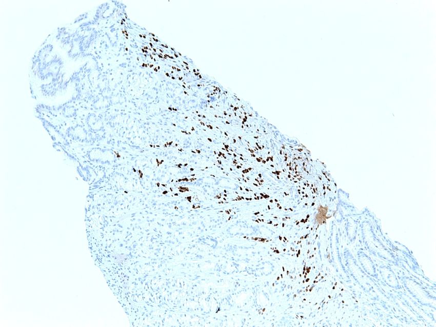
ago. She had undergone a right mastectomy, and her sentinel lymph nodes, which were
                                   sampled during surgery, were negative for metastases. Previously, she had been treated with
                                   adjuvant anastrozole for five years, and yearly mammograms had been negative for recurrence.

                                   One year prior to this presentation, she was evaluated at our hospital for similar complaints of
                                   nausea and vomiting. A computed tomography (CT) scan of her abdomen and pelvis on
                                   admission revealed a mass-like thickening of the gastric antrum and distension of the proximal
                                   stomach, as illustrated in Figure 1.

                                     FIGURE 1: A computed tomography (CT) scan of the abdomen
                                     and pelvis showing a dilated stomach with gastric outlet
                                     obstruction.

                                   An upper endoscopy (EGD) was performed, which revealed esophagitis and gastric stenosis.
                                   This was dilated using a through-the-scope controlled radial expansion (CRE) balloon (Boston
                                   Scientific Inc., MA, US) to a maximum balloon size of 12 mm without fluoroscopic guidance.
                                   Biopsy of the gastric stenosis revealed gastric mucosa of antral type with minimal chronic
                                   inactive gastritis. No morphologic evidence of a Helicobacter pylori infection was detected. The
                                   patients’ symptoms of nausea and vomiting improved following balloon dilation. She was
                                   subsequently discharged on a daily proton pump inhibitor.

2019 Mohy-ud-din et al. Cureus 11(4): e4533. DOI 10.7759/cureus.4533                                                           2 of 6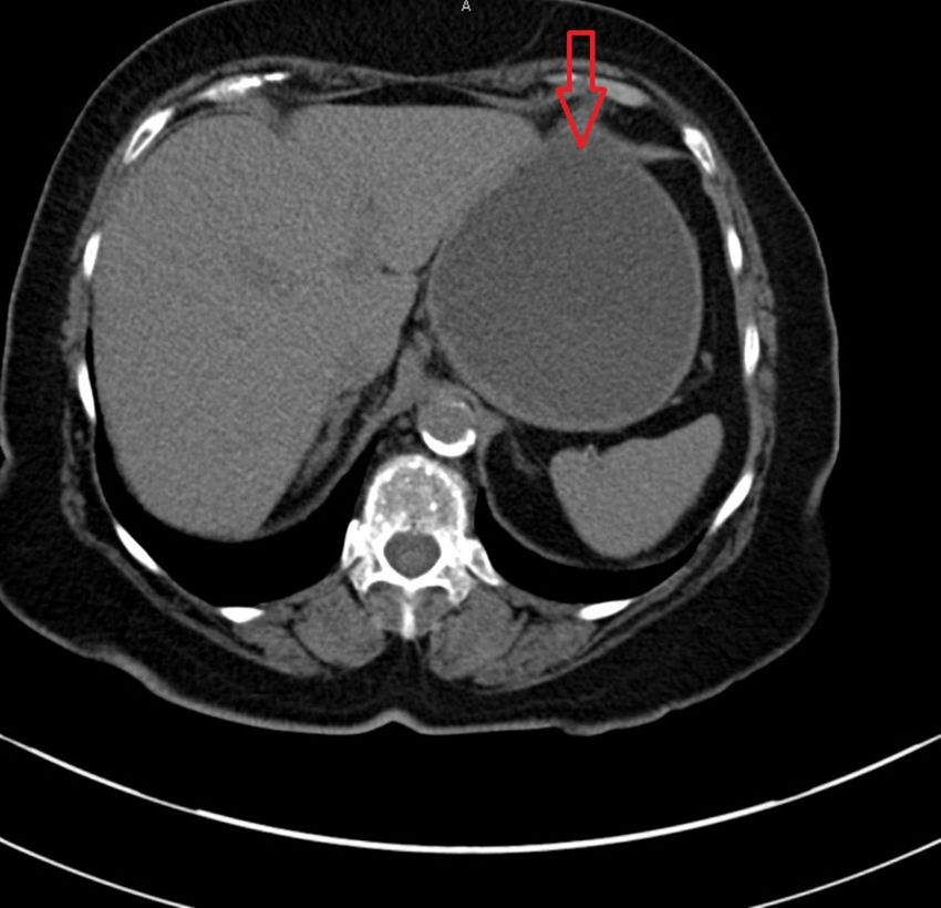
The patient underwent endoscopic ultrasound (EUS) 12 weeks later. Gastric stenosis was found
at the pylorus and duodenal bulb, which was dilated again with a CRE balloon to a maximum
dilation of 13.5 mm. Diffuse wall thickening of the antrum of the stomach was visualized
endosonographically. The gastric wall measured up to 11 mm in thickness. Thickening within
the deep mucosa, submucosa, and muscularis propria was noted. EUS-guided biopsies were
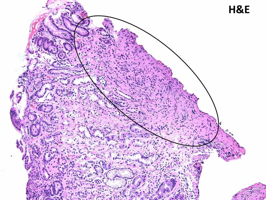
taken, which revealed invasive poorly differentiated metastatic breast adenocarcinoma, as
shown in Figure 2.
FIGURE 2: Histological findings from the gastric biopsy
specimen: Encircled area with H&E staining revealing
infiltration by poorly differentiated adenocarcinoma cells.
H&E: Hematoxylin and eosin
Tumor immunohistochemistry and morphology revealed ER+, PR+, and HER2- negative lobular
breast adenocarcinoma as demonstrated in Figure 3 and Figure 4.
2019 Mohy-ud-din et al. Cureus 11(4): e4533. DOI 10.7759/cureus.4533 3 of 6FIGURE 3: Immunohistochemical examination of the biopsy of
the adenocarcinoma of the lobular breast, demonstrating ER
positivity.
ER: Estrogen receptor
2019 Mohy-ud-din et al. Cureus 11(4): e4533. DOI 10.7759/cureus.4533 4 of 6FIGURE 4: Immunohistochemical examination of the biopsy of
the adenocarcinoma of the lobular breast, demonstrating PR
positivity.
PR: Progesterone receptor
Discussion
This case highlights the importance of keeping a broad differential diagnosis for cases
presenting with gastric outlet obstruction, especially in patients with a history of cancer. A
diagnosis of breast cancer metastases to the gastrointestinal tract is often challenging because
of its low incidence. Distinguishing between metastatic breast carcinoma and primary gastric
adenocarcinoma cannot always be done using histological examination alone, and superficial
biopsies may not be sufficient. Invasive lobular breast carcinoma and primary gastric cancer
can have similar histopathology, as both can show signet ring cells. Immunohistochemistry is
needed to make a distinction between the two, based on staining for estrogen and progesterone
receptors [5]. As in our patient, the initial endoscopic mucosal biopsy reported chronic inactive
gastritis, whereas a EUS-guided biopsy then revealed metastasis from breast cancer.
An important differential diagnosis for patients with concurrent gastric and breast
adenocarcinoma includes hereditary diffuse gastric cancer (HDGC), which is an inherited
cancer syndrome associated with an increased risk for primary gastric cancer and lobular breast
carcinoma and is characterized by a poor prognosis. CDH1 gene mutations are frequently
associated with HDGC and patients with this syndrome may benefit from genetic counseling.
Ulmer et al. described a similar case to ours where previous gastric mucosal biopsies had been
negative; however, a EUS-guided fine needle aspiration clinched the final diagnosis of
metastatic lobular breast carcinoma causing gastric outlet obstruction [6]. Of note, the EUS-
guided biopsy in our case was a mucosal biopsy and not a fine needle aspiration (FNA) biopsy.
It is vital to distinguish between breast cancer metastasis to the stomach and primary gastric
cancer because treatment for the metastatic tumor usually involves systemic chemotherapy
rather than a local treatment for gastric lesions [7]. Taal et al. reported that in a large series of
2000 endoscopic examinations, biopsies were negative in 20% patients with gastric metastases,
which supports the notion that the differential diagnosis of the disease is an important strategy
[8].
Conclusions
Although case reports of breast cancer metastasis causing GOO do exist in the literature, our
case emphasizes the need to be diligent and thorough with a low threshold for EUS-guided
biopsies if the initial endoscopic biopsy is inconclusive. This is critical in order to avoid delay in
care for a patient with metastatic carcinoma.
Additional Information
Disclosures
Human subjects: Consent was obtained by all participants in this study. Conflicts of interest:
In compliance with the ICMJE uniform disclosure form, all authors declare the following:
Payment/services info: All authors have declared that no financial support was received from
any organization for the submitted work. Financial relationships: All authors have declared
2019 Mohy-ud-din et al. Cureus 11(4): e4533. DOI 10.7759/cureus.4533 5 of 6that they have no financial relationships at present or within the previous three years with any
organizations that might have an interest in the submitted work. Other relationships: All
authors have declared that there are no other relationships or activities that could appear to
have influenced the submitted work.
Acknowledgements
The authors would like to acknowledge Qiuhong Zhang for her help in obtaining histopathology
images for this case report.
References
1. Shone DN, Nikoomanesh P, Smith-Meek MM, Bender JS: Malignancy is the most common
cause of gastric outlet obstruction in the era of H2 blockers. Am J Gastroenterol. 1995,
90:1769-1770.
2. Awan A, Johnston DE, Jamal MM: Gastric outlet obstruction with benign endoscopic biopsy
should be further explored for malignancy. Gastrointest Endosc. 1998, 48:497-500.
3. Taal BG, den Hartog Jager FC, Steinmetz R, Peterse H: The spectrum of gastrointestinal
metastases of breast carcinoma: II. The colon and rectum. Gastrointest Endosc. 1992, 38:136-
141.
4. Pectasides D, Psyrri A, Pliarchopoulou K, et al.: Gastric metastases originating from breast
cancer: report of 8 cases and review of the literature. Anticancer Res. 2009, 29:4759-4763.
5. Koike K, Kitahara K, Higaki M, Urata M, Yamazaki F, Noshiro H: Clinicopathological features
of gastric metastasis from breast cancer in three cases. Breast Cancer. 2014, 21:629-634.
10.1007/s12282-011-0284-3
6. Ulmer LL, Cormier I, Jha LK, Singh S, Fisher KW, Hewlett AT: Use of endoscopic ultrasound in
a diagnostic dilemma: metastatic breast cancer to the stomach. Case Rep Gastrointest Med.
2018, 2018:2820352. 10.1155/2018/2820352
7. Kita M, Furukawa M, Iwamuro M, et al.: Breast cancer metastasis to the stomach that was
diagnosed after endoscopic submucosal dissection. Case Rep Gastrointest Med. 2016,
2016:2085452. 10.1155/2016/2085452
8. Taal BG, Peterse H, Boot H: Clinical presentation, endoscopic features, and treatment of
gastric metastases from breast carcinoma. Cancer. 2000, 89:2214-2221.
2019 Mohy-ud-din et al. Cureus 11(4): e4533. DOI 10.7759/cureus.4533 6 of 6You can also read