Updated Guidelines to the Standards for Recording Human Remains - Editors: Piers D Mitchell and Megan Brickley
←
→
Page content transcription
If your browser does not render page correctly, please read the page content below
Updated Guidelines to the
Standards for Recording Human Remains
Editors: Piers D Mitchell and Megan Brickley
Updated Guidelines to the Standards for Recording
Human Remains
Editors: Piers D Mitchell and Megan Brickley

Contents
The Contributors                                                                                                                         3

1      Introduction                                                                                                                      5
       Piers D Mitchell and Megan Brickley

2      Compiling a skeletal inventory: articulated inhumed bone                                                                          7
       Megan Brickley

3      Recording and analysing the human dentition                                                                                      10
       Daniel Antoine

4      Compiling a skeletal inventory: cremated human bone                                                                              14
       Jacqueline I McKinley

5      Compiling a skeletal inventory: disarticulated and commingled remains                                                            20
       Jacqueline I McKinley and Martin Smith

6      Guidance on recording age at death in adult human skeletal remains                                                               25
       Linda O’Connell

7      Estimation of juvenile age at death                                                                                              30
       Jo Buckberry and Megan Brickley

8      Undertaking sex assessment                                                                                                       33
       Megan Brickley and Jo Buckberry

9      Guidance on recording ancestry in adult human skeletal remains                                                                   35
       Linda O’Connell

10     Metric and non-metric studies of archaeological human bone                                                                       39
       Sonia Zakrzewski

Front cover image Excavation of human burials from a medieval Augustinian friary in Cambridge. Image courtesy of the Cambridge
Archaeological Unit.

ISBN 978-0-948393-27-3

                                                                      Updated Guidelines to the Standards for Recording Human Remains    1
11 Guidance on recording palaeopathology (abnormal variation) 44
Charlotte Roberts
12 Recording of interpersonal violent trauma 49
Louise Loe
13 Sampling guidelines for bone chemistry 52
Mike Richards
14 Sampling human remains for evidence of intestinal parasites 54
Piers D Mitchell
15 After the osteological report: the long-term fate of skeletal collections 57
Simon Mays
Appendices 60
List of illustrations
Chapter 3 Figure 3.1 Upper right first molar destroyed by tooth decay 12
Chapter 4 Figure 4.1 Remains of urned cremation burial 15
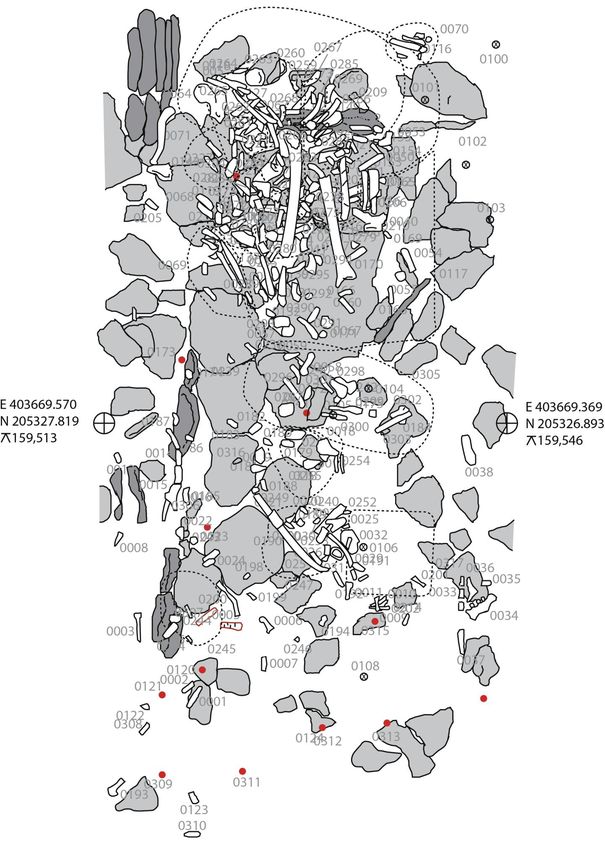
Chapter 5 Figure 5.1 Fragmented and commingled skeletal remains 21
Chapter 10 Figure 10.1 Example of ASU UM parastyle 39
Chapter 12 Figure 12.1 Peri-mortem sharp force trauma to the inferior mandible 50
Chapter 14 Figure 14.1 Decorticated roundworm egg 55
Appendix 1a & b Recording sheet for infant human remains 60
Appendix 2 Recording sheet for juvenile human remains 62
Appendix 3a & b Recording sheet for adult skeletal remains 63
2 Updated Guidelines to the Standards for Recording Human Remains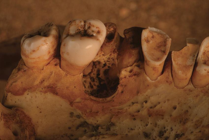
The contributors

Daniel Antoine
Daniel is the British Museum’s Curator of Physical Anthropology, with responsibility for the Museum’s collection of human
remains. Before joining the Museum in 2009, Daniel was at the Institute of Archaeology, University College London, where
he gained his PhD in 2001. He has published widely on dental anthropology, his main area of interest, and bioarchaeology,
including several books: Egyptian Mummies: Exploring Ancient Lives (2016) with Marie Vandenbeusch, Ancient Lives,
New Discoveries: Eight Mummies, Eight Stories with John Taylor (2014) and Regarding the Dead: Human Remains in the
British Museum (2014) with Alexandra Fletcher and J D Hill. He will be president of the Dental Anthropology Association
from 2019–2021, and is an Honorary Senior Research Fellow at the Institute of Archaeology, University College London.

Jo Buckberry
Jo completed her PhD, on later Anglo-Saxon funerary archaeology and osteology, at the University of Sheffield in 2004.
She joined the University of Bradford later that year, where she is now a Reader and programme leader for the MSc in
Human Osteology and Palaeopathology. She continues her research into Anglo-Saxon funerary archaeology, from a
bioarchaeological perspective, alongside research into age estimation and sex assessment, aspects of palaeopathology,
and evidence of violence-related trauma. She is currently analysing the human remains from Stirling Castle.

Megan Brickley
Megan is full professor in the Department of Anthropology, McMaster University, Canada and holds a Tier One Canada
Research Chair in the Bioarchaeology of Human Disease. Research interests include use of palaeopathology in
bioarchaeology and interdisciplinary research. Over the years Megan has undertaken research on a wide range of
bioarchaeological and forensic anthropological projects, including leading analysis of the human bone from St Martin’s
Birmingham, but most research focuses on metabolic bone diseases. Megan is co-author of The Bioarchaeology of
Metabolic Bone Disease (2008) and numerous papers on age-related bone loss, scurvy and vitamin D deficiency.

Louise Loe
Louise has been Oxford Archaeology’s head of Heritage Burial Services since 2006. She holds a PhD from the University
of Bristol in Biological Anthropology. She manages the excavation and post-excavation of archaeological burials, dating
from the Mesolithic to early Modern periods, across the country. In this role she has led teams in a variety of projects,
including the recovery and analysis of WWI soldiers from mass graves in Fromelles, Northern France and of a mass grave
of executed Vikings from Ridgeway Hill, Dorset. She has contributed to numerous site monograph publications and has
recently published on protocols for analysing peri-mortem trauma, trauma patterns in WWI soldiers killed in action and the
role of anthropology in identifying the soldiers from Fromelles.

Simon Mays
Simon gained his PhD at the Department of Archaeology, University of Southampton in 1987. In 1988 he joined English
Heritage as their human skeletal biologist, a post he still holds with the organisation (now called Historic England). Since
1999 Simon has been a visiting lecturer at the Department of Archaeology, University of Southampton, and is also an
Honorary Fellow in the Department of Archaeology, University of Edinburgh. His research interests cover all areas of human
osteoarchaeology, particularly material from England. Simon is the author of The Archaeology of Human Bones (2010,
Routledge) and with Ron Pinhasi is co-editor of Advances in Human Palaeopathology (2008, Wiley).

Jacqueline McKinley
Jackie is currently the Principal Osteoarchaeologist at Wessex Archaeology, and has worked predominantly in the
commercial sector since she graduated in 1981 (Archaeological Sciences, Bradford University) as both a field archaeologist
and osteoarchaeologist. Covering sites across a wide temporal and geographic range throughout the British Isles, she has
produced in excess of 400 osteological and archaeological site reports, and has acted as a visiting lecturer (on cremation)
at several English universities. Her specialist interest lies in mortuary rites, particularly cremation.

                                                              Updated Guidelines to the Standards for Recording Human Remains   3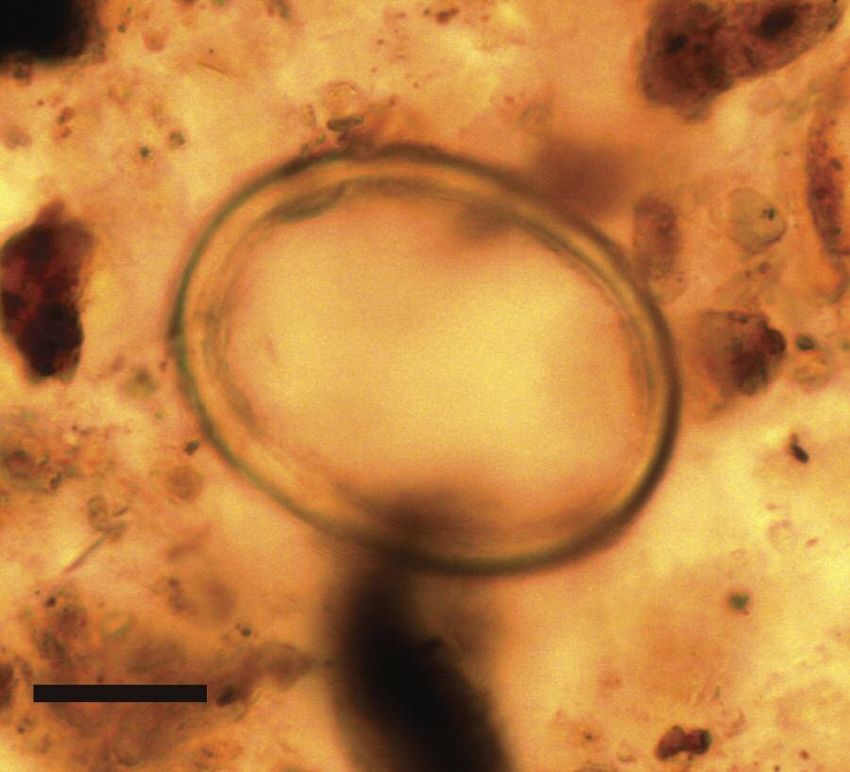
Piers Mitchell
Piers teaches palaeopathology in the Department of Archaeology and Anthropology at the University of Cambridge. He has
trained in medicine, archaeology, medical history, and education. Piers has a strong research interest in ancient infectious
diseases, especially parasites. He has been President of BABAO (2012–2015) and President of the Paleopathology
Association, the worldwide organisation for the study of disease in the past (2015–2017). Piers has published six books,
including Sanitation, Latrines and Intestinal Parasites in Past Populations (2015, Routledge). He is also editor-in-chief of the
book series Cambridge Texts in Human Bioarchaeology and Osteoarchaeology (Cambridge University Press).

Linda O’Connell
Linda is a qualified medical doctor, specialising in forensic and biological anthropology. Together with Prof. Margaret Cox,
she helped establish and direct Bournemouth University’s internationally acclaimed postgraduate provision in Forensic
Archaeology and Anthropology. Consultancy incorporates reporting from archaeological sites, as well as assisting the police
and related professionals with individuals recovered from forensic contexts. In 2009, she was employed by Oxford
Archaeology on the Fromelles Project and has since become a visiting lecturer at Reading University and a member of
the Blake Emergency Services International Response Team. She also works for Silva Legal Services as a Medical Record
Analyst in clinical negligence cases.

Mike Richards
Mike is a professor in the Department of Archaeology at Simon Fraser University in Canada. He obtained his DPhil from
the Research Laboratory for Archaeology and the History of Art at the University of Oxford in 1998, and a BA and MA
from the Department of Archaeology, Simon Fraser University, Canada in 1992 and 1994. He specialises in
bioarchaeology, particularly in bone chemical studies, such as stable isotope studies of past human diets. He is a Fellow
of the Royal Society of Canada and a Fellow of the Society of Antiquaries.

Charlotte Roberts
Charlotte has been Professor of Archaeology in the Department of Archaeology, Durham University since 2004,
teaching undergraduate and postgraduate students and particularly the MSc in Palaeopathology. A bioarchaeologist,
Charlotte began her career as a State Registered Nurse, subsequently gaining her PhD in Bioarchaeology in 1988
(Bradford). She is a Fellow of the British Academy (from 2014) and is currently President of BABAO. Her books include:
Health and Disease in Britain: from Prehistory to the Present Day (2003), The Bioarchaeology of Tuberculosis: a Global
View on a Re-emerging Disease (2003), The Archaeology of Disease (2005), Human Remains in Archaeology: a
Handbook (2009), and The Global History of Palaeopathology (2012). Her webpage provides fuller details:
https://www.dur.ac.uk/archaeology/staff/?id=163

Martin Smith
Martin is Principal Academic in the Department of Archaeology, Anthropology and Forensic Science at Bournemouth
University. He has particular interests in taphonomic changes and injuries to the skeleton. Martin has worked on a broad
range of prehistoric material but has worked most extensively on collective burials from the Early Neolithic. He is co-author
of People of the Long Barrows: Life, Death and Burial in the Earlier Neolithic (2009) and co-editor of the Routledge
Handbook of the Bioarchaeology of Human Conflict (2013). Martin has published papers ranging from vertebrate
scavenging, ballistic injuries, mummified remains and ethical issues surrounding human skeletal remains.

Sonia Zakrzewski
Sonia obtained her PhD in Biological Anthropology at the University of Cambridge, and is now an Associate Professor of
Archaeology at the University of Southampton, focusing on bioarchaeology. Her main research interests are in
morphological population variation in relation to aspects of human identity, including migration, religion, disability and race
within a variety of regions, including Egypt, the Caribbean and Britain. She has also looked at changes in social identity and
its identification through other aspects of bioarchaeology, such as sexual dimorphism or changes in activity patterning, and
has linked these through funerary archaeology and artistic representation with the wider burial record. From 2014 to 2017
she was Vice-President of the Paleopathology Association.

4   Updated Guidelines to the Standards for Recording Human Remains
1      Introduction
Piers D Mitchell, Megan Brickley

In 2004 the British Association for Biological Anthropology and Osteoarchaeology (BABAO) published the first
edition of Guidelines to the Standards for Recording Human Remains in the Institute for Field Archaeologists
publication series. It was edited by Megan Brickley and Jacqueline McKinley, and its aim was to provide a
guidance document to give specialists in osteoarchaeology and burial archaeology a framework within which
to work while maintaining a high level of professionalism. It was primarily aimed at those engaged in recording
human skeletal remains from commercial excavation projects, so ensuring standardised recording and greater
comparability between the reports of human bone assemblages from different sites. The 2004 guidelines
followed on from guidance on assessment and analytical reports on human remains produced in 2002 and
reprinted in 2004 (Mays et al 2004).

Since that time these guidelines have supported those working in the field and when compiling skeletal reports for their
clients, as well as being of great use to researchers in an academic environment and to museum curators. They have
assisted practitioners to ensure their professional activities meet the BABAO Code of Ethics and Code of Practice. The
guidelines also ensure practitioners can meet Principles 3 and 4 of the CIfA Code of Conduct (CIfA, 2014) regarding the
quality of their work, and the various standards and guidance documents published by CIfA
(http://www.archaeologists.net/codes/cifa). Having guidelines that specify what should be included in a skeletal report helps
to ensure that sufficient time and funding is allocated by clients engaging the services of a commercial archaeological
service.

It should be understood that those osteoarchaeologists in commercial units might not have the funding available to
organise some of the more expensive analyses such as ancient DNA, isotopes or radiological imaging. However, what is
important is that all involved are aware when such analysis can be helpful, and when samples could be stored for analysis
at a later date.

The authors of the 2004 guidelines anticipated that the document would probably have a lifespan of ten to fifteen years
(Brickley 2004), and they were correct. Over the last 14 years there have been advances in research methodology that
have necessitated an update to this volume. Following consultation with the BABAO membership, it was decided to create
updates for each chapter that focus on those advances published since 2004, together with changes in ideas and
approaches over this time. Due to work commitments Jacqueline McKinley was not able to act as editor on this update, so
her role has been taken over by Piers Mitchell. It is fitting that the update should be published once again by the (renamed)
Chartered Institute for Archaeologists (CIfA). This is, in effect, a refresher on all that is cutting edge in the field.

An additional chapter has been added on the topic of sampling human burials for the eggs of parasitic worms that caused
gastrointestinal infection when the individual was alive. This type of analysis has become a more common practice than was
the case ten or twenty years ago.

The volume has passed through an intensive peer review process. Every chapter has been reviewed by at least 15 experts,
some based in Britain and others internationally. This will ensure that the views expressed in the guidelines represent a
broad spectrum of opinions in the field.

This guidance is primarily targeted towards the needs of osteoarchaeologists in Britain, but we also envisage it being of use
to those excavating and analysing human skeletal remains across the world.

                                                              Updated Guidelines to the Standards for Recording Human Remains   5
References BABAO. Code of Ethics. http://www.babao.org.uk BABAO. Code of Practice. http://www.babao.org.uk Brickley, M 2004 ‘Introduction’, in M Brickley and J I McKinley (eds) Guidelines to the Standards for Recording Human Remains, IfA Paper no.7, BABAO/Institute of Field Archaeologists: Reading, 5 CIfA 2014 Code of Conduct. http://www.archaeologists.net/codes/cifa Mays, S, Brickley, M and Dodwell, N 2004 Human Bones from Archaeological Sites: Guidelines for Producing Assessment Documents and Analytical Reports. English Heritage 6 Updated Guidelines to the Standards for Recording Human Remains
2 Compiling a skeletal inventory: articulated inhumed
  bone
Megan Brickley

First questions to be asked of any assemblage of human bone will be: how many individuals are present and how well
preserved is the skeletal material?

With most assemblages, a minimum level of recording of numbers of individuals and levels of preservation set out in Mays
et al (2004) should have been undertaken at the assessment stage. However, for the production of a human bone report
the exact number of individuals present should be calculated, and the condition of the bone of each individual should be
analysed and recorded.

2.1    Completeness

There are many systems for recording the completeness of a skeleton, for example those outlined in Buikstra and Ubelaker
(1994). The system selected will largely depend on the specific research questions to be addressed but, as a minimum,
numbers of each bone type and all major joint surfaces should be recorded in such a way as to allow prevalence of
pathological conditions to be calculated (see Chapter 11). A clear reference should be provided for any system used to
describe the completeness of a skeleton (or the full methodology employed set out in the case of unpublished techniques).
Use of visual recording forms such as those included as appendices of the 2004 version of this document will allow not
only the completeness but also the amount of fragmentation to be recorded.

2.2    Fragmentation

Fragmentation has important implications for the amount of metric data that can be recorded. Systems of recording should
be made clear and should be fully referenced, if applicable, in the final report. In the case of highly fragmented skeletons,
refer to Chapter 5 for aspects of fragmented bone that should be considered. Recording features such as abrasion/erosion
and the characteristics of broken ends may assist in determining the cause of fragmentation in articulated skeletons.

2.3    Surface preparation

Previously it was recommended that Behrensmeyer (1978) was used to record surface preservation, but human bone
weathers differently to animal bone (which tends to have a much denser cortex) and the varied burial environments
encountered within contexts across the British Isles result in different mechanisms acting on the bone. The surface
preservation of bone should be recorded following published guidelines, and the system set out by McKinley (2004) is
recommended, since statements such as ‘the bone was well preserved’ are almost meaningless unless they have been
clearly defined, as there will be discrepancies in the way different researchers apply and interpret such a statement.
Information on the surface preservation of bone is important for interpretations of the prevalence of many pathological
changes in bone, for example periosteal new bone formation.

2.4    Exclusion of skeletons with less than ideal preservation

Recent work has demonstrated that human skeletal remains may be partial and poorly preserved due to underlying
pathological processes (eg, see Brickley and Buckberry 2015). Those undertaking recording of human remains should
consider that exclusion of less well-preserved skeletons may lead to the loss of significant information on pathological
conditions that result in loss of bone density and structure (eg, age-related bone loss, deficiency of vitamin C and D, and
neoplastic conditions). Individuals buried at earlier dates may be more likely to be disturbed in some settings and
stratigraphic data should be carefully considered before decisions on recording are made. Results from investigations that
exclude poorly preserved remains will be biased. Recording using true prevalence rates as recommended by Mays et al
(2004) will allow missing elements to be accounted for during data analysis.

                                                              Updated Guidelines to the Standards for Recording Human Remains   7
2.5 Recording sheets and archiving The use of paper or electronic means for recording skeletal completeness, or a combination of these two media, will depend largely on the circumstances of the individual undertaking the recording. However, the durability of records and their accessibility to future researchers should be carefully considered; rapid computer development has rendered many programmes and operating systems obsolete in recent years. Any system used should allow information on the bones present to be accurately recorded in a format that will allow reporting of the true prevalence of pathological and traumatic lesions, and differentiation between undetermined and ambiguous individuals in evaluation of sex and ancestry (see appropriate chapters of this volume). Generating backups and having ‘disaster management’ plans for digital data should be part of the process of setting up any digital recording system. Records should be prepared in line with current standards and guidance on the archiving of paper and digital data (Brown 2007 http://www.archaeologists.net/sites/default/files/ifa_practice_archives.pdf). Where work is to be deposited in a regional archive, records should also be prepared to local, documented standards. Archiving reports that fall within the grey literature with the Archaeology Data Service (ADS) is considered best practice. A number of recording sheets depicting complete skeletons and individual bones are presented in Buikstra and Ubelaker (1994). Whilst some of these are useful and enable detailed recording of individual elements and features observed on bones, the complete skeleton sheets (both adult and juvenile) are felt to lack the detail useful as a means of recording. An updated set of recording sheets is provided in the appendix of this document (Appendices 1–3), for those wishing to record greater detail. Additional forms for perinatal, early childhood and late childhood cranial bones and skeletal completeness are provided in Chapter 9 of Schaefer et al (2009). 2.6 Visual recording (illustrations) Various means of visual recording are available: photographs, radiographs, professional drawings and sketches. It is recommended that as many visual records as possible are obtained during the recording of skeletal and dental material, although the purpose of such recording, to assist in diagnosis or illustrate a point, should always be kept in mind. Clearly, the extent of this type of recording will depend on factors such as the nature of the assemblage and the research questions posed. However, such recording should be considered a vital part of any project (especially primary recording of skeletal material on a commercial basis). Costings for adequate recording of this nature should always be made whether the project is research or commercially funded. As a minimum, photographs of publishable quality should be obtained for any item discussed in the report produced. Although drawings and photographs produced by professionals are indispensable for final reports, the value of images made by the person undertaking the recording should not be underestimated and photographs of the complete skeleton and individual elements for further reference during the writing of a report can be very valuable. Illustrations form a particularly important part of the archive where skeletal material is to be reburied. Photographs should always be viewed in the format they are to be produced in before being submitted for publication. For example, some of the detail visible on a colour picture may be far less clear if reproduced in black and white. Monochrome photographs are often more appropriate than colour images to illustrate fine surface details, such as cut-marks, abrasions or surface etching. Colour images may, however, illustrate some pathological lesions better than a monochrome image. The possibility of obtaining images from microscopic examination should also be considered. In many instances it may be possible to observe and record the features of interest using light or digital microscopy, and many microscopes have camera attachments or digital recording features. Basic digital microscopes are now priced such that they will be accessible to many organisations. At the assessment stage of a project the possibility that microscopic examination of material may be required should be considered. Early planning will allow funds to be requested and/or suitable equipment to be located prior to the start of recording. Useful information on procedures for obtaining various types of visual record are contained in Buikstra and Ubelaker (1994, 10–14), Bruwelheide et al (2001) and White (2000, 517–518). However, the quantity of images – particularly radiographic – 8 Updated Guidelines to the Standards for Recording Human Remains
required will normally be less as these guidelines assume that material will be reburied after primary analysis and this is not
normal practice with British archaeological material.

Additional information on visual recording of various types can be found in Williams (2001). Full visual recording will enhance
both the quality of the report or paper published, and form a valuable resource in the archive. The need for long-term
accessibility and practicalities of archiving visual records of various types should be considered at the planning stages of
any project.

Long-term archiving of visual records should be considered; as set out in Section 2.5, plans should be made at the start of a
project.

2.7    3D laser scanning

Recent projects, such as Digitised Diseases, run from the Biological Anthropology Research Centre, University of Bradford,
show the ways in which technological developments allow the recoding of detailed information on pathological and
taphonomic changes to bone. Digital archives such as that created as part of the Digitised Diseases project also allow
widespread access to material without causing further damage that comes from handling bone.
http://www.digitiseddiseases.org/alpha/.

Technologies such as 3D printing of scanned items are developing rapidly. At present the quality of prints is not sufficient to
accurately record pathological and taphonomic change, but this is likely to change in the future.

References

Behrensmeyer, A K 1978 ‘Taphonomic and ecologic information from bone weathering’ Paleobiology 4: 150–162

Brickley, M B and Buckberry, J 2015 ‘Picking up the pieces: utilizing the diagnostic potential of poorly preserved remains’ International
Journal of Paleopathology 8: 51–54

Brown, D H 2007 'Archaeological Archives: A Guide to Best practice in Creation, Compilation, Transfer and Curation' Archaeological
Archives Forum

Bruwelheide, K S, Beck, J and Pelot, S 2001 ‘Standardized protocol for radiographic and photographic documentation of human
skeletons’, in E Williams (ed.) Human remains: conservation, retrieval and analysis. Proceedings of a conference held in Williamsburg,
VA, Nov 7–11th 1999, BAR International Series 934, Archaeopress: Oxford 53–165

Buikstra, J E and Ubelaker, D H (eds) 1994 Standards for data collection from human skeletal remains, Arkansas Archeological Survey
research series No. 44: Fayetteville, AR

Mays, S, Brickley, M and Dodwell, N 2004 Human bones from archaeological sites: guidelines for producing assessment documents
and analytical reports. Centre for Archaeology Guidelines English Heritage/BABAO: London

McKinley, J 2004 ‘Compiling a skeletal inventory: disarticulated and co-mingled remains’ in M Brickley and J I McKinley (eds)
Guidelines to the standards for recording human remains, IfA Paper no.7, BABAO/Institute of Field Archaeologists: Reading, 5

Schaefer, M, Black, S and Scheuer, L 2009 Juvenile osteology: A laboratory and field manual. Academic Press: Burlington, MA.

White, T 2000 Human osteology, second edition. Academic Press: New York

Williams, E 2001 Human remains: conservation, retrieval and analysis. Proceedings of a conference held in Williamsburg, VA, Nov
7th–11th 1999, BAR International Series 934, Archaeopress: Oxford

                                                                    Updated Guidelines to the Standards for Recording Human Remains     9
3 Recording and analysing the human dentition Daniel Antoine Subtle morphological variations can be used to identify and side individual teeth (see Hillson 1996, 14–67; Lease 2016). The human dentition is usually comprised of 20 deciduous (or milk) teeth that are gradually replaced with 32 permanent teeth. The permanent dentition starts to form just before birth and ends with the development and eruption of the third molars in the late teens to early twenties. During long periods of a child’s life, both deciduous and permanent teeth are present in various states of development (see Hillson 1996 for recording dental development). The number of teeth in an adult dentition can occasionally vary. In some, teeth such as the third permanent molars can be congenitally absent. Disease, trauma or cultural practices may also lead to the loss of teeth during life, whilst some are lost post-mortem. Extra (supernumerary) teeth are less common and usually have a highly irregular form (Nelson 2016). Although most of the methods employed to identify and label teeth have remained the same since the last edition, many of the approaches used to analyse and interpret the human dentition and its supporting alveolar bone have been re-evaluated and improved upon (eg, Irish and Scott 2016). 3.1 Inventories Dental inventories are used to record the presence of individual teeth. As teeth can be lost pre- or post-mortem, the presence of their supporting structures (ie, tooth positions or the root sockets into which they may have once fitted) should also be recorded when observable. Most systems divide teeth into four quadrants that mirror each other: the maxillary right, maxillary left, mandibular left and mandibular right (see van Beek 1983, 3–6; Hillson 1996, 6–12). The upper and lower quadrants are divided into left and right by an imaginary line that passes between the central incisors. When all teeth are present and developed, each quadrant of the permanent dentition is made up of two incisors, one canine, two premolars and three molars. Many recording systems number the teeth in each quadrant from one to eight respectively from the central incisor to the third molar. In the deciduous dentition, each quadrant is made up of two incisors, one canine and two molars labelled from ‘a’ to ‘e’ or 1 to 5 respectively from the central incisor to the second molar. 3.2 Labelling systems Most labelling systems make use of these numbers or letters to avoid using lengthy tooth names. Quadrants are simply identified by adding ‘U’ for upper or ‘L’ for lower, with ‘L’ and ‘R’ used to distinguish left and right. Hence, ‘UR3’ would represent the upper right permanent canine and ‘LLd’ (or ‘dec. LL4’) used to denote the lower left first deciduous molar. Alternatively, teeth can be identified by their initials, with ‘I1’ and ‘I2’ for the central and lateral incisors, ‘C’ for the canine, ‘P1’ and ‘P2’ for the first and second premolars (also labelled ‘P3’ and ‘P4’ in some evolutionary systems), and ‘M1’, ‘M2’ and ‘M3’ for the first, second and third molars respectively (eg, ‘ULP2’ represents the upper left second premolar and ‘dec. LRC’ the deciduous lower right canine). This system is used in most publications (eg, American Journal of Physical Anthropology; Hillson 2014; Irish and Scott 2016). Many variants exist and, as with all recording methods, great care should be taken to note the labelling system used. Permanent and deciduous teeth should also be clearly distinguished, particularly when numbers (and not letters) are used to identify the deciduous teeth. The Zsigmondy system (van Beek 1983, 5; Hillson 1996, 8–9) provides a shorthand alternative that is particularly useful when labelling bags. As above, the teeth of each quadrant are identified using the 1–8 numbering for the permanent dentition and a–e lettering for the deciduous teeth. Quadrants are simply identified by framing the number or letter with a vertical and horizontal bar. If the number or letter is below the horizontal bar, it is a lower tooth, and when above it, an upper tooth. As the dental arcade is being observed head-on in the correct anatomical position, if the vertical bar is to the right, it is a right tooth and vice versa. An upper right permanent canine would be labelled: 3 These labelling systems cannot be inserted into a database and the FDI (Fédération Dentaire International) system provides the most suitable computer-friendly labelling method. Here, the first number denotes the quadrant (numbered clockwise from the upper right) and the second number identifies the tooth (as above, 1–8 for permanent and 1–5 for deciduous). For 10 Updated Guidelines to the Standards for Recording Human Remains
example, 16 represents the upper right first permanent molar and 62 the upper left deciduous lateral incisor. As with the
Zsigmondy system, the viewer is observing the body facing the skull, with left and right reversed from the viewer’s point
of view:

                                Upper right permanent               Upper left permanent

      1      1     1       1     1      1      1      1             2       2      2       2      2       2      2       2
      8      7     6       5     4      3      2      1             1       2      3       4      5       6      7       8

      4      4     4       4     4      4      4      4             3       3      3       3      3       3      3       3
      8      7     6       5     4      3      2      1             1       2      3       4      5       6      7       8

                                Lower right permanent               Lower left permanent

                                Upper right deciduous               Upper left deciduous

                           5     5      5      5      5             6       6      6       6      6
                           5     4      3      2      1             1       2      3       4      5

                           8     8      8      8      8              7      7      7       7      7
                           5     4      3      2      1              1      2      3       4      5

                                Lower right deciduous               Lower left deciduous

The FDI system allows each tooth to have an easily determined and unique number, making it possible to calculate tooth-
specific prevalence rates. Alternatively, Buikstra and Ubelaker’s (1994) numbering system labels the permanent dentition
from 1 to 32 and the deciduous dentition from 51 to 70.

Recording the presence or absence of individual teeth does not usually suffice, as teeth are often absent or non-recordable
for a number of reasons. Forms should ideally differentiate between ante- and post-mortem loss, and record the number
and position of all observable teeth. The simplest recording forms strike through the tooth to indicate post-mortem loss.
Buikstra and Ubelaker (1994, 47–49) recommend the following codes: 1: ‘Present, but not in occlusion’; 2: ‘Present,
development complete, in occlusion’; 3: ‘Missing, with no associated alveolar bone’; 4: ‘Missing, with alveolus resorbing or
fully resorbed: pre-mortem loss’; 5: ‘Missing, with no alveolar resorption: post-mortem loss’; 6: ‘Missing, congenital absence’;
7: ‘Present, damage renders measurement impossible, but other observations are recorded’; 8: ‘Present, but unobservable
(eg, deciduous or permanent tooth in crypt)’. Codes 3–6 can be used to calculate the prevalence of ante-mortem tooth loss
as long the codes are interpreted in a manner that allows for such calculations (eg, code 3 should be equivalent to ‘no
data’). The presence of the supporting alveolar bone is, however, often recorded separately by tooth position (or the root
sockets into which they once fitted) in order to determine the prevalence of periapical cavities (see below).

3.3       Dental disease

When appropriate, dental disease (see Hillson 2005, 286–318; Hillson 2008b; Nelson 2016), dental measurements (see
Hillson 1996; 2005) and, should time allow, morphological crown and root traits (see Scott, Maier and Heim 2016) should be
recorded. Buikstra and Ubelaker’s code 7 (above) raises a very important point; poor preservation, as well as advanced
dental wear, can affect some observations. Calculating the prevalence of any pathological changes (eg, hypoplasia, caries)

                                                               Updated Guidelines to the Standards for Recording Human Remains   11
should take preservation (ie, how complete teeth are) and wear into account (see methods for recording wear in Hillson 1996; Burnett 2016) as these have an impact on the surfaces observed (Hillson 2001). Individuals with highly worn crowns, for example, are unlikely to show signs of occlusal surface caries and most hypoplastic defects are no longer visible if the enamel surface is worn (eg, via brushing or cleaning), absent (ie, attritional wear) or covered by calculus. Overzealous post- excavation cleaning can also damage the enamel surface or remove dental calculus, which has become a valuable source of biomolecular information (eg, Warinner et al 2014; for recording calculus see Hillson 1996, 255–260; Hillson 2008b). With regard to hypoplasia, the degree of magnification is also likely to have a major impact on the number of defects observed and one may question whether it is possible to record these in a way that allows for comparisons between populations (see detailed review in Hillson 2014). The prevalence of dental pathology should not be calculated by aggregating all teeth within an assemblage but should – as a minimum – be divided by tooth type or class and, where possible, be subdivided into wear groups to account for different wear patterns between assemblages. Summaries that report the total number of teeth affected within a population unfortunately combine tooth classes with differing wear patterns and susceptibilities to disease. This is particularly problematic for caries, with the deep fissures and crevices present in posterior teeth, particularly molars, making them more susceptible to tooth decay (see discussion in Temple 2016). To provide greater specificity and generate comparable prevalence data, tooth decay should – time permitting – ideally be recorded by tooth surface and take into account morphological differences between tooth classes (see Hillson 2001; 2008a; 2008b). When all teeth are grouped together and tooth decay is presented as a summary of total tooth count, assemblages with higher numbers of posterior teeth are likely to be biased when compared to samples with higher numbers of anterior teeth, or differing wear patterns. Without considering these factors, differences in dental disease prevalence may simply reflect tooth-class preservation bias or variations in ante-mortem tooth loss, age distribution, patterns of dental wear or tooth-surface preservation. Though time consuming, recording carious lesions by tooth surface (eg, the number of occlusal surface caries in lower first molars with an observable occlusal fissure system) allows such differences to be taken into account when comparing assemblages. The bone supporting teeth should also be carefully scored for periodontal disease (see Hillson 1996, 260–269; Kerr 1988), which is often linked to root exposure, root surface caries and ante-mortem tooth loss (Nelson 2016). The presence of periapical cavities or voids should also be recorded but, as they do not always involve an externally visible sinus, their prevalence can be hard to establish. All root sockets should be examined by carefully lifting the teeth out (something that is not always possible) or by using radiographs to image the root apices (should time and finances allow). Their location and size, as well as the appearance of the cavity wall and (when present) sinus margins, should be carefully documented and used to distinguish abscesses from granulomas and cysts (Figure 3.1) (see Hillson 2008b; Ogden 2008; Nelson 2016). Figure 3.1 Upper right first molar destroyed by tooth decay, with a periapical cavity in the underlying bone. The roughened appearance of the periapical cavity wall indicates an ongoing infection and identifies it as an abscess (rather than a smooth- walled granuloma or cyst). Skull from site 3-J-23, Grave 7, 4th Nile Cataract, Sudan. Medieval period, 4th–15th century AD. Image by R. Whiting, courtesy of the Trustees of The British Museum 12 Updated Guidelines to the Standards for Recording Human Remains
References

Buikstra, J E and Ubelaker, D H (eds) 1994 Standards for Data Collection from Human Skeletal Remains, Arkansas Archeological
Survey Research Series No. 44: Fayetteville, AR

Burnett, S 2016 ‘Crown wear: identification and categorization’ in J D Irish and G R Scott (eds) A Companion to Dental Anthropology.
Wiley Blackwell: Oxford 416–432

Hillson, S 1996 Dental Anthropology. Cambridge University Press: Cambridge

Hillson, S 2001 ‘Recording dental caries in archaeological human remains’ International Journal of Osteoarchaeology 11: 249–289

Hillson, S 2005 Teeth, second edition. Cambridge University Press: Cambridge

Hillson, S 2008a ‘The current state of dental decay’, in J D Irish and C G Nelson (eds) Technique and Application in Dental
Anthropology. Cambridge University Press: Cambridge, 111–135

Hillson, S 2008b ‘Dental pathology’ in M A Katzenberg and S R Saunders (eds) Biological Anthropology of the Human Skeleton,
second edition. Wiley Blackwell: Oxford, 301–340

Hillson, S 2014 Tooth Development in Human Evolution and Bioarchaeology. Cambridge University Press: Cambridge

Irish, J D and Scott, G R (eds) 2016 A Companion to Dental Anthropology. Wiley Blackwell: Oxford

Kerr, N W 1988 ‘A method for assessing periodontal status in archaeologically derived material’ Journal of Paleopathology 2: 67–78

Lease, L R 2016 ‘Anatomy of individual teeth and tooth classes’ in J D Irish and G R Scott (eds) A Companion to Dental Anthropology.
Wiley Blackwell: Oxford, 94–107

Nelson, G C 2016 ‘A host of other dental diseases and disorders’ in J D Irish and G R Scott (eds) A Companion to Dental
Anthropology. Wiley Blackwell: Oxford, 465–483

Ogden, A 2008 ‘Advances in the palaeopathology of teeth and jaws’ in R Pinhasi and S Mays, Advances in Human Palaeopathology.
Wiley: Chichester, 283–307

Scott, G R, Maier, C and Heim, K 2016 ‘Identifying and recording key morphological (nonmetric) crown and root traits’ in J D Irish and
G R Scott (eds) A Companion to Dental Anthropology. Wiley Blackwell: Oxford, 247–264

Temple, D H 2016 ‘Caries: the ancient scourge’ in J D Irish and G R Scott (eds) A Companion to Dental Anthropology. Wiley Blackwell:
Oxford, 433–44

van Beek, G C 1983 Dental Morphology: an Illustrated Guide. Wright: Oxford

Warinner, C, Rodrigues, J F M, Vyas, R, Trachsel, C, Shved, N, Grossmann, J et al 2014 ‘Pathogens and host immunity in the ancient
human oral cavity’ Nature Genetics 46: 336–44

                                                                   Updated Guidelines to the Standards for Recording Human Remains       13
4 Compiling a skeletal inventory: cremated human bone Jacqueline I McKinley Since the 2004 edition of this volume there has been a marked increase of interest in cremated bone and the mortuary rite of cremation in the UK. This has led to the publication of several volumes of work dedicated to cremated/burnt human remains, their context and/or the mortuary rite (eg, Artelius and Svanbery 2005; Davies and Mates 2005; Schmidt and Symes 2008; 2015; Thompson 2015a), adding to the small number of existing volumes of this nature (eg, Holck 1986; Lange et al 1987; McKinley 1994a; Sigvallius 1994; Smits et al 1997). There has, in addition, been a growing frequency of contributions on the subject included in more holistic publications on mortuary studies (eg, Tarlow and Nilsson Stutz 2013). The basic aims of the osteological analysis remain much the same as they have been in the past. Technological advancement and increased accessibility to specialist equipment have led to new techniques being applied to cremated bone. This has broadened the scope of study and had some radical effects on our understanding of the use of the mortuary rite in antiquity. Whilst the specialist aims to recover the basic osteological data pertaining to the cremated individual, they also seek to recover information relevant to the technological aspects and rites of cremation. The systematic recording of data from individual cremation-related deposits enables subsequent analysis to detect variations and similarities in the rite, which may be influenced by the age or sex of the individual, or cultural, temporal or geographic factors. The ancient mortuary rite of cremation was a complex and multi-faceted mode of disposal of the dead. It had the potential to create a variety of deposit and feature types for which we may recover archaeological evidence (eg, McKinley 2013). Consequently, analysis of the cremated remains by an osteologist is inextricably linked with the context of origin. The form and nature of the archaeological deposit will affect the condition of the cremated bone and both data sets (collected in the field and the laboratory) are vital in interpretation of the type of deposit represented. A range of cremation-related features and deposits is commonly encountered in close association as part of the ‘mortuary landscape’, but the ‘transportable’ nature of cremated remains means that some deposits are, and others potentially may be, found outside this arena (Eriksson 2005; McKinley 1994b, 70–71; 2006; Metcalf and Huntington 1991, 102; Oestigaard 1999; van Gennep 1977, 152). Analysis of cremated remains also requires an understanding of the cremation process. Modern crematoria offer the most effective and efficient environment in which cremation is undertaken, but it is also important to consider those factors which may have influenced the equally sophisticated but potentially less controllable environment of an open pyre in the past, including accidental or deliberate curtailing of the process/cooling of the pyre, and secondary (ie, post-cremation) rites (DeHaan 2008; McKinley 1994a, 72–76; 2016, 19–26; Symes et al 2008; Thompson 2015b; Walker et al 2008). For an overview of the weights of bone recovered by various workers from modern crematoria see Gonçalves 2012; Gonçalves et al 2013. 4.1 Recording For those working with cremated remains for the first time (and even thereafter), it is advisable to have a full skeleton accessible for comparative purposes. Correct identification of the skeletal element represented by small, heat-altered fragments can be difficult and it is always wise to check to avoid mis-identification that may contribute to subsequent misinterpretation. Section 4.3 in the 2004 edition presented the four categories of ‘identifiable’ bone; within these categories individual elements should be recorded as closely as possible, such as ‘right nasal process’, ‘left petrous temporal (anterior portion)’, ‘proximal foot phalanx head and shaft’, together with data pertaining to age/sex/pathological lesions and unusual fragmentation or colouring (outside the white of full oxidation). The occasional use of radiographs and computerised tomography (CT) scans for the initial examination of the remains of urned cremation burials (lifted en masse from site for laboratory excavation) prior to excavation of the remains has been undertaken for some years (eg, Anderson and Fell 1995). CT scans are of greater assistance than plain film radiographs, although ready and frequent access to the necessary expensive equipment is likely to be severely limited for many osteologists, especially in the commercial sector; often one has to engage with an accommodating hospital department 14 Updated Guidelines to the Standards for Recording Human Remains
which may be happy to undertake small numbers of urns but which may balk at several dozen. It should be recognised that
CT scanners in National Health Service premises are naturally prioritised for patients. There are times when this technique
can be of particular assistance, most pertinently where the soil acidity (eg, in the case of clay, silty clays, siliceous sands,
peat) can cause destruction of much of the trabecular bone; the latter will be apparent in the CT scan and visible during
excavation but would crumble to dust on excavation (eg, Harvig 2015). Elsewhere, such as when an unusual vessel was
used as the container for burial, a CT scan will give a view of the contents to assist with recording in excavation (Figure 4.1).
Such images alone cannot, however, provide answers to all the aims of analysis.

A

B

                                                    Figure 4.1 Remains of urned cremation burial from Grave 42001, East Kent Access
                                                    Road: A) vessel showing broken neck through which bone was inserted into vessel,
                                                    B) computer tomography (CT) scan of vessel prior to excavation of contents (by kind
                                                    permission Oxford Wessex Archaeology)

In addition to the level of disturbance/truncation, derived from the archaeological records, a note of the condition of the
bone itself needs to be made. As with unburnt bone, this is primarily affected by the burial environment. Well-preserved
bone will have sharply defined surface morphology, but trabecular bone suffers preferentially in an acidic burial
environment, often crumbling to a ‘dust’ fraction, whilst compact bone will appear progressively more eroded and ‘chalky’
(slight/moderate/heavy).

4.2    Demographic data

A major problem with cremated remains – with both age and sex estimation of adults – is the characteristic incomplete
recovery of bone for burial by those performing the rite and the frequent absence of the skeletal elements most useful to
the osteologist. The condition of the bone and level of disturbance to the deposit (with associated loss/increased
fragmentation) are also major factors influencing our ability to estimate both the age and assess the sex of an individual.
It will generally be possible to at least distinguish between ‘immature’ (< approximately 18 years of age) and ‘adult’
(> approximately 18 years) bone, and though a substantial minority will inevitably fall within the broader ‘sub-adult/adult’
range (>12 years), age ranges of varying size will be attributable in many instances. The future wider application of
histological ageing methods may help eventually overcome these difficulties (eg, Cox 2000; Cuijpers 1997; Herrmann 1977;
Hummel and Schutkowski 1993), although qualitative rather than quantitative methods need to be employed with some
techniques to overcome the potential effects of shrinkage.

                                                                 Updated Guidelines to the Standards for Recording Human Remains          15
Most assemblages will include a substantial proportion of unsexed adults and even where sex can be indicated confidence levels may vary. The use of osteological data in the analysis of other archaeological data from the site, such as pyre/grave good associations, should always consider this shortfall to ensure the results from such analyses are not potentially misleading. 4.3 Cremation technology Analytical techniques to explore the nature of cremated bone have been developed over many years, much of the analysis being associated with advances in forensic science (Ubelaker 2015). New approaches to understanding the effects of temperature and oxygen supply on the macroscopic (colour, fragmentation, warping – scored as absent/moderate/marked) and microscopic appearance (crystal structure, chemical composition) of the cremated bone have been developed in recent years, with particular emphasis on the latter (Beach et al 2008; Devlin and Herrmann 2008; Schultz et al 2008; Squires 2015; Thompson 2015b; Walker et al 2008). Pertinent to both archaeological and forensic settings, such data aids our understanding of how effective the cremation/burning episode was and what factors may have influenced it. However, it may be apposite to note that the requirement for ‘full’ oxidation of the organic components of the body is largely a requisite of modern Western crematoria, but is not necessarily considered essential within other contemporary cultures nor need it have been in the past (Barber 1990, 381–2; McKinley 2008; Perrin 1998). Not all burnt bone will have necessarily gone through the cremation process or have been burnt green. Secondary mortuary procedures in prehistory – Neolithic, Late Bronze Age and Iron Age in particular – could involve burning or heating of dry, potentially disarticulated and fragmented bone. The classic dehydration fissures will not be present and colour changes to the bone (indicative of level of oxidation) tend to follow a less consistent pattern (see Baby 1954; Binford 1963). A note of the type and extent of fissuring should be made (curvilinear/angular/crazed; light/moderate/heavy; see also the previous edition of this chapter and above) together with a comment on colour (see previous edition of this chapter). 4.4 Radiocarbon dating, isotope and DNA analyses A major breakthrough in the last decade or so has been the development of a reliable and accessible radiocarbon technique for use on cremated remains, which utilises carbonates trapped within the altered crystal structure of the bone during cremation (Lanting et al 2001; van Strydonck 2016). The introduction of this technique has released a massive, previously untapped resource and allows the routine analysis of samples from deposits devoid of datable artefactual material, enabling the bone and the mortuary rite to be placed in its correct temporal phase (particularly pertinent for large parts of the prehistoric period). Radiocarbon analysis should include all unaccompanied singletons and targeted samples of small, related groups that may potentially reveal a temporal sequence; such selection would be undertaken in corroboration with other archaeological data to best serve the needs of the project as a whole. In some cases, such as for parts of the Early Bronze Age, it may be pertinent to undertake analysis of bone samples from urned burial remains to assist in more secure dating of the ceramics at the request of the pottery specialist. Care should be taken to select samples from appropriate deposit types. Fully oxidised bone (white throughout) is needed for dating, a 2g sample being the standard requirement, and it goes without saying that bone should be recorded prior to submission for any form of destructive analysis. The analysis of stable isotopes (reflecting dietary intake and mobility history) from cremated bones and teeth is being developed but on current evidence is likely to be limited in its scope and application (Schurr et al 2008). Experimental studies, primarily aimed at forensic cases, found that strontium remained unaltered at high temperatures but that other isotope signatures were lost where bone was heated above 300°C (Harbeck et al 2011). Unerupted tooth crowns hold an as yet untapped potential for study. Experimental work has suggested that the petrous portion of the temporal bone may be suitable for such analysis (Harvig et al 2014). However, given that the technique is destructive of potentially important diagnostic elements, which can be few and of significant value within some cremation burials, careful consideration would need to be given as to the value in individual cases of undertaking such analysis at such an early stage in its development. It is possible to source δ13Capatite from tooth enamel, giving a potential for a dietary signature from remains burnt at relatively high temperatures where other normal – collagen-based – C and N isotopes will degrade. The recovery of fragments of enamel from erupted teeth amongst archaeological cremated remains is, however, relatively rare. Lacking an organic 16 Updated Guidelines to the Standards for Recording Human Remains
component, enamel tends to shatter as it expands in the heat of the pyre and the small fragments are frequently absent
from burial deposits (not recovered from the pyre site; potentially related to mode of recovery for burial). The application of
this technique may, consequently, be limited and have greater scope amongst the unerupted tooth crowns from younger
individuals.

Although the study of the survival, recovery and analysis of the organic materials necessary for aDNA analysis have been
undertaken on cremated bone (eg, Cattaneo 1994; Wahl 2008; Walker et al 2008), aDNA does not survive at temperatures
greater than 600°C (ibid; Harbeck et al 2011), and potentially no greater than 300–400°C, at which point much of the
organic component is oxidised.

4.5    Reports

Publication reports should include a summary, by context, inclusive of: the deposit type and its condition at the time of
excavation (eg, highlighting totally undisturbed deposits/only slightly disturbed deposits), condition of the bone,
quantification data (bone weight), age/sex, pathology (bone element affected, type of lesion/differential diagnosis), and the
presence and type of pyre goods (including cremated animal bone).

Following presentation and interpretation of the demographic and pathological data, there should be sections considering
aspects of the cremation technology and the mortuary rite including formation processes. In all areas of study the context of
origin is vital, both in an archaeological and forensic setting. Improvements over the last few decades in excavation and
post-excavation procedures, with greater consistency and objectivity in approach, are providing better quality site recording
to assist in interpretation. Adoption, by both excavators and osteologists, of common (or at least commensurate)
terminology, excavation methodology and analytical methods will allow comparison of data across broader geographic and
temporal areas.

Acknowledgements

Figure 4.1 is reproduced with kind permission of Oxford Wessex Archaeology.

References

Anderson, T and Fell, C 1995 ‘Analysis of Roman cremation vessels by Computerized Tomography’ Journal of Archaeological Science
22: 609–17

Artelius, T and Svanbery, F 2005 (eds) Dealing with the Dead: Archaeological Perspectives on Prehistoric Scandinavian Burial Ritual.
National Heritage Board Stockholm

Baby, R S 1954 Hopewell cremation practices, Papers in Archaeology: 1–7. Ohio Historical Society

Barber, P T 1990 ‘Cremation’ The Journal of Indo-European Studies 18 (3–4): 379–88

Beach, J J, Passalacqua, N V and Chapman, E N 2008 ‘Heat-related changes in tooth colour: temperature versus duration of
exposure’ in C W Schmidt and S A Symes (eds) The Analysis of Burnt Human Remains. Academic Press: London 137–145

Binford, L R 1963 ‘An analysis of cremations from three Michigan sites’ Wisconsin Archaeologist 44: 98–110

Cattaneo, C 1994 ‘Preliminary investigations on the potential of cremated bone for the recovery of human blood samples’ in J I
McKinley Spong Hill Part VIII: The Cremations, East Anglian Archaeology 69, East Dereham, Norfolk 138

Cox, M 2000 ‘Ageing adults from the skeleton’ in M Cox and S Mays (eds) Human Osteology. Greenwich Medical Media: London 61–82

Cuijpers, S A G F M 1997 ‘Possibilities of histological research on diaphyseal fragments in cremated remains’ in E Smits, E Iregren and
A G Drusini (eds) Cremation Studies in Archaeology (Symposium Proceedings). Logos Edizioni: Saonara 73–86

Davies, D with Mates, L H (eds) 2005 Encyclopaedia of Cremation. Ashgate: Aldershot

                                                                   Updated Guidelines to the Standards for Recording Human Remains   17
You can also read