Usher I syndrome: unravelling the mechanisms that underlie the cohesion of the growing hair bundle in inner ear sensory cells
←
→
Page content transcription
If your browser does not render page correctly, please read the page content below
Commentary                                                                                                                                     4593

                          Usher I syndrome: unravelling the mechanisms that
                          underlie the cohesion of the growing hair bundle in
                          inner ear sensory cells
                          Aziz El-Amraoui and Christine Petit
                          Unité de Génétique des Déficits Sensoriels, INSERM U587, Institut Pasteur, 25 rue du Dr Roux, 75724 Paris Cedex 15, France
                          (e-mail: elaz@pasteur.fr; cpetit@pasteur.fr)

                          Journal of Cell Science 118, 4593-4603 Published by The Company of Biologists 2005
                          doi:10.1242/jcs.02636

                          Summary
                          Defects in myosin VIIa, the PDZ-domain-containing                                    stereocilia. Myosin VIIa and Sans are both involved in the
                          protein harmonin, cadherin 23 and protocadherin 15 (two                              sorting and/or targeting of harmonin b to the stereocilia.
                          cadherins with large extracellular regions), and the                                 Together, this suggests that the disorganisation of the hair
                          putative scaffolding protein Sans underlie five genetic                              bundles observed in mice mutants lacking orthologues of
                          forms of Usher syndrome type I (USH1), the most frequent                             USH1 proteins may result from a defect of hair-bundle-
                          cause of hereditary deafness-blindness in humans. All                                link-mediated adhesion forces. Moreover, several recent
                          USH1 proteins are localised within growing stereocilia                               evidences suggest that some genes defective in Usher type
                          and/or the kinocilium that make up the developing                                    II syndrome also encode interstereocilia links, thus
Journal of Cell Science

                          auditory hair bundle, the mechanosensitive structure                                 bridging the pathogenic pathways of USH1 and USH2
                          receptive to sound stimulation. Cadherin 23 has been                                 hearing impairment. Additional functions of USH1
                          shown to be a component of fibrous links interconnecting                             proteins in the inner ear and the retina are evident from
                          the growing stereocilia as well as the kinocilium and the                            other phenotypic abnormalities observed in these mice. In
                          nearest tall stereocilia. A similar function is anticipated for                      particular, myosin VIIa could act at the interface between
                          protocadherin 15. Multiple direct interactions between                               microtubule- and actin-based transport.
                          USH1 proteins have been demonstrated. In particular,
                          harmonin b can bind to the cytoplasmic regions of cadherin
                          23 and protocadherin 15, and to F-actin, and thus probably                           Key words: Usher syndrome, Myosin VIIa, Harmonin, Cadherin 23,
                          anchors these cadherins to the actin filaments filling the                           Protocadherin 15, Sans, Usherin, Vlgr1

                          Introduction                                                                         Usher syndrome type I genes
                          In recent years, unravelling the molecular bases of                                  Usher syndrome is the most frequent cause of hereditary
                          hereditary diseases has revealed numerous functional                                 deafness and blindness in humans, affecting one child in
                          modules underlying developmental and physiological                                   25,000. Three clinical subtypes, USH1, USH2 and USH3, can
                          processes. A good example is the study of genetically                                be defined according to the severity of the hearing impairment,
                          heterogeneous syndromes that include primary cilium                                  the presence or absence of vestibular dysfunction and the age
                          abnormalities, such as polycystic kidney disease, primary                            of onset of retinitis pigmentosa (Petit, 2001). USH1 is the most
                          ciliary dyskinesia, nephronophtisis, Bardet-Biedl syndrome                           severe form, characterised by severe to profound congenital
                          and oro-facio-digital syndrome. Studies of these syndromes                           deafness, balance deficiency and prepubertal-onset retinitis
                          have helped uncover the mechanosensory function of the                               pigmentosa. USH2 patients have a moderate-to-severe hearing
                          primary cilium and how ciliary proteins are targeted to and                          impairment that is in most cases stable, normal vestibular
                          transported within this organelle (Snell et al., 2004).                              function and loss of vision after puberty. USH3 patients have
                          Here, we consider the contribution of studies of Usher                               a progressive hearing impairment, variable vestibular
                          syndrome type I (USH1), a hereditary sensorineural deafness                          dysfunction and retinitis pigmentosa that can occur at various
                          combined with retinitis pigmentosa, to our understanding of                          ages. Retinitis pigmentosa initially manifests as night blindness
                          the development of the mechanosensitive auditory hair                                and a loss of peripheral vision. The progressive degeneration
                          bundles receptive to sound. Previously, we had proposed                              of photoreceptor cells also causes other retinal symptoms,
                          that the various forms of USH1 result from a common                                  including the accumulation of intra-retinal pigment deposits,
                          pathogenic process: the absence of the interstereocilia                              from which the disorder gets its name.
                          links (or connectors) that maintain auditory hair bundle                                Each USH subtype is genetically heterogeneous. Linkage
                          cohesion (Petit, 2001). This proposal is now supported by                            analyses of USH1 families have so far revealed seven distinct
                          additional evidence, which sheds light on the underlying                             USH1 loci (USH1A-USH1G). Five of these genes have been
                          mechanisms.                                                                          identified (see http://webhost.ua.ac.be/hhh/) (Fig. 1A). USH1B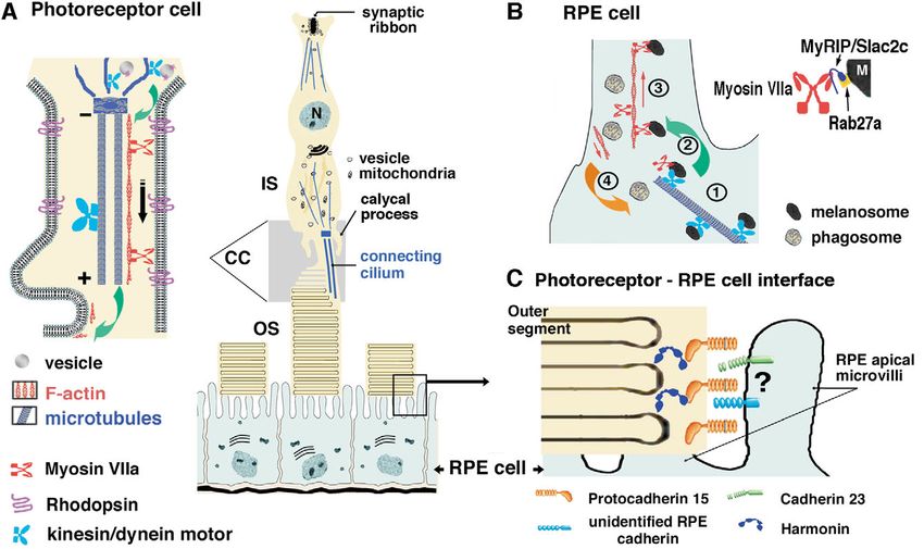
4594      Journal of Cell Science 118 (20)
Journal of Cell Science

                          Fig. 1. (A) Predicted domain structures of the USH1 proteins. Myosin VIIa consists of a spectrin-like SH3 subdomain followed by the motor
                          head, a neck region composed of five IQ (isoleucine-glutamine) motifs and a large tail. The tail starts with an ␣-helical domain, followed by two
                          large repeats, each containing a myosin tail homology 4 (MyTH4) and a 4.1, Ezrin, Radixin, Moesin (FERM)-like domain. These are separated
                          by a poorly conserved Src homology 3 (SH3) domain. Positions of the spliced exon 25 (ex 25) and 34 (ex 34) are indicated. There are three
                          classes of harmonin isoform, depending on the presence of two or three PDZ domains and the presence or absence of a second coiled-coil (CC)
                          domain associated with a proline-, serine- and threonine-rich (PST) domain. The largest cadherin 23 and protocadherin 15 isoforms have 27
                          extracellular cadherin (EC) repeats and 11 EC repeats, respectively. Sans is composed of three ankyrin (ANK)-like repeats and a sterile alpha
                          motif (SAM) domain. (B) Summary of the direct interactions between USH1 proteins. The domains involved in each interaction are indicated by
                          arrows. Harmonin is able to bind to any of the other USH1 proteins. The cytoplasmic regions of cadherin 23a isoforms containing or lacking the
                          exon 68-encoded peptide (ex 68 in A) preferentially bind to the harmonin PDZ1 or PDZ2 domain, respectively. Harmonin can bind to the
                          cytoplasmic region of protocadherin 15 through its first two PDZ domains. It can also bind to the myosin VIIa tail through a PDZ1–C-terminal-
                          MyTH4-FERM domain interaction. Harmonin can also bind to Sans through a PDZ1-SAM region interaction. Finally, the Sans central region
                          can bind to the myosin VIIa N-terminal MyTH4-FERM domain. Harmonin (not shown) and Sans can also form homodimers.

                          encodes the unconventional myosin VIIa (Weil et al., 1995).                Alternatively spliced USH1C transcripts allow us to predict at
                          USH1C encodes harmonin, a PDZ-domain-containing protein                    least ten harmonin isoforms. These form three subclasses (a,
                          (Verpy et al., 2000). USH1D and USH1F encode two large                     b, c), according to their protein domain composition (Verpy et
                          cadherin-related proteins, cadherin 23 and protocadherin 15,               al., 2000) (Fig. 1A). USH1D encodes at least six cadherin 23
                          respectively (Ahmed et al., 2001; Alagramam et al., 2001b;                 isoforms: transmembrane isoforms containing 27 (a isoforms)
                          Bolz et al., 2001; Bork et al., 2001). USH1G encodes a putative            or seven (b isoforms) extracellular cadherin (EC) domains,
                          scaffold protein, Sans, that contains three ankyrin repeats and            each with two different intracellular regions, and two cytosolic
                          a sterile alpha motif (SAM) domain (Weil et al., 2003).                    subtypes (the c isoforms) (Lagziel et al., 2005; Michel et al.,
                             Analysis of the transcripts of these genes in the inner ear and         2005) (Fig. 1A). Finally, USH1F transcripts are predicted to
                          retina, and biochemical studies suggest the existence of several           encode at least two transmembrane isoforms containing 11 (a
                          isoforms of myosin VIIa, harmonin, cadherin 23 and                         isoform) or one (b isoform) extracellular cadherin (EC)
                          protocadherin 15 (Fig. 1A). Splice variants of USH1B lacking               domain(s) (Ahmed et al., 2003) (Fig. 1A).
                          some exons [e.g. exons 8, 9 and 13 (the motor head), exon 25                  Mutations in four of the USH1 genes have also been reported
                          (the first MyTH4 domain) and exon 34 (the first FERM                       to cause isolated recessive (DFNB) or dominant (DFNA) forms
                          domain)] have been detected; they are all predicted to conserve            of deafness: DFNB2 and DFNA11 (USH1B) (Liu et al., 1997a;
                          the myosin VIIa reading frame (Weil et al., 1996) (Fig. 1A). A             Liu et al., 1997b; Weil et al., 1997), DFNB18 (USH1C)
                          shorter USH1B transcript isolated from a human testis cDNA                 (Ahmed et al., 2002; Ouyang et al., 2002), DFNB12 (USH1D)
                          library is predicted to encode a myosin VIIa isoform ending                (Bork et al., 2001) and DFNB23 (USH1F) (Ahmed et al.,
                          after the first MyTH4 domain (Chen et al., 1996). Nevertheless,            2002). The mutations causing these isolated forms of deafness
                          there is no evidence from northern blots or at the protein level           are usually expected to be less deleterious for the protein
                          that confirms the existence of this shorter isoform.                       activities than those observed in USH1 patients.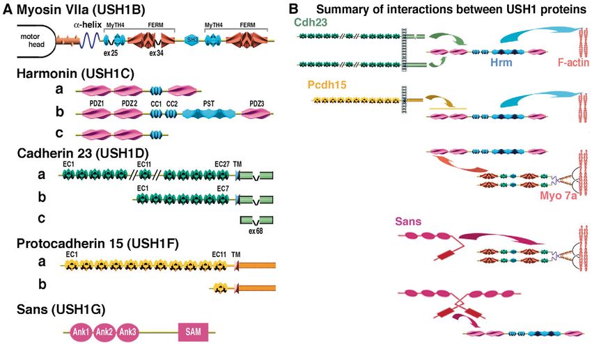
Cell biology of the Usher I syndrome                4595

                          Fig. 2. The mammalian auditory epithelium, the organ of Corti. The sensory inner (IHC) and outer (OHC) hair cells are flanked by various
                          types of supporting cell. Viewed from the top of the organ of Corti, the scanning electron microscopy images show the organisation of the OHC
                          hair bundle from wild-type (source: Marc Lenoir, Montpellier, France), and Myo7a6J (Arg241Pro) shaker-1 mutant mice at P15. In the shaker-1
                          mutant, the stereocilia form clusters of a small number of stereocilia arranged in diverse patterns and orientations (adapted with permission
                          from Self et al., 1998).

                          Animal models of USH1                                                    the apical surface of the cell; (3) they are arranged in three to
                          Mice that have defective myosin VIIa (shaker-1) (Gibson et al.,          four rows, the height the rows rising towards the kinocilium
                          1995), harmonin (deaf circler) (Johnson et al., 2003), cadherin          (Fig. 3A); and (4) they are connected by many lateral links and
                          23 (waltzer) (Di Palma et al., 2001; Wilson et al., 2001),               one tip link. The kinocilium is also connected to the nearest
                          protocadherin 15 (Ames waltzer) (Alagramam et al., 2001a) or             tall stereocilia by fibrous extracellular links, i.e. kinociliary
                          Sans (Jackson shaker) (Kikkawa et al., 2003) have been                   links (Fig. 3A).
                          described. These all exhibit severe hearing impairment and                  Four stages in the formation of the chick auditory hair
                          vestibular dysfunction but, surprisingly, none displays signs of         bundles have been defined by Tilney et al. (reviewed in Tilney
Journal of Cell Science

                          retinitis pigmentosa. EM scanning analysis of the hair bundles           et al., 1992) (see also Fig. 3A, left panels). First, the stereocilia
                          of the auditory sensory cells in mutant mice lacking any of              emerge at the apical surface of the sensory cell as a
                          USH1 proteins showed that this subcellular structure is                  homogeneous group of small, equally sized microvilli,
                          disorganised, and the bundles lack their characteristic cohesion         clustered around a single central kinocilium. Second, the
                          (see Fig. 2). A detailed chronological analysis of                       kinocilium migrates to the peripheral edge thus dictating the
                          morphological abnormalities in the hair bundles of shaker-1              orientation of the hair bundle (i.e. its planar polarity); this is
                          and waltzer mutants has revealed that their fragmentation can            followed by a selective and sequential elongation of the
                          already be detected at embryonic day 18 (E18) in both mutants            stereocilia row (starting with the row closest to the kinocilium)
                          (Self et al., 1998; Holme and Steel, 2002).                              to form a staircase pattern of height-ranked rows. Third, the
                             Zebrafish mutants possessing defective myosin VIIa or                 stereocilia stop elongating and a few central actin filaments of
                          cadherin 23 have also been described. These also have                    the stereocilia extend rootlets deep into a horizontal meshwork
                          disorganised hair bundles, which can easily be observed in the           of actin filaments – the cuticular plate – located beneath the
                          lateral line sensory system (Ernest et al., 2000; Sollner et al.,        apical cell membrane. Then, the stereocilia become wider with
                          2004). Two zebrafish orthologues of the mammalian                        the addition of new actin filaments. Finally, the stereocilia
                          protocadherin 15 gene, pcdh15a and pcdh15b, encoding                     complete their elongation to reach their adult length and any
                          proteins that have highly divergent intracellular domains, have          stereocilia not incorporated into the bundle regress. These steps
                          recently been described. Mutations in pcdh15a result in                  have also been seen in mammals (reviewed in Frolenkov et al.,
                          disorganisation of the hair bundle but cause no visual                   2004), although some differences do exist; for example,
                          abnormalities, whereas pcdh15b inactivation causes only                  elongation and widening of the stereocilium processes, which
                          retinal anomalies: mutants exhibit abnormal alignment of                 are separated in time in chick, occur concomitantly in
                          photoreceptor outer disks with the pigment epithelial cells and          mammals, and the kinocilium disappears in hair bundles of
                          abnormal interdigitation of the outer disks (Seiler et al., 2005).       mammalian auditory cells.
                                                                                                      Distinct specialised links connect the stereocilia (Fig. 3A).
                                                                                                   The tip link is a single, three-stranded interrow link that
                          Morphogenesis of the hair bundle                                         connects the tip of one stereocilium to the side of the nearest
                          The hair bundle is located at the apical surface of the auditory         tall stereocilium. It has a unique direction, following the
                          hair cell (see Fig. 2). It comprises between 20 and 300 actin-           functional axis of the hair bundle. It is already detectable at
                          filled, stiff microvilli – the stereocilia – which contain the           E17 in the mouse and persists throughout life (Geleoc and Holt,
                          mechanoelectrical transduction (MET) machinery and a single              2003; Goodyear et al., 2005). Pickles et al. have proposed that
                          cilium, the kinocilium (Fig. 3A). As in other microvilli, actin          these tip links are a specialisation of the links that join
                          filaments are uniformly polarized, the fast growing ends being           immature stereocilia laterally near their tips (Pickles et al.,
                          located at stereocilia tips (Schneider et al., 2002). However,           1991). Indeed, a dense network of links that connect stereocilia
                          stereocilia in the adult hair bundle have certain specific               within the growing hair bundle and show dramatic changes
                          morphological features (Figs 2, 3): (1) they are wider and               between its early developmental and mature stages. An
                          longer than the microvilli of epithelial cells and, in some              abundant and uniform fibrous network is progressively
                          species, can contain up to 2000 actin filaments (Revenu et al.,          substituted by distinct projecting dense links organised at
                          2004); (2) they taper off at their base, where they insert into          different points over the length of the stereocilia, connecting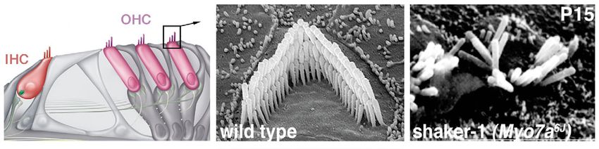
4596 Journal of Cell Science 118 (20)
them within and between adjacent rows. Top connectors couple constant of the transduction process is too short (a few
stereocilia just below the tip link, shaft links connect part or microseconds) to involve a second messenger. In response to
all the length of the stereocilia, and ankle links connect the sound or acceleration stimuli, the hair bundle deflects and pivots
stereocilia at the base (Goodyear and Richardson, 1999; around the basal insertion points of the stereocilia. This causes
Goodyear et al., 2005) (Fig. 3A). The ankle links are first variations in the opening probability of MET channels in the
detectable in the mouse at P0, but they are completely lost at plasma membrane. These thought to result from changes in the
P12 (Goodyear et al., 2005). Most of these links are recognised tension of the tip link, which are proposed to be linked to these
by specific antibodies and characterised by distinct channels (Howard and Hudspeth, 1988) (see Fig. 3A). The rapid
biochemical properties (e.g. resistance to the calcium chelator influx of cations, mostly K+ but also Ca2+ leads to hair cell
BAPTA or the protease subtilisin). Whereas the ankle links are depolarisation followed by neurotransmitter release. Two TRP
sensitive to both treatments, the tip link is only sensitive to channels have been proposed to be components of the MET
BAPTA (see Goodyear et al., 2005). With the exception of the channel: NompC in zebrafish (Sidi et al., 2003) and TRPA1
tip link (see below), the roles of the links in the growing and (also called ANKTM1) in mammals (Corey et al., 2004).
mature hair bundles are unknown. However their conservation Through adaptation processes, the hair bundle preserves its
throughout evolution, from fish (Sollner et al., 2004; Seiler et high sensitivity to extrinsic stimuli, i.e. the MET channel
al., 2005), mice (Alagramam et al., 2001a; Di Palma et al., opening probability (Po) tends to be restored to its resting level
2001; Wilson et al., 2001) to humans (Bork et al., 2001; Ahmed while the mechanical stimulus persists. Fast and slow
et al., 2002), strongly argues for their functional relevance. adaptation processes have been described in the hair cells. Fast
Their molecular composition is a key element because they will adaptation is mediated by Ca2+ influx through the MET
behave as more or less elastic connectors when submitted to channel, which provides a feedback signal acting on the MET
the force of the mechanical stimulation, depending on their channel itself (Fettiplace and Ricci, 2003). Ca2+ also regulates
extracellular domain composition, their possible a slower component of adaptation, which has been
oligomerisation and binding to other ligands. demonstrated to involve myosin 1c (Holt et al., 2002). Myosin
VIIa has also been proposed to contribute to adaptation (Kros
Journal of Cell Science
et al., 2002) (see below).
The mechanoelectrical transduction process
The highly elaborate organisation of the hair bundle is
evolutionarily conserved in vertebrates and probably plays a USH1 protein properties and interactions
crucial role in auditory and balance transduction. The time The tails of unconventional myosins are thought to position
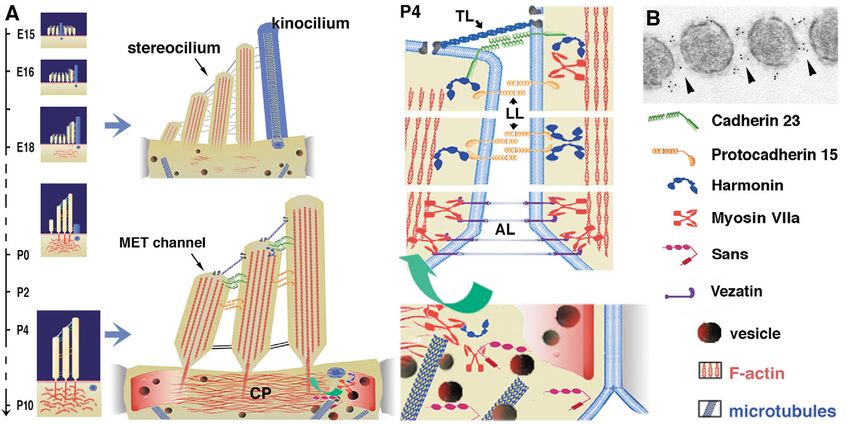
Fig. 3. (A) Early (E15-P0) and late (P4-P10) stages of auditory hair bundle maturation in mice. The stereocilia that form the hair bundle are held
together by various side-links. The number and structure of these links vary during the course of development (top) and in mature (bottom) cells.
At late developmental stages, the most central actin filaments of stereocilia insert their rootlets into another actin-rich structure, the cuticular plate
(CP). Note that the kinocilium is no longer present in mature auditory hair cells, although its basal body persists. The tip link (TL) is believed to
gate the mechanoelectrical transduction (MET) channel. (B) Immunogold labelling showing cadherin 23 is associated with links connecting
growing stereocilia (adapted with permission from Michel et al., 2005). Protocadherin 15 also probably makes interstereocilia lateral links (LL)
in the differentiating hair bundle. During hair bundle maturation, the binding of harmonin b to cadherin 23, protocadherin 15, and F-actin should
anchor the interstereocilia links to the stereocilia actin core. Myosin VIIa is required for transfer of harmonin b up to the stereocilia (green
arrow). Myosin VIIa also interacts with vezatin, a ubiquitous component of adherens junctions, which is associated with the ankle links (AL).
Cell biology of the Usher I syndrome 4597
these motors at a given intracellular location by interacting with Cadherin 23 and protocadherin 15 differ from the classical
protein(s) and/or lipid(s); the motor activity of the head is then cadherins (e.g. E- and N-cadherins) by possessing numerous
harnessed to exert tension on the tethered molecules. These EC repeats (Fig. 1A). Transfected L929 cells expressing full-
molecules (and possibly associated subcellular compartments) length cadherin 23 (L-CDH23 cells) form Ca2+-dependent cell-
can then be moved or not and, in certain cases, can undergo cell contacts, indicating that cadherin 23 can mediate Ca2+-
conformational changes. Myosin VIIa is an actin–plus-end- dependent, homophilic cell-cell adhesion (Siemens et al.,
directed unconventional myosin expected to move from the base 2004). Whether protocadherin 15 also undergoes homophilic
to the tip of stereocilia in the hair bundle. It has been described interactions is unknown. These two cadherin cytoplasmic
as a dimeric motor (Inoue and Ikebe, 2003), but whether (as in regions share no homology with any known protein. In
the case of myosin VI) it can also function as a monomer is particular, they lack the consensus motif for binding to -
unknown. All myosins exert their motile activity during a cyclic catenin (Bershadsky, 2004). This raises the question of how
interaction with actin filaments and ATP. The enzymatic these two cadherins are linked to the actin cytoskeleton.
characteristics of motor proteins are key determinants of their Yeast two-hybrid screenings combined with biochemical
motile activity and underlie their various cellular roles (see De analyses have shown that the five known USH1 proteins each
La Cruz and Ostap, 2004). Although the speed of myosin VIIa directly interact with at least one other USH1 protein (Fig. 1B).
has been estimated at ~160 nm/second (Inoue and Ikebe, 2003), Harmonin can bind to any of the other USH1 proteins through
some of its kinetic parameters are yet to be studied. Hence, its PDZ domains (Boëda et al., 2002; Siemens et al., 2002; Weil
whether or not it is a processive motor remains to be established. et al., 2003; Adato et al., 2005). With the exception of myosin
How proteins associated with its tail might influence its activity VIIa, the four other USH1 proteins harbour class I PDZ-
is another unanswered question. binding motifs at their C-terminal ends. However, harmonin’s
Myosin VIIa has a much higher affinity for ADP (KADP=7 in vitro interactions with either of these proteins is not
M) than for ATP (KATP=200 M) and requires high ATP interrupted by the deletion of these C-terminal PDZ-binding
concentrations for its motile activity. ADP markedly increases motifs (Adato et al., 2005). Therefore, the PDZ interactions of
the binding of myosin VIIa to actin filaments (Inoue and Ikebe, harmonin with USH1 proteins are not characterized by simple
Journal of Cell Science
2003). Therefore, in subcellular compartments that have high classical binding of C-terminal motifs.
ADP concentrations, myosin VIIa may maintain tension rather The Sans central region is involved in the Sans–myosin-VIIa
than move cargo. interaction and Sans homomerisation, whereas the SAM
The involvement of type VII myosins in tension-mediated domain binds to harmonin. The existence of the two distinct
processes seems to be evolutionarily conserved in metazoan and independent interaction motifs is supported by the
organisms, because inactivation of myosin VII in formation of an in vitro Sans-Sans-harmonin tripartite complex
Dictyostelium amoeba inhibits particle ingestion (Titus, 1999) (Adato et al., 2005) (see Fig. 1B). The common binding
owing to defects in the adhesion of particles to the cell surface domains of harmonin and Sans for several other USH1
(Tuxworth et al., 2001). Tuxworth and Titus have proposed two proteins, as well as their involvement in homo-oligomerisation
non-exclusive roles for this motor protein (Tuxworth et al., (Sans) or hetero-oligomerisation (harmonin isoforms) strongly
2001). First, myosin VII dimers capture membrane proteins suggest that some of these interactions are competitive (Adato
‘floating’ at the surface of the cell, allowing their clustering at et al., 2005).
the sites of contact formation. Second, through their motor
domains, myosin VIIa molecules may act locally as force
sensors, determining the strength of the interaction at the USH1 proteins and the cohesion of the growing hair
contact zone. For every receptor that can withstand the strain bundle
imposed by myosin, a non-motile protein anchored to the USH1 proteins are expressed in the hair cells throughout life,
cytoskeleton replaces the myosin. The receptor thus becomes although their subcellular distributions in the stereocilia vary
engaged in a stiff link until the contact area reaches a certain dramatically during development up to the mature stage (Fig.
limit. Through recruitment of additional receptors and their 4). All are expressed in the growing auditory hair bundle.
coupling to the cytoskeleton, myosin VIIa would thus However, whereas myosin VIIa, cadherin 23, harmonin and
contribute to the mechanical stability of the adhesion sites protocadherin 15 are present within the stereocilia (and some
required for efficient cell phagocytosis. Direct evidence for this are also within the kinocilium), Sans has been detected only
model is still missing; however, it can explain the role of the transiently within the auditory kinocilium.
direct interaction between mammalian myosin VIIa and Harmonin, cadherin 23 and protocadherin 15 are present in
vezatin, a component of adherens junctions (Kussel- the hair bundle as soon as it emerges at the apical surface of
Andermann et al., 2000), in the entry of Listeria the sensory cells (Boëda et al., 2002; Ahmed et al., 2003).
monocytogenes into cells (Sousa et al., 2004). Myosin VIIa Harmonin b is present mainly at the tips of stereocilia during
might have a similar role in the auditory hair bundle (see early postnatal stages (Boëda et al., 2002). Cadherin 23 first
below). occupies the entire length of the emerging stereocilia and then
Harmonin b, the longest harmonin isoform subclass, directly becomes progressively restricted to the tip region (see Fig. 4).
binds to F-actin through its C-terminal second coiled-coil Cadherin 23 can no longer be detected in the auditory hair
and/or its PST domain (see Fig. 1A). When overexpressed in bundle after postnatal day 14, but the labelling persists along
cells, it induces the formation of large F-actin bundles resistant the kinocilium of the vestibular organs, where it may
to cytochalasin D or latrunculin A, which suggests that contribute to kinociliary links (Lagziel et al., 2005; Michel et
harmonin b stabilises actin filaments. It also induces bundling al., 2005). Protocadherin 15 has been detected uniformly
of actin filaments in vitro (Boëda et al., 2002). distributed along the growing stereocilia (Ahmed et al., 2003)4598 Journal of Cell Science 118 (20)
Fig. 4. Distributions of USH1 proteins in developing and adult hair cells throughout life, their spatial and temporal subcellular distribution vary
for each molecule. Green arrows indicate the presence of a USH1 protein in the stereocilia. Myosin VIIa (Hasson et al., 1997) and
protocadherin 15 (Ahmed et al., 2003) are present along the entire length of the stereocilia. The precise distribution of harmonin in the
stereocilia at the postnatal stages remains to be established. Notice that, at P30, cadherin 23 disappears from the stereocilia and appears to move
to the pericuticular necklace (Lagziel et al., 2005; Michel et al., 2005). Sans is no longer present in the stereocilia (Adato et al., 2005).
Arrowheads refer to the stage when first signs of hair bundle anomalies have been reported in USH1 mouse mutants; E18 in shaker-1 (Self et
al., 1998) and waltzer (Holme and Steel, 2002) mutants, and P0 in Ames waltzer (Washington et al., 2005). In Jackson shaker and deaf-circler
mutants, the hair bundle disorganisation has been reported only from P10 (Kitamura et al., 1992), and P21 (Johnson et al., 2003), respectively.
(see Fig. 4). Myosin VIIa is present in the emerging hair (Gln520rStop) and have severely disorganized hair bundles
Journal of Cell Science
bundles (El-Amraoui et al., 1996; Boëda et al., 2002) and also (see Kros et al., 2002). In these mice, harmonin b cannot be
within the cuticular plate and the pericuticular necklace detected at the stereocilia tip and accumulates beneath the hair
region, which is characterised by a dense ring of vesicles bundle (Boëda et al., 2002). This indicates that myosin VIIa is
(Hasson et al., 1997). Sans is especially abundant in a vesicle- involved in the sorting and/or targeting of harmonin b to the
rich region in the immediate vicinity of the basal body of the hair bundle. In Jackson-shaker mutants, harmonin b is also
kinocilium (Adato et al., 2005) (see Fig. 3A, Fig. 4). For absent from the hair bundle (Gaëlle Lefèvre, personal
harmonin and protocadherin 15, their precise subcellular communication), which suggests that Sans, which interacts
localisations in the course of auditory hair bundle with myosin VIIa, is also involved in the sorting of harmonin
differentiation remain to be determined. b. These two proteins may also be involved in the sorting of
On the basis of the in vitro direct interaction between other hair bundle proteins. Within the hair bundle, myosin VIIa
cadherin 23 and harmonin, the co-localisation of cadherin 23 might have other functions. It may cluster cadherin-
and harmonin b in the forming hair bundle and the actin 23–protocadherin-15–harmonin complexes, thereby organising
binding activity of harmonin b, we have proposed that the spatial distribution of the hair bundle links. Myosin VIIa
cadherin-23-containing stereocilia connectors are anchored to may also act as a sensor, providing a dynamic link between
the actin core of the stereocilium through harmonin b, thereby interstereocilia connectors and the actin filaments, through
ensuring hair bundle cohesion during early morphogenesis which the cell may measure the tension force at a given
(Fig. 3A) (Boëda et al., 2002). Hair bundle disorganisation has position along the stereocilium membrane.
since been reported in the deaf circler 2J mouse mutant, which The crucial role of interstereocilia and kinociliary links in
lacks harmonin b isoforms (Johnson et al., 2003). Also, almost the cohesion of the growing hair bundles discussed here
all DFNB18 patients carry mutations in USH1C that selectively manifests itself when stereocilia have no or underdeveloped
affect the harmonin b subclass (Ouyang et al., 2002). actin rootlets. At that time, the connection between the hair
Moreover, ultrastructural studies have recently established that bundle and the apical region of the hair cell body seems mainly
cadherin 23 is a component of both interstereocilia and to involve microtubules that arise from the kinocilium basal
kinociliary links of the growing hair bundle (Michel et al., body (Tilney et al., 1992) (see Fig. 3A). Subsequently, the
2005) (see Fig. 3A,B). Protocadherin 15, which can bind to anchoring of stereocilia actin rootlets in the cuticular plate
harmonin b (Adato et al., 2005), may play a role similar to that confers additional stability upon the hair bundle; fibrous links
of cadherin 23 in hair bundle morphogenesis (see Fig. 3A). that connect these rootlets to horizontal actin filaments in the
Whether protocadherin 15 and cadherin 23 can form links cuticular plate horizontal actin filaments also participate
through heterophilic interactions is not yet known. Notice that, (Tilney et al., 1992). Unconventional myosins, including
genetic evidence in mice and humans supports the involvement myosin VIIa, in the cuticular plate may regulate the stability
of these two cadherins in the same pathway, although it is of the stereocilium–cuticular-plate anchor.
unclear whether the late effect reported reflects the early roles In addition, myosin VIIa can bind to vezatin, a
of the two cadherins (Zheng et al., 2005). transmembrane protein that colocalises with ankle links and an
Since it is present throughout the entire hair cell, myosin ubiquitous protein component of adherens junctions (Kussel-
VIIa probably has several functions. We analysed harmonin b Andermann et al., 2000). Myosin VIIa may also strengthen the
distribution in the Myo7a4626SB shaker-1 mice, which carry a adhesion at the junctions between the hair cells and supporting
premature stop codon in the motor domain of myosin VIIa cells and thus contribute to the stability of the cuticular plate.Cell biology of the Usher I syndrome 4599
How these elements are subsequently involved in the electroretinographic abnormalities (a reduction of the
development and/or the maintenance of hair bundle cohesion amplitude of the wave response to light) have been reported in
remains to be clarified. some shaker-1 and waltzer mutants (Libby et al., 2003). These
have not been seen in Ames waltzer (Ball et al., 2003) and deaf
circler mutant mice (Johnson et al., 2003). Slight peripheral
Other roles of USH1 proteins in the hair cell retinal degeneration has been found in 9-month-old deaf circler
Seven mutations in the Myo7a gene have been described and mutants, however (Johnson et al., 2003).
have varying degrees of phenotypic severity (Mburu et al., Because opsin accumulates in the photoreceptor inner
1997). In the original shaker-1 mutant, the Myo7ash1 mutation is segment and connecting cilium in shaker-1 mice, myosin VIIa
an Arg502Pro substitution in a poorly conserved surface loop of could function in the transport of opsin (Liu et al., 1999) (Fig.
the motor domain. This change is associated with the mildest of 5A). Moreover, in the absence of myosin VIIa, there is a
the pathological effects seen among shaker-1 alleles (Self et al., significant decrease in outer disk phagocytosis by pigment
1998). EM scanning analysis of the hair bundles in these epithelial cells (Gibbs et al., 2003). The renewal of
Myo7ash1shaker-1 mutants revealed normal stereocilia photoreceptor outer disk membranes involves continual
development in contrast to other shaker-1 alleles – for example, addition of new disks at the base of the outer segment, the
Myo7a6J or Myo7a4626SB (Self et al., 1998; Kros et al., 2002). shedding of the most distal ones and their phagocytosis by the
Also, whereas in Myo7a4626SB mice (Gln520rStop) harmonin b pigment epithelial cells (Fig. 5). In pigment epithelial cells of
is absent from the stereocilia, normal harmonin b distribution is shaker-1 mice, the transfer of ingested phagosomes from the
observed in the stereocilia tip of Myo7ash1 mutants (A.E.-A., apical cell processes to the cell body is delayed (Gibbs et al.,
unpublished). The mutant myosin VIIa molecule in the 2003). Also, EM studies in shaker-1 mice showed that
Myo7ash1 mice seems to have conserved sufficient activity to melanosomes are not spread throughout the apical projections
support normal early development of the stereocilia and proper of these cells but cluster around the nucleus. This suggests that
harmonin b targeting. This further supports the correlation myosin VIIa is necessary for the transfer of these organelles
between the presence of harmonin b in the stereocilia and normal from the cell periphery to the apical microvilli (Liu et al.,
Journal of Cell Science
hair bundle organisation. However, the progressive hearing loss 1998).
manifested by Myo7ash1 mutant mice (Self et al., 1998) indicates MyRIP/Slac2c, a protein that binds to myosin VIIa tail and
that, besides its function in stereocilia development, myosin VIIa directly interacts with Rab27a (El-Amraoui et al., 2002;
plays unidentified roles in the hair cell. Fukuda and Kuroda, 2002) and with F-actin (Desnos et al.,
Kros et al. have shown that, in Myo7a6J and Myo7a4626SB 2003) might be involved in this transport process (see Fig. 5B).
shaker-1 mice, the auditory mechanotransduction slow- Among the 18 classes that constitute the myosin superfamily,
adaptation process is impaired at an early postnatal stage (Kros myosins I, II, V and VI have been implicated in the transport
et al., 2002). Whether the defective adaptation in these two of organelles such as vacuoles, recycling endosomes,
mutants is due to a failure of myosin VIIa function at the lysosomes, secretory granules and melanosomes (reviewed in
mechano-electrical transduction machinery itself or from a Seabra and Coudrier, 2004). In particular, actin-dependent
more general perturbation of the dynamic properties of the capture of the melanosomes has been shown to involve a
entire hair bundle is not known. Irrespective of the exact myosin V motor in skin melanocytes (see Seabra and Coudrier,
underlying mechanism, it will therefore be interesting to 2004). In these cells, a complex formed by Rab27a, a MyRIP
characterise hair cell adaptation in Myo7ash1 shaker-1 mutants. homolog called melanophilin and myosin Va is involved. In
The various isoforms of cadherin 23 may also perform melanocytes isolated from mice that have defective Rab27a
additional functions beyond the maintenance of the growing (leaden mutants), melanophilin (ashen mutants), or myosin Va
hair bundle cohesion. Two recent studies using antibodies (dilute mutants), melanosomes are abnormally clustered
directed against part of its cytoplasmic region localized around the nucleus, as in retinal pigment epithelial cells in
cadherin 23 to the tip of stereocilia in the mature hair bundle, shaker-1 mutants. In melanophilin-depleted skin melanocytes,
which suggests that cadherin 23 dimers may form the tip links ectopic overexpression of myosin VIIa alone does not rescue
(Siemens et al., 2004; Sollner et al., 2004). By contrast, as the clustering phenotype, whereas overexpression of myosin
mentioned above, two other groups failed to detect cadherin 23 VIIa together with MyRIP restores normal peripheral
at the tip of stereocilia from mature hair bundles by antibodies melanosome distribution (Kuroda and Fukuda, 2005). MyRIP
raised against an extracellular epitope [EC11, epitope N1 thus acts as a myosin VIIa linker in vivo, and a
(Michel et al., 2005)] or other parts of its cytoplasmic region Rab27A–MyRIP–myosin-VIIa tripartite complex appears to
(Boëda et al., 2002; Lagziel et al., 2005; Michel et al., 2005). function in the transfer of retinal melanosomes to the actin
Notice that cadherin 23 does not completely disappear from the cytoskeleton, a prerequisite for their entry into the apical
auditory hair cell, because labelling is observed in the vesicle- microvilli.
rich pericuticular region of the hair cell. This suggests that it Despite the apparent diversity of the phenotypic
still plays a role in adult (Michel et al., 2005). Definition of the abnormalities observed in shaker-1 mice, we can speculate that
temporal and spatial subcellular localisation of the different some of these abnormalities are related to the same myosin
cadherin 23 isoforms might help clarify the above observations. VIIa activity. An attractive possibility is that the failure of
protein/organelle translocation (harmonin b sorting, opsin
transport, melanosome delivery and phagosome transfer) is due
USH1 proteins in the retina to a defect in the transport role of myosin VIIa at the interface
Although none of the mice that have mutations in USH1 between microtubules and actin filaments (green arrows, Figs
orthologues display retinitis pigmentosa, minor 3, 5). Indeed, myosin VIIa has been reported to interact with4600 Journal of Cell Science 118 (20)
the microtubule-associated protein MAP2B (Todorov et al., by contrast, present along the entire length of the outer segments
2001). The MyTH4-FERM domains of myosin X interact (Ahmed et al., 2003). Protocadherin 15 may thus connect
directly with microtubules (Weber et al., 2004). The myosin adjacent outer disks through homophilic interactions.
VIIa MyTH4-FERM domains might therefore bind to Protocadherin 15 may also engage in heterophilic
microtubules in a similar way. interactions with an as yet unknown cadherin present at the
The presence of all USH1 proteins at the photoreceptor membrane of pigment epithelial cells (see Fig. 5C). In the latter
synaptic active zone strongly suggests they contribute to the case, protocadherin 15 may contribute to photoreceptor-RPE
trafficking of synaptic vesicles (Reiners et al., 2003; Reiners et adhesion by anchoring the membranes of these two cell types.
al., 2005). The bulk of protocadherin 15 labelling is, however, Whether it also interacts with other cell surface or extracellular
present in the photoreceptor outer segments in the monkey and matrix proteins cannot be excluded. The failure of such
human retina (Ahmed et al., 2003), which is consistent with its interactions may explain the abnormal alignment of
having a role in the organisation of the photoreceptor outer disks. photoreceptor outer disks with the pigment epithelial cells in
During photoreceptor outer disk outgrowth, protocadherin 15, the pcdh15b-deficient zebrafish (Seiler et al., 2005). Although
through its extracellular domain, may provide the cell with little is known about the cytoskeletal elements and/or
information about its environment, in particular its proximity to associated proteins within the outer disk, harmonin b has been
the apical microvilli of the pigment epithelial cells. Targeted shown to be restricted to the photoreceptor outer segment
ablation of a photoreceptor-specific member of the cadherin (Reiners et al., 2003). Therefore, it is worth considering the
superfamily, prCAD, results in outer disk disorganisation and possibility that protocadherin-15–harmonin-b complexes play
fragmentation. PrCAD is confined to the base of the a role in the organisation of the hair bundle in the inner ear
photoreceptor outer segment, where it may interact either with (Fig. 3A) and the photoreceptor outer disks in the retina (Fig.
extracellular matrix components or with photoreceptor 5C).
membrane proteins (Rattner et al., 2001). Protocadherin 15 is, It is unclear why mice lacking orthologues of USH1 proteins
Journal of Cell Science
Fig. 5. (A) Photoreceptor and retinal pigment epithelial (RPE) cells. At the tip of the photoreceptor inner segment (IS), myosin VIIa may be
involved in opsin transfer (green arrow) to the connecting cilium and its transport to the outer segments (OS). (B) In RPE cells, melanosomes
(M) display fast, bidirectional microtubule-dependent long-range movements in the cell body driven by kinesin/dynein motor proteins (1).
Upon reaching the plus end of the microtubule at the periphery, myosin VIIa may be involved in the transfer (green arrow) of these organelles
towards the actin filaments (2). Rab27a, which is targeted to the melanosome membrane, interacts with its effector, MyRIP/Slac2c, which in
turn binds to myosin VIIa. Myosin VIIa then enables the retention and/or local movement of the melanosomes along the actin filaments of the
microvilli (3). Myosin VIIa also plays a role in the transfer of phagosomes from the microvilli to the cell body (4; orange arrow). (C) At outer-
disk–RPE-cell interface, protocadherin 15 present at the membrane of photoreceptor outer segments may serve to sense its immediate
environment. For instance, through its extracellular domain, it may engage in heterophilic interactions with unidentified cadherin(s) in RPE
cells to ensure proper alignment of the outer disks and apical microvilli. Harmonin b expressed in the photoreceptor outer disks is expected also
to play a role in disk structure. N, nucleus.Cell biology of the Usher I syndrome 4601
do not suffer from retinitis pigmentosa. Several explanations Finally, the studies discussed here are fuelled largely by the
could account for this difference between humans and mice. need for a deeper understanding of the roles played by USH1
These include the shorter lifespan of the mouse, differences in proteins in the two sensory organs so that we can develop
light exposure experienced by humans and mice, functional therapy or prevent (in the case of the visual defect) Usher
redundancy of USH1 proteins in the human but not the mouse syndrome in humans. Clarification of the pathogenesis of the
retina, or the presence of highly developed calycal processes in retinal degeneration observed in humans suffering from USH1
human but not mouse photoreceptor cells. The calycal processes is required and calls for more favourable animal models.
are microvillus-like extensions of the photoreceptor inner
segment that cup the base of the outer segment (see Fig. 5A) We are grateful to Jean-Pierre Hardelin, Gaelle Lefèvre and
and are connected to the connecting cilium stalk plasma Raphaël Étournay for stimulating discussions, to Avital Adato for her
membrane by fibrous links. They share common components critical reading of the manuscript, to Jacqueline Levilliers for help in
with hair bundle links. For instance, ALA (ankle link antigen), the preparation of the document and to Marc Lenoir for the SEM
image. We apologise for not citing all the relevant references because
which associates with the ankle links in the hair bundle, also of space limitations. The work in the authors’ laboratory is supported
colocalises with links bridging the calycal processes and the by the R. and G. Strittmatter Foundation, the A. and M. Suchert
connecting cilium (Goodyear and Richardson, 1999). The Forschung contra Blindheit-Initiative Usher Syndrom.
functions of calycal processes, first described more than 40
years ago (Cohen, 1963), have received little attention, however.
References
Adato, A., Kikkawa, Y., Reiners, J., Alagramam, K. N., Weil, D.,
Perspectives Yonekawa, H., Wolfrum, U., El-Amraoui, A. and Petit, C. (2005).
Interactions in the network of Usher syndrome type 1 proteins. Hum. Mol.
The study of USH1 proteins strongly suggests that some Genet. 14, 347-356.
fibrous hair bundle extracellular links have a crucial role in the Ahmed, Z. M., Riazuddin, S., Bernstein, S. L., Ahmed, Z., Khan, S.,
cohesion of the growing hair bundle during auditory hair cell Griffith, A. J., Morell, R. J., Friedman, T. B., Riazuddin, S. and Wilcox,
differentiation. This raises the question of the respective E. R. (2001). Mutations of the protocadherin gene PCDH15 cause Usher
Journal of Cell Science
contribution of interstereocilia and kinociliary links to the syndrome type 1F. Am. J. Hum. Genet. 69, 25-34.
Ahmed, Z. M., Smith, T. N., Riazuddin, S., Makishima, T., Ghosh, M.,
cohesion of the forming hair bundle. We must also establish Bokhari, S., Menon, P. S. N., Deshmukh, D., Griffith, A. J., Riazuddin,
that hair bundles are disorganised in the early developmental S. et al. (2002). Nonsyndromic recessive deafness DFNB18 and Usher
stages of all mutants that have defective USH1 proteins. syndrome type IC are allelic mutations of USHIC. Hum. Genet. 110, 527-
Additional USH1 proteins will undoubtedly be discovered in 531.
the near future. This should help us further decipher this Ahmed, Z. M., Riazuddin, S., Ahmad, J., Bernstein, S. L., Guo, Y., Sabar,
M. F., Sieving, P., Riazuddin, S., Griffith, A. J., Friedman, T. B. et al.
molecular network, which at present contains no known (2003). PCDH15 is expressed in the neurosensory epithelium of the eye and
regulatory molecule. Moreover, recent evidence suggests that ear and mutant alleles are responsible for both USH1F and DFNB23. Hum.
the transmembrane (TM) usherin isoforms, encoded by the Mol. Genet. 12, 3215-3223.
USH2A gene, are found within other interstereocilia links Alagramam, K. N., Murcia, C. L., Kwon, H. Y., Pawlowski, K. S., Wright,
C. G. and Woychik, R. P. (2001a). The mouse Ames waltzer hearing-loss
(Avital Adato, personal communication). Furthermore, in the mutant is caused by mutation of Pcdh15, a novel protocadherin gene. Nat.
hair cells of mutant mice lacking Vlgr1 (very large G-protein- Genet. 27, 99-102.
coupled receptor-1, also called Mass1), a transmembrane Alagramam, K. N., Yuan, H., Kuehn, M. H., Murcia, C. L., Wayne, S.,
protein defective in USH2C (Weston et al., 2004), the hair Srisailpathy, C. R. S., Lowry, R. B., Knaus, R., Van Laer, L., Bernier,
bundle appear rounded and deformed, with a loss of stereocilia F. P. et al. (2001b). Mutations in the novel protocadherin PCDH15 cause
Usher syndrome type 1F. Hum. Mol. Genet. 10, 1709-1718.
cohesion (Johnston et al., 2005). Similarly to TM usherin, Ball, S. L., Bardenstein, D. and Alagramam, K. N. (2003). Assessment of
Vlgr1 has a long extracellular modular ectodomain (~180 nm), retinal structure and function in Ames waltzer mice. Invest. Ophthalmol. Vis.
and is thus qualified to form interstereocilia links. Whether the Sci. 44, 3986-3992.
other USH2 or USH3 proteins are also involved in connecting Bershadsky, A. (2004). Magic touch: how does cell-cell adhesion trigger actin
stereocilia is similarly worth considering. assembly? Trends Cell Biol. 14, 589-593.
Boëda, B., El-Amraoui, A., Bahloul, A., Goodyear, R., Daviet, L.,
Adhesion contacts and associated cytoskeletal networks Blanchard, S., Perfettini, I., Fath, K. R., Shorte, S., Reiners, J. et al.
have been shown to operate as tension-sensing devices that (2002). Myosin VIIa, harmonin and cadherin 23, three Usher I gene products
trigger signal transduction to orchestrate the cellular response that cooperate to shape the sensory hair cell bundle. EMBO J. 21, 6689-
(see Ingber, 2003). Such signaling cascades, however, have not 6699.
Bolz, H., von Brederlow, B., Ramirez, A., Bryda, E. C., Kutsche, K.,
yet been addressed in adhesion sites at the level of stereocilia Nothwang, H. G., Seeliger, M., del C-Salcedo Cavrera, M., Vila, M. C.,
connecting links. It is worth considering that the developing Molina, O. P. et al. (2001). Mutation of CDH23, encoding a new member
hair bundle is a tensegrity-based structure and that link- of the cadherin gene family, causes Usher syndrome type 1D. Nat. Genet.
mediated adhesion forces can act as active elements that affect 27, 108-112.
the tensegrity force balance that controls and preserves hair Bork, J. M., Peters, L. M., Riazuddin, S., Bernstein, S. L., Ahmed, Z. M.,
Ness, S. L., Polomeno, R., Ramesh, A., Schloss, M., Srisailpathy, C. R.
bundle three-dimentional architecture, differentiation and S. et al. (2001). Usher syndrome 1D and nonsyndromic autosomal recessive
function. deafness DFNB12 are caused by allelic mutations of the novel cadherin-like
Most of the USH1 proteins are expressed in mature sensory gene CDH23. Am. J. Hum. Genet. 68, 26-37.
cells, both in the inner ear and in the retina. Their roles in fully Chen, Z.-Y., Hasson, T., Kelley, P. M., Schwender, B. J., Schwartz, M. F.,
differentiated sensory cells are not known. However, this could Ramakrishnan, M., Kimberling, W. J., Mooseker, M. S. and Corey, D.
P. (1996). Molecular cloning and domain structure of human myosin-VIIa,
also be addressed by genetic approaches, provided new tools the gene product defective in Usher syndrome 1B. Genomics 36, 440-448.
are developed to circumvent the early abnormal phenotype we Cohen, A. I. (1963). Vertebrate retinal cells and their organization. Biol. Rev.
discuss here. Cambridge Philos. Soc. 38, 427-459.4602 Journal of Cell Science 118 (20)
Corey, D. P., Garcia-Anoveros, J., Holt, J. R., Kwan, K. Y., Lin, S. Y., M., Taya, C., Kamiya, K., Yoshikawa, Y., Tokano, H. et al. (2003).
Vollrath, M. A., Amalfitano, A., Cheung, E. L., Derfler, B. H., Duggan, Mutations in a new scaffold protein Sans cause deafness in Jackson shaker
A. et al. (2004). TRPA1 is a candidate for the mechanosensitive transduction mice. Hum. Mol. Genet. 12, 453-461.
channel of vertebrate hair cells. Nature 432, 723-730. Kitamura, K., Kakoi, H., Yoshikawa, Y. and Ochikubo, F. (1992).
De La Cruz, E. M. and Ostap, E. M. (2004). Relating biochemistry and Ultrastructural findings in the inner ear of Jackson shaker mice. Acta
function in the myosin superfamily. Curr. Opin. Cell Biol. 16, 61-67. Otolaryngol. 112, 622-627.
Desnos, C., Schonn, J.-S., Huet, S., Tran, V. S., El-Amraoui, A., Raposo, Kros, C. J., Marcotti, W., van Netten, S. M., Self, T. J., Libby, R. T., Brown,
G., Fanget, I., Chapuis, C., Ménasché, G., de Saint Basile, G. et S. D., Richardson, G. P. and Steel, K. P. (2002). Reduced climbing and
al. (2003). Rab27A and its effector MyRIP link secretory granules to F-actin increased slipping adaptation in cochlear hair cells of mice with Myo7a
and control their motion towards release sites. J. Cell Biol. 163, 559-570. mutations. Nat. Neurosci. 5, 41-47.
Di Palma, F., Holme, R. H., Bryda, E. C., Belyantseva, I. A., Pellegrino, Kuroda, T. S. and Fukuda, M. (2005). Functional analysis of Slac2-c/MyRIP
R., Kachar, B., Steel, K. P. and Noben-Trauth, K. (2001). Mutations in as a linker protein between melanosomes and myosin VIIa. J. Biol. Chem.
Cdh23, encoding a new type of cadherin, cause stereocilia disorganization 30, 30.
in waltzer, the mouse model for Usher syndrome type 1D. Nat. Genet. 27, Kussel-Andermann, P., El-Amraoui, A., Safieddine, S., Nouaille, S.,
103-107. Perfettini, I., Lecuit, M., Cossart, P., Wolfrum, U. and Petit, C. (2000).
El-Amraoui, A., Sahly, I., Picaud, S., Sahel, J., Abitbol, M. and Petit, C. Vezatin, a novel transmembrane protein, bridges myosin VIIA to the
(1996). Human Usher IB/mouse shaker-1; the retinal phenotype discrepancy cadherin-catenins complex. EMBO J. 19, 6020-6029.
explained by the presence/absence of myosin VIIA in the photoreceptor Lagziel, A., Ahmed, Z. M., Schultz, J. M., Morell, R. J., Belyantseva, I. A.
cells. Hum. Mol. Genet. 5, 1171-1178. and Friedman, T. B. (2005). Spatiotemporal pattern and isoforms of
El-Amraoui, A., Schonn, J.-S., Küssel-Andermann, P., Blanchard, S., cadherin 23 in wild type and waltzer mice during inner ear hair cell
Desnos, C., Henry, J.-P., Wolfrum, U., Darchen, F. and Petit, C. (2002). development. Dev. Biol. 280, 295-306.
MyRIP, a novel Rab effector, enables myosin VIIa recruitment to retinal Libby, R. T., Kitamoto, J., Holme, R. H., Williams, D. S. and Steel, K. P.
melanosomes. EMBO Rep. 3, 463-470. (2003). Cdh23 mutations in the mouse are associated with retinal
Ernest, S., Rauch, G.-J., Haffter, P., Geisler, R., Petit, C. and Nicolson, T. dysfunction but not retinal degeneration. Exp. Eye Res. 77, 731-739.
(2000). Mariner is defective in myosin VIIA: a zebrafish model for human Liu, X.-Z., Walsh, J., Mburu, P., Kendrick-Jones, J., Cope, M. J. T. V.,
hereditary deafness. Hum. Mol. Genet. 9, 2189-2196. Steel, K. P. and Brown, S. D. M. (1997a). Mutations in the myosin VIIA
Fettiplace, R. and Ricci, A. J. (2003). Adaptation in auditory hair cells. Curr. gene cause non-syndromic recessive deafness. Nat. Genet. 16, 188-190.
Opin. Neurobiol. 13, 446-451. Liu, X.-Z., Walsh, J., Tamagawa, Y., Kitamura, K., Nishizawa, M., Steel,
Frolenkov, G. I., Belyantseva, I. A., Friedman, T. B. and Griffith, A. J. K. P. and Brown, S. D. M. (1997b). Autosomal dominant non-syndromic
Journal of Cell Science
(2004). Genetic insights into the morphogenesis of inner ear hair cells. Nat. deafness caused by a mutation in the myosin VIIA gene. Nat. Genet. 17,
Rev. Genet. 5, 489-498. 268-269.
Fukuda, M. and Kuroda, T. S. (2002). Slac2-c (synaptotagmin-like protein Liu, X., Ondek, B. and Williams, D. S. (1998). Mutant myosin VIIa causes
homologue lacking C2 domains-c), a novel linker protein that interacts with defective melanosome distribution in the RPE of shaker-1 mice. Nat. Genet.
Rab27, myosin Va/VIIa, and actin. J. Biol. Chem. 277, 43096-43103. 19, 117-118.
Geleoc, G. S. and Holt, J. R. (2003). Developmental acquisition of sensory Liu, X., Udovichenko, I. P., Brown, S. D., Steel, K. P. and Williams, D. S.
transduction in hair cells of the mouse inner ear. Nat. Neurosci. 6, 1019- (1999). Myosin VIIa participates in opsin transport through the
1020. photoreceptor cilium. J. Neurosci. 19, 6267-6274.
Gibbs, D., Kitamoto, J. and Williams, D. S. (2003). Abnormal phagocytosis Mburu, P., Liu, X. Z., Walsh, J., Saw, D., Jr, Cope, M. J., Gibson, F.,
by retinal pigmented epithelium that lacks myosin VIIa, the Usher syndrome Kendrick-Jones, J., Steel, K. P. and Brown, S. D. (1997). Mutation
1B protein. Proc. Natl. Acad. Sci. USA 100, 6481-6486. analysis of the mouse myosin VIIA deafness gene. Genes Funct. 1, 191-203.
Gibson, F., Walsh, J., Mburu, P., Varela, A., Brown, K. A., Antonio, M., Michel, V., Goodyear, R. J., Weil, D., Marcotti, W., Perfettini, I., Wolfrum,
Beisel, K. W., Steel, K. P. and Brown, S. D. M. (1995). A type VII myosin U., Kros, C., Richardson, G. P. and Petit, C. (2005). Cadherin 23 is a
encoded by the mouse deafness gene Shaker-1. Nature 374, 62-64. component of the transient lateral links in the developing hair bundles of
Goodyear, R. and Richardson, G. (1999). The ankle-link antigen: an epitope cochlear sensory cells. Dev. Biol. 280, 281-294.
sensitive to calcium chelation associated with the hair-cell surface and the Ouyang, X. M., Xia, X. J., Verpy, E., Du, L. L., Pandya, A., Petit, C.,
calycal processes of photoreceptors. J. Neurosci. 19, 3761-3772. Balkany, T., Nance, W. E. and Liu, X.-Z. (2002). Mutations in the
Goodyear, R. J., Marcotti, W., Kros, C. J. and Richardson, G. P. (2005). alternatively spliced exons of USH1C cause non-syndromic recessive
Development and properties of stereociliary link types in hair cells of the deafness. Hum. Genet. 111, 26-30.
mouse cochlea. J. Comp. Neurol. 485, 75-85. Petit, C. (2001). Usher syndrome: from genetics to pathogenesis. Annu. Rev.
Hasson, T., Gillespie, P. G., Garcia, J. A., MacDonald, R. B., Zhao, Y., Yee, Genomics Hum. Genet. 2, 271-297.
A. G., Mooseker, M. S. and Corey, D. P. (1997). Unconventional myosins Pickles, J. O., von Perger, M., Rouse, G. W. and Brix, J. (1991). The
in inner-ear sensory epithelia. J. Cell Biol. 137, 1287-1307. development of links between stereocilia in hair cells of the chick basilar
Holme, R. H. and Steel, K. P. (2002). Stereocilia defects in waltzer (Cdh23), papilla. Hear. Res. 54, 153-163.
shaker1 (Myo7a) and double waltzer/shaker1 mutant mice. Hear. Res. 169, Rattner, A., Smallwood, P. M., Williams, J., Cooke, C., Savchenko, A.,
13-23. Lyubarsky, A., Pugh, E. N. and Nathans, J. (2001). A photoreceptor-
Holt, J. R., Gillespie, S. K., Provance, D. W., Shah, K., Shokat, K. M., specific cadherin is essential for the structural integrity of the outer segment
Corey, D. P., Mercer, J. A. and Gillespie, P. G. (2002). A chemical-genetic and for photoreceptor survival. Neuron 32, 775-786.
strategy implicates myosin-1c in adaptation by hair cells. Cell 108, 371-381. Reiners, J., Reidel, B., El-Amraoui, A., Boëda, B., Huber, I., Petit, C. and
Howard, J. and Hudspeth, A. J. (1988). Compliance of the hair bundle Wolfrum, U. (2003). Differential distribution of harmonin isoforms and
associated with gating of mechanoelectrical transduction channels in the their possible role in Usher-1 protein complexes in mammalian
bullfrog’s saccular hair cell. Neuron 1, 189-199. photoreceptor cells. Invest. Ophthalmol. Vis. Sci. 44, 5006-5015.
Ingber, D. E. (2003). Tensegrity I. Cell structure and hierarchical systems Reiners, J., Marker, T., Jurgens, K., Reidel, B. and Wolfrum, U. (2005).
biology. J. Cell Sci. 116, 1157-1173. Photoreceptor expression of the Usher syndrome type 1 protein
Inoue, A. and Ikebe, M. (2003). Characterization of the motor activity of protocadherin 15 (USH1F) and its interaction with the scaffold protein
mammalian myosin VIIA. J. Biol. Chem. 278, 5478-5487. harmonin (USH1C). Mol. Vis. 11, 347-355.
Johnson, K. R., Gagnon, L. H., Webb, L. S., Peters, L. L., Hawes, N. L., Revenu, C., Athman, R., Robine, S. and Louvard, D. (2004). The co-
Chang, B. and Zheng, Q. Y. (2003). Mouse models of USH1C and workers of actin filaments: from cell structures to signals. Nat. Rev. Mol.
DFNB18: phenotypic and molecular analyses of two new spontaneous Cell. Biol. 5, 635-646.
mutations of the Ush1c gene. Hum. Mol. Genet. 30, 3075-3086. Schneider, M. E., Belyantseva, I. A., Azevedo, R. B. and Kachar, B. (2002).
Johnson, K. R., Zheng, Q. Y., Weston, M. D., Ptacek, L. J. and Noben- Rapid renewal of auditory hair bundles. Nature 418, 837-838.
Trauth, K. (2005) The Mass1frings mutation underlies early onset hearing Seabra, M. C. and Coudrier, E. (2004). Rab GTPases and myosin motors in
impairment in BUB/BnJ mice, a model for the auditory pathology of Usher organelle motility. Traffic 5, 393-399.
syndrome IIC. Genomics 85, 582-590. Seiler, C., Finger-Baier, K. C., Rinner, O., Makhankov, Y. V., Schwarz, H.,
Kikkawa, Y., Shitara, H., Wakana, S., Kohara, Y., Takada, T., Okamoto, Neuhauss, S. C. and Nicolson, T. (2005). Duplicated genes with splitYou can also read


