Wearable Motion Sensor Device to Facilitate Rehabilitation in Patients With Shoulder Adhesive Capsulitis: Pilot Study to Assess Feasibility
←
→
Page content transcription
If your browser does not render page correctly, please read the page content below
JOURNAL OF MEDICAL INTERNET RESEARCH                                                                                                           Chen et al

     Original Paper

     Wearable Motion Sensor Device to Facilitate Rehabilitation in
     Patients With Shoulder Adhesive Capsulitis: Pilot Study to Assess
     Feasibility

     Yu-Pin Chen1,2,3, MD; Chung-Ying Lin4, PhD; Ming-Jr Tsai5, MD; Tai-Yuan Chuang2,3, MD; Oscar Kuang-Sheng
     Lee1,6,7, PhD, MD
     1
      Institute of Clinical Medicine, National Yang Ming University, Taipei, Taiwan
     2
      Department of Orthopedic Surgery, Wan Fang Hospital, Taipei Medical University, Taipei, Taiwan
     3
      Department of Orthopedic Surgery, School of Medicine, College of Medicine, Taipei Medical University, Taipei, Taiwan
     4
      Department of Rehabilitation Sciences, Faculty of Health and Social Sciences, The Hong Kong Polytechnic University, Hung Hom, China (Hong
     Kong)
     5
      Department of Orthopedic Surgery, Puli Christian Hospital, Nantou, Taiwan
     6
      Department of Medical Research, Taipei Veterans General Hospital, Taipei, Taiwan
     7
      Department of Orthopedics, China Medical University Hospital, Taichung, Taiwan

     Corresponding Author:
     Oscar Kuang-Sheng Lee, PhD, MD
     Institute of Clinical Medicine
     National Yang Ming University
     No.155, Sec.2, Linong Street
     Taipei, 112
     Taiwan
     Phone: 886 2 28200183
     Email: oscarlee9203@gmail.com

     Abstract
     Background: Adhesive capsulitis (AC) of the shoulder is a common disorder that painfully reduces the shoulder range of motion
     (ROM) among middle-aged individuals. Although physical therapy with home-based exercises is widely advised to restore ROM
     in the treatment of AC, clinical results vary owing to inconsistent patient compliance.
     Objective: In this study, we aimed to verify the feasibility of a treatment model that involves applying a wearable motion sensor
     device to assist patients conduct home-based exercises to improve training compliance and the accuracy of exercises, with the
     ultimate goal of improving the functional recovery of patients with AC.
     Methods: The motion sensor device was comprised of inertial measurement unit–based sensors and mobile apps for patients
     and physicians, offering shoulder mobility tracing, home-based exercise support, and progress monitoring. The interrater reliability
     of shoulder mobility measurement using the motion sensor device on 10 healthy participants and 15 patients with AC was obtained
     using an intraclass correlation coefficient analysis and compared with the assessments performed by two highly experienced
     physicians. A pilot prospective control trial was then carried out to allocate the 15 patients with AC to two groups: home-based
     exercise group and motion sensor–assisted rehabilitation group. Changes in active and passive shoulder ROM, pain and functional
     scores, and exercise completion rates were compared between the groups during a treatment period of 3 months.
     Results: Shoulder ROM, as measured using the motion sensor device, exhibited good to excellent reliability based on the
     comparison with the measurements of two physicians (intraclass correlation coefficient range, 0.771 to 0.979). Compared with
     patients with AC in the home-based exercise group, those in the motion sensor–assisted rehabilitation group exhibited better
     shoulder mobility and functional recovery and a higher exercise completion rate during and after 3 months of rehabilitation.
     Conclusions: Motion sensor device–assisted home-based rehabilitation for the treatment of AC is a useful treatment model for
     telerehabilitation that enhances the compliance of patients through training, thus improving functional recovery. This helps
     overcome important obstacles in physiotherapy at home by providing comprehensible and easily accessible exercise instructions,
     enhancing compliance, ensuring the correctness of exercise, and monitoring the progress of patients.

     http://www.jmir.org/2020/7/e17032/                                                                   J Med Internet Res 2020 | vol. 22 | iss. 7 | e17032 | p. 1
                                                                                                                        (page number not for citation purposes)
XSL• FO
RenderX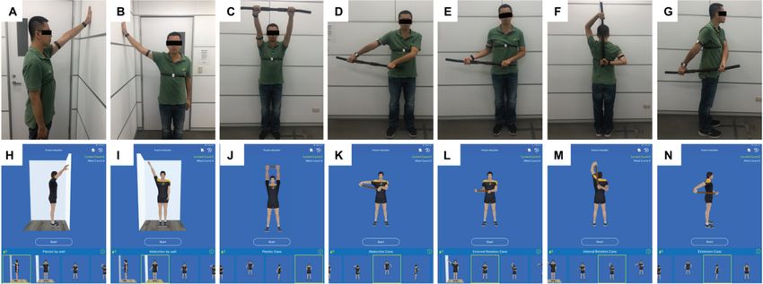
JOURNAL OF MEDICAL INTERNET RESEARCH                                                                                                    Chen et al

     (J Med Internet Res 2020;22(7):e17032) doi: 10.2196/17032

     KEYWORDS
     motion sensor; adhesive capsulitis; rehabilitation; home-based exercise; telerehabilitation; telehealth, telemonitoring

                                                                          including accelerometers and gyroscopes, have been extensively
     Introduction                                                         used in technology-assisted rehabilitation, with sufficient
     Adhesive capsulitis (AC) of the shoulder, which occurs in            efficacy [14-16]. However, despite the potential of using
     approximately 2% to 5% of the general population, is an              IMU-based sensors in neurorehabilitation and for treating
     idiopathic, progressive, and painful restriction of the active       musculoskeletal impairments [13,17,18], few such sensors have
     range of motion (aROM) and passive range of motion (pROM)            been used in clinical studies, especially those involving
     [1,2]. AC typically affects patients older than 50 years, and        physiotherapy for AC.
     involvement of both shoulders is noted in 20% to 30% of cases        Through this study, we tested wearable IMU-based sensors that
     [1,2]. Although symptomatic improvement tends to occur               integrated with interactive mobile apps using wireless
     naturally within years even with minimal treatment [3],              telecommunication technology to enable physiotherapists to
     approximately 50% of patients experience pain or some mild           monitor the progress of patients with AC who were conducting
     restriction of movement, and 11% experience some residual            home-based self-exercise and improve their training compliance.
     disability even several years after the resolution of their other    We hypothesized that the use of a wearable IMU-based motion
     symptoms [4,5]. Although AC is a common condition,                   sensor device could help patients perform home-based exercises
     high-quality evidence of successful treatment methods for AC         correctly and increase their motivation and compliance regarding
     has not yet been obtained [6]. The well-accepted standard            training, thereby improving functional outcomes.
     treatment for AC mostly involves physical therapy and home
     exercises to restore ROM [7,8]. Evidence suggests that,              Methods
     compared with less frequent self-exercise in a home setting or
     joint mobilization sessions in a hospital setting, regular and       Motion Sensor Device
     daily self-exercise in a home setting could contribute to greater    A wearable motion sensor device (BoostFix wearable
     improvement in shoulder ROM and a shorter duration of                self-training kit, COMPAL Electronics Inc, Taipei, Taiwan)
     symptoms [9].                                                        was used (see Multimedia Appendix 1). The motion sensor
     Despite realizing the importance of daily physiotherapy,             device was comprised of wearable IMU-based sensors that
     including mobilization and strength exercises, clinicians have       record the angular shoulder motion of a patient, a mobile app
     achieved varied outcomes in patients who are trained in a            termed Patient App for use by the patient, and a mobile app
     home-based exercise program [10]. Challenges that affect the         termed Doctor App for use by a qualified health care
     effectiveness of home-based exercises for AC may include             professional.
     training compliance and exercise correctness. Patients with AC
                                                                          Wearable IMU–Based Sensors
     do not maintain their training frequency and duration at home
     because the prescribed exercise programs are typically not           The IMU-based sensors were comprised of 6-axis
     followed without the constant supervision of a physiotherapist.      microelectromechanical systems that included accelerometers
     Failure to incorporate exercises into daily life is the main form    and gyroscopes. These devices collect information about the
     of noncompliance, as reported in up to 60% of patients whose         angular motion of the shoulder of interest. Three sensors are
     treatment plan included home-based exercise [11]. Whether            required for angular measurements of the shoulder. In this study,
     patients can correctly perform exercises at home after initial       they were strapped to the sternum, distal third of the lateral
     instruction by physiotherapists is also a concern [12]. Therefore,   upper arm approximately 5 cm above the lateral epicondylar of
     the development of methods to improve compliance and exercise        the humerus bone, and dorsal wrist (Figure 1). Three sensors
     correctness for patients with AC is worthwhile to maximize the       were calibrated in position A once the measurement began
     effectiveness of home-based physiotherapy.                           (Figure 1A). The initial calibration process involved placing
                                                                          the sensor on a horizontal fixture to measure the offset for each
     Techniques for detecting bodily motions are extensively used         axis, to align the initial difference for each sensor. The
     in health care to monitor and rehabilitate disabled patients. With   calibration process involved obtaining the initial value of the
     the evolution of sensing and body area network technologies,         sensor for relative offset correction instead of correcting the
     wearable rehabilitation technology has opened up the possibility     deviation caused by incorrect sensor placement. In addition, the
     of independent training, which has advantages over traditional       process entailed identifying the original offset of the sensors to
     rehabilitation services [13]. Inertial measurement units (IMUs),     each other and applying the zero setting using an algorithm.

     http://www.jmir.org/2020/7/e17032/                                                            J Med Internet Res 2020 | vol. 22 | iss. 7 | e17032 | p. 2
                                                                                                                 (page number not for citation purposes)
XSL• FO
RenderX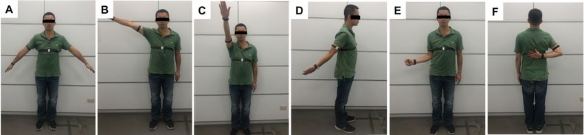
JOURNAL OF MEDICAL INTERNET RESEARCH Chen et al
Figure 1. Angular measurement of the shoulder with motion sensors worn on the body for (A) calibration, (B) shoulder abduction, (C) shoulder flexion,
(D) shoulder extension, (E) shoulder external rotation, (F) shoulder internal rotation.
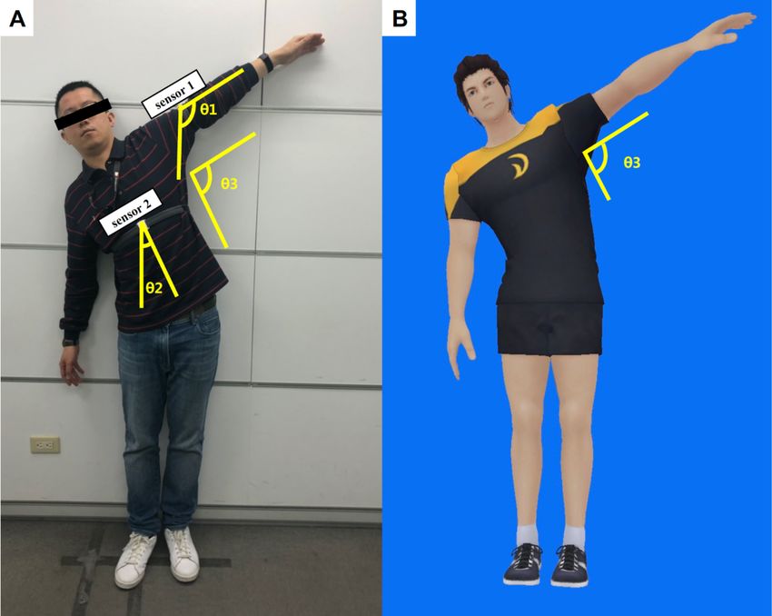
Dual-sensor systems were used in the motion sensor device to between the arm and axial body (by compensating for the angle
accurately record the shoulder ROM angle. Using the sensors of the body tilting relative to the ground), which was superior
on the sternum as a reference datum point, the raw data from for eliminating the error in the shoulder ROM measurement
each sensor can be converted into a quaternion algorithm to attributed to body tilting from a one-sensor system, especially
convert the relative angle changes of the sensors on the upper for patients with AC and painful shoulder movement (Figure
arm and wrist into a 3-dimensional (3D) motion of the shoulder 2). Additionally, repeated angular measurements with the
structure. Although one sensor can provide the angular change dual-sensor system on simulated shoulder motion was validated
with the gyroscopes on one plane of motion, dual-sensor systems as highly accurate. This is referred to in Multimedia Appendix
can provide relative angular changes in shoulder movements 2.
Figure 2. Dual-sensor system simulates the motion of (A) patients with adhesive capsulitis performing shoulder abduction using a (B) 3-dimensional
avatar. θ1: angular motion reported by the sensor on the upper arm; θ2: axial body tilting angle reported by the sensor on the sternum; θ3: shoulder
abduction angle (ie, θ1 - θ2).
http://www.jmir.org/2020/7/e17032/ J Med Internet Res 2020 | vol. 22 | iss. 7 | e17032 | p. 3
(page number not for citation purposes)
XSL• FO
RenderX
JOURNAL OF MEDICAL INTERNET RESEARCH                                                                                                                  Chen et al

     Mobile Phone App for Patients                                                  stretch, lateral wall walking stretch, and cane stretch for shoulder
     To design the mobile app for patients with AC for use on mobile                flexion, extension, abduction, internal rotation, and external
     phones, a cocreation process was used. The app has modes for                   rotation (Figure 4). The mobile app assigns each set of exercises
     measuring shoulder mobility, generating historical records of                  as a “daily task” assigned by the supervising physiotherapist or
     angular measurements and exercise completion rates, and                        physicians and provides a demonstration using a 3D avatar
     providing daily shoulder exercises with detailed instructions                  (Figure 3E). The app mirrors the user’s shoulder movement
     (Figure 3). The mobile app determines shoulder ROM in all                      during the exercise using angular information collected from
     directions by calculating the relevant information regarding the               the worn motion sensors. Each user has an account in the Patient
     angle measurement reported by the 3 wearable sensors (Figures                  App to access his or her records, which includes daily progress
     2B and 3B). The Patient App provides 7 sets of home-based                      and the completion rate of the daily exercises (Figures 3C and
     shoulder exercises for training, including a forward wall walking              3D).

     Figure 3. Examples of screens on the mobile app for patients, including the (A) functional introduction, (B) shoulder mobility measurement, (C)
     historical records of shoulder range of motion, (D) historical records of daily exercise completion rate, (E) daily home-based exercise tasks.

     Figure 4. Sets of home-based shoulder exercises displayed on the mobile app and demonstrated by participants: (A, H) forward wall walking stretch;
     (B, I) lateral wall walking stretch; (C, J) cane stretch for shoulder flexion; (D, K) cane stretch for shoulder abduction; (E, L) cane stretch for shoulder
     external rotation; (F, M) cane stretch for shoulder internal rotation; (G, N) cane stretch for shoulder extension.

                                                                                    personalized daily home-based exercises with adjustable targeted
     Mobile App for Physiotherapists and Physicians                                 angles, numbers of repetitions, and holding times for each
     The Doctor App was designed for physiotherapists and                           patient based on the angular status of the affected shoulder
     physicians for use on a mobile tablet. This app provides each                  (Figure 5B). Physiotherapists/physicians can directly
     patient’s information, including the latest shoulder ROM                       communicate with the patients by sending text messages through
     measurements and exercise completion rates for the previous                    the Doctor App.
     week (Figure 5A). Physiotherapists and physicians can assign

     http://www.jmir.org/2020/7/e17032/                                                                          J Med Internet Res 2020 | vol. 22 | iss. 7 | e17032 | p. 4
                                                                                                                               (page number not for citation purposes)
XSL• FO
RenderX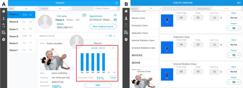
JOURNAL OF MEDICAL INTERNET RESEARCH                                                                                                              Chen et al

     Figure 5. Examples of screens on the mobile app for physiotherapists and physicians, including the (A) daily and weekly exercise completion rate of
     each patient displayed with patient information and (B) doctor-assigned daily exercises with specified targeted angles, holding times, and numbers of
     repetitions.

                                                                                 For angle measurements, aROM and pROM for flexion,
     Study Protocol                                                              extension, abduction, and internal and external rotation at a
     This study included two investigations. The first evaluated the             neutral position for both shoulders of healthy participants and
     reliability of measurements of shoulder ROM using the motion                for the affected shoulder of the patients with AC were assessed.
     sensor device, and the second confirmed the effectiveness of                The shoulder ROM in all directions for each participant was
     using the motion sensor device for the rehabilitation training of           first assessed using the motion sensor device. Two examiners
     patients with AC. The Ethical Committee of Taipei Medical                   (one well-trained physiotherapist and one highly experienced
     University approved the entire protocol and instrumentation                 orthopedic surgeon) who were blinded to the angle
     (TMU-JIRB N201708048). All participants consented to                        measurements by the motion sensor device measured shoulder
     participation in the study and the publication of data.                     ROM in all directions using a universal goniometer.
     Reliability of Shoulder ROM Measurements Using a                            Each participant stood straight, and scapular rotation was
     Motion Sensor Device                                                        permitted to measure abduction, flexion, and extension (Figures
     From January 2017 to August 2017, two groups of participants                1B, 1C, 1D). External rotation was measured in a neutral
     — 10 healthy participants and 15 patients with a confirmed                  position with the shoulder abducted, the elbow flexed at 90°,
     diagnosis of AC — were prospectively enrolled for measurement               and the forearm in a neutral position; the angle between the
     of shoulder angular motion.                                                 long axis of the forearm and the sagittal plane of the trunk was
                                                                                 measured as external rotation (Figure 1E). Internal rotation was
     Volunteers aged 20 to 70 years were included in the group of                determined by having the participants reach up the back to the
     healthy participants. Participants who reported discomfort or               highest point along the midline (Figure 1F). Six levels of internal
     limited ROM of the shoulder within the preceding year were                  rotation were defined according to the areas that the participant’s
     excluded. Patients were included in the AC group who (1) were               hand reached, as follows: interscapular region (T7), T12
     aged between 40 and 70 years, (2) had spontaneous onset of a                vertebra, waist (L3), lumbosacral junction, buttock, and lateral
     painful stiff shoulder and marked loss of active and passive                thigh. A high reach corresponded to better internal rotation. The
     global shoulder motion, (3) had reported local shoulder pain                aROM was measured by instructing participants to move their
     that was frequently present either over the anteromedial aspect             arm as far as they could, and the pROM was measured as the
     of the shoulder extending distally into the biceps region or over           examiner moved the subject’s arm to its mechanical limit or a
     the lateral aspect of the shoulder extending into the lateral               limit imposed by pain.
     deltoid region, and (4) had symptoms for at least 3 months, with
     normal findings on anteroposterior and axillary radiographs of              Effectiveness of Motion Sensor Device for Rehabilitation
     the glenohumeral joint. Patients were excluded if they (1) had              Training of Patients with AC
     pathological findings of rotator cuff tear on physical examination          The 15 patients with confirmed diagnoses of AC were allocated
     (eg, abduction weakness and positive dropped arm test); (2) had             to 2 intervention groups: 7 patients to the home-based exercise
     glenohumeral osteoarthritis upon radiographic evaluation; (3)               group and 8 patients to the motion sensor–assisted rehabilitation
     had clinical evidence of severe cervical spine disease; (4) had             group. The patients were allocated according to their willingness
     a history of severe trauma to the shoulder; (5) received a local            to use, and familiarity with, mobile apps. The patients in both
     corticosteroid injection or any physiotherapeutic intervention              groups received 3 months of rehabilitation.
     to the affected shoulder in the 3 months preceding the study
     start date; (6) had a history of inflammatory joint disease,                Patients with AC in the home-based exercise group received
     infection, stroke, or thyroid diseases that affected the shoulder;          comprehensive instructions on the daily shoulder exercises from
     or (7) were unwilling to undergo an intervention or participate             an experienced orthopedic surgeon before beginning the exercise
     in the trial.                                                               program. The information included instructions for home-based

     http://www.jmir.org/2020/7/e17032/                                                                      J Med Internet Res 2020 | vol. 22 | iss. 7 | e17032 | p. 5
                                                                                                                           (page number not for citation purposes)
XSL• FO
RenderX
JOURNAL OF MEDICAL INTERNET RESEARCH Chen et al
exercises; a description of frozen shoulder; and advice on sleep, the 1-month, 2-month, and 3-month follow-ups by an
posture, and pain relief. Home-based exercises included independent physiotherapist who was not involved in treating
Codman’s or pendulum exercises (circumduction) and passive any patients and was blinded to the treatment allocation. This
stretching exercises, including “wall walking stretch exercises” questionnaire was based on a pain severity visual analog scale
and “cane stretch exercises” (Figure 4). Further information (VAS) and the Quick Disabilities of the Arm, Shoulder, and
regarding the standard protocol of the home-based exercises is Hand (qDASH) questionnaire. The monthly exercise completion
available in Multimedia Appendix 3. The patients were rate, as reported by all participants and by the motion sensor
instructed to perform these exercises within the painless range, devices in the motion sensor–assisted rehabilitation group, was
maintain the maximal achievable angle for at least 10 seconds recorded every month.
in each exercise, and repeat each exercise at least 10 times daily.
All patients were instructed to follow the standard home-based
Instruments
exercise protocol, but individualized modification was permitted Measurement of Shoulder Function
based on health professionals’ judgment. In practice, an
The self-reported qDASH, consisting of 11 items, was developed
orthopedic surgeon reviewed the progress of each patient every
to measure the relevant physical function and symptoms of
month. They gave advice and modified the exercise program
patients with upper limb disorders [19]. The Chinese version
based on the patient’s compliance and AC status. Therefore, if
of qDASH has excellent reliability (Cronbach α=.818, intraclass
a patient had poor performance on a specific direction of
correlation coefficient [ICC]=0.907) [20]. A higher qDASH
shoulder ROM after the exercise program of the previous month
score indicates poorer shoulder function.
(eg, on shoulder abduction), the orthopedic surgeon could make
adjustments to the next month’s exercise protocol by increasing Measurement of Pain
the daily exercise frequency to 15-20 times and the holding time A VAS is an instrument that is regularly used to measure the
to 15-20 seconds per session for the “lateral wall walking intensity of pain [21]. A researcher asked the patients, “On a
stretch” and “cane stretch for shoulder abduction” exercises. scale of 0 to 10, how severe has been the worst pain that you
All patients in the motion sensor–assisted rehabilitation group have experienced within the last week?” This question was
received detailed instructions on operating the motion sensor repeated in the follow-up sessions.
device and Patient App on their own mobile phones before they Measurement of Exercise Completion Rate
began the rehabilitation program. The aforementioned
orthopedic surgeon assigned the standard protocol of daily The exercise completion rate reported by the motion sensor
home-based shoulder exercises to each patient via the Patient device was calculated by dividing the number of exercises
App with individualized modifications according to the patient’s completed daily by the number of assigned exercise tasks daily.
performance. The completion rate of these daily tasks was The monthly exercise completion rate was the average of the
calculated using the app for each patient and sent to the Doctor daily exercise completion rates over an entire month. The
App over the internet, enabling the supervisor to immediately patient-reported exercise completion rate in each month was
discuss the progress of each patient with them. In addition, obtained by asking each patient, “On a scale of 0%-100%, what
patients in the motion sensor–assisted rehabilitation group were was your average exercise completion rate in the previous
individually assigned each set of stretching exercises with the month?”
target angle on the mobile app by the orthopedic surgeon based Statistical Analysis
on the patients’ previous shoulder angular performance (Figure
The ICC was used to calculate the interrater reliability between
5B). For example, if the patient had 100° shoulder abduction
the shoulder aROM and pROM outcomes measured by the
before starting the shoulder exercise program, the target angle
motion sensor device and the two experienced examiners. The
for the “lateral wall walking stretch” and “cane stretch for
ICC was obtained by conducting a 2-way analysis of variance
shoulder abduction” would be assigned as 120° with an
in a random effects model. ICCs >0.8 and >0.9 were considered
acceptable range of 30°. Achievement of the target angle of the
to indicate good and excellent reliability, respectively [22]. The
assigned exercise could be recorded as a successful count for
ICC was further used to determine the minimal clinically
calculating the daily exercise completion rate on the mobile app
important difference (MCID) in a distribution-based method.
(Figure 3D). In this manner, the patient could therefore stretch
Specifically, the standard error of the measurement (SEM) was
the involved shoulder to the maximal limit of the angle on each
first computed using the following equation: SEM = SD of the
attempt, thereby effectively maximizing the training effect of
baseline measures × 0√1 − ICC; then, the MCID was computed
stretching exercises.
as follows: SEM × 1.96 × √2 [23,24].
Outcome Measurement In the second part of the study, in which the home-based
Basic demographic data, including age, gender, and educational exercise and motion sensor–assisted rehabilitation groups were
status, were collected for each patient with AC through compared, the Mann-Whitney U test was used to compare two
face-to-face interviews. For all patients, shoulder aROM and independent, ordinal groups, regardless of the normal
pROM in all directions of the affected shoulder were assessed distribution of the metric of interest owing to small sample sizes.
and recorded by the motion sensor device at baseline and each The Wilcoxon signed-rank test was used for within-group
month for 3 months following the initiation of the rehabilitation comparisons of the dependent variables at follow-up. The MCID
program. All patients received and completed questionnaires computed from the first part of the study was used to illustrate
that allowed relevant metrics to be evaluated at baseline and at whether the changes in aROM and pROM were meaningful.
http://www.jmir.org/2020/7/e17032/ J Med Internet Res 2020 | vol. 22 | iss. 7 | e17032 | p. 6
(page number not for citation purposes)
XSL• FO
RenderXJOURNAL OF MEDICAL INTERNET RESEARCH Chen et al
Along with descriptive statistics concerning participant
characteristics, generalized estimating equations were used to
Results
compare the two groups with respect to improvements in several Regarding the first part of the study on the reliability of
outcomes (including aROM, pROM, VAS, and qDASH). All measurement by the motion sensor device, Table 1 presents the
generalized estimating equations were analyzed using the results for shoulder aROM and pROM in all directions for
restricted maximum likelihood estimation. They all controlled healthy participants and for patients with AC, as obtained by
for the effects of time during follow-up. SPSS 23.0 (IBM Corp, the motion sensor device and the two examiners.
Armonk, NY) was used for all data analyses. P≤.05 was
considered to indicate statistical significance.
Table 1. Active and passive ranges of motion of the shoulder measured by different examiners.
Examiner Abduction (°), mean (SD) Flexion (°), mean (SD) Extension (°), mean External rotation (°), Internal rotation
(SD) mean (SD) (level)a
Active Passive Active Passive Active Passive Active Passive Active Passive
Healthy participants (10 patients, 20 shoulders)
Examiner 1 167.4 (9.1) 173.2 (5.2) 161 (8.4) 164.9 (5.6) 61.5 (7.9) 74.7 (8.7) 74.7 (6.6) 85.6 (4.6) 13b, 6c, 18b, 2c
1d
Examiner 2 167.1 (8.5) 171.6 (6.7) 161.8 (9.9) 166 (7.1) 59.3 (5.4) 71.3 (8.0) 74.3 (8.7) 84.6 (4.8) 11b, 9c 17b, 3c
Motion sen- 162.4 (9.1) 171.7 (7.3) 156.6 (9.4) 162.9 (6.4) 56.2 (7.9) 69.1 77.6 (9.9) 86.2 (6.2) 15b, 5c 19b, 1c
sor device (10.8)
Patients with adhesive capsulitis (15 patients, 15 shoulders)
Examiner 1 117.6 (18.1) 128.9 (18.1) 130.7 (13.8) 141.8 (10.5) 56.7 (11.7) 65.7 (9.5) 49.3 61.3 1b, 2c, 3b, 7c,
(11.6) (12.1) d
12 5d
Examiner 2 118.8 (17.4) 129.5 (18.3) 132 (14.2) 144.9 (9.4) 56.3 (9.1) 64.7 (9.3) 51.4 60.1 1b, 3c, 3b, 7c,
(10.8) (11.0) d
11 5d
Motion sen- 118.7 (17.8) 131.7 (18.7) 133.7 (13.2) 146.7 (11.4) 55.7 (9.8) 63.9 (8.5) 49.5 59.6 1b, 2c, 3b, 7c,
sor device (11.1) (12.2) d e
11 , 1 5d
a
Reported at the internal rotation level.
b
Interscapular (T7).
c
T12 vertebra.
d
Waist (L3).
e
Lumbosacral junction.
Table 2 shows whether the interrater reliability for the 0.899-0.979). The interrater reliability of the measurement of
measurement of shoulder aROM and pROM in all directions, active and passive shoulder extension was fair to good (aROM:
except for shoulder extension, as assessed by the motion sensor 0.771; pROM: 0.799).
device and the two examiners was good or excellent (ICC range
Table 2. Interobserver reliability between angle measurements obtained by different examiners.
Values Abduction Flexion Extension External rotation Internal rotation
Active Passive Active Passive Active Passive Active Passive Active Passive
ICCa among examiners 0.971 0.979 0.952 0.899 0.771 0.799 0.951 0.960 0.914 0.966
95% lower limit 0.950 0.964 0.918 0.832 0.640 0.680 0.916 0.931 0.855 0.941
95% upper limit 0.984 0.989 0.974 0.944 0.867 0.885 0.973 0.978 0.952 0.981
a
ICC: intraclass correlation coefficient.
In the second part of the study on the effectiveness of the motion rehabilitation. The patient received a subsequent diagnosis of
sensor device in rehabilitation training, one patient in the motion a full-thickness rotator cuff tear based on shoulder magnetic
sensor–assisted rehabilitation group was excluded from the resonance imaging. Therefore, the comparison was of 7 patients
analysis owing to progressive shoulder pain and newly in the motion sensor–assisted rehabilitation group and 7 patients
developed shoulder abduction weakness 1 month after in the home-based exercise group. Table 3 presents the patient
http://www.jmir.org/2020/7/e17032/ J Med Internet Res 2020 | vol. 22 | iss. 7 | e17032 | p. 7
(page number not for citation purposes)
XSL• FO
RenderXJOURNAL OF MEDICAL INTERNET RESEARCH Chen et al
demographics, which did not differ significantly at baseline between the two groups.
Table 3. Comparison of patient demographics between groups.
Demographic characteristics Motion sensor–assisted rehabilitation group (n=7) Home-based exercise group (n=7) P value
Lesion side, n (%)
Left 3 (42.9) 4 (57.1) .59
Right 4 (57.1) 3 (42.9)
Age (years), mean (SD) 53.0 (6.2) 56.1 (13.3) .35
Gender, n (%)
Male 4 (57.1) 5 (71.4) .58
Female 3 (42.9) 2 (28.6)
Education, n (%)
Senior high 2 (28.6) 4 (57.1) .28
Bachelor’s degree and higher 5 (71.4) 3 (42.9)
Symptom duration (months), n (%)
3-6 4 (57.1) 3 (42.9) .59
6-12 3 (42.9) 4 (57.1)
Range of motion, mean (SD)
Active abduction (°) 122.4 (15.7) 113.1 (20.3) .48
Passive abduction (°) 126.9 (16.9) 131.6 (17.1) .65
Active flexion (°) 138.4 (14.4) 127.9 (11.0) .28
Passive flexion (°) 146.4 (15.7) 146.7 (7.3) .75
Active extension (°) 56.4 (12.6) 54.1 (5.0) .95
Passive extension (°) 63.9 (11.0) 63.1 (6.6) .95
Active external rotation (°) 52.4 (11.6) 49.0 (9.7) .48
Passive external rotation (°) 61.7 (14.0) 58.0 (11.9) .52
Active internal rotation (°)a 1b, 4c, 2d 1b, 2c, 4d .16
Passive internal rotation (°)a 2b, 4c, 1d 1b, 4c, 2d .67
VASe score, mean (SD) 5.3 (1.3) 6.1 (1.8) .37
qDASHf, mean (SD) 30.6 (18.1) 23.3 (7.2) .90
a
Reported at the internal rotation level.
b
Interscapular (T7).
c
T12 vertebra.
d
Waist (L3).
e
VAS: visual analogue scale.
f
qDASH: Quick Disabilities of the Arm, Shoulder, and Hand questionnaire.
After 3 months of follow-up, patients in the motion aROM and pROM in shoulder extension, and VAS score (Table
sensor–assisted rehabilitation group exhibited a significant 5). Additionally, when the improvements in the motion
improvement from baseline in terms of shoulder aROM and sensor–assisted rehabilitation and home-based exercise groups
pROM in all directions, pain score, and qDASH (Table 4). By were compared, the motion sensor–assisted rehabilitation group
contrast, patients in the home-based exercise group exhibited had achieved better and faster meaningful changes than the
significant improvements only in aROM in shoulder abduction, home-based exercise group.
http://www.jmir.org/2020/7/e17032/ J Med Internet Res 2020 | vol. 22 | iss. 7 | e17032 | p. 8
(page number not for citation purposes)
XSL• FO
RenderXJOURNAL OF MEDICAL INTERNET RESEARCH Chen et al
Table 4. Improvement in parameters compared with baseline in the motion sensor–assisted rehabilitation group.
Parameters Baseline 1 month P value 2 months P value 3 months P value MCIDa
Abduction (°), mean (SD)
Active 122.4 (15.7) 148.1 (17.5) .04 154.1 (11.9) .02 156.7 (9.9) .02 5.99
Passive 126.9 (16.9) 152.9 (17.1) .03 157.6 (10.7) .02 161.6 (10.3) .02 4.63
Flexion (°), mean (SD)
Active 138.4 (14.4) 149.1 (11.4) .02 151.0 (5.4) .04 159.6 (8.5) .02 6.78
Passive 146.4 (15.7) 162.0 (11.9) .02 164.7 (5.6) .03 170.1 (8.1) .02 7.14
Extension (°), mean (SD)
Active 56.4 (12.6) 62.7 (12.6) .06 65.9 (10.8) .03 73.6 (11.9) .03 11.15
Passive 63.9 (11.0) 69.3 (12.2) .11 78.1 (11.4) .03 84.3 (6.4) .02 11.36
External rotation (°), mean (SD)
Active 52.4 (11.6) 66.4 (15.4) .02 64.7 (14.8) .02 64.6 (18.8) .04 5.88
Passive 61.7 (14.0) 67.6 (18.6) .20 71.9 (15.2) .03 76.7 (15.1) .02 4.44
Internal rotation (level)b, mean (SD)
Active 1c, 5d, 1e 1f, 4c, 2d .01 3f, 2c, 2d .04 3f, 2c, 2d .04 —g
Passive 1f, 3c, 3d 2f, 4c, 1d .08 3f, 4c .06 3f, 4c .06 —g
VASh score, mean (SD) 5.3 (1.3) 4.1 (1.1) .12 2.7 (1.6) .03 2.0 (0.6)+ .02 —g
qDASHi score, mean (SD) 30.6 (18.1) 15.5 (7.5) .03 11.4 (9.5) .02 9.8 (12.4) .02 —g
a
MCID: minimal clinically important difference. Computed using a distribution-based method using the standard error of measurement (SEM). SEM
= SD of the baseline measurements * √1-reliability, where the reliability used was retrieved from the intraclass correlation coefficient, and MCID =
SEM * 1.96 * √2.
b
Internal rotation level.
c
T12 vertebra.
d
Waist (L3).
e
Lumbosacral junction.
f
Interscapular (T7).
g
Not applicable.
h
VAS: visual analogue scale.
i
qDASH: Quick Disabilities of the Arm, Shoulder, and Hand questionnaire.
http://www.jmir.org/2020/7/e17032/ J Med Internet Res 2020 | vol. 22 | iss. 7 | e17032 | p. 9
(page number not for citation purposes)
XSL• FO
RenderXJOURNAL OF MEDICAL INTERNET RESEARCH Chen et al
Table 5. Improvement in parameters compared with baseline and with time in the home-based exercise group.
Parameters Baseline 1 month P value 2 months P value 3 months P value MCIDa
Abduction (°), mean (SD)
Active 113.1 (20.3) 115.9 (26.4) .35 127.9 (21.6) .04 134.3 (21.8) .051 5.99
Passive 131.6 (17.1) 133.9 (15.9) .40 135.3 (15.6) .18 142.7 (18.2) .13 4.63
Flexion (°), mean (SD)
Active 127.9 (11.0) 132.3 (15.1) .13 138.0 (13.7) .03 140.0 (11.2) .03 6.78
Passive 146.7 (7.3) 148.6 (9.5) .35 152.7 (8.3) .07 154.3 (8.1) .13 7.14
Extension (°), mean (SD)
Active 54.1 (5.0) 55.7 (6.9) .50 58.3 (4.6) .26 56.0 (5.9) .35 11.15
Passive 63.1 (6.6) 63.3 (8.0) .93 65.9 (6.4) .50 68.3 (6.3) .27 11.36
External rotation (°), mean (SD)
Active 49.0 (9.7) 50.9 (8.7) .46 51.1 (8.4) .45 53.1 (12.9) .13 5.88
Passive 58.0 (11.9) 58.7 (10.2) .67 59.1 (10.2) .40 60.9 (11.5) .13 4.44
Internal rotation (level)b, mean (SD)
Active 1c, 2d, 4e 1c, 2d, 3e,1d .56 1c, 2d, 4e >.99 1c, 2d, 3e, 1f .56 —g
Passive 1c, 4d, 2e 1c, 3d, 3e .56 2c, 3d, 2e .66 3c, 2d, 2e .41 —g
VASh score, mean (SD) 6.1 (1.8) 5.6 (1.7) .46 4.1 (1.1) .04 3.3 (1.1) .02 —g
qDASHi score, mean (SD) 23.3 (7.2) 24.4 (18.7) .80 19.8 (12.1) .35 19.1 (13.7) .25 —g
a
MCID: minimal clinically important difference. Computed using a distribution-based method using the standard error of measurement (SEM). SEM
= SD of the baseline measurements * √1-reliability, where the reliability used was retrieved from the intraclass correlation coefficient, and MCID =
SEM * 1.96 * √2.
b
Internal rotation level.
c
Interscapular (T7).
d
T12 vertebra.
e
Waist (L3).
f
Lumbosacral junction.
g
Not applicable.
h
VAS: visual analogue scale.
i
qDASH: Quick Disabilities of the Arm, Shoulder, and Hand questionnaire.
A comparison of the improvements in the motion the changes in shoulder ROM in most of the directions at
sensor–assisted rehabilitation and home-based exercise groups different follow-up time points from baseline were correlated
over time revealed that, compared with patients in the with improvements in qDASH score, inferring that the motion
home-based exercise group, those in the motion sensor–assisted sensor device is a reliable tool for evaluating treatment efficacy
rehabilitation group had significantly better pROM in shoulder in patients with AC (Multimedia Appendix 4). Table 7 shows
abduction, flexion, extension, and external rotation; better that, compared with patients in the home-based exercise group,
aROM in shoulder extension and internal rotation; and a better those in the motion sensor–assisted rehabilitation group had a
qDASH score at the 3-month follow-up (Table 6). In addition, significantly higher patient-reported exercise completion rate.
http://www.jmir.org/2020/7/e17032/ J Med Internet Res 2020 | vol. 22 | iss. 7 | e17032 | p. 10
(page number not for citation purposes)
XSL• FO
RenderXJOURNAL OF MEDICAL INTERNET RESEARCH Chen et al
Table 6. Generalized estimating equation analysis for improvements between groups with time (reference: home-based exercise group).
Dependent variables 1 month, beta (SE) P value 2 months, beta (SE) P value 3 months, beta (SE) P value
Abduction (o)
Active 23.00 (10.49) .03 17.00 (8.09) .04 13.14 (9.83) .19
Passive 23.71 (8.37) .006 27.00 (5.49)JOURNAL OF MEDICAL INTERNET RESEARCH Chen et al
difficult to access, and requires expertise to operate, preventing can be applied in conjunction with telerehabilitation.
its general clinical use. Wearable IMU-based sensors are Telerehabilitation is the provision of rehabilitation services at
becoming popular and have the potential to measure the joint a distance; it can be image-based or sensor-based and can be
angles of upper limbs with acceptable reliability [28]. Studies delivered using virtual environments or virtual reality [32]. A
have already revealed that wearable IMU-based sensors can motion sensor device can support image-based and sensor-based
track shoulder movements with high accuracy [29]. In the telerehabilitation using an activity recognition model, an
present study, shoulder ROM measurement by IMU sensors interactive 3D avatar in mobile phone apps, and a wireless
yielded good to excellent interrater reliability with reports from telecommunicated network, supporting medical treatment for
well-experienced physicians who were using goniometers; it patients with AC. The future use of motion sensor devices in
can therefore provide reliable angular information that telerehabilitation for various muscle skeletal disorders is
physicians can use to monitor the progress of shoulder expected after its development reaches maturation.
rehabilitation and facilitate the design of upper limb exercises
that promote the rehabilitation of patients with AC.
Limitations
The main limitations of this study were the relatively small
An important aspect of the motion sensor device used in this number of patients, short follow-up period, and consequent lack
study is the integration of a mobile phone–based system. Mobile of information on the long-term effects of the intervention. The
phone apps for medical and rehabilitation purposes that are allocation of patients was not randomized, owing to their varying
adaptable and easily accessible are being intensively researched degrees of familiarity with mobile apps and motion sensor
[29,30]. Such apps may help in the development of a platform devices. Studies have found that new rehabilitation technology
for delivering self-management interventions, thus improving may be unlikely to be accepted by many elderly patients [33].
the delivery of health care and outcomes [28]. Evidence has In this study, the targeted patients with AC were 40-70 years
also revealed that app-based exercise instructions and tools can old. Within this population, younger and highly educated
help patients to monitor their training compliance and progress patients are generally accepting of motion sensor device–assisted
with high acceptance and usability [29]. In this study, the mobile rehabilitation and so were likely to be allocated to the motion
apps with the motion sensor device system provided an sensor–assisted rehabilitation group. Researchers of future
informative patient interface with comprehensible exercise studies should consider a training program for motion sensor
instructions and simple progress monitoring; they support device usage before enrollment to prevent any potential selection
self-managed health care anywhere through the monitoring of bias. Finally, although this study revealed that the IMU-based
rehabilitation exercise performance. Wireless telecommunication sensors that were used to measure shoulder ROM were as
is used to synchronize with the Doctor App, send information reliable as goniometers used by two well-experienced
regarding each patient’s progress with home-based exercises, physicians, the true accuracy of IMU-based sensors for shoulder
enable physicians to remotely supervise rehabilitation, and ROM measurement is still unclear. Evidence has confirmed that
provide instant feedback to patients. Studies have established the reliability of shoulder and elbow ROM measurement using
that the delivery of simple text message reminders to mobile the goniometer varies (interrater ICC, 0.36-0.91) [25]. Further
phones increases the compliance of AC patients to a shoulder assessment may be required to compare IMU-based sensors and
exercise regimen [31]. With the assistance of a mobile app that the 3DMA system in the measurement of shoulder ROM with
is used in conjunction with a motion sensor device for shoulder regard to accuracy and reliability.
rehabilitation, physiotherapists or physicians can not only send
text messages to remind patients to perform their daily exercises Conclusions
but also assign personal home-based exercises based on their Wearable IMU-based sensors can reliably record the angular
training performance, thus increasing their motivation. motion of shoulders. Motion sensor device–assisted home-based
Overall, the positive results justify further work on motion rehabilitation can increase patient compliance with a daily
sensor devices for treating AC. Further investigations of the self-exercise regime and facilitate the early stage of functional
usability of mobile apps and the development of auto-adjusted recovery for patients with shoulder AC. Collectively, motion
rehabilitation programs that are based on a user’s personal sensor device–assisted home-based rehabilitation is a useful
performance are warranted before well-controlled research is treatment model for AC with telerehabilitation to overcome
conducted with large patient cohorts to demonstrate the viability obstacles to physiotherapy at home; it provides comprehensive
of motion sensor devices in real-world environments. The and easily accessible exercise instructions, increases compliance,
motion sensor device–assisted rehabilitation model in this study and enhances exercise correctness through progress monitoring.
Acknowledgments
The authors acknowledge financial support from the Ministry of Science and Technology (MOST 109-2926-I-010-501, MOST
107-2314-B-010-015-MY3, MOST 109-2926-I-010-502, MOST 109-2321-B-010-005 and MOST 108-2923-B-010-002-MY3).
This work was specifically supported by the “Development and Construction Plan” of the School of Medicine, National Yang-Ming
University (107F-M01-0504) and Aiming for the Top University Plan, a grant from the Ministry of Education.
Conflicts of Interest
None declared.
http://www.jmir.org/2020/7/e17032/ J Med Internet Res 2020 | vol. 22 | iss. 7 | e17032 | p. 12
(page number not for citation purposes)
XSL• FO
RenderXJOURNAL OF MEDICAL INTERNET RESEARCH Chen et al
Multimedia Appendix 1
BoostFix Quick Guide.
[DOCX File , 7159 KB-Multimedia Appendix 1]
Multimedia Appendix 2
Accuracy of Angular Measurement obtained with Single-Sensor and Dual-Sensor Systems.
[DOCX File , 2261 KB-Multimedia Appendix 2]
Multimedia Appendix 3
Home-based Exercise Protocol for Shoulder Adhesive Capsulitis.
[DOCX File , 839 KB-Multimedia Appendix 3]
Multimedia Appendix 4
Correlation between changes in qDASH score and shoulder ROM in the different directions at different time points of follow-up
from baseline.
[DOCX File , 14 KB-Multimedia Appendix 4]
References
1. Neviaser AS, Hannafin JA. Adhesive capsulitis: a review of current treatment. Am J Sports Med 2010 Nov;38(11):2346-2356.
[doi: 10.1177/0363546509348048] [Medline: 20110457]
2. Robinson CM, Seah KTM, Chee YH, Hindle P, Murray IR. Frozen shoulder. J Bone Joint Surg Br 2012 Jan;94(1):1-9.
[doi: 10.1302/0301-620X.94B1.27093] [Medline: 22219239]
3. Hsu JE, Anakwenze OA, Warrender WJ, Abboud JA. Current review of adhesive capsulitis. J Shoulder Elbow Surg 2011
Apr;20(3):502-514. [doi: 10.1016/j.jse.2010.08.023] [Medline: 21167743]
4. Shaffer B, Tibone JE, Kerlan RK. Frozen shoulder. A long-term follow-up. J Bone Joint Surg Am 1992 Jun;74(5):738-746.
[Medline: 1624489]
5. Neviaser AS, Neviaser RJ. Adhesive capsulitis of the shoulder. J Am Acad Orthop Surg 2011 Sep;19(9):536-542. [doi:
10.5435/00124635-201109000-00004] [Medline: 21885699]
6. Rookmoneea M, Dennis L, Brealey S, Rangan A, White B, McDaid C, et al. The effectiveness of interventions in the
management of patients with primary frozen shoulder. J Bone Joint Surg Br 2010 Sep;92(9):1267-1272. [doi:
10.1302/0301-620X.92B9.24282] [Medline: 20798446]
7. Maund E, Craig D, Suekarran S, Neilson A, Wright K, Brealey S, et al. Management of frozen shoulder: a systematic review
and cost-effectiveness analysis. Health Technol Assess 2012;16(11):1-264 [FREE Full text] [doi: 10.3310/hta16110]
[Medline: 22405512]
8. Hanchard NCA, Goodchild L, Thompson J, O'Brien T, Davison D, Richardson C. Evidence-based clinical guidelines for
the diagnosis, assessment and physiotherapy management of contracted (frozen) shoulder: quick reference summary.
Physiotherapy 2012 Jun;98(2):117-120. [doi: 10.1016/j.physio.2012.01.001] [Medline: 22507361]
9. Tanaka K, Saura R, Takahashi N, Hiura Y, Hashimoto R. Joint mobilization versus self-exercises for limited glenohumeral
joint mobility: randomized controlled study of management of rehabilitation. Clin Rheumatol 2010 Dec;29(12):1439-1444.
[doi: 10.1007/s10067-010-1525-0] [Medline: 20585816]
10. Russell S, Jariwala A, Conlon R, Selfe J, Richards J, Walton M. A blinded, randomized, controlled trial assessing conservative
management strategies for frozen shoulder. J Shoulder Elbow Surg 2014 Apr;23(4):500-507. [doi: 10.1016/j.jse.2013.12.026]
[Medline: 24630545]
11. Campbell R, Evans M, Tucker M, Quilty B, Dieppe P, Donovan JL. Why don't patients do their exercises? Understanding
non-compliance with physiotherapy in patients with osteoarthritis of the knee. J Epidemiol Community Health 2001
Feb;55(2):132-138 [FREE Full text] [Medline: 11154253]
12. Faber M, Andersen MH, Sevel C, Thorborg K, Bandholm T, Rathleff M. The majority are not performing home-exercises
correctly two weeks after their initial instruction-an assessor-blinded study. PeerJ 2015;3:e1102 [FREE Full text] [doi:
10.7717/peerj.1102] [Medline: 26244112]
13. Wang Q, Markopoulos P, Yu B, Chen W, Timmermans A. Interactive wearable systems for upper body rehabilitation: a
systematic review. J Neuroeng Rehabil 2017 Dec 11;14(1):20 [FREE Full text] [doi: 10.1186/s12984-017-0229-y] [Medline:
28284228]
14. Ongvisatepaiboon K, Vanijja V, Chignell M, Mekhora K, Chan J. Smartphone-Based Audio-Biofeedback System for
Shoulder Joint Tele-Rehabilitation. J Med Imaging Hlth Inform 2016 Aug 01;6(4):1127-1134. [doi: 10.1166/jmihi.2016.1810]
http://www.jmir.org/2020/7/e17032/ J Med Internet Res 2020 | vol. 22 | iss. 7 | e17032 | p. 13
(page number not for citation purposes)
XSL• FO
RenderXJOURNAL OF MEDICAL INTERNET RESEARCH Chen et al
15. Timmermans AAA, Seelen HAM, Geers RPJ, Saini PK, Winter S, te Vrugt J, et al. Sensor-based arm skill training in
chronic stroke patients: results on treatment outcome, patient motivation, and system usability. IEEE Trans Neural Syst
Rehabil Eng 2010 Jun;18(3):284-292. [doi: 10.1109/TNSRE.2010.2047608] [Medline: 20388603]
16. Lemmens RJM, Janssen-Potten YJM, Timmermans AAA, Smeets RJEM, Seelen HAM. Recognizing complex upper
extremity activities using body worn sensors. PLoS One 2015;10(3):e0118642 [FREE Full text] [doi:
10.1371/journal.pone.0118642] [Medline: 25734641]
17. O'Reilly MA, Whelan DF, Ward TE, Delahunt E, Caulfield BM. Technology in S&C: Assessing Bodyweight Squat
Technique with Wearable Sensors. J Strength Cond Res 2017 Apr 15:2303-2312. [doi: 10.1519/JSC.0000000000001957]
[Medline: 28426512]
18. Whelan DF, O'Reilly MA, Ward TE, Delahunt E, Caulfield B. Technology in Rehabilitation: Evaluating the Single Leg
Squat Exercise with Wearable Inertial Measurement Units. Methods Inf Med 2016 Oct 26;55(6):88-94. [doi:
10.3414/ME16-02-0002] [Medline: 27782290]
19. Beaton DE, Wright JG, Katz JN, Upper Extremity Collaborative Group. Development of the QuickDASH: comparison of
three item-reduction approaches. J Bone Joint Surg Am 2005 May;87(5):1038-1046. [doi: 10.2106/JBJS.D.02060] [Medline:
15866967]
20. Cao S, Zhou R, Zhou H, Chen Y, Cui H, Lu Z, et al. Reliability and validity of Simplified Chinese version of Quick
Disabilities of the Arm, Shoulder, and Hand (QuickDASH) questionnaire: cross-cultural adaptation and validation. Clin
Rheumatol 2019 Jul 03:3281-3287. [doi: 10.1007/s10067-019-04661-8] [Medline: 31270698]
21. Flandry F, Hunt JP, Terry GC, Hughston JC. Analysis of subjective knee complaints using visual analog scales. Am J Sports
Med 1991;19(2):112-118. [doi: 10.1177/036354659101900204] [Medline: 2039061]
22. Landis JR, Koch GG. The measurement of observer agreement for categorical data. Biometrics 1977 Mar;33(1):159-174.
[Medline: 843571]
23. Su C, Ng H, Yang A, Lin C. Psychometric evaluation of the Short Form 36 Health Survey (SF-36) and the World Health
Organization Quality of Life Scale Brief Version (WHOQOL-BREF) for patients with schizophrenia. Psychol Assess 2014
Sep;26(3):980-989. [doi: 10.1037/a0036764] [Medline: 24796341]
24. Cheng C, Luh W, Yang A, Su C, Lin C. Agreement of Children and Parents Scores on Chinese Version of Pediatric Quality
of Life Inventory Version 4.0: Further Psychometric Development. Applied Research Quality Life 2015 Apr 24;11(3):891-906.
[doi: 10.1007/s11482-015-9405-z]
25. Muir SW, Corea CL, Beaupre L. Evaluating change in clinical status: reliability and measures of agreement for the assessment
of glenohumeral range of motion. N Am J Sports Phys Ther 2010 Sep;5(3):98-110 [FREE Full text] [Medline: 21589666]
26. McGinley JL, Baker R, Wolfe R, Morris ME. The reliability of three-dimensional kinematic gait measurements: a systematic
review. Gait Posture 2009 Apr;29(3):360-369. [doi: 10.1016/j.gaitpost.2008.09.003] [Medline: 19013070]
27. Rau G, Disselhorst-Klug C, Schmidt R. Movement biomechanics goes upwards: from the leg to the arm. J Biomech 2000
Oct;33(10):1207-1216. [Medline: 10899329]
28. Walmsley CP, Williams SA, Grisbrook T, Elliott C, Imms C, Campbell A. Measurement of Upper Limb Range of Motion
Using Wearable Sensors: A Systematic Review. Sports Med Open 2018 Nov 29;4(1):53 [FREE Full text] [doi:
10.1186/s40798-018-0167-7] [Medline: 30499058]
29. Lin H, Chiang S, Lee K, Kan Y. An activity recognition model using inertial sensor nodes in a wireless sensor network for
frozen shoulder rehabilitation exercises. Sensors (Basel) 2015 Jan 19;15(1):2181-2204 [FREE Full text] [doi:
10.3390/s150102181] [Medline: 25608218]
30. Whitehead L, Seaton P. The Effectiveness of Self-Management Mobile Phone and Tablet Apps in Long-term Condition
Management: A Systematic Review. J Med Internet Res 2016;18(5):e97 [FREE Full text] [doi: 10.2196/jmir.4883] [Medline:
27185295]
31. Chen H, Chuang T, Lin P, Lin Y, Chuang Y. Effects of Messages Delivered by Mobile Phone on Increasing Compliance
With Shoulder Exercises Among Patients With a Frozen Shoulder. J Nurs Scholarsh 2017 Jul;49(4):429-437. [doi:
10.1111/jnu.12308] [Medline: 28632975]
32. Russell TG. Physical rehabilitation using telemedicine. J Telemed Telecare 2007;13(5):217-220. [doi:
10.1258/135763307781458886] [Medline: 17697506]
33. Axelrod L, Fitzpatrick G, Burridge J, Mawson S, Smith P, Rodden T, et al. The reality of homes fit for heroes: design
challenges for rehabilitation technology at home. Jnl of Assistive Technologies 2009 Jul 10;3(2):35-43. [doi:
10.1108/17549450200900014]
Abbreviations
AC: adhesive capsulitis
aROM: active range of motion
3D: 3-dimensional
3DMA: 3-dimensional motion analysis
ICC: intraclass correlation coefficient
http://www.jmir.org/2020/7/e17032/ J Med Internet Res 2020 | vol. 22 | iss. 7 | e17032 | p. 14
(page number not for citation purposes)
XSL• FO
RenderXJOURNAL OF MEDICAL INTERNET RESEARCH Chen et al
IMU: inertial measurement unit
MCID: minimal clinically important difference
pROM: passive range of motion
qDASH: Quick Disabilities of the Arm, Shoulder, and Hand
ROM: range of motion
SEM: standard error of the measurement
VAS: visual analogue scale
Edited by G Eysenbach; submitted 25.11.19; peer-reviewed by J Richards, H Lin; comments to author 13.01.20; revised version
received 04.03.20; accepted 14.05.20; published 23.07.20
Please cite as:
Chen YP, Lin CY, Tsai MJ, Chuang TY, Lee OKS
Wearable Motion Sensor Device to Facilitate Rehabilitation in Patients With Shoulder Adhesive Capsulitis: Pilot Study to Assess
Feasibility
J Med Internet Res 2020;22(7):e17032
URL: http://www.jmir.org/2020/7/e17032/
doi: 10.2196/17032
PMID:
©Yu-Pin Chen, Chung-Ying Lin, Ming-Jr Tsai, Tai-Yuan Chuang, Oscar Kuang-Sheng Lee. Originally published in the Journal
of Medical Internet Research (http://www.jmir.org), 23.07.2020. This is an open-access article distributed under the terms of the
Creative Commons Attribution License (https://creativecommons.org/licenses/by/4.0/), which permits unrestricted use, distribution,
and reproduction in any medium, provided the original work, first published in the Journal of Medical Internet Research, is
properly cited. The complete bibliographic information, a link to the original publication on http://www.jmir.org/, as well as this
copyright and license information must be included.
http://www.jmir.org/2020/7/e17032/ J Med Internet Res 2020 | vol. 22 | iss. 7 | e17032 | p. 15
(page number not for citation purposes)
XSL• FO
RenderXYou can also read