West Nile Virus: An Update on Pathobiology, Epidemiology, Diagnostics, Control and "One Health" Implications
←
→
Page content transcription
If your browser does not render page correctly, please read the page content below
pathogens
Review
West Nile Virus: An Update on Pathobiology,
Epidemiology, Diagnostics, Control and “One
Health” Implications
Gervais Habarugira 1 , Willy W. Suen 2 , Jody Hobson-Peters 3,4 , Roy A. Hall 3,4 and
Helle Bielefeldt-Ohmann 1,3,4, *
 1   School of Veterinary Science, University of Queensland Gatton Campus, Queensland, QLD 4343, Australia;
     g.habarugira@uq.net.au
 2   Australian Centre for Disease Preparedness, The Commonwealth Scientific and Industrial Research
     Organization, Geelong, VIC 3219, Australia; Willy.Suen@csiro.au
 3   School of Chemistry &Molecular Biosciences, The University of Queensland, St. Lucia, QLD 4072, Australia;
     j.peters2@uq.edu.au (J.H.-P.); roy.hall@uq.edu.au (R.A.H.)
 4   Australian Infectious Diseases Research Centre, University of Queensland, St. Lucia, QLD 4072, Australia
 *   Correspondence: h.bielefeldtohmann1@uq.edu.au

 Received: 28 June 2020; Accepted: 16 July 2020; Published: 19 July 2020

 Abstract: West Nile virus (WNV) is an important zoonotic flavivirus responsible for mild fever to
 severe, lethal neuroinvasive disease in humans, horses, birds, and other wildlife species. Since its
 discovery, WNV has caused multiple human and animal disease outbreaks in all continents, except
 Antarctica. Infections are associated with economic losses, mainly due to the cost of treatment of
 infected patients, control programmes, and loss of animals and animal products. The pathogenesis of
 WNV has been extensively investigated in natural hosts as well as in several animal models, including
 rodents, lagomorphs, birds, and reptiles. However, most of the proposed pathogenesis hypotheses
 remain contentious, and much remains to be elucidated. At the same time, the unavailability of
 specific antiviral treatment or effective and safe vaccines contribute to the perpetuation of the disease
 and regular occurrence of outbreaks in both endemic and non-endemic areas. Moreover, globalisation
 and climate change are also important drivers of the emergence and re-emergence of the virus and
 disease. Here, we give an update of the pathobiology, epidemiology, diagnostics, control, and “One
 Health” implications of WNV infection and disease.

 Keywords: West Nile virus; pathogenesis; control; one health

1. Introduction
     West Nile Virus (WNV) is a zoonotic, mosquito-borne flavivirus, one of about 75 virus species of the
Flaviviridae family [1]. WNV belongs to the Japanese encephalitis virus [2] serocomplex together with St.
Louis encephalitis virus (SLEV), Murray Valley encephalitis virus (MVEV), and Alfuy virus (ALFV) [2,3].
It was first isolated in the West Nile Province of Uganda in 1937 from a febrile patient [4–6]. Initially,
the virus was considered of less human importance as it only caused mild, subclinical infections [7].
However, the virus has been responsible for many cases of morbidity and mortalities in different
animal species, including birds [8–17], horses [6,18,19], sheep [20], reptiles [21–23], cats [24], and
rodents [6,25–29]. Over the last two decades, there have been notable increases in human and
equine cases.
     WNV is transmitted by a mosquito vector of the genus Culex through hematophagy [30–32].
However, some other ways of transmission, including ingestion, aerosol, and direct contact,
have been reported in experimental settings [33,34], in humans (intrauterine and breastfeeding

Pathogens 2020, 9, 589; doi:10.3390/pathogens9070589                           www.mdpi.com/journal/pathogens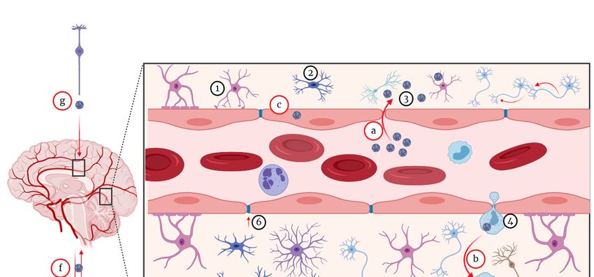
Pathogens 2020, 9, x FOR PEER REVIEW 2 of 51
Pathogens 2020, 9, 589 2 of 51
been reported in experimental settings [33,34], in humans (intrauterine and breastfeeding
transmission) [35–37], and recently also in farmed alligators and crocodiles [38–41]. To date, only one
transmission) [35–37], and recently also in farmed alligators and crocodiles [38–41]. To date, only one
case of vertical transmission has been reported in humans [36,42,43]. WNV transmission through
case of vertical transmission has been reported in humans [36,42,43]. WNV transmission through
blood transfusion and organ transplant have also been reported in humans [44,45]. The shedding of
blood transfusion and organ transplant have also been reported in humans [44,45]. The shedding of
the virus in urine during the acute phase of infection also suggests that transmission through contact
the virus in urine during the acute phase of infection also suggests that transmission through contact
with environmentally contaminated material might be possible [46]. Despite much effort invested in
with environmentally contaminated material might be possible [46]. Despite much effort invested in
vaccine development, there is currently no registered vaccine against WNV for use in humans.
vaccine development,
There have been there is reviews
several currentlycovering
no registered
variousvaccine
aspectsagainst
of WNV,WNV for usevirus
including in humans.
ecology and
There have been several reviews covering various aspects of WNV, including virus
pathobiology [1,47–50], epidemiology [1,51–53], medicine and clinical pathology [1,44,52,54], ecologyand
and
pathobiology [1,47–50],[55–57].
vaccine development epidemiology [1,51–53],
This review medicine
provides and clinical
a comprehensive pathology
update on WNV,[1,44,52,54],
focusing and
on
vaccine development [55–57]. This review provides a comprehensive update on WNV,
virus biology and pathobiology, epidemiology, diagnostics, public and One Health importance focusing
andon
virus biology and pathobiology, epidemiology, diagnostics, public and One Health importance
control, including new approaches made towards vaccine development, as well as other modes of and
control, including
prevention new approaches made towards vaccine development, as well as other modes of
and treatment.
prevention and treatment.
2. Virus Biology
2. Virus Biology
2.1. Genetic Organisation and Virus Replication
2.1. Genetic Organisation and Virus Replication
Like all flaviviruses, WNV contains a positive‐sense, single‐stranded RNA [ssRNA(+)] genome
Like all flaviviruses,
of approximately WNV
11 kb. The contains
genome a positive-sense,
is enclosed within ansingle-stranded RNA [ssRNA(+)]
enveloped, icosahedral nucleocapsidgenomewithof
approximately 11 kb. The genome is enclosed within an enveloped, icosahedral
mature virions appearing spherical in morphology with an approximate diameter of 50 nm [58,59]. nucleocapsid with
mature virions appearing spherical in morphology with an approximate diameter
The viral genome contains a single open reading frame (ORF) coding for a polyprotein that is cleaved of 50 nm [58,59]. The
viral
bothgenome
co‐ andcontains a single open[60].
post‐translationally reading frame (ORF)
The cleavage coding
of the for a polyprotein
polyprotein that
is facilitated by is cleaved
both both
the host
co- and post-translationally [60]. The cleavage of the polyprotein is facilitated by
cell and the viral proteases and gives rise to structural and non‐structural proteins [61]. The threeboth the host cell and
the viral proteases and gives rise to structural and non-structural proteins [61].
structural proteins include the capsid (C), pre‐membrane (prM), and the envelope (E) proteins [62] The three structural
proteins
(Figure include
1). There thearecapsid
seven(C), pre-membrane
non‐structural (NS) (prM), and encompassing
proteins, the envelope (E) proteins
NS1, NS2A,[62] (Figure
NS2B, NS3,1).
There are seven non-structural (NS) proteins, encompassing NS1, NS2A, NS2B,
NS4A, NS4B, and NS5, and they all play a crucial role in genome replication [63–69] (Figure 1; Table NS3, NS4A, NS4B,
and
1). NS5, and they
The open all play
reading a crucial
frame role in
is flanked ongenome replication
either side by 5′ and[63–69] (Figure 1; Table
3′ untranslated 1). The
regions open
(UTRs),
reading frame[70–72].
respectively is flankedTheonWNVeithergenome
side byhas 50 and 30 untranslated
96 nucleotides in theregions
5′ NCR(UTRs),
and 632respectively
nucleotides [70–72].
in the
The WNV genome has 96 nucleotides 0 NCR and 632 nucleotides in the 30 NCR with some
in the 5strains
3′ NCR with some variations between various [73]. Each of the viral proteins, either structural
or non‐structural,
variations between play variousa different and specific
strains [73]. Each of role in theproteins,
the viral biology either
and/orstructural
the pathogenesis of WNV
or non-structural,
infections
play (Table
a different and 1).specific role in the biology and/or the pathogenesis of WNV infections (Table 1).
Figure1.1.The
Figure Thestructure
structureof
ofWNV
WNVvirion
virion (A) and 11
(a) and 11 kb
kb long
long viral
viral genome
genomerepresented
representedwith
withone
oneORF
ORF
encoding 3 structural and 7 non-structural proteins (B) Source: adapted from De Filette et al. [74].
encoding 3 structural and 7 non‐structural proteins (b) Source: adapted from De Filette et al. [74].
2.1.1. The Capsid (C) Protein
2.1.1. The Capsid (C) Protein
The C protein is the WNV core protein and is made up of about 105 amino acid residues, most of
The C protein is the WNV core protein and is made up of about 105 amino acid residues, most
which are charged and distributed across the protein [48,75], with some genetic variations occurring
of which are charged and distributed across the protein [48,75], with some genetic variations
amongst WNV linages, strains, and isolates [76–79]. The C protein plays an important role in virus
occurring amongst WNV linages, strains, and isolates [76–79]. The C protein plays an important role
replication through its interaction with E3 ligases such as HDM2 [80]. The C protein also plays a role
in virus replication through its interaction with E3 ligases such as HDM2 [80]. The C protein also
inplays
the degradation of its binding proteins through the proteasome pathway [81–83]. The key role of the
a role in the degradation of its binding proteins through the proteasome pathway [81–83]. The
Ckey
protein
role of the C protein is assembly;
is in nucleocapsid thus, the
in nucleocapsid C protein
assembly; thus,shelters the viral
the C protein geneticthe
shelters material (RNA).
viral genetic
During
material (RNA). During viral replication, the RNA encapsidation and uncoating is enhanced bythe
viral replication, the RNA encapsidation and uncoating is enhanced by the binding of theC
protein
binding toofthe
theviral genomic
C protein RNA.
to the viralItgenomic
has been suggested
RNA. that suggested
It has been the encapsidation and uncoating
that the encapsidation andare
achieved through recruiting and releasing the viral genome [81,82]. Like in other
uncoating are achieved through recruiting and releasing the viral genome [81,82]. Like in otherflaviviruses, the
WNV C protein is functionally flexible; hence, it can survive and adapt to severe or harmful mutations
that would be fatal to other species of viruses [84]. The presence of the viral C protein in the host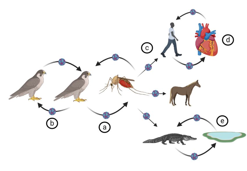
Pathogens 2020, 9, 589                                                                               3 of 51

cell also plays a role in disease pathogenesis. The C protein induces cytotoxic effects in infected cells
exhibiting cell cycle arrest in G2 phase [84,85]. Moreover, the C protein instigates the upregulation of
caspase-9 and activation of the apoptosis pathway and subsequent cell death [85].

2.1.2. The Envelope (E) Protein
      The E protein is a transmembrane protein and has a protective role for other viral components
by maintaining the envelope integrity [48,86]. The architecture of the E protein is conserved among
different flavivirus species, including WNV [87]. The E protein has three domains: domain I (DI), DII,
and DIII. They all are interconnected by a flexible, pH-dependent hinge region. At the surface of the
virions (mature or immature), the three distinct domains are arranged in an antiparallel dimer [86,88].
It is the most immunogenic of the flaviviral proteins and due to its critical role in virus entry of
the target cell, it is the principal target for most vaccines and curative drug designs mainly through
immunotherapeutic approaches. The most potent neutralising antibodies to WNV have been mapped
to EDIII [89]. The neutralising antibodies act either by inhibiting virion-cell attachment, endocytosis,
or membrane fusion [48,86].
      Like in many flaviviruses, the E protein of most WNV strains contains a conserved N-linked
glycosylation site at the 154–156 amino acid position in DI [90,91]. However, some WNV strains
contain no N-linked glycosylation site in E [90,92]. For example, the 1937 prototype strain of WNV [93]
and the 1960 prototype isolate of the WNV Kunjin strain (WNVKUN ) E protein were both shown to be
unglycosylated [94–96]. There is a putative association between glycosylation of the E protein and
neuro-invasiveness of WNV in various host species [94], although that has recently been disputed [96].
N-linked glycosylation is apparently not a requirement for WNV virulence in avian species [97].

2.1.3. The prM/M Protein
     The membrane (M) protein results from the cleavage of the glycosylated prM protein by the
trans-Golgi resident enzyme furin. The ‘pr’ segment is secreted, while the M protein, with two
membrane-spanning domains, forms part of the virion membrane. Exposed on the surface of the
immature virion, prM is believed to play a critical role in preventing the premature fusion of the E
proteins with the membrane of the host cell [94]. The cleavage of prM by furin is essential for the virus
maturity [89].

2.1.4. NS1 Protein
      Non-structural protein 1 (NS1) has a molecular weight of approximately 46–55 kDA [98,99]. NS1
occurs as a dimer and is secreted as a soluble high-density lipoprotein hexamer of three more stable
dimeric subunits. The dimeric form is critical for efficient virus replication [100]. In the infected cell,
the NS1 is present both extracellularly [cell-membrane-associated (mNS1)] as well as intracellularly
and its role varies according to the location. The extracellular form of NS1 plays a role in the regulation
and evasion of the innate immune system through modulation of complement. The intracellular form
is indirectly involved in the virus replication and maturation. It has been reported that NS1 enhances
the attachment of the virus onto the endoplasmic reticulum and ensures the stability of the virus in
the host cell [98,101,102]. NS1 protein is critical to WNV replication due to its ability to evade the
host’s immune system through inhibition of complement activation and inhibition of TLR3 [103].
NS1 is actively secreted during WNV infection; thus, it is a useful serological marker. Moreover,
NS1 is a potential diagnostic marker for differentiating infected from vaccinated animals [104] when
vaccines used do not contain WNV-NS1 [105]. In addition, intracellular NS1 is a valuable target for
immunohistochemistry applied to tissues collected at necropsy [106–110].
      NS 1 prime (NS1’) is an extension of NS1 protein and has been reported in various flaviviruses
including WNV, Japanese encephalitis virus [2] and dengue virus (DENV) [71,98]. NS1’ has a molecular
weight of about 52–53 kDa. It has been hypothesised that the extension of NS1 is caused by the cleavage
at an alternative site in NS2A due to the −1 programmed ribosomal frameshift slippage, downstream of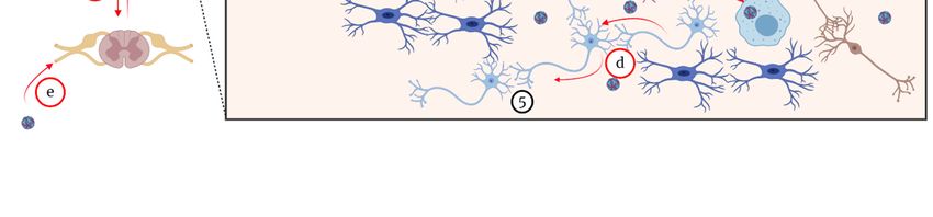
Pathogens 2020, 9, 589                                                                             4 of 51

the NS2A protein gene [98]. However, attempts to localise the cleavage site have all been in vain [111].
It has been demonstrated that NS1’ plays a key role in WNV neuroinvasiveness [98,112].

2.1.5. NS2A Protein
    The NS2A protein of the flaviviruses is a membrane-associated small molecule made up of 231
amino acids. This protein plays a key role in virus replication, virus assembly, and host immune
modulation by disrupting the host’s interferon (IFN) response [113].

2.1.6. NS2B Protein
     NS2B is a small, hydrophobic protein, and an essential co-factor of NS3 to fulfil viral protease
activity [114,115]. These two proteins are highly conserved across different flaviviruses of clinical
interest and are essential to virus replication [48,67].

2.1.7. NS3 Protein
      NS3 is the second largest flaviviral protein after NS5 with a molecular weight of approximately
69 kDa. NS3 is multifunctional including a serine protease at the N-terminal end [116,117] and a
RNA helicase at the C-terminal end [118–120]. The NS3 protease activity is dependent on NS2B as a
co-factor [67]. The NS2B-NS3 protease is crucial for viral replication and cleaves the newly translated
polyprotein at the junctions NS2A/NS2B, NS2B/NS3, NS3/NS4A, and NS4B/NS5 as well as internal
sites within C, and NS4A [62]. The NS3 RNA helicase interaction with NTPase is essential for viral
RNA replication and virion assembly [121]. Due to its multifunctional role in virus replication, NS3 has
been suggested as a good target for antiviral drug development [114].

2.1.8. NS4A Protein
     NS4A is a small hydrophobic, non-conserved and solely a transmembrane protein that plays
a role in the virus replication process through rearranging the viral membrane [67,122]. Moreover,
NS4A-NS1 interaction is needed for viral RNA synthesis [62]. It also has been suggested that NS4A
plays various roles during virus replication depending on where it is cleaved [119,123]. It has also been
speculated that NS4A may play a role as cofactor regulating ATPase activity of the NS3 helicase [119].
NS4A protein is also associated with immune evasion [48].

2.1.9. NS4B Protein
     The NS4B protein plays a crucial role in immune evasion through inhibition of WNV interferon
signalling [124]. In addition, the attenuation of WNV replication in vivo due to various mutations in
NS4B suggests its role in virus replication [124]. Although there have not been strong evidence, it is
believed that the interaction of NS1 and NS4B modulates WNV replication [125].

2.1.10. NS5 Protein
     The NS5 protein is the largest and most conserved among the non-structural proteins
(approximately 96 kDa). Like most flaviviruses, the WNV NS5 protein is comprised of a N-terminal
methyltransferase (MTase) and a C-terminal RNA-dependent RNA polymerase (RdRP) domains. The
two enzymes play a crucial role in virus replication [126,127]. During viral replication process, the
NS5 MTase domain is involved in RNA capping [127]. NS5 is also an IFN-α and β antagonist, hence a
virulence determinant via evasion of the innate immune response. It also inhibits the translation of
IFN stimulated genes (ISGs) [67,128–131].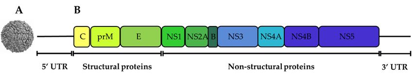
Pathogens 2020, 9, 589                                                                                    5 of 51

                               Table 1. Summary of WNV proteins and their function.

   Viral       Position in the Genome
                                           Main Role                                           References
  Protein           (Nucleotides)

                                           -    RNA encapsidation and uncoating
     C                    97-465           -    Activation of apoptosis pathway and          [62,81,82,85,132]
                                                cell death

                                           -    Virion assembly
  prM/M             466-741-742-966        -    Virus—host cell fusion                         [62,94,133]

                                           -    Viral binding and entry to host
                                                cell receptors
     E                   967-2469          -    Virus particle protection                       [48,62,86]
                                           -    Viral membrane—host cell fusion

                                           -    Viral RNA replication
                                           -    Enhancement of the attachment of the virus
                                                onto the endoplasmic reticulum
    NS1                  2470-3525         -    Virus stability                              [62,98,101–103]
                                           -    Immune evasion (inhibition of
                                                complement activation)

                                           -    Viral RNA replication and virions assembly
   NS2A                  3526-4218         -    Immune evasion (disruption of                  [62,113,134]
                                                IFN transcription)

                                           -    Cofactor for NS3 protease activity
   NS2B                  4219-4611         -    Virus replication and assembly                  [48,62,67]

                                           -    Serine protease (N-terminal)
    NS3                  4612-6468         -    RNA helicase (C-terminal)                    [48,62,118–120]

                                           -    Viral membrane rearrangement
   NS4A                  6469-6915         -    Inhibitor of interferon α/β host response          [62]

                                           -    Immune evasion (inhibitor of interferon
                                                α/β host response)
   NS4B                  6916-7680         -    Viral replication (enhancer of                 [62,125,135]
                                                NS3hel activity)

                                           -    IFN-α and β antagonist
                                           -    Evasion of the innate immune response
                                                (IFN antagonist)
    NS5                  768-10395         -    Methyltransferase (N-terminal),              [62,67,128–131]
                                                RNA-dependent RNA polymerase
                                                (C-terminal)
Pathogens 2020, 9, 589 6 of 51
2.2. The Life Cycle of WNV
The WNV life cycle involves virus reservoirs (mainly birds, which can harbour the virus without
signs of clinical disease), mosquito vectors (which also support viral replication), as well as final or
incidental hosts. The latter are mainly infected during a mosquito blood meal, if the mosquito saliva
titre exceeds 104 TCID50 /mL and may then develop clinical disease [42] (Figure 2). Final hosts are
generally dead-end-hosts, except for the crocodilians which, unlike other final hosts, also amplify the
virus [39,41]. Competent mosquito vectors acquire the virus from a viraemic vertebrate host during
their blood meal. Following ingestion of the blood meal, the WNV reaches the mosquito midgut where
the virus is amplified and spreads to the salivary glands prior to infecting the final host during the
mosquito’s subsequent blood meal [136,137]. It was initially thought that, in the mosquito, the virus
replication is strictly limited to the midgut. This was hypothesised based on failure to detect the virus
out of peritrophic matrix barriers made of chitin and various other proteins [138]. However, WNV-NS1
has also been detected by immunohistochemistry in salivary glands, neurons in the ganglia, and eye
cells in addition to the midgut tissues [137,139]. Interestingly, as is the case for other flaviviruses, WNV
does not cause apparent disease in the mosquitoes [138]. After replication in the midgut, and other
tissues, the virus starts a retrograde journey to the mosquito salivary glands via hemolymph. In the
mosquito salivary glands, virus particles aggregate pending the mosquito’s feeding on a definitive
host [136,140].
WNV vertebrate hosts, including reservoirs and incidental hosts, are infected during the uptake
of a blood-meal by a WNV-infected mosquito. Mosquitos probe their blood vessels by injecting its
saliva prior to sucking its blood meal. In addition to anticoagulation properties, the injected saliva
contains proteins that interfere with the host’s T cell response, hence, initial cell mediated immune
evasion and virus spread [141].
The virus infects the vertebrate host cell via cell receptor mediated endocytosis following cell-virus
fusion [142]. Although other receptors, such as the mannose receptor and several glycosaminoglycans,
have been suggested, dendritic cell-specific intercellular adhesion molecule 3-grabbing non-integrin
receptor (DC-SIGNR) has been shown to be the main mediator for WNV cell entry [142–144].
Once the virus gets into the host cell endosomal vesicles, the viral E protein acidifies, triggering
conformational changes and the viral and cellular membranes fuse [142]. Optimal viral membrane and
cell endosomes/liposomes fusion is achieved at pH 6.3–6.9. After the fusion is optimally achieved,
the nucleocapsid and viral RNA are released into host cell cytoplasm to initiate replication [145].
After replication in the cytoplasm of the infected cell, new virus particles acquire a lipid envelope by
budding into the lumen of the ER and are matured via cleavage of prM (removal of pr by furin) during
exocytosis and release from the cell.
It has been speculated that the DC-SIGN receptor is a key factor during vector-dependent infection
of cutaneous macrophage and dendritic cells, although the subsequent virus spread within infected
host could be DC-SIGN independent [142].lipid envelope by budding into the lumen of the ER and are matured via cleavage of prM (removal
of pr by furin) during exocytosis and release from the cell.
It has been speculated that the DC‐SIGN receptor is a key factor during vector‐dependent
infection of cutaneous macrophage and dendritic cells, although the subsequent virus spread within
Pathogenshost
infected 9, 589 be DC‐SIGN independent [142].
2020, could 7 of 51
Figure 2. WNV lifecycle and transmission. (a) WNV maintenance between birds (reservoir) and
competent
Figure mosquito
2. WNV vector,
lifecycle and(b)transmission.
WNV transmission via direct
(a) WNV between between
maintenance birds in commercial farm setting,
birds (reservoir) and
(c) WNV transmission
competent to various
mosquito vector, (b) WNVhoststransmission
(human, horse
via and crocodile)
direct betweenvia mosquito
birds bite, (d) farm
in commercial WNV
transmission
setting, viatransmission
(c) WNV blood transfusion and organ
to various transplant
hosts (human, in human,
horse (e) WNVvia
and crocodile) infection
mosquitoin crocodile
bite, (d)
through WNV contaminated water.
WNV transmission via blood transfusion and organ transplant in human, (e) WNV infection in
crocodile through WNV contaminated water.
3. Genetic Diversification within the WNV Species
Based on biology, evolution, pathogenicity, and geographic distribution, WNV has been grouped
into nine lineages [146–151] (Figure 3). Except for Koutango virus, the only member of lineage 7, WNV
3.strains
Genetic
of Diversification within
lineage 1 and 2 are the WNV
the most Species
virulent and have been responsible for several outbreaks with
severe neurological disease worldwide [152]. Lineage 1 is subdivided into 3 sub-lineages, including
sub-lineage 1a encompassing the African, European, and Middle Eastern isolates [148,153]. The
sub-lineage 1b comprises WNVKUN strains from Australasia and the sub-lineage 1c, also known as
lineage 5, comprises virus isolates from India [146,154,155].
Lineage 2, also neurotropic but with lesser virulence, comprises isolates from Sub-Saharan Africa,
Madagascar, and Europe. Viruses in this lineage have caused several outbreaks in humans, horses, and
birds [76,146,150,156–160].
Lineage 3 and lineage 4 comprise one virus isolate each. Lineage 3, also known as
Rabensburg virus, has only one strain from the Czech Republic [161,162]. Lineage 4 consists of
one WNV strain (LEIV-Vlg99-27889-human, LEIV-Vlg00-27924-human, Ast99-90 I-human) isolated
from Russia [163,164]. A putative lineage 6 has been suggested based on NS5 gene sequence in WNV
isolated from Spain (HU2925/06) [165].
Lineage 7 consists of Koutango virus (Flavivirus), first isolated in 1968 in Koutango, Senegal,
and later in Somalia [166]. The Koutango virus was initially classified as an independent flavivirus
species but later classified as a WNV strain [167,168]. Koutango virus was demonstrated to be more
pathogenic than other virulent strains [107]. A putative lineage 8 virus was isolated from Culex perfuscus
in Kedougou, Senegal [169].
The WNV-Uu-LN-AT-2013 strain, isolated in Austria, was proposed to form lineage 9 or to be
part of lineage 4 as sub-lineage 4c. Although it was concluded that this isolate is not insect or mosquito
specific, there are no reports about its in vivo pathogenicity either in humans or animals [151].Pathogens 2020, 9, 589 8 of 51
Pathogens 2020, 9, x FOR PEER REVIEW 8 of 51
Figure 3.
Figure 3. Maximum‐likelihood
Maximum-likelihood phylogenetic
phylogenetic tree
tree of
of estimating
estimating the
the relationships
relationships ofof selected
selected West Nile
West Nile
virus isolates. The tree was constructed with MEGA‐X software version 10.1.8. The optimal
virus isolates. The tree was constructed with MEGA-X software version 10.1.8. The optimal tree was tree was
obtained using Maximum likelihood method, Nearest Neighbour Interchange
obtained using Maximum likelihood method, Nearest Neighbour Interchange (NNI) inference method. (NNI) inference
method.
The The phylogeny
phylogeny was tested was
withtested with bootstrap
bootstrap replicates replicates
method (Nmethod = 1000).
= 1000).(NThe The evolutionally
evolutionally distances
distances
were were calculated
calculated with the
with the general general
time time(GTR)
reversible reversible (GTR)
model, model,
uniform uniform
rates. rates.
The tree wasThe treewith
edited was
edited with
FigTree FigTree
v1.4.3 v1.4.3
software software (http://tree.bio.ed.ac.uk/software/figtree/).
(http://tree.bio.ed.ac.uk/software/figtree/). The scaleatrepresents
The scale represents the bottomat
the bottom represent divergence time in millions of years ago (MYA). Each sequence
represent divergence time in millions of years ago (MYA). Each sequence used was labelled by GenBank used was
labelled by
accession GenBank accession number_isolate/strain
number_isolate/strain name_country
name_country of isolation_year of isolation_year of isolation.
of isolation.
4. WNV Ecology
4. WNV Ecology
This section describes
This section describesthe
thevirus
virusbiology
biology including
including vectors
vectors andand transmission,
transmission, reservoirs,
reservoirs, and
and host
host interactions.
interactions.
4.1. Virus Transmission
4.1. Virus Transmission
WNV
WNV is is primarily
primarily transmitted
transmittedbiologically
biologicallyby
bycompetent
competentmosquitoes.
mosquitoes.Mosquitoes
Mosquitoesnotnotonly
onlyplay
playa
vectorial role but are also intermediate hosts, with some level of virus amplification prior to
a vectorial role but are also intermediate hosts, with some level of virus amplification prior to infecting
definitive hosts [42,170–172].
infecting definitive WNV can WNV
hosts [42,170–172]. also becan
nosocomially acquired mainly
also be nosocomially via organ
acquired mainlytransplants,
via organ
needlestick, haemodialysis, and blood transfusion in humans. These modes of transmission
transplants, needlestick, haemodialysis, and blood transfusion in humans. These modes were first
of
reported during the first WNV outbreak in the USA [173–178]. Oral-faecal route of transmission
transmission were first reported during the first WNV outbreak in the USA [173–178]. Oral‐faecal has
route of transmission has also been confirmed in American alligators and saltwater crocodiles [39–
41,179]. Contact transmission in commercial geese farming has also been documented. It was thoughtPathogens 2020, 9, 589 9 of 51
also been confirmed in American alligators and saltwater crocodiles [39–41,179]. Contact transmission
in commercial geese farming has also been documented. It was thought to be generally associated
with cannibalism and feather picking of infected birds [180]. There has been one reported case of
WNV transplacental transmission in human [36]. Plausible breastfeeding transmission of WNV has
also been documented [37]. Aerosol transmission among animal handlers and laboratory workers
has also been hypothesised [174]. Behavioural risk factors have also been documented. A study by
Lindsey et al. [181] reported alcohol abuse as a major risk factor for WNV infection and disease.
Blood transfusions and organ transplants from previously infected individuals are other sources
of WNV infection [182]. WNV has been diagnosed in people who received whole blood as well as
blood components including red blood cells, plasma, and platelets [45,177,183,184]. It has been shown
that the virus might be present and viable in solid organs despite negative serology results. Thus, solid
organ transplants pose a potential risk to recipients [44,173,182,183,185–189].
4.2. Biological Vectors of WNV
Culex mosquitoes are reported to be the primary competent vectors of WNV. However, several
other mosquito species have been suggested to be vectors, although their competency varies [190–194].
There are geographical variations in vectors of WNV across the globe. In Africa, where the virus was
first isolated, Cx. univittatus is the most competent vector of WNV-transmission to humans [195–197].
Following WNV discovery, there have been several experimental transmission experiments in various
mosquito species. The first successful experimental transmission was in 1942 in Aedes albopictus,
thus, a potential competent vector [198] Several other experimental infections were reported in two
mosquito species, Culex pipiens and Cx. tritaeniorhynchus, most abundant in Africa [197]. However,
other mosquito species such as Cx. antennatus, Cx. univittatus, Cx. theileri, Cx. neavei, Ae. caballus,
Ae. circumluteolus, Coquillettidia spp., Cx. poicilipes, Ae. albocephalus, Cx. quinquefaciatus, Mansonia
spp., and Cx. neavei also play a significant role in the transmission of the virus to both humans and
horses in different parts of Africa such as South Africa, Egypt, Senegal, and Sudan [169,199,200]. Culex
interrogator and Cx. nigripalpus were reported to transmit the virus in Mexico and other parts of Latin
America [201]. When WNV was introduced into North America in 1999, two mosquito species, Cx.
restuans and Cx. salinarius, were incriminated in the transmission of the virus [202]. However, later
studies confirmed the role of other mosquitoes including Ochlerotatus triseriatus, Ochlerotatus japonicus
japonicus, Aedes albopictus, and Cx. pipiens [203]. Further studies have detected WNV in about 150
mosquito species [204]; however, it was concluded that the key vectors of WNV in the USA are Cx.
pipiens, Cx. tarsalis, and Cx. quinquefasciatus [205,206].
The main WNV vectors in Europe include Cx. pipiens, Cx. modestus, Cx. molestus, Ochlerotatus
caspius, Cx. torrentium, Anopheles maculipennis, and Coquillettidia richiardii [198,207,208]. Culex
annulirostris, a freshwater mosquito, is the main competent vector of WNV in Australia. The species is
also the most laboratory competent vector [148,209]. However, WNV has been recovered from other
mosquito species in Australia including Aedes alternans, Ae. nomenensis, Ae. tremulus, Ae. vigilax, Cx.
australicus, Cx. squamosus, Anopheles amictus, and Cx. quinquefasciatus. None of the latter mosquitoes
are as competent as Cx. annulirostris [209–211].
WNV is endemic in The Middle East countries, including Israel, Turkey, Jordan, Iran, and Lebanon.
In that region, WNV is largely transmitted by Cx. pipiens, Cx. perexiguus, and Ae. caspius [212]. WNV
vectors have also been documented in Asia, mainly in Pakistan and India where WNV is endemic.
The main reported vectors are Cx. vishnui complex, Cx. fatigans, Cx. tritaeniorhynchus, Cx. vishnui, Cx.
bitaeniorhynchus and Cx. univittatus, Cx. pipiens fatigans, Ae. albopictus, and Cx. tritaeniorhyncus [213].
WNV has been isolated from arthropods other than mosquitoes. These include hard ticks
(Hyalomma marginatum and Rhipicephalus sanguineus), soft ticks (Ornithodoros maritimus and Argas
hermanni), swallow bugs (Oeciacus hirundinis), and chicken mite (Ornithonyssus sylviarum) [198,214,215].Pathogens 2020, 9, 589 10 of 51
4.3. WNV Reservoirs
Several studies have demonstrated that various animal species such as Indian elephant (Elephas
maximus indicus), Indian rhinoceros (Rhinoceros unicornis), ring-tailed lemur (Lemur catta), red panda
(Ailurus fulgens fulgens), snow leopard (Panthera uncia), and babirusa (Babyrousa babyrousa) are susceptible
to WNV infection [216–218]. However, it was concluded that only bird species can produce high
enough virus titres to infect mosquitoes, which is a key requirement for the sustainability of the
infection cycle. Birds not only play a role as reservoir but also are virus amplifiers and source
of infection for dead-end-hosts [219]. Interest in researching the role of birds in the pathogenesis
of WNV resulted from the detection of the virus in blood, spleen, and brain of pigeons from the
Nile Delta in Egypt [217,220,221]. Subsequent susceptibility and permissiveness studies have been
conducted in domestic and wild birds. Severe WNV disease has been diagnosed in chukar partridge
(Alectoris chukar) [222,223], domestic geese (Anser anser domesticus) [222], domestic Impeyan pheasants
(Lophophorus impeyanus) [223], Strigiformes (owls), Columbiformes (pigeons), Cathartidae (vultures),
Corvidae (crows and related species), Gruidae (cranes), Pelicanidae (pelicans), turtle doves (Streptopelia
turtur), bald eagle (Haliaeetus leucocephalus), a snowy owl (Nyctea scandiaca), flamingos (Phoenicopterus
spp.), cormorants (Phalacrocorax spp.), American crows (Corvus brachyrhynchos), bald eagle (Haliaeetus
leucocephalus), and cormorants (Phalacrocorax spp.) [15,218,224–226].
Although bird species are generally WNV reservoirs, investigations conducted during the WNV
outbreak between 1999 and 2001 in the USA revealed Corvus species are the most susceptible to the
diseases and the main amplifier [226–229]. Following the 1999 WNV outbreak in American alligators
in the Americas and WNV associated “pix” lesions in saltwater crocodiles in Australia, experimental
studies suggested that American alligators and saltwater crocodiles are also WNV amplifiers with
high enough titres in their blood to potentially transmit the virus to mosquitoes [39,41].
Raccoons (Procyon lotor) were thought to be potential reservoirs and amplifiers of WNV in
Europe but that hypothesis is still surrounded by controversies and requires more studies [230,231].
Seroprevalences studies of WNV in raccoons in the USA have reported WNV seroprevalence ranging
between 34–54% [230,232,233]. Viremia and virus shedding profiles in experimentally infected Fox
squirrels (Sciurus niger) suggested their ability in WNV infection maintenance and spread to final
hosts [234].
5. Pathogenesis of WNV
The pathobiology of WNV infection in human and other mammalian, avian and reptilian species
has been extensively studied. There is no single proven pathogenesis of WNV; however, some theories
of the pathogenesis of WNV in mammals have been suggested. Following an infectious mosquito
bite, the virus replicates locally at the injection site in the keratinocytes and Langerhans cells of
the epidermis, a specialised type of dendritic cells associated with the skin [1,235]. The local virus
replication is enhanced due to the immune modulation of the host response by the mosquito saliva
through two mechanisms, including alteration of leukocyte proliferation and recruitment to the site of
bite, and cytokine signalling by suppressing the production of interleukin (IL) 2 and IFNγ [141,236].
It is thought that dendritic cells could be among the early primary targets of WNV infection. This
hypothesis was supported by the expression of DC-SIGN (also known as CD219) by dendritic cells
during viral infection [144,237]. It has been hypothesised that infected Langerhans cells migrate to the
draining lymph nodes in which the virus replicates further. Infected cells and free virus particles are
picked up by macrophages and cleared either directly through phagocytosis or indirectly enhanced
antigen presentation, cytokine, and chemokine secretion [238]. While macrophages clear the infection,
virus replication continues in dendritic cells in the lymph nodes [237,239,240]. WNV replication in vitro
in B and T lymphocytes suggests that these cells are potentially among the primary target during early
stage of infection in vivo [241]. The infection of the cells of the immune system and virus replication in
the cells is associated with immune modulation and appearance of primary clinical signs at the end
of the incubation period, which is in the range of 2 to 14 days post infection [42]. From the lymphPathogens 2020, 9, 589 11 of 51
nodes, the virus is spread to peripheral organs hematogenously (Figure 4). In some hosts such as
avian species, the virus has a wide range of tissue tropism and can replicate in nearly all the body
systems [110,242–244].
Pathogens 2020, 9, x FOR PEER REVIEW 12 of 51
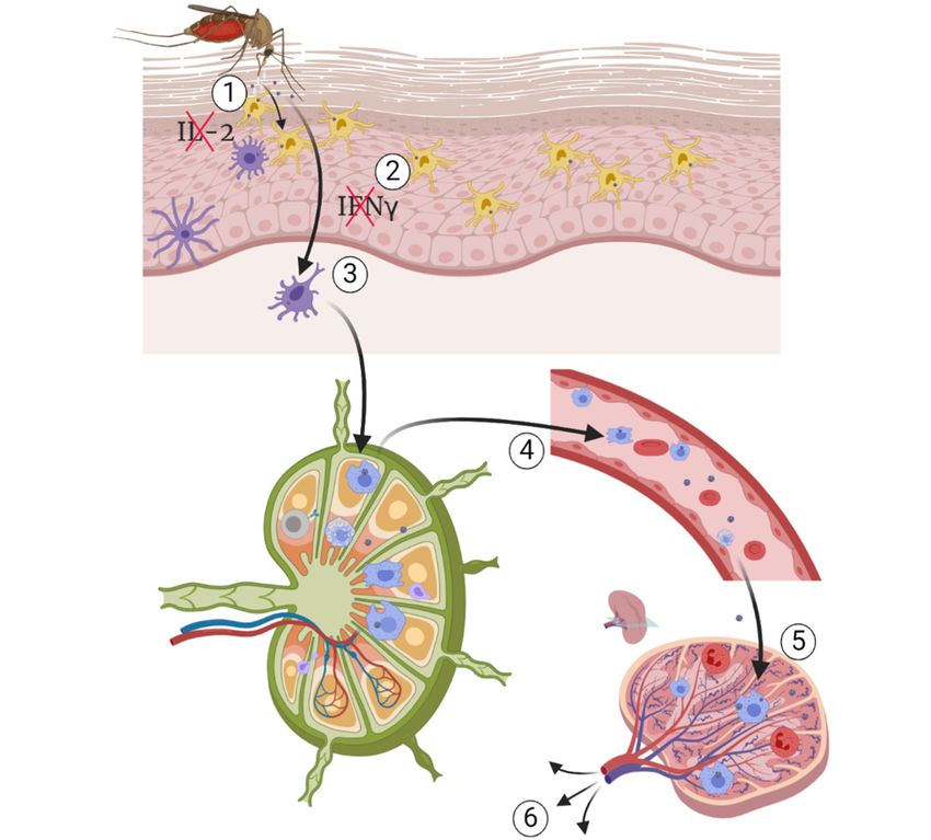
Figure 4. Figure
Pathogenesis of WNV
4. Pathogenesis infection.
of WNV (1)(1)
infection. Culex
Culexquinquefasciatus transmitting
quinquefasciatus transmitting WNVWNV duringduring
a blood a blood
meal on meal
susceptible host and
on susceptible releasing
host and itsitsinfectious
releasing saliva,(2)(2)
infectious saliva, immunomodulation
immunomodulation by mosquito’s
by mosquito’s
saliva followed by infection of keratinocytes and Langerhans cells, (3) migration
saliva followed by infection of keratinocytes and Langerhans cells, (3) migration of infected of infected cells to cells to
nearby draining lymph nodes, (4) viremia followed by migration of infected macrophage from the
nearby draining lymph nodes, (4) viremia followed by migration of infected macrophage from the
lymph nodes, and (5) spleen from which the virus spread to other organs of tropism. Source: Adapted
lymph nodes, and (5)etspleen
from Petersen al. [1]. from which the virus spread to other organs of tropism. Source: Adapted
from Petersen et al. [1].
Based on clinical presentation, there are several forms of WNV infection, including the
neuroinvasive form previously reported in many hosts, the gastrointestinal form, hepatic form,
5.1.1. Hematogenous Route
pancreatic form, cardiovascular form, and a cutaneous form characterised by erythematous macules in
humanshaematogenous
Although and lymphohistiocytic-plasmacytic
WNV spread inflammation in crocodilians
has been suggested, it [60,229,232–236].
has not yet been definitively
Presently, based on the clinical presentation and pathology,
demonstrated in vivo; thus, this mechanism remains controversial. The active three main forms have been suggested
transportation by
and studied in various host species (animal models). These forms include the neuroinvasive form,
infected blood cells is the most plausible since the migration of infected cells begins before blood
the cutaneous form, and the gastrointestinal form [60,229,233,235–237]. The form and severity of
vessels leak [257]. However,
WNV infection passive
depend on several haematogenous
factors, including the WNV route following
strain and anhost
lineage, the increased
species andvascular
permeability during
intrinsic acute and
susceptibility, phase inflammation
the viral tropism as well could
as somebeextrinsic
another possibility
factors [258,259].and
such as environment It is also
plausiblecoinfection [230,238–240]. can
that neuroinvasion Following WNVoccur
initially infection,
bya acascade of proinflammatory cytokines
non‐haematogenous and othercausing
route initially,
neuroinflammation, which then subsequently increased vascular permeability triggering
haematogenous invasion. Given the complexity of cardiovascular anatomy and physiology, there is
a need to elucidate the spreading mechanisms of WNV in animal models.Pathogens 2020, 9, 589 12 of 51
protein genes are upregulated as part of the innate immune response [108,110,241]. The activation
and release of these chemicals are essential in the initiation and maintenance of inflammation in the
control of viral infections, including WNV [74,145,242]. However, overexpression and continuous
upregulation of inflammatory cytokine genes, may be detrimental in some viral infections including
WNV, by enhancing the severity of infection and/or inflammation leading to death, chronic or permanent
morbidity and or sequelae such as immunopathology. This is a phenomenon commonly observed
with increased upregulation of cytokines such as IL-2, IL-6, IL-12, IL-17A, IFN- γ, IFN-γ induced
protein 10 (IP-10), granulocyte-macrophage colony-stimulating factor (GM-CSF) and proinflammatory
chemokine osteopontin (OP). The cytokines and chemokines may remain upregulated, even long after
recovery from WNV infection [243–247].
5.1. Pathogenesis of Neuroinvasive Form
The neuroinvasive form of WNV infection is the most severe form of the disease. It occurs
in about 1% of human and equine cases of WNV infection [235,247,248]. In humans as
well as in equine species, this form of disease is characterised by syndromes of meningitis,
encephalitis, and acute flaccid paralysis/poliomyelitis [249]. The lesions include granulocytic
meningitis, lymphoplasmacytic-histiocytic perivascular cuffing, and lymphoplasmacytic meningo-
encephalomyelitis. Similar pathology is reported in naturally and experimentally infected
alligators [38,40,179]. Currently, there are no reports of the neuroinvasive form of WNV infection in
other crocodilian species.
The lesions in infected birds with the neuroinvasive form are like those in other hosts. They
include meningoencephalitis as characterised by lymphoplasmacytic-histiocytic perivascular cuffing,
mild to diffuse gliosis and glial nodules. Rarely, multifocal necrosis (malacia) in the gray matter of the
brain has been observed [243,250,251].
The mechanism of neuroinvasion by WNV has been debated over the years, with two scenarios
still receiving foremost consideration. These include haematogenous and transneural routes of
neuroinvasion (Figure 5). For both routes, several mechanisms have been proposed [252–254]. The
first hypothesis consists of direct invasion of the CNS via a transendothelial mechanism following the
infection of the endothelial cells. However, this mechanism may be host specific and even specific to
some WNV strains [253,255]. It has also been demonstrated that brain endothelial cells are not the
main target in the neuroinvasive form in the horse, human, or mouse [107,108,256]. Lim et al. [235]
hypothesised that there is an association between levels and duration of viraemia and the WNV
neuroinvasion. However, species susceptible to the neuroinvasive form, such as the horse, primates,
and crocodilians, generally do not develop substantial and sustained viraemia. This therefore begs
more questions regarding the link between viraemia and neuroinvasion, thus, requiring further studies.Pathogens
Pathogens2020,
2020,9,9,x589
FOR PEER REVIEW 1313ofof51
51
Figure 5. WNV neuroinvasive mechanisms. (a) Passive migration of free virus particles across
the disrupted blood-brain barrier (BBB) through a “transudative” mechanism following increased
vascular permeability, (b) “Trojan horse” mechanism through migration of infected macrophages into
Figure 5. WNV neuroinvasive
brain parenchyma, mechanisms.
(c) direct infection (a) Passive
of endothelial migration
cell. of free axonal
(d) retrograde virus particles
transportacross the
of WNV,
disrupted blood‐brain
(e) WNV migration intobarrier
spinal(BBB) through
cord, (f) WNV amigration
“transudative” mechanism
from spinal following
cord to brain increased
and vice versa,
vascular permeability, (b) “Trojan horse” mechanism through migration of infected macrophages
(g) neuroinvasive mechanism by transneural mechanism via olfactory nerve, (1) astrocyte, (2) microglia, into
brain parenchyma, (c) direct infection of endothelial cell. (d) retrograde axonal transport of
(3) WNV particles, (4) transmigrating macrophage, (5) motor neuron, (6) blood-brain barrier (BBB) tightWNV, (e)
WNV migration into spinal cord, (f) WNV migration
junction. Source: Adapted from Petersen et al. [1]. from spinal cord to brain and vice versa, (g)
neuroinvasive mechanism by transneural mechanism via olfactory nerve, (1) astrocyte, (2) microglia,
5.1.1.(3)Hematogenous
WNV particles, Route
(4) transmigrating macrophage, (5) motor neuron, (6) blood‐brain barrier (BBB)
tight junction. Source: Adapted from Petersen et al. [1].
Although haematogenous WNV spread has been suggested, it has not yet been definitively
demonstrated in vivo; thus, this mechanism remains controversial. The active transportation by
5.1.2. Virus Passive Migration
infected blood cells is the most plausible since the migration of infected cells begins before blood vessels
leakAnother hypothesis
[257]. However, is the haematogenous
passive passive migration of free
route virus particles
following acrossvascular
an increased the disrupted blood‐
permeability
brain
duringbarrier
acute(BBB)
phasethrough a “transudative”
inflammation could be mechanism. This disruption
another possibility [258,259].of the
It isBBB
alsoisplausible
a result ofthat
an
increased vascular
neuroinvasion can permeability
initially occurdueby atonon-haematogenous
the acute phase pro‐inflammatory cytokines
route initially, causing and chemokines
neuroinflammation,
[258–262].
which thenHowever, this hypothesis
subsequently has not yet
increased vascular been validated
permeability due to
triggering the lack of a universal
haematogenous invasion.inGiven
vitro
or incomplexity
the vivo model.of Moreover, there isanatomy
cardiovascular another and
controversy about
physiology, thisishypothesis.
there Using a mouse
a need to elucidate model,
the spreading
JEV neuroinvasion
mechanisms of WNV wasindemonstrated
animal models. to occur prior to the production of inflammatory cytokines and
chemokines, which disrupted the BBB [263]. Similar observations were made in the WNV mouse
5.1.2. Virus
model [260].Passive Migration
Another hypothesis is the passive migration of free virus particles across the disrupted blood-brain
5.1.3. The
barrier “Trojan
(BBB) Horse”
through Mechanism mechanism. This disruption of the BBB is a result of an increased
a “transudative”
vascular permeability
The “Trojan horse”due to the acute
mechanism hasphase pro-inflammatory
also been cytokines
hypothesised in and chemokines
several WNV studies and[258–262].
reviews
[235,241]. It has been suggested that WNV neuroinvasion by the “Trojan Horse” mechanism in
However, this hypothesis has not yet been validated due to the lack of a universal in vitro or is vivo
the
model.of Moreover,
result there of
the expression is another
lymphocyte controversy about this
and monocyte hypothesis.
chemokines Using a the
triggering mouse model, JEV
recruitment of
neuroinvasion
infected wasleukocytes
peripheral demonstrated into to
theoccur prior
cerebral to the production
vasculature. of inflammatory
The infected cytokines
leukocytes then reach and
the
chemokines,
brain parenchymawhich
viadisrupted
the leaking theBBB,
BBBresulting
[263]. Similar observations
in appearance were madeclinical
of neurological in the signs
WNVand mouse
the
model [260]. of lesions in the CNS [258,259]. Neuroinvasion by a “Trojan horse” mechanism was
development
demonstrated for a pestivirus (of the Flaviviridae family), bovine viral diarrhea virus (BVDV), where,
during transplacental infection of the fetus, infected microglial precursor cells, also known as
amoeboid glial cells, brought the virus into the brain via the periventricular germinal zone fromPathogens 2020, 9, 589 14 of 51
5.1.3. The “Trojan Horse” Mechanism
The “Trojan horse” mechanism has also been hypothesised in several WNV studies and
reviews [235,241]. It has been suggested that WNV neuroinvasion by the “Trojan Horse” mechanism
is the result of the expression of lymphocyte and monocyte chemokines triggering the recruitment
of infected peripheral leukocytes into the cerebral vasculature. The infected leukocytes then reach
the brain parenchyma via the leaking BBB, resulting in appearance of neurological clinical signs and
the development of lesions in the CNS [258,259]. Neuroinvasion by a “Trojan horse” mechanism was
demonstrated for a pestivirus (of the Flaviviridae family), bovine viral diarrhea virus (BVDV), where,
during transplacental infection of the fetus, infected microglial precursor cells, also known as amoeboid
glial cells, brought the virus into the brain via the periventricular germinal zone from where it spread to
differentiating neurons and glial cells [264]. However, because the innate immune response is still not
fully developed in a mid-gestation fetus, this scenario may not be applicable to WNV infection of adult
humans and animals, and more studies therefore should be conducted to elucidate this phenomenon.
5.1.4. Transneuronal Mechanism
The transneural mechanism has also been proposed as another potential route of invasion of the
brain by WNV. The transneural mechanism consists of virus migration following motor and sensory
nerves from the point of entry. This mechanism is understudied as reflected in the scarcity of literature
about this mechanism. Currently, two entry points have been suggested and these include from
the peripheral somatic nerves into the CNS and from the olfactory nerve into the CNS [108,265,266].
It has been proposed that WNV spread from the point of entry to the central and peripheral nervous
system by retrograde and anterograde axonal transport and/or via non-neural cells such as glial cells
(astrocytes and microglial cells). By this mechanism, it has been suggested that the virus spread is
mediated by viral release from distal axon to infect adjacent neurons leading to flaccid paralysis in
some hosts when the sciatic nerve has been directly infected [265,267]. Flaccid limb paralysis is a result
of neuronal injury associated with a severe infection and necrosis of the anterior horn neurons of the
lumbosacral region (L5-S1) of the spinal cord [265].
Although several pathogenesis hypotheses have been proposed, none of them has been conclusive
so far. The use of labelled infectious virus clones could potentially shed more light on different WNV
pathogeneses, mainly the neuroinvasion mechanism.
5.2. Pathogenesis of the Cutaneous Form
Cutaneous manifestation of WNV infections have been extensively documented in humans.
It is mainly characterised by an erythematous, maculopapular rash [268], and punctate exanthem
affecting the extremities and the trunk [269]. Several reports have demonstrated the permissiveness
of both keratinocytes, skin fibroblasts, and epithelial cells to WNV replication in vitro as well as
in vivo [242,270–278]. The infection of skin epithelial cells is believed to be the cause of skin rash
in 25–45% of the neuroinvasive cases in humans [271]. Viral antigen has been detected in the
skin of naturally infected goshawks [278]. The replication of WNV in skin keratinocytes has been
demonstrated at various stages of WNV infection and these are thought to be the primary target of
WNV [271]. WNV replication was confirmed by immunohistochemistry (IHC) and virus titration at
the subcutaneous injection site for up to 3 dpi [242]. Appler et al. [270] demonstrated the persistence
of WNV in mouse skin for up to 14 dpi and WNV RNA for up to 30 dpi with or without clinical
disease. Nevarez et al. [279], using RT-PCR confirmed the potential role of WNV in the development
of lymphohistiocytic proliferative syndrome in farmed American alligators (Alligator mississippiensis).
However, virus isolation was not successful; therefore, it could not be confirmed if the viral RNA
detected was representing infectious or defective/inactivated virus. Similarly, WNVKUN RNA was
detected in skin lesions from farmed saltwater crocodiles (Crocodylus porosus) but live virus has so far
not been isolated from these lesions [280]. In addition, WNV was successfully grown in a human skinPathogens 2020, 9, 589 15 of 51
primary cell line, the foreskin fibroblasts (HFF) [272,281]. Despite all the evidence of the susceptibility
and permissiveness of skin cells, the pathogenesis of the cutaneous form of the WNV infection has not
yet been elucidated.
5.3. Pathogenesis of the Gastrointestinal Form
WNV infection has been associated with gastrointestinal syndrome in humans as characterised
by diarrhoea, enteritis, gastritis, pancreatitis, and hepatitis [282,283]. The gastrointestinal form of
WNV infection has been reported in up to 30% of human cases [284–287]. However, to date, WNV
replication in the human gastrointestinal tract (GIT) has not been investigated. Therefore, it would be
worthwhile to test this either via WNV isolation, detection of viral antigen or viral RNA from faecal
samples or GIT biopsy. The detection of viral antigen in the duodenal and proventricular enterocytes
of naturally infected goshawks suggests replication of WNV could occur in the gastrointestinal tract
of infected avian hosts [278]. Klenk et al. [39] demonstrated oral transmission of WNV in Alligator
mississippiensis through feeding the latter with infected mice. WNV was isolated from cloacal samples
from approximately 84% of the infected A. mississippiensis. The high virus titres (106.2 PFU/swab)
strongly suggested replication of the virus in the GIT. Alligators infected with WNVNY99 displayed
a gastrointestinal clinical and pathological picture as characterised by inappetence, diarrhea, and
bloating, preventing the affected animals from submerging into water [40]. Lesions in infected alligators
include haemorrhagic enteritis, proliferative enteritis, ulcerative-proliferative esophagitis, fibrinous
and necrotising colitis, histiocytic and necrotising hepatitis, and necrotising pancreatitis. WNV antigen
positive cells were detected in the gastrointestinal tract of infected alligators by IHC [40,288]. However,
these lesions are neither specific nor pathognomonic to WNV. Interestingly, although WNV genomic
RNA was detected in both oral and cloacal swabs of saltwater crocodiles experimentally infected with
WNVKUN as well as in their un-infected pen-mates, C. porosus does not present with gastrointestinal
clinical signs or pathological changes, and it remains unknown where exactly in the alimentary tract
the virus is replicating in these animals [41].
5.4. Renal Form
The detection of WNV viral RNA in the urine of infected hamsters and humans raises concerns
about a potential permissiveness of kidney cells and a renal form of the disease. Barzon et al. [46]
detected WNV RNA from urine samples during the acute phase of infection in humans. In half of the
investigated patients, viral RNA was detected in urine for much longer than in plasma. Moreover,
the viral RNA load was higher in urine than in plasma. The renal form hypothesis is also supported
by the detection of viral antigen by IHC in kidneys from patients who succumbed to the WNV
infection [289–291]. WNV RNA was also detected in urine of infected patients for up to a month post
infection [292]. The recovery of virus in urine from experimentally WNV infected hamsters for up
to two months post recovery supports renal infection [293]. However, while hamster was a good
animal model to investigate this form, the use of the WNV 385-99 strain, which has been adapted to
hamster through serial passage of the virus isolated from urine, might have influenced these findings.
Therefore, it would be worthwhile to investigate this form of the diseases in other animal model system
such as rabbits and mice.
Although there is strong evidence for renal WNV infection in infected individuals, this form often
exists in association with other forms of the disease, mainly the neuroinvasive form. Although the
reasons for this association has not been elucidated, the immunocompromised status of the infected
humans could be one explanation. Thus, the pathogenesis of the renal form is unclear, hence, to
elucidate this, there is a need for modelling the infections in various laboratory animal models.You can also read