Workers' Compensation Guidelines for Determining Impairment - First Edition, November 22, 2017 - Workers' Compensation Board
←
→
Page content transcription
If your browser does not render page correctly, please read the page content below
Workers’ Compensation
Guidelines for
Determining Impairment
First Edition, November 22, 2017

                                   1|Page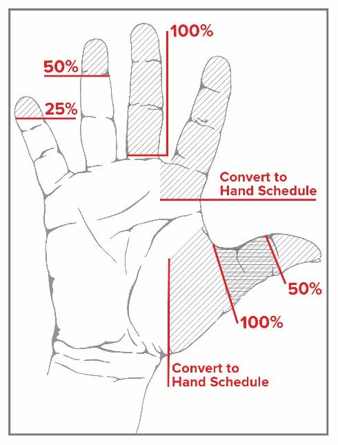
Forward
Legislation enacted in April 2017 [WCL§15(3)(x)] directed the Board to consult with
“representatives of labor, business, medical providers, insurance carriers, and self-insured
employers regarding revisions to permanency impairment guidelines, including permitting
review and comment by such representatives’ chosen medical advisors…”, to adopt revised
guidelines for the evaluation of medical impairment and determination of permanency with
respect to injuries which are amenable to a schedule loss of use award pursuant to
paragraphs (a) through (v) of subdivision 3 of section 15 of the WCL. As the law directs, these
Guidelines are to be “…reflective of advances in modern medicine that enhance healing and
result in better outcomes.” [WCL§15(3)(x)]

Therefore, these revised permanency guidelines supersede those sections of the Board’s
2012 Impairment Guidelines concerning medical evaluation of injuries amenable to a
schedule loss of use (chapters 1 through 8 of the 2012 Guidelines), as well any other provision
of the 2012 Impairment Guidelines which are inconsistent with these Guidelines.

                                                                                   2|Page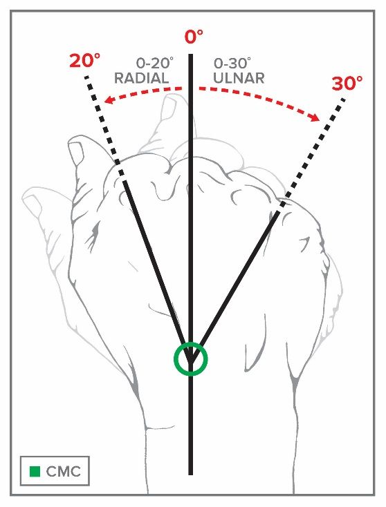
Table of Contents
Chapter 1:    Introduction ......................................................................................................6
  1.1   Types of Disability Under the Workers’ Compensation Law .................................... 6
  1.2   Maximum Medical Improvement (MMI) ................................................................... 6
  1.3   Role of Examining Medical Providers ...................................................................... 6
  1.4   Types of Final Evaluation Examinations.................................................................. 7
  1.5   Schedule Awards ....................................................................................................8
  1.6   Non-Schedule Awards (Classification) .................................................................... 8
  1.7   Abbreviation Codes.................................................................................................9
Chapter 2:    Upper Extremities – Thumb and Fingers ........................................................ 10
  2.1   Objectives for Determining Impairment for Thumb and Fingers............................. 10
  2.2   Methods Available to Assess Permanent Impairment ........................................... 10
  2.3   Maximum Rating of Body Part............................................................................... 10
  2.4   Thumb ..................................................................................................................11
  2.4   (A) Thumb Range of Motion .................................................................................. 11
  2.4   (B) Calculating Loss of Use of Thumb ................................................................... 13
  2.4   (C) Thumb Special Considerations ....................................................................... 15
  2.5   Fingers..................................................................................................................15
  2.5   (A) Finger Range of Motion ................................................................................... 15
  2.5   (B) Calculating Loss of Use of Finger .................................................................... 16
  2.5   (C) Finger Special Considerations ........................................................................ 17
  2.6   Loading .................................................................................................................18
  2.7   Amputation ...........................................................................................................20
Chapter 3:    Upper Extremities – Hand and Wrist .............................................................. 22
  3.1   Objectives for Determining Impairment for Hand and Wrist ................................... 22
  3.2   Methods Available to Assess Permanent Impairment ........................................... 22
  3.3   Wrist Range of Motion .......................................................................................... 22
  3.4   Calculating Loss of Use ........................................................................................ 24
  3.5   Special Considerations ......................................................................................... 25
  3.6   Amputation ...........................................................................................................25
Chapter 4:    Upper Extremities - Elbow.............................................................................. 26
  4.1   Objectives for Determining Impairment for Elbow ................................................. 26
  4.2   Methods Available to Assess Permanent Impairment ........................................... 26
  4.3   Elbow Range of Motion ......................................................................................... 26
  4.4   Calculating Loss of Use ........................................................................................ 27
  4.5   Special Considerations ......................................................................................... 27

                                                                                                                   3|Page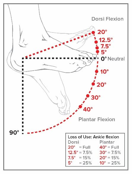
4.6      Amputation ...........................................................................................................28
Chapter 5:        Upper Extremities - Shoulder ......................................................................... 29
   5.1      Objectives for Determining Impairment for Shoulder ............................................. 29
   5.2      Methods Available to Assess Permanent Impairment ........................................... 29
   5.3      Shoulder Range of Motion .................................................................................... 29
   5.4      Calculating Loss of Use ........................................................................................ 30
   5.5      Special Considerations ......................................................................................... 32
   5.6      Amputation ...........................................................................................................33
Chapter 6: Hip and Femur .................................................................................................. 34
   6.1      Objectives for Determining Impairment for Hip and Femur .................................... 34
   6.2      Methods Available to Assess Permanent Impairment ........................................... 34
   6.3      Hip Range of Motion ............................................................................................. 35
   6.4      Calculating Loss of Use ........................................................................................ 36
   6.5      Special Considerations ......................................................................................... 38
   6.6      Amputation ...........................................................................................................40
Chapter 7:        Knee and Tibia............................................................................................... 41
   7.1      Objectives for Determining Impairment for Knee and Tibia ................................... 41
   7.2      Methods Available to Assess Permanent Impairment ........................................... 41
   7.3      Knee Range of Motion .......................................................................................... 41
   7.4      Calculating Loss of Use ........................................................................................ 42
   7.5      Special Considerations ......................................................................................... 43
   7.6      Amputation ........................................................................................................... 45
Chapter 8:        Lower Extremities – Ankle and Foot............................................................... 46
   8.1      Objectives for Determining Impairment for Ankle and Foot ................................... 46
   8.2      Methods Available to Assess Permanent Impairment ........................................... 46
   8.3      Ankle Range of Motion .......................................................................................... 47
   8.4      Calculating Loss of Use of the Foot ...................................................................... 48
   8.5      Special Considerations ......................................................................................... 49
   8.6      Amputation ...........................................................................................................49
Chapter 9:        Lower Extremity – Great and Lesser Toes ..................................................... 50
   9.1      Objectives for Determining Impairment for Toes ................................................... 50
   9.2      Methods Available to Assess Permanent Impairment ........................................... 50
   9.3      Maximum Rating of Body Part............................................................................... 50
   9.4      Great Toe ............................................................................................................. 51
   9.4      (A)Calculating Loss of Use of Great Toe ............................................................... 51
   9.5      Smaller Toes (Second, Third, Fourth & Fifth) ........................................................ 52

                                                                                                                      4|Page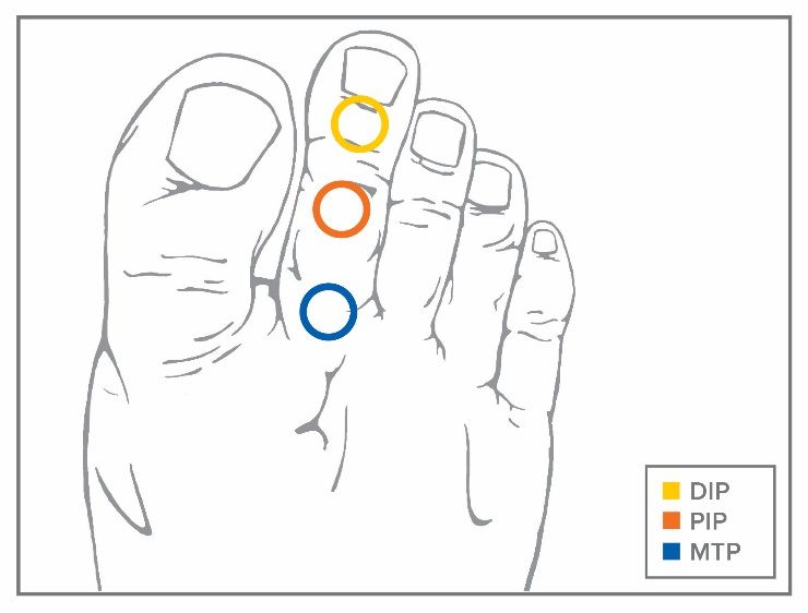
9.5      (A) Calculating Loss of Use of Smaller Toes ......................................................... 52
   9.6      Amputations and Loading ..................................................................................... 53
Chapter 10:   Central Nervous System Conditions, Peripheral Nerve Injuries and
Entrapment / Compression Neuropathies............................................................................ 54
   10.1     Central Nervous System - Cranial Nerves ............................................................ 54
   10.2     Peripheral Nervous System .................................................................................. 55
   10.3     Entrapment / Compression Neuropathies ............................................................. 56
Chapter 11:           Visual System/Auditory System/Facial Scars and Disfigurement ................ 59
   11.1     Visual System Introduction.................................................................................... 59
   11.1.1 Criteria and Methods for Evaluating Permanent Impairment ................................. 59
   11.2     Loss of Hearing ..................................................................................................... 62
   11.2.1 Occupational Loss of Hearing ............................................................................... 62
   11.2.2 Traumatic Loss of Hearing .................................................................................... 62
   11.3     Facial Scars and Disfigurement ............................................................................ 63
APPENDIX A ......................................................................................................................64

                                                                                                                    5|Page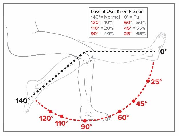
Chapter 1:             Introduction
Disability is a legal determination that reflects the impact of a workplace injury on a claimant’s
ability to work. The Workers’ Compensation Law Judge establishes the level of disability
based on the available medical evidence and other relevant information. Medical evidence
may be submitted by the claimant’s health provider, a medical consultant for the employer
and/or an independent medical examiner.

A distinction is made between disability and impairment. Impairment is a purely medical
determination made by a medical professional, and is defined as any anatomic or functional
abnormality or loss. Competent evaluation of impairment requires a complete medical
examination and accurate objective assessment of function. These Guidelines provide the
medical provider with a uniform process for evaluating an individual's impairment resulting
from a medically documented work related injury or illness.

1.1    Types of Disability Under the Workers’ Compensation Law

This law establishes the following types of disability in workers’ compensation cases:

       1.   Temporary total disability
       2.   Permanent total disability
       3.   Temporary partial disability
       4.   Permanent partial disability

Evaluation of permanent disability occurs when there is a permanent impairment remaining
after the claimant has reached maximum medical improvement (MMI). These Guidelines were
created for purposes of determining impairment for permanent disabilities.

1.2    Maximum Medical Improvement (MMI)

A finding of MMI is based on a medical judgment that (a) the claimant has recovered from the
work injury or illness to the greatest extent that is expected and (b) no further improvement is
reasonably expected. The need for palliative or symptomatic treatment does not preclude a
finding of MMI. In cases that do not involve surgery or fractures, MMI cannot be determined
prior to 6 months from the date of injury or disablement, unless otherwise stated or agreed to
by the parties.

1.3    Role of Examining Medical Providers

Medical providers are obligated to provide the Board and the parties their best professional
opinion of the claimant’s medical condition, degree of impairment, and functional abilities.
These Guidelines provide detailed criteria for determining the severity of a medical
impairment, with a greater weight given to objective findings. It is the responsibility of the
medical provider to submit medical evidence that the Board will consider in making a legal
determination about disability.

Medical providers should not infer findings or manifestations that are not drawn from the
physical examination or test reports, but rather medical providers should look to the objective

                                                                                      6|Page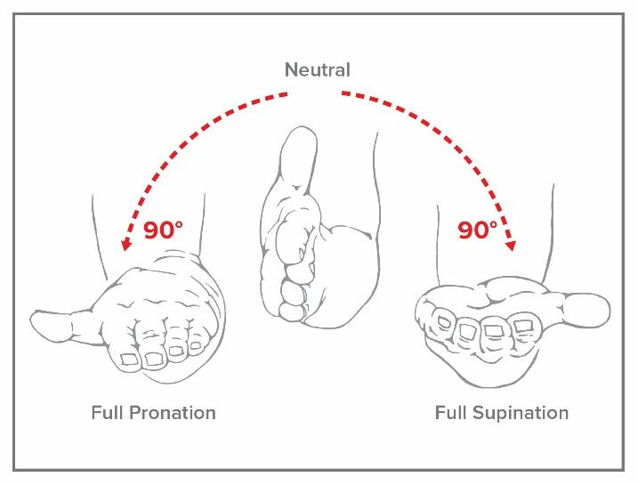
findings of the physical examination and data contained within the medical records of the
patient. This methodology is intended to foster consistency, predictability and inter-rater
reliability for determining impairment.

In order to prepare a report on permanent impairment, the medical provider should do the
following:
     1. Identify the affected body part or system (include chapter, table number, class, and
        severity level for non-schedule disabilities) and review the Guidelines (for body parts
        not covered by the Guidelines, see Chapter on Other Injuries and Occupational
        Diseases [Default Guideline]).
     2. Review the relevant medical records and medical history.
     3. Perform a thorough physical examination.
            a. To measure active range of motion (ROM), medical providers should generally
                 utilize a goniometer. In order to measure the maximum range of active motion,
                 three repeat measurements should be taken.
            b. Deficits should be measured by comparing to the baseline reading of the
                 contralateral member, if appropriate. Using the contralateral is not appropriate
                 where the opposite side has been previously injured or is not otherwise
                 available for comparison.
     4. Report the work-related medical diagnosis(es) and examination findings, including
        appropriate specific references to the relevant medical history, examination, and test
        results.
     5. Follow the recommendations to establish a level of impairment.
     6. For a non-schedule permanent disability, evaluate the impact of the impairment(s) on
        claimant’s functional and exertional abilities. See Medical Impairment and Functional
        Assessment Guidelines in the 2012 New York State Guidelines for Determining
        Permanent Impairment and Loss of Wage Earning Capacity.
     7. When determining the value of a schedule loss of use, the total value of several
       range of motion deficits should not exceed the value of full ankylosis of the joint.
       The sum of multiple ankylosed joints of a major member cannot exceed the
       value of amputation. However, digits may exceed these values due to loading.

1.4    Types of Final Evaluation Examinations
Examining medical providers will conduct final evaluation examinations in connection with
the following categories of awards:

   1. A Schedule Award for:
         a. Impairment of extremities (including nervous system impairment that impacts
            use of extremities)
         b. Loss of vision
         c. Loss of hearing
         d. Facial disfigurement
   2. Non-Schedule Award for:
         a. Classification as permanent partial disability
         b. Classification as permanent total disability

                                                                                     7|Page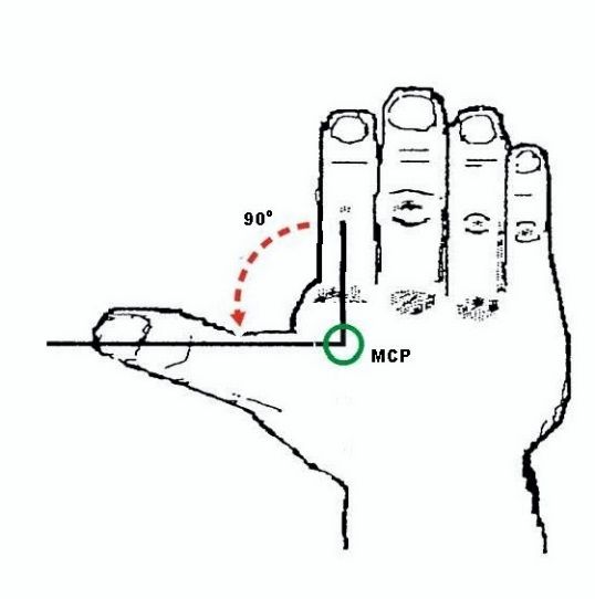
Medical providers evaluating a claimant located in New York, and medical providers located
in New York who perform evaluations, must be authorized by the Workers’ Compensation
Board. For medical providers outside of New York, any evaluation performed must comport
with these Guidelines, including the use of any forms prescribed by the Chair.

1.5    Schedule Awards

A schedule award is given not for an injury sustained but for the residual permanent physical
and functional impairments. Final adjustment of a claim by a schedule award must comply
with the following medical requirements:

   1. There must be a permanent impairment of an extremity, permanent loss of vision or
      hearing, or permanent facial disfigurement, as defined by law.
   2. The impairment must involve anatomical or functional loss such as physical damage
      to bone, muscles, cartilage, tendons, nerves, blood vessels, and other tissues.
   3. The claimant must have reached maximum medical improvement.
   4. No residual impairments must remain in the systemic area (i.e., head, neck, back, etc.)
      before the claim is considered suitable for schedule evaluation of an extremity or
      extremities involved in the same accident.
Workers' Compensation Law Section 15 prescribes the value for a percentage loss or loss of
use of body members. See Appendix A: Weeks by Percentage Loss of Use of Body Part for
a table containing the appropriate number of weeks of compensation provided by percentage
of loss.

1.6    Non-Schedule Awards (Classification)

Non-schedule awards include permanent impairments that are not covered by a schedule,
such as conditions of the spine and pelvis, lungs, heart, skin, and brain, as well as impairments
of the extremities that are not amenable to a schedule award as described below.

Schedule Impairments Subject to Classification
Examples of impairments of the extremities not amenable to a schedule award:

   1. Progressive and severe painful conditions of the major joints of the extremities such
      as the shoulders, elbows, hips and knees with one or more of the following:
          a. Objective findings of acute or chronic inflammation of one or more joints such
              as swelling, effusion, change of color or temperature, tenderness, painful range
              of motion, etc.
          b. X-ray evidence of progressive and severe degenerative arthritis.
          c. Minimal or no improvement after all modalities of medical and surgical
              treatment have been exhausted.
   2. Chronic painful condition of an extremity commonly affecting the distal extremities
      such as the hands and feet, with one or more of the following:
          a. Complex regional pain syndrome (reflex sympathetic dystrophy), Sudeck’s
              atrophy or chronic painful extremity syndrome.
          b. Objective findings or chronic swelling, atrophy, dysesthesias, hypersensitivity
              or changes of skin color and temperature such as mottling.

                                                                                     8|Page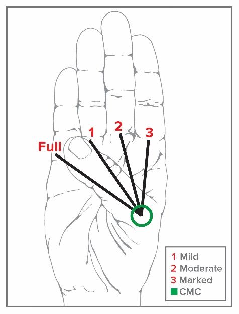
c. X-ray evidence of osteoporosis.
          d. Minimal or no reported improvement after claimant has undergone all
              modalities of chronic pain treatment.
  3. Mal-union of the long bones.
  4. Aseptic necrosis of the head of the femur or other bones.
  5. Severe and persistent instability of the knee joint or other major joints.
  6. Advanced Paget’s disease.
  7. Tumors.
  8. Caisson’s disease involving the joints.
  9. Persistent ulcerations, draining sinuses.
  10. Recurrent dislocations (shoulders).
  11. Amputees with neuromas or poorly healed stumps.
  12. Failed joint replacement such as total hip, total knee and shoulder replacements.

1.7   Abbreviation Codes

                 Acronym/Term                            Definition
      Mi                             Mild
      Mo                             Moderate
      Ma                             Marked
      F                              Flexion deficits
      E                              Extension deficits
      DIP                            Distal interphalangeal joint
      PIP                            Proximal interphalangeal joint
      MCP                            Metacarpophalangeal joint
      CMC                            Carpo-metacarpal joint
      MTP                            Metatarsophalangeal joint
      SLU                            Schedule loss of use
      ANCR                           Accident Notice Casual Relation
      ODNCR                          Occupational Disease Notice Casual Relation
      Per NYS Statute:
      Thumb                          Thumb
      First finger                   Index finger
      Second Finger                  Middle / Long Finger
      Third Finger                   Ring Finger
      Fourth Finger                  Small / Little / Pinky Finger
      Symbols
      <                              Less than
      ≤                              Less than or equal to
      >                              Greater than
      ≥                              Greater than or equal to

                                                                               9|Page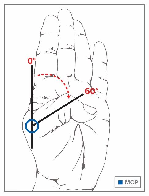
Chapter 2:             Upper Extremities – Thumb and Fingers
2.1 Objectives for Determining Impairment for Thumb and
Fingers

The objective is to accurately assess the permanent residual physical deficit a claimant
suffered as a result of his/her injury. To the degree possible, the assessment should be based
on objective findings determined by the history and physical examination, as well as the
results of any appropriate diagnostic testing.

2.2    Methods Available to Assess Permanent Impairment
Determination of the degree of permanent residual physical deficit should be performed at the
time of maximum medical improvement, the point at which no further healing is expected.
Maximum medical improvement should be determined based on the outcome of the clinical
course of treatment, the medical provider’s expertise and any further treatment options
available to the claimant. When evaluating the level of permanent residual physical deficit, the
medical provider should consider the contralateral extremity where appropriate and
expected/normal values. The duration of time from the injury to maximum medical
improvement may vary, but in most cases is one year from the injury or last surgery.

The severity of the permanent residual physical deficit is not based on the mechanism of
injury. It reflects the permanent residual physical deficit at the time of medical maximum
improvement and may include physical damage to bone, muscles, cartilage, tendons, nerves,
blood vessels, and other tissues.

2.3    Maximum Rating of Body Part
These Guidelines are to be used for evaluating permanent residual physical deficits of the
thumb and fingers. Single digit loss/impairment must be determined based on the impairment
to the digit alone and not as part of the hand. The total value for the range of motion, if multiple
joints are affected, cannot exceed the maximum value of the digit.

When multiple digit impairments are considered together in one comprehensive rating, the
total impairment cannot exceed 100% of the next largest major member. Therefore, the loss
of multiple digits, resulting in conversion to a hand impairment, may not exceed 100%
schedule loss of use of the hand.

                                                                                      10 | P a g e
2.4 Thumb
The thumb works in conjunction with other fingers to reach,
pinch, grasp or grip and manipulate objects. Thumb flexion,
opposition and adduction are required for pinch, precision and
some power grips.
The thumb deserves special consideration; it is the highest
valued digit and the most important. The functional units of the
thumb are the proximal and distal phalanges, the
interphalangeal, metacarpal, and carpometacarpal joints.
Impairment of hand function with loss of pinch and reduced
grasping power is a significant presentation; furthermore,
opportunity for reconstructive surgery is eliminated.
Figure 2.4 Joints of Thumb
2.4 (A) Thumb Range of Motion
1. Measurement Position for Interphalangeal
Joint (IP)
The hand is supine, palm up; the thumb is placed
in full extension. The IP joint of the thumb is
flexed to full extent. Normal range of motion is
80 degrees.
Figure 2.4 (A)(1) IP Joint
11 | P a g e2. Measurement Position for
Metacarpophalangeal Joint
(MCP)
The hand is supine, palm up; the thumb is
placed in full extension. Measure the angle
between the first metacarpal and the proximal
phalanx as the MCP joint is flexed to the full
extent. Normal range of motion is 60 degrees.
Figure 2.4 (A)(2) MCP Joint
3. Measurement Position for CMC Joint
Flexion/Adduction/Opposition
These motions (flexion/adduction/opposition) are responsible for most of the activities
of the hand. They allow for tip to tip contact for pinch, prehension and object
manipulation. The measurement is as follows:
The hand is supine, palm up. As the thumb is flexed
across the palm to the fullest extent possible, the thumb
rotates. This is apparent as the thumbnail position
changes. In this maneuver, the transition from full
flexion to opposition is observed as additional degrees
of rotation bring the thumb tip in contact with the MCP
joints.
- Opposition is full if the tip of the thumb contacts the
MCP of the pinky or little finger (4th finger).
- In a mild deficit (1) the tip of the thumb contacts the
MCP of the ring finger (3rd finger), but not the MCP of
the pinky.
- In a moderate deficit (2) the tip of the thumb contacts
the MCP of the middle finger (2nd finger), but not the
MCP of the ring finger.
- In a marked deficit (3) the tip of the thumb contacts
the MCP of the index finger (1st finger), but not the
MCP of the middle finger.
Figure 2.4 (A)(3) CMC Joint
12 | P a g e4. Measurement Position for Radial Abduction
With the forearm resting in a neutral
position, with thumb in contact with the first
finger, the thumb is moved outward to
maximum movement. Measure the angle as
the MCP joint is abducted to the full extent.
Normal range of motion is 90 degrees
Figure 2.4 (A)(4) Radial Abduction
5. Measurement Position for Isolated Opposition
The hand is supine, palm up; all thumb
joints are extended to the fullest extent
possible along the supine plane (radial
extension) and the thumb is positioned
perpendicular to the palm. Measure the
thumb’s ability to touch the tip of each
finger.
Figure 2.4 (A)(5) Isolated Opposition
2.4 (B) Calculating Loss of Use of Thumb
To determine the overall schedule loss of use of the thumb, first assess whether any special
considerations apply. If not, to calculate the schedule loss of use of the thumb, each joint
should be measured individually and any deficit values (found in table below) are added
together. If a single motion deficit is involved (flexion or extension), the lower figure applies.
If both flexion and extension are involved, the higher figure applies.
When using range of motion to determine schedule loss of use, a reduction to the sum of
two major values may be in order and the total schedule loss of use of the thumb cannot
exceed the value of ankylosis.
13 | P a g eTable 2.4(B) Thumb
Percent Loss of Use of Thumb
Instructions: To the extent there are deficits, add A+B unless a reduction
to the sum of two major values is in order.
Maximum value cannot exceed the value for ankylosis.
Schedule loss of use percentages for ranges of motion values above/below
those depicted here should be adjusted proportionally.
ROM Mild Moderate Marked Ankylosis
IP* 10 - 15% 20 - 25% 40 - 45% 50% loss of
use of the
(ROM =0-80o) (ROM 60o) (ROM 40o) (ROM 25o) thumb
A MCP* 15 - 20% 25 - 30% 45 - 50% 75% loss of
use of the
(Select (ROM =0-60o) (ROM 45o) (ROM 30o) (ROM 15o) thumb
one)
Both (IP and
MCP) – utilized
in the event a 20 - 30% 40 - 50% 80 - 90%
deficit exists in
both the IP and
MCP joints
CMC Flexion 20 - 25% 30 - 40% 50 - 90% 80% -100%
as defined
B above (Figure To 3rd Finger To 2nd Finger To 1st Finger
2.4(A)(3))
*Use lower figure for one deficit and higher when both are affected
(flexion/extension)
NOTE: If no other deficits exist:
• A mild impairment of thumb adduction is equal to 7½% loss of use of the
thumb.
• A mild impairment of thumb opposition is equal to 10% loss of use of the
thumb.
• A mild impairment of the radial abduction is equal to 10% loss of use of the
thumb.
• More significant deficits in adduction, opposition, or abduction may be given
a higher schedule.
14 | P a g e2.4 (C) Thumb Special Considerations
The following are special considerations in the final adjustment of the fingers.
1. Loss of active flexion or ankylosis at CMC joint is 100% loss of use of the thumb and
is usually associated with a wrist deficit in which case it becomes a hand schedule.
2. Abduction and opposition of the thumb is mainly centered on the CMC joint with
possible deficits at the MCP and IP joints, resulting in mild, moderate or marked
impairment of pinch and grasp power of the hand. Such cases are given a hand
schedule.
2.5 Fingers
The index through the small finger (fingers 1 – 4
as defined by WCB) each have three
joints, the metacarpophalangeal (MCP joint),
the proximal interphalangeal (PIP) and the
distal interphalangeal (DIP). The MCP joint
enables finger flexion and extension and is
important for both grip and pinch activities.
Figure 2.5 Fingers
2.5 (A) Finger Range of Motion
1. Measurement Position for Distal Interphalangeal (DIP) Joint
(Normal range of motion is 90 degrees)
Flexion: Hand prone, fingers extended, measure the
angle between the middle phalanx and the distal phalanx
as the stabilized DIP joint is flexed towards the palm.
Block flexion of the PIP during this maneuver.
Extension: Hand prone, DIP joints fully flexed. Measure
the angle at the middle phalanx and distal phalanx joint
as it is extended away from the palm.
Figure 2.5(A)(1) DIP Joint
15 | P a g e2. Measurement Position for Proximal Interphalangeal (PIP) Joint
(Normal range of motion is 100 degrees)
Flexion: Fingers extended horizontal with palm and wrist.
Measure the angle between the middle phalanx and the
proximal phalanx as the PIP joint is flexed towards the
palm. Block flexion of the MCP during this maneuver.
Extension: PIP joints fully flexed. Measure the angle as
the PIP joint is extended towards horizontal position of the
fingers.
Figure 2.5(A)(2) PIP Joint
3. Measurement Position for Metacarpophalangeal Joint (MCP)
(Normal range of motion is 90 degrees)
Flexion: Fingers extended, measure the
angle between the metacarpal bone and
the proximal phalanx as the MCP joint is
flexed towards the palm.
Extension: MCP joints fully flexed.
Measure the angle as the MCP joint is
extended to the maximum extent possible.
Figure 2.5(A)(3)
MCP Joint
2.5 (B) Calculating Loss of Use of Finger
To determine the overall schedule loss of use of the finger, first assess whether any special
considerations apply. If not, to calculate the schedule loss of use of the finger, each joint
should be measured individually and the values (found in the table below) added together.
If a single motion deficit is involved (flexion or extension), the lower figure applies. If both
flexion and extension are involved, the higher figure applies.
16 | P a g eWhen using range of motion to determine schedule loss of use, a reduction to the sum of
two major values may be in order for losses in all three joints and the total schedule loss of
use of the finger cannot exceed of the value of ankylosis.
Table 2.5(B) Finger
Percent Loss of Use of Finger
Instructions: To the extent there are deficits, add A+B+C unless a reduction
to the sum of two major values is in order.
Maximum value cannot exceed the value for ankylosis.
Schedule loss of use percentages for ranges of motion values above/below
those depicted here should be adjusted proportionally.
ROM Mild Moderate Marked Ankylosis
DIP 10 - 15% 20 - 25% 40 - 45%
A*
(ROM 0 – 90o) ROM 75o ROM 45o ROM 25o
PIP 15 - 20% 25 - 30% 45 - 50% Ankylosis of the
DIP joint (loss of
B* active flexion) is a
50% loss of use of
(ROM 0 – 100o) ROM 75o ROM 45o ROM 25o
the finger.
Ankylosis of
MCP 20 - 25% 30 - 40% 50 - 90% multiple joints
cannot exceed
100%.
C*
(ROM 0 – 90o) ROM 75o ROM 45o ROM 25o
*Use lower figure for one deficit and higher figure when both are affected.
2.5 (C) Finger Special Considerations
The following are special considerations that have enumerated schedule loss of use values.
Other deficits may be added when specified. However, the maximum schedule loss of use
value cannot exceed the value of ankylosis except under special consideration number 5
below.
1. Mallet deformity: Up to 33⅓% loss of finger depending on degree.
2. Trigger finger: Up to 33⅓% loss of finger. Use the maximum value (33⅓%) if the thumb
or index finger is involved.
3. Flail DIP joint: 50% loss of finger.
4. Loss of half or more of the distal phalanx equals 50% loss of use of the finger.
5. Dupuytren’s Contracture - There must be an ODNCR and/or ANCR for Dupuytren's
Contracture before schedule evaluation thereof. Schedule loss of use should be limited
to the accident or occupational disease. There is a 5% to 7½% loss of use of the hand if
impairment is found in one finger only. A larger schedule may be given if two or three
17 | P a g efingers are involved and function of the hand is compromised, such as grasp power. It
is recognized that this may exceed the value of ankylosis of the affected finger.
2.6 Loading
Loading is the amount added to a schedule to allow for weakness of grasp or other major loss
of function when multiple digits are affected. Where indicated, convert multiple digit loss to an
overall hand schedule. Schedules below 50% in one or two digits remain in the digits.
To calculate the overall loading value:
1. Determine the number of weeks per digit by multiplying the percentage loss of use
per digit by the statutory maximum weeks allowed per digit1:
2. Add the number of weeks per digit together to arrive at “total digit weeks”;
3. Multiply the “total digit weeks” by the appropriate loading percentage2 to arrive at the
overall number of “loading weeks” and add to “total digit weeks”; and
4. Divide this value by the maximum statutory weeks for a hand (2441). Multiply the
quotient by 100 to arrive at the percentage loss of the hand.
Example: a 50% loss of use of the index finger and a 60% loss of use of the
thumb is given a 60% load and converted to a hand schedule (Scenario C
below).
1. (A) 50% Loss of Index 23 weeks (50% of 46 weeks)
(B) 60% Loss of Thumb 45 weeks (60% of 75 weeks)
2. Total Digit Weeks (A plus B) 68 weeks (23 + 45)
3. Total Weeks including 60% load 108.8 weeks ((68 x 60%)+68)
4. Converted to Hand Schedule 44.6% ((108.8/244) x 100)
1
See Appendix A.
2 See tables 2.6 [a], [b], and [c]
18 | P a g eTable 2.6(a) – Loading when two digits are affected
Loading for Two Digits Scenario A Scenario B Scenario C Scenario D
One-digit loss > 50% 100% > 50% Thumb or Index
with 100% bone
loss.
Second digit loss < 50% > 50% > 50% < 50%
Loading 0 60% 60% 60%
Convert to Hand No Yes Yes Yes
Per table above:
• Scenario A - No load is given when one digit has 50% loss of use and another has
less than 50% loss of use; instead a separate percentage is given for each finger.
• Scenario B - The load is 60% and converted to a hand schedule when one digit has
100% loss of use and another digit has 50% loss of use.
• Scenario C - Schedules of 50% or more in two digits are loaded 60% and converted
to a hand schedule.
• Scenario D - The load is 60% and converted to a hand schedule when there is a 100%
bone loss in either the thumb or index finger and a second digit has less than 50%
loss of use.
Table 2.6(b) Loading when three or more digits are affected
Loading for Three Digits Scenario A Scenario B
One-digit loss < 50% > 50%
Second digit loss < 50% > 50%
Third digit loss < 50% With or without loss
Fourth digit loss n/a With or without loss
Loading 30% 60%
Convert to Hand Yes Yes
Per table above:
• Scenario A - In cases of loss of three fingers with less than 50% loss of use in each
finger, are given a 30% load and converted to a hand schedule.
• Scenario B - Schedules of 50% or more loss in two or more digits are loaded 60% and
converted to a hand schedule.
19 | P a g eTable 2.6(c) Loading for Amputations
Conversion
Loading for Amputation Loading to Hand
Amputation of ½ the distal
phalanges or ankylosis of the DIP Yes
Scenario A 60%
plus loss of active flexion of > two
fingers
Amputation mid phalanges > two
Scenario B digits 60% Yes
Amputation through the proximal
Scenario C phalanges of > two or more digits 120% Yes
Amputation at first metacarpal of
Scenario D the thumb 120% Yes
Per table above:
• Scenario A - Amputation of half of the distal phalanges of two or more digits or
ankylosis of the DIP joints of two or more digits and loss of active e flexion of two or
more digits is loaded 60% and given a hand schedule.
• Scenario B - Amputation through the middle phalanges of two or more digits is loaded
60% and given a hand schedule.
• Scenario C - Amputation through the proximal phalanges of two or more digits is
loaded 120% and given a hand schedule.
• Scenario D - An amputation involving the first metacarpal of the thumb is loaded 120%
and given a hand schedule.
2.7 Amputation
Determination of residual impairment and functional loss depends on the level of amputation.
Reliance on initial x-rays or reports may be misleading. The operative amputation is frequently
performed at a higher level in order to obtain adequate closure or better function. If in doubt,
new post-operative x-rays are needed to determine the degree of bone loss and the final level
of amputation. This information will be needed in the calculation of schedule loss.
20 | P a g eFigure 2.7 Schedule Loss of Use of the Fingers Due to an Amputation
1. Loss of all fingers at proximal phalanges equals 100% loss of use of the hand.
2. Loss of tip of tuft (with bone loss) of the distal phalanx equals 15% to 20% loss of use
of the finger. Add percentage for mobility deficit at the DIP joint if present.
3. Loss through the base of the tuft equals 33 ⅓% loss of use of the finger.
4. Loss of half or all of the distal phalanx of the finger equals 50% loss of use of finger (no
additional values added for mobility impairment at the DIP joint).
5. Amputation through the DIP joint equals 50% loss of use of the finger.
6. Loss of any portion of the middle phalanx equals 100% loss of use of the finger.
7. Loss involving the proximal phalanx equals 100% loss of use of the finger.
8. A 100% loss of use of the thumb equals 75 weeks. In cases of amputation proximal to
the MCP joint, there is a load of 120% and converted to a hand schedule
9. Loss involving the entire finger and any part of the ray (metacarpal) equals 100% loss
of use of the digit and is loaded 120% and converted to a hand schedule.
10. In cases where 100% was given for a member, additional schedules may be given for
subsequent injuries under certain circumstances, e.g., amputation above the elbow
receives 100% loss of the arm.
21 | P a g eChapter 3: Upper Extremities – Hand and Wrist
3.1 Objectives for Determining Impairment for Hand and Wrist
The hand and wrist are an integral part of the finger and thumb motion. In addition, the wrist
acts as a bridge between the associated structures of the hand and the forearm. The wrist
enables the hand to perform complex flexion/extension and radial/ulnar movements.
The objective is to accurately assess the permanent residual physical deficits a claimant
suffered as a result of his/her injury. To the degree possible, the assessment should be based
on objective findings determined by the history and physical examination, as well as the
results of any appropriate diagnostic testing.
3.2 Methods Available to Assess Permanent Impairment
Determination of the degree of permanent residual physical deficit should be performed at the
time of maximum medical improvement, the point at which no further healing is expected.
Maximum medical improvement should be determined based on the outcome of the clinical
course of treatment, the medical provider’s expertise and any further treatment options
available to the claimant. When evaluating the level of permanent residual physical deficit, the
medical provider should consider the contralateral extremity where appropriate and
expected/normal values. The duration of time from the injury to maximum medical
improvement may vary, but in most cases is one year from the injury or last surgery.
The severity of the permanent residual physical deficit is not based on the mechanism of
injury. It reflects the permanent residual physical deficit at the time of medical maximum
improvement and may include physical damage to bone, muscles, cartilage, tendons, nerves,
blood vessels, and other tissues.
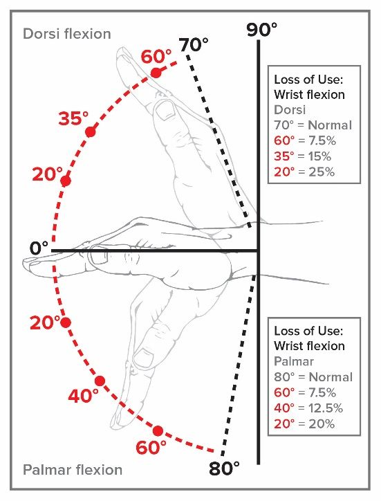
3.3 Wrist Range of Motion
Figure 3.3 (a) Dorsi flexion and Palmar flexion of
the Wrist (Percent Loss of Use of the Hand)
22 | P a g eFigure 3.3 (b) Pronation – Supination of the Wrist (as measured with the elbow flexed to 90 degrees, and
arm adducted along the chest wall).
Figure 3.3 (c) Lateral Wrist Motion
23 | P a g e3.4 Calculating Loss of Use
To determine the overall schedule loss of use of the wrist, first assess whether any special
considerations apply. If not, to calculate the overall schedule loss of use of the wrist, add
Palmar Flexion (A) + Dorsi Flexion (B) + Pronation/Supination (C), to the extent there are
deficit in these ranges of motion. However, where marked deficits are present in all wrist
motions the schedule loss of use cannot exceed 55%. For other deficits see notes.
Table 3.4 – Wrist
Percent Loss of Use of Wrist
Instructions: To the extent there are deficits, add A+B+C or values as indicated per notes.
Maximum value for marked cannot exceed 55%.
Schedule loss of use percentages for ranges of motion values above/below those
depicted here should be adjusted proportionally.
ROM Mild Moderate Marked Ankylosis
Position of
Palmar Flexion 7½% 12½% 20% function
A (mild dorsi
flexion):
ROM 0 - 80o ROM 60o ROM 40o ROM 20o 60% loss of
the hand.
Dorsi Flexion 7½% 15% 25% In any other
B position
(palmar,
ROM 0 – 70o ROM 60o ROM 35o ROM 20o marked
dorsi flexion
Pronation / Supination 7½ - 10% 17½ - 20% 25 - 30% or lateral
C* deviation):
70 – 90%
ROM 0 – 90o ROM 75o ROM 45o ROM 25o loss of the
hand.
* Use lower figure for one deficit and higher figure when both are affected.
Notes:
• Complete loss of Palmar flexion equals 25% loss of the hand.
• Complete loss of Dorsi flexion equals 33⅓ % loss of the hand.
• Complete loss of both pronation and supination equals 35% loss of use of the hand.
• Deficits in radial-lateral motion and ulnar motion may be separately considered if other
findings in the wrist are normal.
24 | P a g e3.5 Special Considerations
The following are special considerations that have enumerated schedule loss of use values.
Other deficits may be added when specified or when no schedule value is provided. However,
the maximum schedule loss of use value cannot exceed the value of ankylosis except for
special consideration number one.
1. Complete wrist drop or radial nerve palsy equals 66 ⅔% loss of use of the hand; less is
given for partial wrist drop.
2. Darrach procedure (resection distal ulna) equals 10% loss of use of the hand for bone
loss and add for mobility deficits.
3. Resection "proximal row" carpal bones equals 20% loss of use of the hand for bone loss
alone and add for mobility deficits if present.
4. Navicular fracture - Hold non-union cases for two years. Give a schedule loss of use of
the hand if the X-rays provide evidence of clinical union (fibrous) and if the pain is not
severe. In rare, very painful conditions, consider classification.
5. Kienböck's Disease (aseptic necrosis of carpal lunate): Hold until X-rays show static
condition. Consider classification if condition is symptomatic.
6. Carpal Tunnel Syndrome: Schedule one-year post decompression if asymptomatic. If
symptoms persist and become severe and disabling, consider classification. [For values
see Nerve Section 10.3A]
7. De Quervain's Disease with or without surgical release equals 7½-20% loss of use of
the thumb depending on impairments. If there is a residual deficit of the wrist and the
grip power of the hand is impaired, give a schedule loss of use of the hand.
8. Ganglion of wrist equals 0-7½% of hand depending on clinical findings.
3.6 Amputation
Amputation at the wrist equals 100% loss of use of the hand (80% loss of use of the arm).
25 | P a g eChapter 4: Upper Extremities - Elbow
4.1 Objectives for Determining Impairment for Elbow
The elbow plays an important role in positioning the hand and wrist to allow for functional use
of the upper extremity. The most important function of the elbow joint is to position the hand,
either moving the hand away from the body (elbow extension), towards the body (elbow
flexion) or in a more precise hand movement (supination/pronation).
The objective is to accurately assess the permanent residual physical deficit a claimant
suffered as a result of his/her injury. To the degree possible, the assessment should be based
on objective findings determined by the history and physical examination, as well as the
results of any appropriate diagnostic testing.
4.2 Methods Available to Assess Permanent Impairment
Determination of the degree of permanent residual physical deficit should be performed at the
time of maximum medical improvement, the point at which no further healing is expected.
Maximum medical improvement should be determined based on the outcome of the clinical
course of treatment, the medical provider’s expertise and any further treatment options
available to the claimant. When evaluating the level of permanent residual physical deficit, the
medical provider should consider the contralateral extremity where appropriate and
expected/normal values. The duration of time from the injury to maximum medical
improvement may vary, but in most cases is one year from the injury or last surgery.
The severity of the permanent residual physical deficit is not based on the mechanism of
injury. It reflects the permanent residual physical deficit at the time of maximum medical
improvement and may include physical damage to bone, muscles, cartilage, tendons, nerves,
blood vessels, and other tissues.
4.3 Elbow Range of Motion
Normal Range of Motion 0-150°
o Flexion: From the position of
maximum extension, measure the
angle between the extension position
and the full flexion of the forearm.
Normal flexion is 150 degrees.
Figure 4.3(a) Elbow Flexion
26 | P a g eo Extension: Starting at maximum
flexion, measure the angle between
the flexion position and the full
extension of the forearm. Normal
extension is to the zero degrees
position as indicated in the figure.
Figure 4.3(b) Elbow Extension
4.4 Calculating Loss of Use
To determine the overall schedule loss of use of the elbow, first assess whether any special
considerations apply. If not, overall loss of use of the elbow is calculated by combining any
noted deficits in extension and flexion. When evaluating based on range of motion, the
maximum loss of use for the elbow cannot exceed ankylosis.
Table 4.4 – Elbow
Percent Loss of Use of Elbow
Instructions: To the extent there are deficits, add A+B.
Maximum value cannot exceed the value for ankylosis.
Schedule loss of use percentages for ranges of motion values above/below those
depicted here should be adjusted proportionally.
ROM Mild Moderate Marked Ankylosis
Position of function: 66
Extension 25% 50% 85% ⅔% loss of the arm.
A
Higher percentage is
ROM: Full ROM 450 ROM 900 ROM 1250 given for extremes of
Flexion to 0° flexion or rotation of
the forearm.
Flexion 7½% 33⅓% 66⅔% Ankylosis at 00 (full
B
extension) is a 90%
ROM: Full ROM 1250 ROM 90o ROM 45o loss of the arm.
Extension to 150°
4.5 Special Considerations
The following are special considerations that have enumerated schedule loss of use values.
Other deficits may be added when specified or when no schedule value is provided. However,
the maximum schedule loss of use value cannot exceed the value of ankylosis.
1. Loss of head of the radius equals 10% loss of use of the arm and add for mobility
deficits.
2. Laxity of the elbow with hyperextension deficit equals 10-15% loss of use of the arm.
3. Medial and lateral epicondylitis are usually given a schedule, but if it becomes chronic,
severe and disabling, consider classification.
27 | P a g e4. Olecranon fracture and olecranon bursitis. Schedules depend on residual deficits.
5. Olecranon excision equals 10% loss of the use of the arm for bone loss and add for
mobility deficits.
4.6 Amputation
Table 4.6 Percent Loss of Use of the Arm: Amputation at Different Levels
Amputation % Loss of Use of the Arm
At Elbow or Above 100%
Three Inches Below Elbow 95%
Mid-Forearm 90%
At Wrist Joint 80%
28 | P a g eChapter 5: Upper Extremities - Shoulder
5.1 Objectives for Determining Impairment for Shoulder
The shoulder and elbow play important roles in positioning the hand in space to allow for
functional use of the upper extremity. Injury can result in significant limitations and negatively
impact the ability to perform work responsibilities.
The objective is to accurately assess the permanent residual physical deficit a claimant
suffered as a result of his/her injury. To the degree possible, the assessment should be based
on objective findings determined by the history and physical examination, as well as the
results of any appropriate diagnostic testing.
5.2 Methods Available to Assess Permanent Impairment
Determination of the degree of permanent residual physical deficit should be performed at the
time of maximum medical improvement, the point at which no further healing is expected.
Maximum medical improvement should be determined based on the outcome of the clinical
course of treatment, the medical provider’s expertise and any further treatment options
available to the claimant. When evaluating the level of permanent residual physical deficit, the
medical provider should consider contralateral extremity where appropriate and
expected/normal values. The duration of time from the injury to maximum medical
improvement may vary, but in most cases is one year from the injury or last surgery.
The severity of the permanent residual physical deficit is not based on the mechanism of
injury. It reflects the residual physical deficit at the time of maximum medical improvement
and may include physical damage to bone, muscles, cartilage, tendons, nerves, blood
vessels, and other tissues.
5.3 Shoulder Range of Motion
Shoulder motions include:
1. Flexion (forward elevation) - Range of motion that
is in the sagittal plane rotating about an axis of an
imaginary line through the glenoid fossae with the
arm moving in front of and above the body. The
normal range of motion is to 180 degrees.
Extension - Range of motion that is in the sagittal
plane rotating about an axis of an imaginary line
through the glenoid fossae with the arm moving
behind the body to 60 degrees.
Figure 5.3(1) Shoulder
Flexion and Extension
29 | P a g e2. Abduction - Range of motion in the coronal plane
rotating about an imaginary line of an axis through
the glenohumeral joint. The normal range of
motion is to 180 degrees. (The arm moves to the
side away from the body.)
Adduction - Range of motion in the coronal plane
rotating about an imaginary line of an axis through
the glenohumeral joint. The normal range of
motion is to 30 degrees. (The arm moves across
in front of the body.)
Figure 5.3(2) Shoulder
Abduction and Adduction
3. External Rotation - The arm is at the side, the
elbow is flexed to 90 degrees and rotated about an
imaginary line along the axis of the humerus. The
normal range of motion is 90 degrees.
Internal Rotation - The arm is at the side, the
elbow is flexed to 90 degrees and rotated about the
imaginary line along the axis of the humerus. The
normal range of motion is 70 degrees.
Figure 5.3(3) Shoulder
External (Up) and
Internal (Down)
5.4 Calculating Loss of Use
To determine the overall schedule loss of use of the shoulder, first assess whether any special
considerations apply. If not, where deficits are present in abduction and flexion see table
below and use whichever deficit is higher. See notes for additional considerations. When
evaluating based on range of motion, the overall deficit, when combined, cannot exceed the
value of ankylosis.
30 | P a g eTable 5.4(a) Shoulder
Percent Loss of Use of Shoulder
Instructions: To the extent there are deficits select values per the chart and/or notes below.
Maximum value cannot exceed the value for ankylosis.
Schedule loss of use percentages for ranges of motion values above/below those
depicted here should be adjusted proportionally.
ROM Mild Moderate Marked Ankylosis
Flexion/Abduction 20% 40% 60% Ankylosis at the scapulo-
ROM: 0 – 180o humeral joint at 0
(use greater deficit) ROM: 135o ROM: 90o ROM: 45o degrees equals 80%
loss of use of the arm.
Notes:
• If a deficit of both flexion (forward elevation) and abduction are documented, the
greater of the two deficits must be utilized, not both. However, if the deficit in both
ranges of motion are moderate or higher, and the measures are within 100 of each
other, up to 10% may be added to the overall schedule loss of use, not to exceed
ankylosis.
• Do not add mild deficits of internal and external rotation to avoid cumulative values.
May add 10-15% for marked deficits of rotation and muscle atrophy, not to exceed
ankylosis.
• Mild deficits of adduction equal 7½-10% loss of use of an arm.
• Mild deficits of posterior extension equal 7½-10% loss of use of an arm.
• For isolated internal/external ROM deficits use the table below.
Table 5.4(b) Shoulder
Internal and External Rotation Only
Internal/External Rotation
(Values below are only considered where no other ROM deficits exist.)
ROM Mild Moderate Marked Complete Loss
Internal Rotation 7½% 10% 12½% 15%
ROM: 0 – 70o ROM: 55o ROM: 35o ROM: 20o
External Rotation 7½% 10% 12½% 15%
ROM: 0 – 90o ROM: 75o ROM: 45o ROM: 25o
Both Internal and 10% 15% 20 - 25% 30%
External
31 | P a g e5.5 Special Considerations
The following are special considerations that have enumerated schedule loss of use values.
Other deficits may be added when specified or when no schedule value is provided. However,
the maximum schedule loss of use value cannot exceed the value of ankylosis.
1. Dislocation of the shoulder may be amenable for a schedule loss of use evaluation
provided that it has been at least one year from corrective surgery, or recurrent
dislocation and that permanent impairment exists after one year. Pre-existent recurrent
dislocation of the shoulder calls for an overall schedule and apportionment.
2. Fracture of the clavicle may equal 0-10% depending on degree of impairment.
3. Acromio-clavicular or sterno-clavicular separation equals 7½-10% loss of use of the arm.
4. Winged scapula due to Serratus Anterior Palsy and/or Trapezius Palsy may be given 15-
20% loss of use of the arm depending on degree of functional impairment. For such
cases do not evaluate for a schedule loss until two years’ post-surgical repair of a major
nerve.
5. Resection of the clavicle, either end, equals 10% for bone loss; entire clavicle equals
15% loss of use of the arm. Add for mobility deficits of the single most notable in relation
to functional deficit if present.
6. Non-surgical rupture of the long head of the biceps muscle is equal to 10-15% loss of
use of the arm. Rupture at distal point of insertion of the biceps is equal to 20% loss of
use of the arm. Taking into consideration mobility and muscle weakness, the schedule
can vary up to 33 ⅓% loss of use of the arm depending on degree of impairment found.
7. Frozen shoulder and adhesive capsulitis (with or without surgery): if the condition is
asymptomatic give a schedule loss of use of the arm. If extremely painful and all
modalities of treatment exhausted, consider classification after two years.
8. The schedule given is focused on the highest valued part of the extremity. In case of a
high schedule for one given part of the extremity calculate first for the major loss in part
involved. For example, amputation at the wrist equals 100% loss of use of the hand or
equals 80% loss of use of the arm. If there are additional deficits of the elbow and/or
shoulder add 10% to the 80% loss of use of the arm and the final schedule would be
90% loss of use of the arm.
9. Full or partial shoulder arthroplasty or replacement results are assessed no sooner than
twelve months after surgery, as clinically significant changes in functions can occur
before this timeframe. The schedule is given based upon the medical assessment of:
• range of motion as measured by flexion or abduction, using the greatest degree
of impairment;
• atrophy as measured at the level of the mid arm and compared to the contralateral
side, and
• presence of chronic complications according to the table below (unless
appropriate for classification).
32 | P a g eYou can also read