Yeast Species in the Oral Cavities of Older People: A Comparison between People Living in Their Own Homes and Those in Rest Homes - MDPI
←
→
Page content transcription
If your browser does not render page correctly, please read the page content below
Journal of
           Fungi
Article
Yeast Species in the Oral Cavities of Older People:
A Comparison between People Living in Their Own
Homes and Those in Rest Homes
Nurulhuda Mohd Thiyahuddin, Erwin Lamping, Alison M. Rich                  and Richard D. Cannon *
 Sir John Walsh Research Institute, Faculty of Dentistry, University of Otago, Dunedin 9054, New Zealand;
 nurul.thiyahuddin@otago.ac.nz (N.M.T.); erwin.lamping@otago.ac.nz (E.L.); alison.rich@otago.ac.nz (A.M.R.)
 * Correspondence: richard.cannon@otago.ac.nz; Tel.: +64-3-479-7081

 Received: 26 February 2019; Accepted: 9 April 2019; Published: 12 April 2019

 Abstract: Oral candidiasis is prevalent among older people due to predisposing factors such as
 impaired immune defenses, medications and denture use. An increasing number of older people live
 in rest home facilities and it is unclear how this institutionalized living affects the quantity and type
 of fungi colonizing these people’s oral cavities. Smears and swabs of the palate and tongue and saliva
 samples were taken from participants residing in rest homes (RH; n = 20) and older people living
 in their own homes (OH; n = 20). Yeast in samples were quantified and identified by culturing on
 CHROMagar Candida and sequencing the ITS2 region of rDNA. A higher proportion of RH residents
 had Candida hyphae present in smears compared to OH participants (35% vs. 30%) although this
 difference was not statistically significant (p = 0.74). RH residents had, on average, 23 times as
 many yeast per mL saliva as OH participants (p = 0.01). Seven yeast species were identified in OH
 samples and only five in RH samples, with Candida albicans and Candida glabrata being the most
 common species isolated from both participant groups. The results indicate that older people living
 in aged-care facilities were more likely to have candidiasis and have a higher yeast carriage rate
 than similarly aged people living at home. This may be due to morbidities which led to the need for
 residential care and/or related to the rest home environment.

 Keywords: oral candidiasis; older people; elderly; rest home; yeast carriage; Candida albicans

1. Introduction
     The oral cavity is home to many hundreds of species of microorganisms. The majority of these
microbes are present in biofilms on the various oral surfaces. The immune systems of healthy individuals
maintain stable biofilm communities that help prevent oral infections. When immune defenses are
impaired this can cause dysbiosis of the microbiota and lead to bacterial, viral or fungal infections.
     Candida species are a normal component of the oral microbiota [1] that are carried in the mouths
of about 40–60% of healthy individuals [2]. While Candida albicans is the most prevalent species [3,4],
non-albicans Candida species that can be found in healthy individuals include Candida glabrata, Candida
tropicalis, Candida parapsilosis, and Candida krusei [5]. Other species such as Candida guilliermondii,
Candida kefyr, Candida rugosa, Candida dubliniensis and Candida famata have also been reported, but less
frequently [6,7]. Oral candidiasis is the most common type of fungal infection of the oral cavity
and may present as pseudomembranous candidiasis, erythematous candidiasis, plaque-like/nodular
candidiasis, denture-associated candidiasis, angular cheilitis, median rhomboid glossitis and chronic
mucocutaneous candidiasis [8]. Due to its prevalence as a commensal microbe, C. albicans is the most
frequent cause of oral candidiasis [9]. A preliminary clinical diagnosis of candidiasis can be made based
on the medical history of the individual, their symptoms and an oral examination. This diagnosis can

J. Fungi 2019, 5, 30; doi:10.3390/jof5020030                                        www.mdpi.com/journal/jof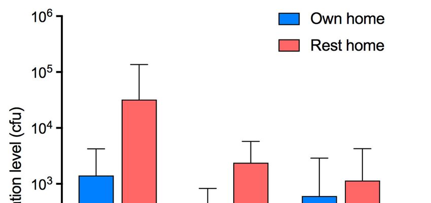
J. Fungi 2019, 5, 30                                                                              2 of 10

be confirmed histologically by the presence of hyphae in mucosal smears. Patients with oral candidiasis
can be treated with antifungal agents such as polyenes (e.g., amphotericin B and nystatin) and azole
derivatives (e.g., fluconazole, ketoconazole, clotrimazole and miconazole). It has been reported that
20% of patients with oral candidiasis experience recurrence of infection, and around 30% of the
recurrences were caused by Candida strains different from the first episode of infection [10]. Candida
species differ in their susceptibility to antifungal agents and C. glabrata and C. krusei show reduced
susceptibility to azoles [4,11] and Candida lusitaniae can develop resistance to amphotericin B [12].
In addition, C. parapsilosis has been reported to cause outbreaks of candidemia in immunocompromised
patients in hospital settings [13] where it is thought to be transmitted by the hands of health care
workers. Therefore, in institutionalized settings it is important to know what species of Candida are
colonizing individuals.
       Colonization of the oral cavity by Candida depends on the entry of yeast cells, attachment and
growth, and cell removal from the oral cavity—the rate of yeast replication must at least match
the rate of clearance [1]. Key factors that predispose for Candida colonization include systemic
factors caused by drug therapy (e.g., broad spectrum antibiotics, corticosteroids), reduced saliva
flow, chronic disease processes, malnutrition, immunosuppression and neoplasia while local factors
include wearing dentures overnight, ill-fitting dentures and poor prosthesis hygiene [14]. In the oral
cavity, components of the innate immune system, such as salivary flow and biological components
within saliva, play a major role in preventing infections. Factors that reduce saliva flow predispose to
infections such as candidiasis. In studies of people with xerostomia (dry mouth) it has been shown
that there is an inverse correlation between salivary flow rate and the number of Candida cells in oral
samples [15,16]. There was also an inverse correlation between the salivary flow rate and the severity
of oral candidiasis [15]. The proportion of the population receiving medications increases with age
and older people are often prescribed multiple medications, many of which reduce saliva flow rates.
Denture wearing also increases with age and dentures obstruct saliva flow across mucosal surfaces
reducing the clearance of microbes and creating environments that promote fungal growth. Therefore,
it is not surprising that older people are more likely than younger people to have oral mucosal lesions,
including candidiasis [17].
       Around the world, populations are ageing. The WHO estimates that by 2050 the proportion of
the population that is ‘older’ (in this case above the age of 60) will have increased from 12% in 2015
to 22%, that is, from 900 million to 2 billion people [18]. Studies have shown elderly patients living
in institutional care facilities, such as nursing homes and hospitals, are more susceptible to oral and
systemic Candida infections [19], mainly due to comorbidities, in-dwelling devices and common care
facility processes. What is concerning is the emergence of non-albicans Candida strains with lower
antifungal susceptibility in self-caring, nursing home residents [4]. C. glabrata and C. krusei strains
showed reduced susceptibility to azoles and C. rugosa and C. krusei strains were less susceptible
to flucytosine than other yeast. The presence of Candida strains with reduced susceptibility to
antifungal agents could pose a problem for older people if they become immune compromised and
develop candidiasis.
       A significant number of older people live in residential aged care facilities [20], which in New
Zealand was approximately 32,000 people in 2013 [21]. The objective of this study was to compare the
Candida species colonizing the oral cavities of people living in rest homes with those living in their
own homes.

2. Materials and Methods

2.1. Participants
     This pilot cross-sectional study was a convenience sampling of two groups of older people; people
living at rest home facilities (n = 20) and people living in their own homes (n = 20). Rest home
participants came from either Montecillo Veterans Home and Hospital, or Little Sisters of the Poor,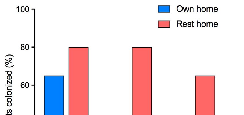
J. Fungi 2019, 5, 30                                                                                 3 of 10

both located in Dunedin, New Zealand. Participants for the control group were members of the public
recruited from posters circulated via Age Concern, Bowling Clubs Dunedin and personal contact.
Inclusion criteria were that participants had to be above the age of 65, healthy or have well controlled
systemic disease, have sufficient mouth opening to allow intraoral access and able to give consent.
Participants were excluded from taking part in this study if they had used antibiotics or antifungals in
the past 2 months, were terminally ill or were current smokers. Ethical approval for this study was
obtained from the University of Otago Human Ethics Committee (approval number H17/081).

2.2. Clinical Examination
     A clinical examination was carried out by direct observation of the condition of the oral mucosa.
Participants who exhibited mucosal inflammation and had hyphae present in the smear test were
prescribed antifungal agents. If they wore dentures they were advised to leave them out at night and
denture hygiene instructions were given. The rest home in-house medical doctor and nurse manager
were involved in this process to further support the participants.

2.3. Collection of Oral Samples
      Unstimulated saliva was collected from participants. Participants were requested to allow their
saliva to pool in their mouths for one minute then expectorate it into a glass measuring cup. This was
done 5 times. The total volume of saliva produced was recorded and the saliva transferred to a sterile
15 mL Falcon tube. Patients who were unable to generate saliva rinsed their mouths with 2 mL of
sterile water. This was to enable analysis of salivary gland function and the yeast present in saliva.
Participants’ palates and tongues were swabbed with sterile cotton buds and each swab was placed in
2 mL sterile water in a sterile 15 mL Falcon tube.
      Smears of the palate and tongue were taken by gently rubbing a wooden spatula on the
dorsal surface of the tongue and hard palate, avoiding the area that had just been swabbed, but
immediately adjacent to it. The material obtained on the spatula was smeared onto a glass slide and
the slides immersed in Coplin jars filled with 95% ethanol. The Coplin jars were transported to the
University of Otago Faculty of Dentistry Oral Pathology Centre where they were stained with periodic
acid-Schiff (PAS).

2.4. Analysis of Samples
      Saliva and mucosal swabs were transported on ice to the Faculty of Dentistry Molecular Biosciences
Laboratory for analysis and storage (4 ◦ C). Whole saliva samples and suspensions of palate and tongue
swabs were vortexed for 30 s and portions (100 µL) were spread on CHROMagar Candida agar
plates (Becton Dickinson & Co., Franklin Lakes, NJ, USA) which were incubated at 37 ◦ C for 48 h.
Candida species were presumptively identified according to colony color, and colony forming units
(cfu) per ml sample were determined. When the numbers of cfu on plates were too many to count,
portions of the stored yeast suspensions were diluted in sterile water 100- or 1000-fold and plated
and incubated as described above. The numbers of colonies on plates were converted to cfu/mL
saliva by multiplying by the dilution factor and then by 10 (as 100 µL was plated). Yeast species
were confirmed by colony polymerase chain reaction (PCR) amplification and sequencing of the
rDNA internal transcribed spacer region ITS2 [22]. Colony PCR was carried out using KOD FX Neo
DNA Polymerase (Toyobo, Osaka, Japan), where each 20 µL PCR reaction contained 0.6 µL sterile
water, 10 µL 2 × PCR Buffer for KOD FX Neo, 4 µL dNTPs (2 mM), 2 µL each of forward and reverse
primers ITS3(F) 50 -GCATCGATGAAGAACGCAGC and ITS4(R) 50 -TCCTCCGCTTATTGATATGC [23]
(3.2 µM), 1 µL of template cells (suspended in 20 µL sterile water) and 0.4 µL KOD FX Neo (1.0 U/µL).
The PCR cycle conditions were: initial denaturation at 98 ◦ C for 30 s (1 cycle); followed by 44 cycles of
the following steps: denaturation at 98 ◦ C for 10 s; annealing at 55 ◦ C for 10 s; elongation at 68 ◦ C for
40 s; and final elongation step at 68 ◦ C for 1 min.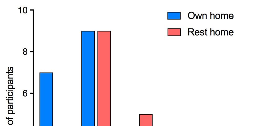
J. Fungi 2019, 5, 30                                                                                                               4 of 10

     The presence of PCR products was confirmed by running 1 µL of colony PCR sample with 5 µL of
1× loading dye on agarose gel (1.5% w/v) through electrophoresis at 100 V for 28 min with ethidium
bromide staining. DNA oligomer primers were removed from colony PCR products with ExoSAP-IT™
(ThermoFisher Scientific, Christchurch, New Zealand) according to the manufacturer’s instructions.
DNA sequencing was carried out using primer ITS3(F) by the Genetic Analysis Service, Anatomy
Department, University of Otago. Results were analysed with the computer programs FinchTV
(version 1.5.0) and UniPro UGENE (version 1.29.0) and the sequences compared with the publicly
available database at https://www.ncbi.nlm.nih.gov/BLAST/.
     PAS-stained slides were examined by a consultant oral pathologist to determine the presence or
absence of fungal hyphae.

2.5. Statistical Analysis
     The t-test was used to compare parametrically distributed data. The Mann-Whitney U test
was used to compare non-parametrically distributed data. Differences were considered significant if
p < 0.05. For binary dependent variables, SPSS software (version 25, IBM Corporation) was used for
cross-tabulations, with Chi-square tests used to determine statistical significance (at the 0.05 level).

3. Results

3.1. Participant Demographics
     The participants in the two groups (those who lived in their own homes, and those who lived
in rest homes) were sex-matched and although the participants from rest homes were, on average,
2.9 years older than those who lived in their own homes, this difference was not statistically significant
(Table 1).

              Table 1. The demographics, clinical features, and colonization status of the participants.

                            Variable                                          Participant Location                    p Value
                                                               Own Home (n = 20)           Rest Home (n = 20)
    Gender
       Female                                                            13                          13
       Male                                                               7                           7
    Mean age (range)                                                83.1 (71–92)               86.0 (72–94)               0.50 a
    Proportion with dentures                                            55%                        70%                    0.33 b
    Mean salivary flow rate [mL/min] (range)                        0.25 (0–0.7)               0.23 (0–0.6)               0.35 a
    Proportion with hyphae in smears                                    30%                        35%                    0.74 b
    Mean number of medications (range)                               5.4 (0–11)                 6.6 (1–12)                0.25 a
    Proportion with saliva colonized by yeast                           65%                        80%                    0.29 b
    Proportion with tongue colonized by yeast                           35%                        80%                    0.004 b
    Proportion with palate colonized by yeast                           25%                        65%                    0.01 b
    Level of yeast colonization of saliva (cfu/mL)
        Mean                                                            1410                     32,240                   0.01 c
        Median (range)                                              50 (0–9160)            2260 (0–4.72 × 10⁵ )
    Level of yeast colonization of tongue (cfu/swab)
        Mean                                                             271                      2400                    0.001 c
        Median (range)                                               0 (0–2200)              1970 (0–14,760)
    Level of yeast colonization of palate (cfu/swab)
        Mean                                                            606                        1150                   0.03 c
        Median (range)                                              0 (0–10,240)              200 (0–14,100)
               a   t-test; b Chi-square test; c Mann–Whitney U test. p values in bold typeface are significant (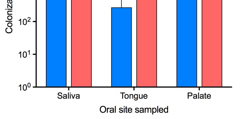
J. Fungi 2019, 5, x FOR PEER REVIEW                                                                                5 of 11

       Proportion with hyphae in smears                               30%                      35%               0.74 b
J. Fungi 2019, 5, 30                                                                                                5 of 10
       Mean number of medications (range)                          5.4 (0–11)               6.6 (1–12)           0.25 a
       Proportion with saliva colonized by yeast                      65%                      80%               0.29 b
      The   participants
      Proportion            from colonized
                     with tongue  rest homes by were
                                                 yeast more likely to35%  wear dentures (full80%  or partial) than   those
                                                                                                                 0.004   b

living in their own homes, and they had, on average, a slightly lower salivary flow rate, although this
      Proportion with palate colonized by yeast                         25%                     65%               0.01 b
difference was not statistically significant (Table 1). Fifty-five percent of the rest home participants had
      Level
salivary      of yeast
           gland        colonization(defined
                    hypofunction     of saliva as
                                               (cfu/mL)
                                                   an unstimulated whole salivary flow rate < 0.2 mL/min [24])
as did 40%Mean of those living at home, this difference was not 1,410   statistically significant   (p = 0.34, chi-square
                                                                                               32,240             0.01 c
test). RestMedian
              home participants
                       (range)       were more likely to be diagnosed
                                                                   50 (0–9,160)with candidiasis
                                                                                        2,260 (0–4.72  × 105) present in
                                                                                                     (hyphae
smears of the palate and tongue; Table 1) than participants living in their own homes (difference not
      Level of yeast colonization of tongue (cfu/swab)
statistically  significant; p = 0.74 Table 1).
             Mean
      The participants      were taking a variety of, and usually       271multiple, medications.
                                                                                                2,400            0.001 c of
                                                                                                          The number
medications      being
             Median       taken by people living in their own
                       (range)                                          homes ranged1,970
                                                                    0 (0–2,200)            from(0–14,760)
                                                                                                  0 to 11 with a mean
of 5.4 medications (Table 1). Rest home participants were taking, on average, more medications
      Level of yeast colonization of palate (cfu/swab)
(6.6, range: 1–12), but this difference was not statistically significant (p = 0.25; t-test). If, however,
             Mean were categorized as either having a normal
all participants                                                        606salivary flow rate1,150
                                                                                                 or a low salivary0.03flow
                                                                                                                        c

             Median (range)
rate (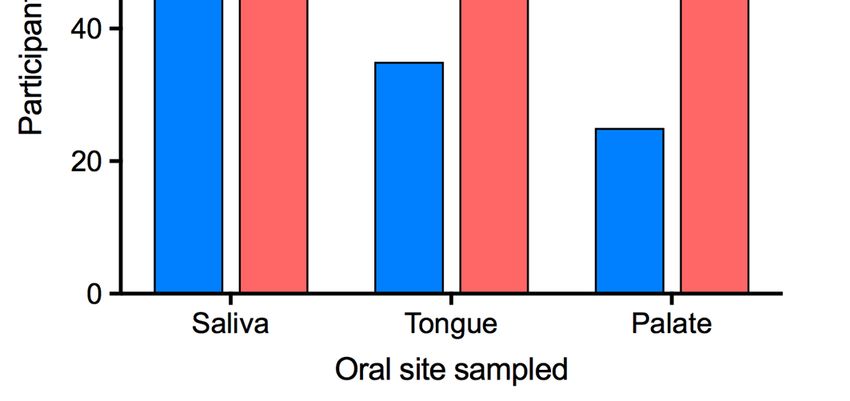
J. J.Fungi
Fungi2019,
2019,5,5,x xFOR
FORPEER
PEERREVIEW
REVIEW 6 6ofof1111
J.swab samples
swab2019,
Fungi 5, 30 inina afurther
samples further1111participants
participantswho
whowere
werenot
notPAS-positive.
PAS-positive.For
Forthose
thosepeople
peoplecolonized
colonized
6 of 10
with
withyeast,
yeast,the
themean
meannumber
numberofofcfu cfuper
permlmlofofsaliva
salivawas
was40,305
40,305(median:
(median:4,425;
4,425;range:
range:410–472,000)
410–472,000)
for
forparticipants
participantsfrom
fromrest
resthomes,
homes,whereas
whereasthe themean
meanfor forparticipants
participantsliving
livinginintheir
theirown
ownhomes
homeswas was
20–9160).
only 2,171 As several230;
(median: of the participants
range: 20–9,160).living
As in theirofown
several the homes had no
participants detectable
living in their yeast
own inhomes
their
only 2,171 (median: 230; range: 20–9,160). As several of the participants living in their own homes
saliva
had the mean colonization levelsaliva
for the group was 1410 cfu/mL (median: 50 cfu/mL) whereascfu/mL
it was
hadnonodetectable
detectableyeast
yeastinintheir
their salivathe
themean
meancolonization
colonizationlevel
levelfor
forthe
thegroup
groupwaswas1,410
1,410 cfu/mL
32,240 cfu/mL (median:
(median: 2260 cfu/mL) for the rest home group (Table 1,cfu/mL)
Figure 2). The numbers of yeast
(median:5050cfu/mL)
cfu/mL)whereas
whereasititwaswas32,240
32,240cfu/mL
cfu/mL(median:
(median:2,260
2,260 cfu/mL)for forthe
therest
resthome
homegroup
group
in each
(Table type
1, of
Figure samples
2). The from
numbers participants
of yeast from
in rest
each homes
type of were
samples significantly
from higher
participants than
from those
rest from
homes
(Table 1, Figure 2). The numbers of yeast in each type of samples from participants from rest homes
people
were living in their own than
homes (Table 1).
weresignificantly
significantlyhigher
higher thanthose
thosefrom
frompeople
peopleliving
livinginintheir
theirown
ownhomes
homes(Table
(Table1).1).
Figure
Figure 2. Quantity of yeast at oral sites in participants. Level of yeast colonization at each site in each
Figure2.2.Quantity
Quantityofofyeast
yeastatatoral
oralsites
sitesininparticipants.
participants.Level
Levelofofyeast
yeastcolonization
colonizationatateach
eachsite
siteinineach
each
group
group (cfu/mL of saliva samples, cfu/swab for tongue and palate swabs ± standard deviation).
group(cfu/mL
(cfu/mLofofsaliva
salivasamples,
samples,cfu/swab
cfu/swabfor fortongue
tongueand
andpalate
palateswabs
swabs± ±standard
standarddeviation).
deviation).
3.3. Species of Colonizing Yeast
3.3.
3.3.Species
SpeciesofofColonizing
ColonizingYeast
Yeast
Yeast were identified by sequencing their rDNA internal transcribed spacer region ITS2. The yeast
Yeast were identified by
bysequencing their
theirrDNA internal transcribed
transcribedspacerspacerregion ITS2. The
speciesYeast
mostwere identified
frequently isolated sequencing
in each sample rDNA
type internal
from both participant groups region
was C. ITS2. The
albicans
yeast species
yeast species most frequently
most frequently isolated
isolated in each sample type from both participant groups was C.
followed by C. glabrata. Seven yeast speciesinwere
eachidentified
sample type from both
in samples from participant
individuals groups
living inwastheirC.
albicans
albicansfollowed
followed by
byC.C.C.
glabrata.
glabrata. Seven yeast
yeastspecies were
wereidentified ininsamples from individuals
own homes: C. albicans, glabrata, Seven
C. parapsilosis, species
C. lusitaniae, identified
C. guilliermondii, samples
Pichiafrom individuals
fermentans and
living in
living in their own
their own homes:
homes: C. albicans, C. glabrata, C. parapsilosis, C. lusitaniae, C. guilliermondii, Pichia
Yarrowia lipolytica (Figure 3). C. albicans,
Only C. glabrata,
five yeast speciesC.(C.parapsilosis,
albicans, C.C.glabrata,
lusitaniae, C. guilliermondii,
Saccharomyces Pichia
cerevisiae,
fermentans and Yarrowia lipolytica (Figure 3). Only five
five yeast
yeast species (C. albicans, C.C. glabrata,
C.fermentans
dubliniensisand Yarrowia
and C. lipolytica
tropicalis) could be(Figure
identified 3).in Only
rest-home species
participants, even(C.though
albicans,
more glabrata,
samples
Saccharomyces
Saccharomyces cerevisiae,
cerevisiae,C.C.dubliniensis
dubliniensis and
and C.C.tropicalis)
tropicalis)could
could bebeidentified
identified ininrest-home
rest-home participants,
participants,
were yeast-positive.
even
eventhough
thoughmore moresamples
sampleswere wereyeast-positive.
yeast-positive.
Figure
Figure 3. Species of yeast colonizing participants.
Figure3.3.Species
Speciesofofyeast
yeastcolonizing
colonizingparticipants.
participants.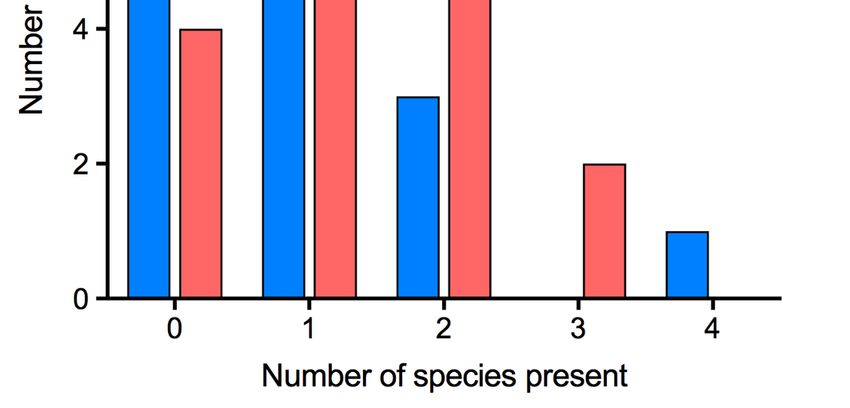
J. Fungi 2019, 5, 30 7 of 10
J. Fungi 2019, 5, x FOR PEER REVIEW 7 of 11
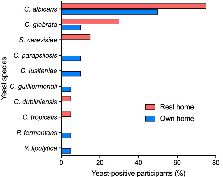
Of theOf60 the
oral60samples
oral samples
takentaken
fromfrom the participants
the participants living
living in in their
their ownown homes,25
homes, 25were
werepositive
positive for
for yeast, whereas 45 of the 60 residential home samples contained yeast. Of the total 70 oral samples
yeast, whereas 45 of the 60 residential home samples contained yeast. Of the total 70 oral samples that
that were yeast-positive, 42 contained only one species of yeast and 28 had more than one species.
were yeast-positive, 42 contained only one species of yeast and 28 had more than one species. The species
The species that were found together most frequently were C. albicans and C. glabrata. The largest
that were found
number oftogether most in
species found frequently
the oral cavity C. aalbicans
were of participant C. glabrata.
and (who The
lived in largest
their number
own home) of four
was species
found in the oral
(Figure 4). cavity of a participant (who lived in their own home) was four (Figure 4).
Figure 4. Number of yeast species colonizing individual participants.
Figure 4. Number of yeast species colonizing individual participants.
4. Discussion
4. Discussion
In this study the types, and quantities, of yeast colonizing the oral surfaces of older people
In this study the types, and quantities, of yeast colonizing the oral surfaces of older people
living in rest homes were compared with those in people living in their own homes. Three oral sites
living in rest homes were compared with those in people living in their own homes. Three oral sites
were sampled to make these measurements, the tongue, the palate, and whole unstimulated saliva.
were sampled to make these measurements, the tongue, the palate, and whole unstimulated saliva.
The tongue was sampled
The tongue was sampledas it has
as itbeen shown
has been shownto betoone of the
be one most
of the frequently
most frequently colonized
colonized oral
oralsites
sites[25],
and the[25],
palate
and isthealso often
palate colonized
is also in patients
often colonized with full
in patients withupper dentures
full upper [5]. [5].
dentures We Wefound
found that inin
that both
groupsbothof participants the tongue
groups of participants was more
the tongue was morefrequently colonized
frequently colonizedthanthanthe palate,and
the palate, and that
that yeast
yeast
were most
werefrequently found
most frequently in saliva;
found all all
in saliva; individuals
individualswithwithyeast
yeast on their
theirtongues
tonguesoror palate
palate also
also hadhad
yeast in saliva, and for some individuals yeast were only detected in their
yeast in saliva, and for some individuals yeast were only detected in their saliva. This is probably saliva. This is probably
because because
if yeastif colonize
yeast colonize oral surfaces,
oral surfaces, they they are likely
are likely to betowashed
be washed offsome
off to to some extent
extent by saliva.
by saliva. Thus,
Thus, for a study of the variety of fungi in people’s mouths, yeast in saliva
for a study of the variety of fungi in people’s mouths, yeast in saliva can be used as a proxy for oralcan be used as a proxy for
oral colonization and saliva may act as a reservoir for colonization of other oral sites. It is difficult to
colonization and saliva may act as a reservoir for colonization of other oral sites. It is difficult to
assign a concentration of yeast in saliva, on swabs, or in smears that might indicate candidiasis as
assign host
a concentration of yeast in saliva, on swabs, or in smears that might indicate candidiasis as host
factors are important in disease progression. The presence of hyphae in smears is a good
factorsindication
are important in disease progression.
of clinically-significant disease The
as thepresence of hyphae
hyphal form in smears
can penetrate is a good
tissues. However,indication
many of
clinically-significant
Candida species, such as C. glabrata, do not form hyphae making the diagnosis of candidiasis fromspecies,
disease as the hyphal form can penetrate tissues. However, many Candida the
C. glabrata,
such asinspection do notmore
of smears formdifficult.
hyphaeThe making the yeast
fact that diagnosis of candidiasis
were cultured from morefrompeople
the inspection
than had of
smearsPAS-positive
more difficult. smears
Themay factreflect the greater
that yeast sensitivityfrom
were cultured of culturing yeast from
more people than swabs or saliva
had PAS-positive
smearssamples,
may reflectbut this
thepositive
greaterculture does not
sensitivity give the clinician
of culturing yeast an indication
from swabs of orthe disease
saliva status. but this
samples,
Theredoes
positive culture are anot
number
give theof factors that
clinician anare known to
indication of favor oral colonization
the disease status. with yeast. These
include reduced salivary flow rates [16,26] and wearing dentures [27]. In the present study we found
There are a number of factors that are known to favor oral colonization with yeast. These include
that people living in their own homes who had a low unstimulated salivary flow rate (J. Fungi 2019, 5, 30 8 of 10
colonization level was higher than that for residents with a reduced salivary flow rate due to two
residents with high salivary yeast concentrations and normal flow rates. Unstimulated saliva flow
rates reduce with age [28] and older people are often prescribed medications which are known to be
xerogenic (cause dry mouth). Due to the number and variety of medications our participants were
prescribed, it was not possible to control for medications between the groups. There was no significant
difference in the mean number of medications taken by the two groups of participants, but there
was a significant association between low salivary flow rate and a higher number of medications.
Participants in both groups who wore dentures had greater concentrations of yeast in their saliva than
those participants who did not wear dentures, but the differences were not statistically significant.
CHROMagar Candida is a useful medium for the presumptive identification of C. albicans from
biological samples and for distinguishing other fungal species. Combining this primary screen with
rDNA ITS2 sequencing of isolates enabled precise identification of the predominant fungal species
present in oral samples without the detection of extremely minor components of the mycobiome as
is often the case with direct next generation sequencing of biological samples. As has been shown
previously [4,5,16], the most common yeast species found in the mouths of participants was C. albicans.
The second most frequently isolated species was C. glabrata. This may reflect the increased prevalence
of this species in clinical samples that has been reported in recent years [29]. It is of concern that of
the 10 yeast species detected in participants, seven (C. albicans, C. glabrata, C. parapsilosis, C. lusitaniae,
C. guilliermondii, C. tropicalis, and Y. lipolytica) have been reported as being potentially resistant to
antifungals [12,29–33]. It was beyond the scope of the present project to measure the antifungal
susceptibility of all the fungal isolates, but this would provide an indication of whether the older adults
sampled were at risk from a fungal infection that would be difficult to treat.
As populations age, an increasing number of older people are being cared for in rest homes,
yet there are few studies on the microbial colonization of people living in these institutions. One
comparison of older people in institutionalized long-term care with those not in such care found that
oral colonization with yeast was more frequent in the institutionalized individuals [34]. This study
did not, however, identify the colonizing yeast. We found that oral sites were more frequently
colonized by yeast in rest-home participants than in people living in their own home and that the
mean concentration of yeast in the saliva of rest-home participants was 23 times higher than that in
non-rest home participants (the median was 45 times higher). This may indicate poorer oral hygiene
in the rest home participants which may be due to the reason they are in rest homes—they are less
capable of independent living. The increased numbers of opportunistic fungal pathogens in rest home
residents suggests that the rest homes would be well advised to pay greater attention to the oral
hygiene of residents.
It was interesting to note that a greater variety of yeast species was detected in people living in
their own homes (7 species) compared to those living in rest homes (5 species) with only two species
(C. albicans and C. glabrata) common to both groups. A possible explanation is that communal living and
shared care assistants results in the sharing of yeast between rest home residents. This possibility could
be investigated further by MLST (multilocus sequence typing) analysis of the predominant species in
both groups, C. albicans, to determine the relatedness of strain types in the two groups, as we have done
previously for people wearing dentures [35]. Alternatively, a higher proportion of people in rest homes
may have morbidities that favor yeast growth, and yeast species particularly well-adapted to the oral
environment in these people may out-compete other species. This could lead to high numbers of a single
yeast species in such individuals. Species with the potential to be drug resistant were present in both the
rest home residents (C. albicans, C. glabrata, C. dubliniensis, and C. tropicalis) and people living in their own
homes (C. albicans, C. glabrata, C. parapsilosis, C. lusitaniae, C. guilliermondii, and Y. lipolytica) emphasizing
the need for all older people to keep in good health and maintain good oral hygiene.
This study has some limitations. One is the small sample size. While a few studies report the
colonization of older people living in institutionalized care by yeast [34,36], there are no quantitative
analyses of the different yeast present. Thus, it was not possible to perform a sample-size estimationJ. Fungi 2019, 5, 30 9 of 10
for this study, and a pilot investigation was undertaken. Also, participants were recruited from two
rest homes which introduced a potential confounder. Despite the small sample size, the present study
revealed some important statistically significant differences between the oral colonization of people
living in rest homes and those living in their own homes. The results from this study, therefore, provide
a justification, and basis, for examining the oral fungal colonization of larger samples of older people
from a single rest home and older people living in their own homes.
Author Contributions: Conceptualization, R.D.C., N.M.T. and A.M.R.; methodology, E.L., A.M.R. and R.D.C.;
investigation, N.M.T.; data curation, N.M.T; writing—original draft preparation, N.M.T.; writing—review and
editing, R.D.C., E.L. and A.M.R.; supervision, E.L., A.M.R. and R.D.C.; project administration, R.D.C.; funding
acquisition, N.M.T., E.L., A.M.R. and R.D.C.
Funding: This research was funded by a New Zealand Ministry of Health Oral Health Research Grant, (grant
number RF8.15).
Acknowledgments: We would like to thank Murray Thomson (Faculty of Dentistry, University of Otago) for his
assistance with statistical analysis, and Trudy Milne (Faculty of Dentistry, University of Otago) for assistance with
the Figures.
Conflicts of Interest: The authors declare no conflict of interest. The funders had no role in the design of the
study; in the collection, analyses, or interpretation of data; in the writing of the manuscript, or in the decision to
publish the results.
References
1. Cannon, R.D.; Chaffin, W.L. Oral colonization by Candida albicans. Crit. Rev. Oral Biol. Med. 1999, 10, 359–383.
[CrossRef]
2. Zegarelli, D.J. Fungal infections of the oral cavity. Otolaryngol. Clin. N. Am. 1993, 26, 1069–1089.
3. Cannon, R.D.; Holmes, A.R.; Mason, A.B.; Monk, B.C. Oral Candida: Clearance, colonization, or candidiasis?
J. Dent. Res. 1995, 74, 1152–1161. [CrossRef]
4. Meurman, J.H.; Parnanen, P.; Seneviratne, C.J.; Samaranayake, L.P.; Saarinen, A.M.; Kari, K. Prevalence and
antifungal drug sensitivity of non-albicans Candida in oral rinse samples of self-caring elderly. Gerodontology
2011, 28, 246–252. [CrossRef] [PubMed]
5. Odds, F.C. Candida and Candidosis, 2nd ed.; Bailliere Tindall: London, UK, 1988.
6. Lopez-Martinez, R. Candidosis, a new challenge. Clin. Dermatol. 2010, 28, 178–184. [CrossRef] [PubMed]
7. Miceli, M.H.; Diaz, J.A.; Lee, S.A. Emerging opportunistic yeast infections. Lancet Infect. Dis. 2011, 11,
142–151. [CrossRef]
8. Cannon, R.D.; Firth, N.A. Fungi and fungal infections of the oral cavity. In Oral Microbiology and Immunology,
2nd ed.; Lamont, R.J., Hajishengallis, G.N., Jenkinson, H.F., Eds.; ASM Press: Washington, DC, USA, 2014;
pp. 343–359.
9. Nobile, C.J.; Johnson, A.D. Candida albicans biofilms and human disease. Annu. Rev. Microbiol. 2015, 69,
71–92. [CrossRef] [PubMed]
10. Galle, F.; Sanguinetti, M.; Colella, G.; Di Onofrio, V.; Torelli, R.; Rossano, F.; Liguori, G. Oral candidosis:
Characterization of a sample of recurrent infections and study of resistance determinants. New Microbiol.
2011, 34, 379–389. [PubMed]
11. Sanguinetti, M.; Posteraro, B.; Lass-Florl, C. Antifungal drug resistance among Candida species: Mechanisms
and clinical impact. Mycoses 2015, 58, 2–13. [CrossRef]
12. Atkinson, B.J.; Lewis, R.E.; Kontoyiannis, D.P. Candida lusitaniae fungemia in cancer patients: Risk factors for
amphotericin B failure and outcome. Med. Mycol. 2008, 46, 541–546. [CrossRef]
13. Pinhati, H.M.; Casulari, L.A.; Souza, A.C.; Siqueira, R.A.; Damasceno, C.M.; Colombo, A.L. Outbreak of
candidemia caused by fluconazole resistant Candida parapsilosis strains in an intensive care unit. BMC Infect. Dis.
2016, 16, 433. [CrossRef] [PubMed]
14. Thomas, J.E.; Lloyd, P.M. Oral candidiasis in the elderly. Spec. Care Dentist 1985, 5, 222–225. [CrossRef]
[PubMed]
15. Shinozaki, S.; Moriyama, M.; Hayashida, J.N.; Tanaka, A.; Maehara, T.; Ieda, S.; Nakamura, S. Close
association between oral Candida species and oral mucosal disorders in patients with xerostomia. Oral Dis.
2012, 18, 667–672. [CrossRef]J. Fungi 2019, 5, 30 10 of 10
16. Nadig, S.D.; Ashwathappa, D.T.; Manjunath, M.; Krishna, S.; Annaji, A.G.; Shivaprakash, P.K. A relationship
between salivary flow rates and Candida counts in patients with xerostomia. J. Oral Maxillofac Pathol. 2017,
21, 316. [CrossRef]
17. Lynge Pedersen, A.M.; Nauntofte, B.; Smidt, D.; Torpet, L.A. Oral mucosal lesions in older people: Relation
to salivary secretion, systemic diseases and medications. Oral Dis. 2015, 21, 721–729. [CrossRef]
18. World Health Organization. Ageing and Health. Available online: https://www.who.int/news-room/fact-
sheets/detail/ageing-and-health (accessed on 20 December 2018).
19. Flevari, A.; Theodorakopoulou, M.; Velegraki, A.; Armaganidis, A.; Dimopoulos, G. Treatment of invasive
candidiasis in the elderly: A review. Clin. Interv Aging 2013, 8, 1199–1208. [CrossRef]
20. Thomson, W.M.; Ma, S. An ageing population poses dental challenges. Singapore Dent. J. 2014, 35C, 3–8.
[CrossRef] [PubMed]
21. Statistics New Zealand. Available online: http://archive.stats.govt.nz/Census/2013-census/profile-and-
summary-reports/outside-norm/residential-old.aspx (accessed on 26 March 2019).
22. Hoggard, M.; Vesty, A.; Wong, G.; Montgomery, J.M.; Fourie, C.; Douglas, R.G.; Biswas, K.; Taylor, M.W.
Characterizing the Human Mycobiota: A Comparison of Small Subunit rRNA, ITS1, ITS2, and Large Subunit
rRNA Genomic Targets. Front. Microbiol. 2018, 9, 2208. [CrossRef]
23. Lott, T.J.; Kuykendall, R.J.; Reiss, E. Nucleotide sequence analysis of the 5.8S rDNA and adjacent ITS2 region
of Candida albicans and related species. Yeast 1993, 9, 1199–1206. [CrossRef] [PubMed]
24. Longman, L.P.; Higham, S.M.; Bucknall, R.; Kaye, S.B.; Edgar, W.M.; Field, E.A. Signs and symptoms in
patients with salivary gland hypofunction. Postgrad. Med. J. 1997, 73, 93–97. [CrossRef]
25. Arendorf, T.M.; Walker, D.M. The prevalence and intra-oral distribution of Candida albicans in man.
Arch. Oral Biol. 1980, 25, 1–10. [CrossRef]
26. Narhi, T.O.; Ainamo, A.; Meurman, J.H. Salivary yeasts, saliva, and oral mucosa in the elderly. J. Dent. Res.
1993, 72, 1009–1014. [CrossRef]
27. Abu-Elteen, K.H.; Abu-Alteen, R.M. The prevalence of Candida albicans populations in the mouths of complete
denture wearers. New Microbiol. 1998, 21, 41–48.
28. Percival, R.S.; Challacombe, S.J.; Marsh, P.D. Flow rates of resting whole and stimulated parotid saliva in
relation to age and gender. J. Dent. Res. 1994, 73, 1416–1420. [CrossRef]
29. Chapman, B.; Slavin, M.; Marriott, D.; Halliday, C.; Kidd, S.; Arthur, I.; Bak, N.; Heath, C.H.; Kennedy, K.;
Morrissey, C.O.; et al. Changing epidemiology of candidaemia in Australia. J. Antimicrob. Chemother. 2017,
72, 1103–1108. [CrossRef]
30. Colombo, A.L.; Junior, J.N.A.; Guinea, J. Emerging multidrug-resistant Candida species. Curr. Opin. Infect. Dis.
2017, 30, 528–538. [CrossRef]
31. Asner, S.A.; Giulieri, S.; Diezi, M.; Marchetti, O.; Sanglard, D. Acquired multidrug antifungal resistance in
Candida lusitaniae during therapy. Antimicrob. Agents Chemother. 2015, 59, 7715–7722. [CrossRef]
32. Whaley, S.G.; Berkow, E.L.; Rybak, J.M.; Nishimoto, A.T.; Barker, K.S.; Rogers, P.D. Azole antifungal resistance
in Candida albicans and emerging non-albicans Candida species. Front. Microbiol. 2016, 7, 2173. [CrossRef]
33. Coleman, D.C.; Moran, G.P.; McManus, B.A.; Sullivan, D.J. Mechanisms of antifungal drug resistance in
Candida dubliniensis. Future Microbiol. 2010, 5, 935–949. [CrossRef]
34. Glazar, I.; Muhvic Urek, M.; Kuis, D.; Prpic, J.; Miskovic, I.; Kovacevic Pavicic, D.; Pezelj-Ribaric, S. Salivary
flow rate, oral yeast colonization and dental status in institutionalized and non-institutionalized elderly.
Acta Clin. Croat. 2016, 55, 390–395. [CrossRef]
35. Choo, K.H.; Lee, H.J.; Knight, N.J.; Holmes, A.R.; Cannon, R.D. Multilocus sequence typing (MLST) analysis
of Candida albicans isolates colonizing acrylic dentures before and after denture replacement. Med. Mycol.
2017, 55, 673–679. [CrossRef]
36. Grimoud, A.M.; Marty, N.; Bocquet, H.; Andrieu, S.; Lodter, J.P.; Chabanon, G. Colonization of the oral cavity
by Candida species: Risk factors in long-term geriatric care. J. Oral Sci. 2003, 45, 51–55. [CrossRef]
© 2019 by the authors. Licensee MDPI, Basel, Switzerland. This article is an open access
article distributed under the terms and conditions of the Creative Commons Attribution
(CC BY) license (http://creativecommons.org/licenses/by/4.0/).You can also read