Zebrafish embryonic explants undergo genetically encoded self-assembly - eLife
←
→
Page content transcription
If your browser does not render page correctly, please read the page content below
RESEARCH ARTICLE

                                    Zebrafish embryonic explants undergo
                                    genetically encoded self-assembly
                                    Alexandra Schauer†, Diana Pinheiro†, Robert Hauschild, Carl-Philipp Heisenberg*

                                    IST Austria, Klosterneuburg, Austria

                                    Abstract Embryonic stem cell cultures are thought to self-organize into embryoid bodies, able
                                    to undergo symmetry-breaking, germ layer specification and even morphogenesis. Yet, it is unclear
                                    how to reconcile this remarkable self-organization capacity with classical experiments
                                    demonstrating key roles for extrinsic biases by maternal factors and/or extraembryonic tissues in
                                    embryogenesis. Here, we show that zebrafish embryonic tissue explants, prepared prior to germ
                                    layer induction and lacking extraembryonic tissues, can specify all germ layers and form a
                                    seemingly complete mesendoderm anlage. Importantly, explant organization requires polarized
                                    inheritance of maternal factors from dorsal-marginal regions of the blastoderm. Moreover,
                                    induction of endoderm and head-mesoderm, which require peak Nodal-signaling levels, is highly
                                    variable in explants, reminiscent of embryos with reduced Nodal signals from the extraembryonic
                                    tissues. Together, these data suggest that zebrafish explants do not undergo bona fide self-
                                    organization, but rather display features of genetically encoded self-assembly, where intrinsic
                                    genetic programs control the emergence of order.

                                    Introduction
                                    Embryonic development entails both progressive specification of cells into distinct cell fates and
                                    their respective allocation into different domains within the embryo. Key signaling molecules, also
*For correspondence:                termed morphogens, play critical roles in eliciting both the genetic programs underlying cell fate
heisenberg@ist.ac.at                specification and the morphogenetic cell properties required to shape the developing embryo
†
 These authors contributed          (Gilmour et al., 2017; Heisenberg and Solnica-Krezel, 2008; Pinheiro and Heisenberg, 2020).
equally to this work                While amphibians critically rely on maternal factors for precise spatiotemporal activation of morpho-
                                    gen signaling during early embryonic development, mammalian embryos mostly depend on recipro-
Competing interests: The
                                    cal interactions between embryonic and extraembryonic tissues (Mummery and Chuva de Sousa
authors declare that no
competing interests exist.          Lopes SM, 2013; Heasman, 2006; Langdon and Mullins, 2011; Stern and Downs, 2012; Tam and
                                    Loebel, 2007). Interestingly, zebrafish embryos combine features characteristic of both amphibian
Funding: See page 25
                                    and mammalian development. Here, maternal factors initially deposited within the oocyte
Received: 15 January 2020           (Langdon and Mullins, 2011; Marlow, 2020) and signals from extraembryonic tissues, namely the
Accepted: 05 April 2020             yolk cell and its yolk syncytial layer (YSL), play important roles in germ layer induction and patterning
Published: 06 April 2020            (Carvalho and Heisenberg, 2010; Chen and Kimelman, 2000; Erter et al., 1998; Fan et al., 2007;
Reviewing editor: Ashley Bruce,     Gagnon et al., 2018; Hong et al., 2011; Mizuno et al., 1999; Mizuno et al., 1996; Ober and
                                    Schulte-Merker, 1999; Schier and Talbot, 2005; van Boxtel et al., 2015; Veil et al., 2018;
   Copyright Schauer et al. This
                                    Xu et al., 2012). Nodal/TGFb ligands, for instance, key signals involved in mesoderm and endoderm
article is distributed under the
                                    (mesendoderm) specification and patterning, are initially expressed under maternal control and then
terms of the Creative Commons
Attribution License, which          secreted by the extraembryonic YSL to pattern the embryonic blastoderm (Chen and Schier, 2002;
permits unrestricted use and        Dougan et al., 2003; Erter et al., 1998; Fan et al., 2007; Fauny et al., 2009; Feldman et al.,
redistribution provided that the    1998; Gagnon et al., 2018; Gore et al., 2005; Gritsman et al., 2000; Gritsman et al., 1999;
original author and source are      Hong et al., 2011; Rebagliati et al., 1998; Rogers et al., 2017; Rogers and Müller, 2019;
credited.                           Thisse and Thisse, 2015; van Boxtel et al., 2015; Xu et al., 2012).

Schauer et al. eLife 2020;9:e55190. DOI: https://doi.org/10.7554/eLife.55190                                                          1 of 30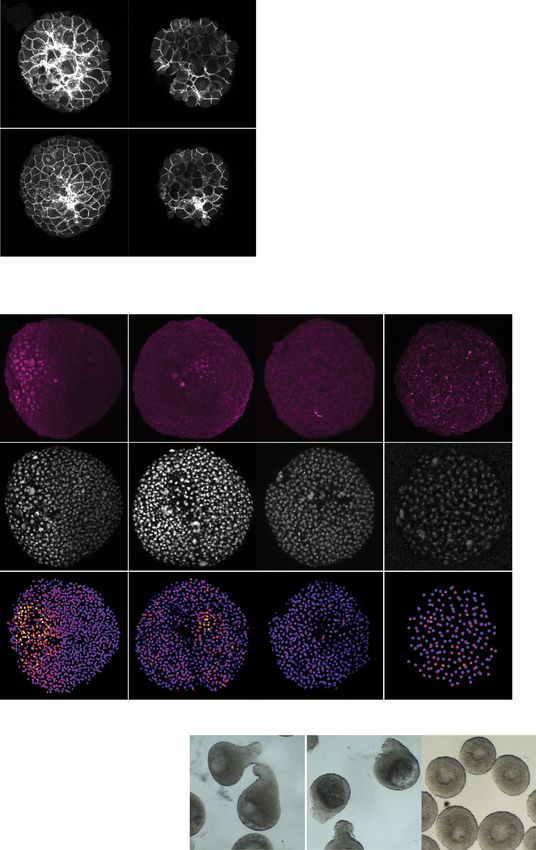
Research article Developmental Biology Stem Cells and Regenerative Medicine
Pioneering studies have suggested that both human and mouse embryonic stem cell (ES) colonies
exhibit a remarkable capacity to self-organize in vitro and recapitulate features associated with early
embryogenesis (Baillie-Johnson et al., 2015; Beccari et al., 2018; Doetschman et al., 1985;
Shahbazi et al., 2019; Simunovic et al., 2019; Simunovic and Brivanlou, 2017; ten Berge et al.,
2008; Turner et al., 2017; Turner et al., 2014; van den Brink et al., 2014; van den Brink et al.,
2020; Warmflash et al., 2014). For instance, homogeneous stimulation of human ES colonies cul-
tured on 2-dimensional circular substrates with BMP4 ligands is sufficient to reproducibly generate
radially symmetric colonies, where cells at the outmost rim differentiate into extraembryonic tissue,
while the remaining cells differentiate progressively into more internal rings composed of the differ-
ent embryonic germ layers - endoderm, mesoderm and ectoderm (Warmflash et al., 2014). These
apparent self-organizing processes are thought to rely on boundary sensing, community effects and
the interplay between activators and inhibitors (Chhabra et al., 2019; Etoc et al., 2016;
Nemashkalo et al., 2017; Sasai, 2013; Xavier da Silveira Dos Santos and Liberali, 2019). While
these pioneering studies suggest that embryonic tissues might have some self-organizing capacity,
classical studies have also established the dependency of embryonic development on both maternal
factors and extraembryonic tissues, thereby raising key questions as to their specific contributions to
robust embryonic development.
Results
To address this question, we established an ex vivo system to culture zebrafish blastoderm tissue
explants lacking the yolk cell and its associated YSL. For this, blastoderm explants were prepared
from embryos at early cleavage stages (256 cell stage), before YSL formation or germ layer induc-
tion, and kept in serum-free medium until stage-matched control embryos had completed gastrula-
tion (bud stage; Figure 1A). We found that these blastoderm explants initially rounded up in
culture, but eventually became ‘pear’-shaped by forming an extension (Figure 1B,C, Video 1). Inter-
estingly, this extension began to form when stage-matched control embryos initiated body axis
extension at shield stage (Sepich et al., 2005; Solnica-Krezel and Sepich, 2012), pointing at the
possibility that these processes might be related. Contrary to intact embryos, however, blastoderm
explants formed a large luminal cavity, reminiscent of a blastocoel (Krens et al., 2017), which was
typically positioned at the opposite end of the explant extension (Figure 1B, Figure 1—figure sup-
plement 1A,B, Video 2). Importantly, cells within explants appeared to normally complete their
rounds of cleavages and divisions following explant preparation, as evidenced by the similar average
diameter of explant and stage-matched embryo cells (Figure 1—figure supplement 1C). Moreover,
we found that the onset of zygotic genome activation (ZGA), as monitored by the expression of the
two zygotic genes tbx16 and chrd (Gagnon et al., 2018; Lee et al., 2013), was similar in stage-
matched embryos and blastoderm explants (Figure 1—figure supplement 1D). Finally, comparable
morphological changes could be observed when blastoderm explants were prepared from earlier
(64 and 128 cell-stage) or later (high stage) embryos (Figure 1—figure supplement 2A–D), suggest-
ing that the exact time point of explant preparation during pre-gastrula stages has no decisive influ-
ence on explant morphogenesis.
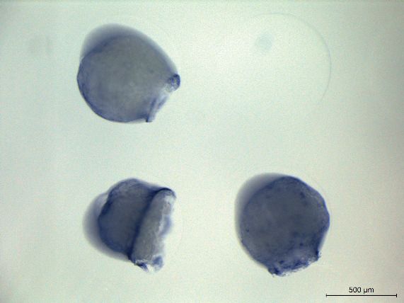
To analyze how these distinct changes in explant morphology relate to the specification of differ-
ent progenitor cell types therein, we analyzed the expression of various progenitor cell marker genes
by whole mount in situ hybridization. Strikingly, we found distinct expression domains for all tested
marker genes outlining ectoderm, neuroectoderm, endoderm and different types of mesoderm pro-
genitor cell populations. While ectoderm (gata2) and neuroectoderm (six3) marker genes were
expressed predominantly at the opposite end of the explant extended portion, the region where
the cavity was also forming (Figure 1D–G, Figure 1—figure supplement 1A,B,H, Figure 1—figure
supplement 3A), all mesendoderm and endoderm marker genes were exclusively found within or
adjacent to the extension (Figure 1D–G, Figure 1—figure supplement 1G–J, Figure 1—figure sup-
plement 3A). Specifically, we found that markers of posterior axial mesoderm (ntl and flh) were
expressed in the central region of the extension, flanked by paraxial and lateral mesoderm (papc,
myoD, tbx6) (Figure 1D–G, Figure 1—figure supplement 1G–I, Figure 1—figure supplement 3A).
In contrast, anterior axial mesendoderm markers (hgg and gsc) were found to be expressed at vari-
able positions along the extended region or directly adjacent to it (Figure 1D–G, Figure 1—figure
supplement 1G–I, Figure 1—figure supplement 3A). Finally, sporadic endoderm progenitors
Schauer et al. eLife 2020;9:e55190. DOI: https://doi.org/10.7554/eLife.55190 2 of 30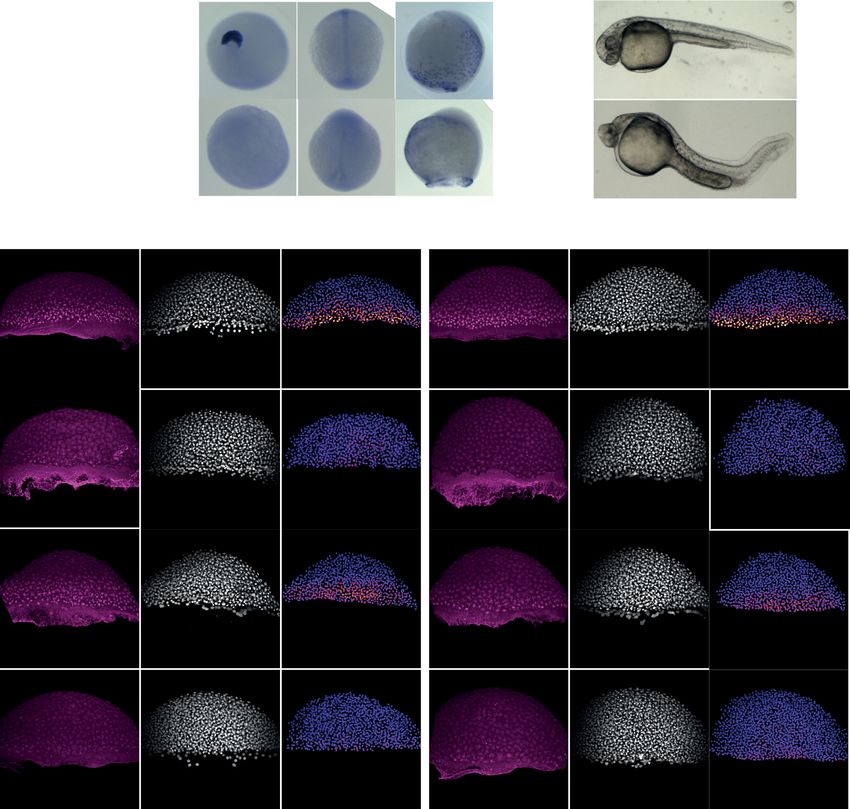
Research article Developmental Biology Stem Cells and Regenerative Medicine
A B Oblong Dome Shield 90% epiboly Bud C
1.00
Preparation of blastoderm
explants
0.95
Embryo
Circularity (±SD)
256c stage 256c stage
embryo explant 0.90
Blastoderm
0.85
B
B
0.80
*
Explant
*Serum-free * 0.75
yolk cell
medium *
*
ep e
ep y
50 D g
7 0 S ly
9 0 e pi d
y
d
*
% bol
ol
% om
on
Bu
l
*
o
% ie
ib
ib
*
h
bl
O
D A Side view
gata2 six3 hgg ntl papc
Animal
V D
view Side view Animal view Animal view Dorsal view Dorsal view
P
Embryo
Dorsal
view
Bud
21/21 20/20 34/34 46/46 43/43
Tip Side view Side view
extension
Explant
* *
Back * 32/33 * 20/26 22/49 43/48 40/55
E 1.0
gata2 F G
six3 Embryo Tip
Explant
A
Normalized expression along
hgg extension ntl & flh
the back-tip axis (±SEM)
0.8 V D
ntl
six3 papc & myoD
papc P hgg & tbx6
0.6
& gsc hgg & gsc sox32
0.4 six3
0.2 gata2 sox32
ntl & flh gata2
0.0
0.0 0.2 0.4 0.6 0.8 1.0 Back
Normalized position along the explant papc & myoD & tbx6
Back Tip extension
Figure 1. Mesendoderm specification and morphogenesis in the absence of the yolk cell and its associated YSL. (A) Schematic representation of the
preparation method of blastoderm explants from 256 cell stage (256 c) stage embryos. (B) Bright-field single-plane images of stage-matched embryo
and blastoderm explants from oblong to bud stage. The white dashed lines outline the shape of the explant. (C) Circularity of blastoderm explants
from oblong to bud stage (oblong: n = 42, dome: n = 34, 50% epiboly: n = 35, shield: n = 40, 70% epiboly: n = 35, 90% epiboly: n = 36, bud: n = 38;
N = 2). (D) Expression of ectoderm (gata2), neuroectoderm (six3) and mesendoderm (hgg, ntl and papc) marker genes as determined by whole mount
in situ hybridization of bud stage embryos and blastoderm explants. Schematic representation of the different views for embryos and blastoderm
explants is shown on the left. The proportion of embryos or blastoderm explants with a phenotype similar to the images shown is indicated in the
lower right corner (gata2: embryos, n = 21, N = 4, explants, n = 33, N = 6; six3: embryos, n = 20, N = 3, explants, n = 26, N = 4; hgg: embryos, n = 34,
N = 5, explants, n = 49, N = 5; ntl: embryos, n = 46, N = 4, explants, n = 48, N = 4; papc: embryos, n = 43, N = 4, explants, n = 55, N = 5). (E)
Normalized expression domain of ectoderm (gata2: n = 31, N = 6), neuroectoderm (six3: n = 20, N = 4) and mesendoderm (hgg: n = 20, N = 5; ntl:
n = 41, N = 4; papc: n = 37, N = 5) marker genes along the back-tip axis of bud stage blastoderm explants. (F-G) Schematic representation of
ectoderm, neuroectoderm, mesendoderm and endoderm marker gene expression domains in intact embryos (F) and blastoderm explants (G). White
asterisks denote the main luminal cavity in explants. Scale bars: 200 mm (B, D).
The online version of this article includes the following source data and figure supplement(s) for figure 1:
Source data 1. Mesendoderm specification and morphogenesis in the absence of the yolk cell and its associated YSL.
Figure supplement 1. Phenotypic characterization of blastoderm explants.
Figure supplement 1—source data 1. Phenotypic characterization of blastoderm explants.
Figure supplement 2. Morphogenesis of blastoderm explants prepared at 64 c, 128 c, 256 c or high stage.
Figure 1 continued on next page
Schauer et al. eLife 2020;9:e55190. DOI: https://doi.org/10.7554/eLife.55190 3 of 30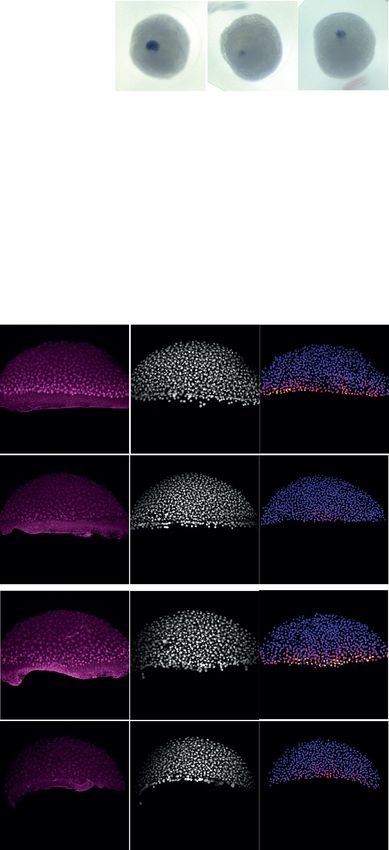
Research article                                                            Developmental Biology Stem Cells and Regenerative Medicine

Figure 1 continued
Figure supplement 2—source data 1. Morphogenesis of blastoderm explants prepared at 64 c, 128 c, 256 c and high stage.
Figure supplement 3. Relative spatial distribution of ectoderm and mesendoderm progenitor cell types in blastoderm explants.
Figure supplement 4. Blastoderm explants exhibit reduced or no YSL features.
Figure supplement 4—source data 1. Blastoderm explants exhibit reduced or no expression of YSL markers.

                                    (sox32) could be found throughout the extension (Figure 1G, Figure 1—figure supplement 1G,H,J,
                                    Figure 1—figure supplement 3A). On the explant surface, much like in the intact embryo, we found
                                    that cells expressed elevated levels of Keratin 4, a marker of enveloping layer (EVL) cell differentia-
                                    tion (Figure 1—figure supplement 1E,F). Collectively, these findings suggest that the blastoderm
                                    explants contain all main germ layer progenitors also found in intact embryos and display a seem-
                                    ingly complete patterning of the mesendoderm anlage. They also indicate that, although explants
                                    undergo extensive morphogenesis, mesendoderm progenitors do not clearly internalize and,
                                    instead, organize into an extension (Video 3). This is highly reminiscent of the ‘exogastrula’ pheno-
                                    type observed in embryos with defective mesendoderm internalization and, more recently, in mouse
                                    gastruloids (Beccari et al., 2018; Branford and Yost, 2002; Turner et al., 2017; van den Brink
                                    et al., 2014).
                                       To ensure that all extraembryonic tissues were efficiently removed during explant preparation
                                    and that explants do not form a secondary YSL, we analyzed the expression of marker genes, nor-
                                    mally present within the YSL in intact embryos, in blastoderm explants. We found that all of these
                                    genes were either not expressed in explants or showed only sporadic expression in small groups of
                                    blastoderm cells (Figure 1—figure supplement 4A,B). Moreover, we failed to detect any clearly rec-
                                    ognizable syncytium in explants, a hallmark of the YSL in intact embryos (Figure 1—figure supple-
                                    ment 4C). Together, these data strongly support the notion that germ layer specification and
                                    mesendoderm patterning in blastoderm explants can occur in the absence of the extraembryonic
                                    yolk and YSL.

                                                                                               Video 2. Formation of the luminal cavity in blastoderm
                                    Video 1. Blastoderm explant morphogenesis in the           explants. High-resolution fluorescence time-lapse
                                    absence of the yolk cell and its associated YSL. Bright-   imaging of a blastoderm explant (cross-section) from
                                    field time-lapse imaging of a blastoderm explant           oblong stage onwards (n = 9, N = 2). Interstitial fluid
                                    (cross-section) from late sphere to bud stage (n = 10,     (green) is marked by dextran Alexa Fluor 647 and the
                                    N = 3). The white dashed lines outline the initial and     cell membranes (white) are marked by Membrane-GFP.
                                    final shape of the explant. White asterisks denote the     White asterisks denote the two luminal cavities. Time in
                                    main luminal cavity. Time in min. Scale bar: 200 mm.       min. Scale bar: 100 mm.
                                    https://elifesciences.org/articles/55190#video1            https://elifesciences.org/articles/55190#video2

Schauer et al. eLife 2020;9:e55190. DOI: https://doi.org/10.7554/eLife.55190                                                                     4 of 30
Research article                                                        Developmental Biology Stem Cells and Regenerative Medicine

                                       To understand how mesendoderm tissues are formed within explants, we first analyzed whether
                                    Nodal ligands, critical for mesendoderm specification and patterning in vivo (Erter et al., 1998;
                                    Feldman et al., 1998; Gritsman et al., 2000; Gritsman et al., 1999; Rebagliati et al., 1998), are
                                    also expressed in explants. We observed expression of both Nodal ligands sqt and cyc in blastoderm
                                    explants (Figure 2—figure supplement 1A,B), consistent with previous observations in intact
                                    embryos (Erter et al., 1998; Fan et al., 2007; Feldman et al., 1998; Rebagliati et al., 1998;
                                    van Boxtel et al., 2015). In addition to this, we detected nuclear localization of phosphorylated
                                    SMAD2/3 (pSMAD2/3), a well-established readout of Nodal/TGFb signaling activation
                                    (Economou and Hill, 2020; Schier, 2009; van Boxtel et al., 2015; van Boxtel et al., 2018), in cells
                                    located close to the wounding site of explants (Figure 2A, Figure 2—figure supplement 1C,D),
                                    indicative of local Nodal signaling activation. A comparison of the spatiotemporal dynamics of Nodal
                                    signaling establishment between explants and intact embryos further revealed that, while blasto-
                                    derm explants showed initially fewer and less bright pSMAD2/3 positive nuclei than intact embryos,
                                    both parameters continued to increase in explants until stage-matched embryos had reached shield
                                    stage (Figure 2A–C, Figure 2—figure supplement 1C–J). This differs from the temporal dynamics
                                    of Nodal signaling in intact embryos, where nuclear accumulation of pSMAD2/3 peaks already by
                                    50% epiboly (Figure 2A–C, Figure 2—figure supplement 1C–J). Notably, the wounding site of blas-
                                    toderm explants, where Nodal signaling was activated corresponds to the former blastoderm margin
                                    where explants were removed from the yolk cell (Figure 2D, Figure 2—figure supplement 1E–J).
                                    Given that Nodal signaling in intact embryos is initiated in the blastoderm margin (Dubrulle et al.,
                                    2015; Harvey and Smith, 2009; Schier, 2009; van Boxtel et al., 2015), this points at the intriguing
                                    possibility that Nodal signaling is active in similar cells in embryos and explants. This notion was fur-
                                    ther substantiated by our observation that cells close to the wounding site in explants display only
                                    very limited dispersal during explant maturation (Figure 2—figure supplement 2A–D). Moreover,
                                    similar to intact embryos, a gradient of BMP signaling was also detectable in blastoderm explants
                                    (Figure 2—figure supplement 2E,F), suggesting
                                    that explants might also use a BMP signaling gra-
                                    dient for patterning along their dorsoventral axis.
                                       To test whether and when Nodal signaling is
                                    required for mesendoderm induction and mor-
                                    phogenesis in blastoderm explants, we prepared
                                    explants from maternal-zygotic (MZ) mutants of
                                    the Nodal co-receptor Oep (MZoep), previously
                                    shown to be essential for Nodal signaling and
                                    proper mesendoderm induction during embryo-
                                    genesis (Gritsman et al., 1999). We found that
                                    blastoderm explants obtained from MZoep
                                    mutants failed to extend, indicative of strongly
                                    reduced mesendoderm formation (Figure 2E–G).
                                    This suggests that, similar to the situation in
                                    intact embryos, proper Nodal signaling is critical
                                    for mesendoderm induction and morphogenesis
                                    in blastoderm explants. To determine when
                                    Nodal signaling is required for mesendoderm
                                    induction and morphogenesis, we cultured
                                    explants in the presence of the selective and
                                    reversible Nodal inhibitor SB-505124 (50 mM)
                                    (DaCosta Byfield et al., 2004; Fan et al., 2007; Video 3. Mesendoderm progenitor cells organize into
                                                                                            an extension without undergoing clear internalization
                                    Hagos and Dougan, 2007) for different time-
                                                                                            movements. High-resolution fluorescence time-lapse
                                    periods during explant maturation. We found
                                                                                            imaging of a blastoderm explant from late 50% epiboly
                                    that blocking Nodal signaling from 256 cell to onwards (n = 14, N = 14). The cell nuclei (white) are
                                    shield stage was sufficient to completely abolish marked by H2A-chFP and the mesendoderm tissue
                                    mesendoderm induction and explant extension (green) by expression of gsc::GFP-CAAX. The explant is
                                    (Figure 2E–G, Figure 2—figure supplement 3A– shown both from the top and from a side view along
                                    D), while blocking Nodal signaling after shield the extension axis. Time in min. Scale bar: 100 mm.
                                    stage had no strong impact on these processes https://elifesciences.org/articles/55190#video3

Schauer et al. eLife 2020;9:e55190. DOI: https://doi.org/10.7554/eLife.55190                                                              5 of 30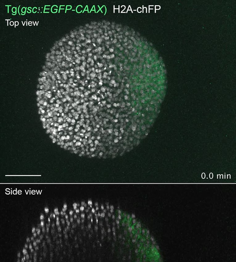
Research article Developmental Biology Stem Cells and Regenerative Medicine
A 50% Epiboly Germ ring Shield
Embryo Explant Embryo Explant Embryo Explant
Dorsal view Top view Dorsal view Top view Dorsal view Top view
pSMAD2/3
12/12 19/21 16/16 22/25 6/6 10/10
DAPI
Masked pSMAD2/3
B C D
80 1.0 6.0
to YSL/wounding site (±SD)
Norm. intensitity of brightest
Cell tier of brighest nuclei
pSMAD2/3 nuclei (±SD)
Embryo Embryo Embryo
Percentage of positive
pSMAD2/3 nuclei (±SD)
**** ****
Explant Explant **** 5.0 Explant
0.8
60 **** ns ns **** ****
**** 4.0
0.6
40 3.0
0.4
2.0
20 0.2
1.0
0 0.0 0.0
50% Germ Shield 50% Germ Shield 50% Germ Shield
epiboly ring epiboly ring epiboly ring
E Nodal inhibitor (SB-505124)
MZoep DMSO (256c > Bud) 256c > Bud 256c > Shield Shield > Bud
Bud
extended not-extended
F G H
**** **** **** * ns
wildtype 1.0 60
Normalized extension
MZoep 0.8
length (%)
40
Circularity
DMSO
0.6
256c > Bud
Nodal inhibitor
20
(SB-505124)
0.4
256c > Shield
0.2 0
ud
Shield > Bud
d
ud
ld
e
SO
SO
p
Bu
yp
oe
hie
>B
>B
DM
DM
ldt
MZ
d>
>S
wi
6c
ld
0 20 40 60 80 100
iel
ie
25
6c
Sh
Sh
Percentage (%)
25
Nodal inhibitor Nodal inhibitor
(SB-505124) (SB-505124)
Figure 2. Nodal signaling is active in blastoderm explants and is required for tissue extension. (A) High-resolution fluorescence images of stage-
matched embryos (dorsal view) and blastoderm explants (top view) at 50% epiboly, germ ring and shield stage stained for both pSMAD2/3 (pink) and
DAPI (grey). Nuclear pSMAD2/3 is color-coded using a fire lookup table (highest intensities in yellow) and was masked based on the DAPI signal. Insets
are zoom-in images of the highlighted regions (dashed boxes). Yellow circles denote the wounding site in explants. The proportion of embryos and
Figure 2 continued on next page
Schauer et al. eLife 2020;9:e55190. DOI: https://doi.org/10.7554/eLife.55190 6 of 30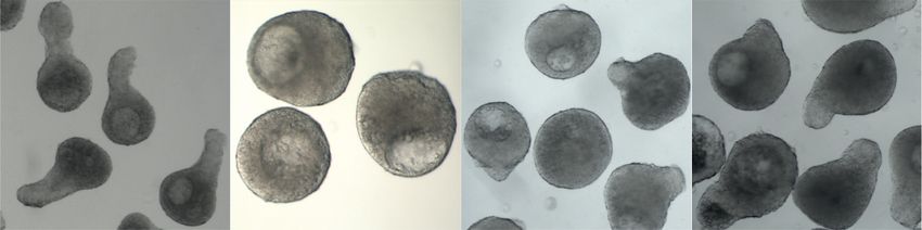
Research article                                                             Developmental Biology Stem Cells and Regenerative Medicine

Figure 2 continued
explants with a phenotype similar to the images shown is indicated in the lower left corner (50% epiboly: embryos, n = 12, N = 4, explants, n = 21,
N = 4; germ ring: embryos, n = 16, N = 4, explants, n = 25, N = 4 and shield: embryos, n = 6, N = 3, explants, n = 10, N = 3). (B) Percentage of
pSMAD2/3 positive nuclei in stage-matched embryos and blastoderm explants at 50% epiboly (embryos: n = 7, N = 3; explants: n = 10, N = 3), germ
ring (embryos: n = 10, N = 4; explants: n = 10, N = 4) and shield stage (embryos: n = 6, N = 3; explants: n = 8, N = 3). ****p
Research article Developmental Biology Stem Cells and Regenerative Medicine
A wildtype MZfz7a/b MZwnt11 MZwnt5b
Bud
B extended not-extended C D
wildtype 1.0 **** **** * 60 **** *
Normalized extension
0.8
MZfz7a/b
length (%)
Circularity
40
0.6
MZwnt11
20
0.4
MZwnt5b
0.2 0
0 20 40 60 80 100
e
e
a/b
t11
t11
t5b
b
yp
yp
nt5
Percentage (%)
fz7
wn
wn
wn
ldt
ldt
w
wi
wi
MZ
MZ
MZ
MZ
MZ
Figure 3. Non-canonical Wnt/PCP signaling is required for blastoderm explant morphogenesis. (A) Bright-field
single-plane images of wildtype (n = 120, N = 8), MZfz7a/b (n = 31, N = 3), MZwnt11 (n = 69, N = 5) and MZwnt5b
(n = 75, N = 4) blastoderm explants at bud stage. Scale bars: 200 mm. (B) Percentage of extended or not-extended
wildtype (n = 120, N = 8), MZfz7a/b (n = 31, N = 3), MZwnt11 (n = 69, N = 5) and MZwnt5b (n = 75, N = 4)
blastoderm explants at bud stage. (C) Circularity of wildtype (n = 120, N = 8), MZfz7a/b (n = 31, N = 3), MZwnt11
(n = 69, N = 5) and MZwnt5b (n = 75, N = 4) blastoderm explants at bud stage. ****p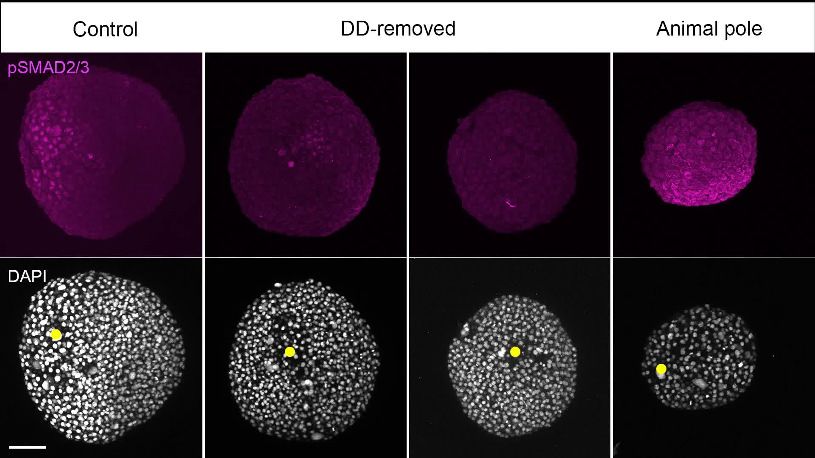
Research article Developmental Biology Stem Cells and Regenerative Medicine
A ß-catenin B 5.0
Top view Substack
positive nuclei (±SD)
Number of ß-catenin
4.0
30 min post-explant
3.0
2.0
1.0
0.0
1 2 3 4
16/20
Cell tier from the wounding site
C Angular dispersion of
ß-catenin positive nuclei
90 min post-explant
90
90°
30
Number of ß-catenin
60°
positive nuclei
20
30°
10
10/16
0 0°
D 50% epiboly
Control DD-removed Animal pole
Top view
pSMAD2/3
59/73 8/30 14/30 18/22
DAPI
Masked pSMAD2/3
E pSMAD2/3 present F Control DD-removed Animal pole
pSMAD2/3 strongly reduced
pSMAD2/3 absent
Control
Bud
DD-
removed
Animal
pole
0 20 40 60 80 100
Percentage (%)
G extended H I
not-extended 1.0 **** **** 60 ****
Normalized extension
Control 0.8
40
Circularity
length (%)
DD- 0.6
removed
20
0.4
Animal
pole
0.2 0
0 20 40 60 80 100
le
ol
ed
ed
ol
po
ntr
ntr
ov
v
Percentage (%)
mo
Co
al
Co
m
im
-re
-re
An
DD
DD
Figure 4. Maternal pre-patterning is required for Nodal signaling in blastoderm explants. (A) High-resolution
fluorescence images of blastoderm explants 30 and 90 min post-explanting stained for both b-catenin (grey) and
DAPI (not shown). Both full projection and substack top views are shown to facilitate simultaneous visualization of
the wounding site (yellow circle) and nuclear accumulation of b-catenin (yellow arrowheads). Insets are zoom-in
Figure 4 continued on next page
Schauer et al. eLife 2020;9:e55190. DOI: https://doi.org/10.7554/eLife.55190 9 of 30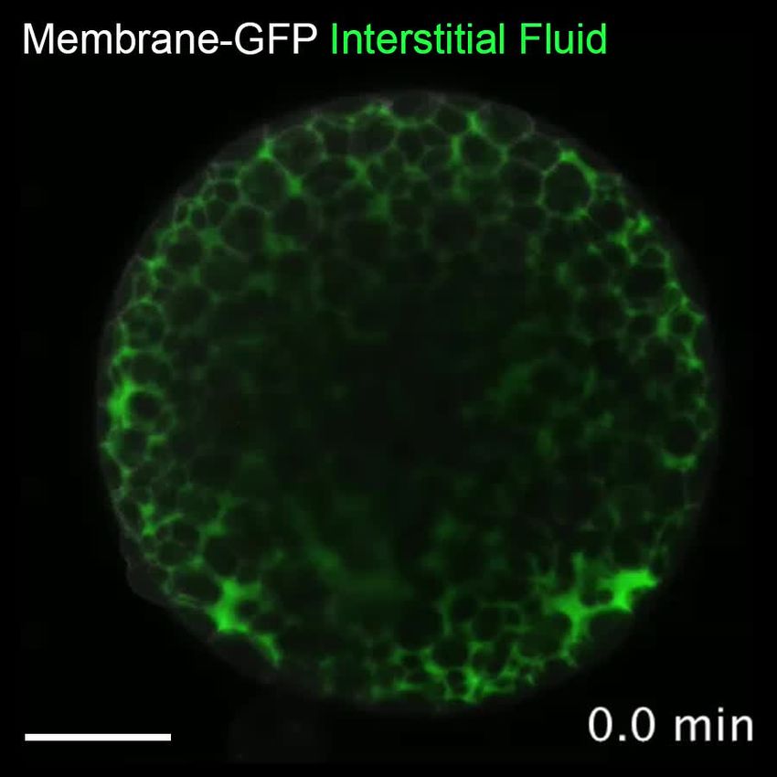
Research article                                                            Developmental Biology Stem Cells and Regenerative Medicine

                                    Figure 4 continued
                                    images of the highlighted regions (dashed boxes). The proportion of blastoderm explants with a phenotype
                                    similar to the images shown is indicated in the lower left corner (30 min: n = 20, N = 6; 90 min: n = 16, N = 4). (B)
                                    Number of b-catenin positive nuclei 30 min post-explant preparation as a function of the distance to the wounding
                                    site, expressed as cell tiers (n = 16, N = 6). (C) Angular dispersion of b-catenin positive nuclei 30 min post-explant
                                    preparation (for details see Materials and methods; n = 16, N = 6). (D) High-resolution fluorescence images of
                                    control, dorsal determinants-removed (DD-removed) and animal pole explants at 50% epiboly stained for both
                                    pSMAD2/3 (pink) and DAPI (grey). Nuclear pSMAD2/3 is color-coded using a fire lookup table (highest intensities
                                    in yellow) and was masked based on the DAPI signal. Insets are zoom-in images of the highlighted regions
                                    (dashed boxes) and the yellow circles denote the wounding site. The proportion of blastoderm explants with a
                                    phenotype similar to the images shown is indicated in the lower left corner (control: n = 73, N = 14; DD-removed:
                                    n = 30, N = 7; animal pole: n = 22, N = 6). (E) Percentage of control (n = 73, N = 14), DD-removed (n = 30, N = 7)
                                    and animal pole (n = 22, N = 6) explants showing a domain of pSMAD2/3 positive nuclei (present), a few sporadic
                                    pSMAD2/3 positive nuclei (strongly reduced) or no positive nuclei (absent) at 50% epiboly (see Materials and
                                    methods for additional details). (F) Bright-field single-plane images of control (n = 228, N = 15), DD-removed
                                    (n = 75, N = 8) and animal pole (n = 42, N = 5) explants at bud stage. Control explants partially correspond to
                                    explants shown in Figure 1—figure supplement 2A–D. (G) Percentage of extended or not-extended control
                                    (n = 228, N = 15), DD-removed (n = 75, N = 8) and animal pole (n = 42, N = 5) explants at bud stage. (H)
                                    Circularity of control (n = 228, N = 15), DD-removed (n = 75, N = 8) and animal pole (n = 42, N = 5) explants at
                                    bud stage. ****p
Research article Developmental Biology Stem Cells and Regenerative Medicine
signaling domain (Figure 4—figure supplement 2G,H), supporting the notion that b-cat does not
affect Nodal signaling through its effect on dorsoventral embryo patterning.
To further challenge the notion that maternal pre-patterning of blastoderm explants is critical for
mesendoderm induction and morphogenesis, we sought to prepare explants lacking all pre-pattern-
ing. To this end, we prepared explants from the animal pole (AP) of the blastoderm, which should
be devoid of marginal cells and dorsal determinants (Xu et al., 2014). In these explants, no accumu-
lation of b-cat was observed in the nuclei of cells close to the wounding site (Figure 4—figure sup-
plement 1C,D). Moreover, AP-derived explants showed strongly reduced nuclear pSMAD2/3
accumulation, mesendoderm induction and tissue extension (Figure 4D–H, Figure 4—figure sup-
plement 1E–H, Video 4). These findings therefore suggest that maternal pre-patterning by dorsal
determinants is needed for proper mesendoderm induction and morphogenesis in blastoderm
explants.
To determine whether positional information retained within the blastoderm and subsequently
wounding site of blastoderm explants is critical for coordinated mesendoderm induction and pat-
terning, we dissociated blastoderm explants obtained from high stage embryos into their constitu-
ent cells and allowed them to reaggregate in culture in the presence of serum-free medium
(Figure 4—figure supplement 3A). These reaggregates are thus expected to consist of a random
mixture of blastomeres, including some marginal cells that are pre-patterned to express Nodal sig-
nals and, thus, induce mesendoderm. We found that blastomere reaggregates underwent progres-
sive compaction and eventually rounded up in culture (Figure 4—figure supplement 3B).
Importantly, reaggregated cells were still capable of undergoing cell divisions (Figure 4—figure
supplement 3B), as evidenced by their progressive reduction in cell diameter (Figure 4—figure sup-
plement 3C), suggesting that blastomere reaggregates are viable under culture conditions. Interest-
ingly, we found that even when cultured until stage-matched embryos had reached bud stage,
blastomere reaggregates failed to form a clearly recognizable extension (Figure 4—figure supple-
ment 3D,E). Consistent with this, we found that expression of the mesodermal marker ntl (Schulte-
Merker, 1995; Schulte-Merker et al., 1994) was restricted to a few sparse cells or seemingly ran-
domly scattered cells within the reaggregates (Figure 4—figure supplement 3F–H). Importantly,
neither increasing the cell number within reaggregates nor changing the culture media conditions
substantially impacted on reaggregate morphogenesis or its ability to undergo coordinated meso-
derm specification (Figure 4—figure supplement 3D–H). Collectively, these findings suggest that
maternal pre-patterning and positional information within the blastoderm and, subsequently wound-
ing site of blastoderm explants, is required for coordinated mesoderm induction and
morphogenesis.
Our analysis of blastoderm explants and reaggregates so far indicates that in isolation from extra-
embryonic signaling sources, the spatially localized pre-patterning within the blastoderm margin can
still drive a large degree of mesendoderm patterning and morphogenesis. This seems to contradict
previous claims from embryo analysis that Nodal signaling from the extraembryonic YSL is needed
for these processes (Carvalho and Heisenberg, 2010; Fan et al., 2007; Gagnon et al., 2018;
Hong et al., 2011; van Boxtel et al., 2015; Veil et al., 2018; Xu et al., 2012). To understand this
apparent discrepancy, we directly compared mesendoderm induction in blastoderm explants with
intact embryos where Nodal signal production within the YSL is blocked. To block Nodal expression
within the YSL, we injected sqt and cyc MOs specifically into the YSL of high stage embryos. Consis-
tent with previous observations (Fan et al., 2007), we found that injection of high amounts of both
sqt (2 ng/embryo) and cyc (4 ng/embryo) MOs impaired induction of endoderm (sox32) and head
mesoderm (hgg and gsc) (Figure 5A, Figure 5—figure supplement 1A,B), the two mesendoderm
progenitor cell types whose induction is thought to require the highest dose of Nodal signaling
(Dougan et al., 2003; Gritsman et al., 2000; Schier et al., 1997; Thisse and Thisse, 1999). The
induction of notochord (ntl), paraxial (papc) and ventrolateral mesoderm (myoD and tbx6), in con-
trast, appeared normal in the morphant embryos (Figure 5A, Figure 5—figure supplement 1A,B).
This also became apparent by 32 hpf, when most morphant embryos displayed severe cyclopia,
indicative of defective induction of anterior mesendoderm structures, but largely normal trunk and
tail structures (Figure 5B). Together, these observations suggest that - consistent with previous find-
ings (Fan et al., 2007) - the YSL is needed for robust induction of endoderm and head mesoderm,
and that this signaling source might be specifically required to achieve peak levels of Nodal signaling
along the blastoderm margin.
Schauer et al. eLife 2020;9:e55190. DOI: https://doi.org/10.7554/eLife.55190 11 of 30Research article Developmental Biology Stem Cells and Regenerative Medicine
A hgg ntl sox32 B Pharyngula stage (32hpf)
A Side view Animal view Dorsal view Side view Side view
YSL-injected
YSL-injected
Animal
Control MO
Control MO
V D
view
P
Dorsal
18/19
Bud
view 68/69 50/50 48/48
sqt + cyc MO
YSL-injected
sqt + cyc MO
YSL-injected
45/57 28/28 20/23 13/14
C pSMAD2/3 DAPI Masked pSMAD2/3 pSMAD2/3 DAPI Masked pSMAD2/3
Dorsal view Lateral view
YSL-injected
Control MO
50% Epiboly
sqt + cyc MO
YSL-injected
YSL-injected
Control MO
Germ ring
sqt + cyc MO
YSL-injected
D Control MO
sqt + cyc MO
Control MO
sqt + cyc MO E Control MO
sqt + cyc MO
Control MO
sqt + cyc MO F Dorsal
G Lateral
80 1.4 50% Control MO 1.4 50% Control MO
Norm. intensity of brightest
1.0
Norm. intensity of positive
Norm. intensity of positive
pSMAD2/3 nuclei (±SD)
pSMAD2/3 nuclei (±SD)
pSMAD2/3 nuclei (±SD)
pSMAD2/3 nuclei (±SD)
epiboly sqt + cyc MO epiboly sqt + cyc MO
Percentage of positive
1.2 1.2
60 ** 0.8 Germ Control MO Germ Control MO
1.0 sqt + cyc MO
1.0 sqt + cyc MO
**** **** **
ring ring
0.6 0.8 0.8
40
**** **** **** *** 0.6 0.6
0.4
20 0.4 0.4
0.2
0.2 0.2
0 0.0 0.0 0.0
50% Germ 50% Germ 50% Germ 50% Germ 1 2 3 4 5 6 7 8 9 10 1 2 3 4 5 6 7 8 9 10
epiboly ring epiboly ring epiboly ring epiboly ring Cell tier from the YSL Cell tier from the YSL
Dorsal Lateral Dorsal Lateral
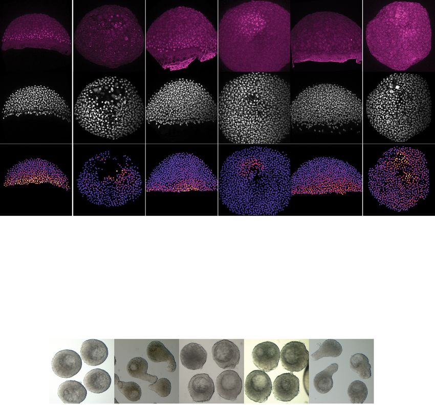
Figure 5. YSL-derived Nodal signals are required for inducing head mesoderm and endoderm in the intact embryo. (A) Expression of mesendoderm
(hgg and ntl) and endoderm (sox32) marker genes, as determined by whole mount in situ hybridization in control MO (6 ng) or sqt (2 ng) + cyc (4 ng)
MO YSL-injected embryos at bud stage. Schematic representation of the embryo views is shown on the left. The proportion of embryos with a
phenotype similar to the images shown is indicated in the lower right corner (hgg: control, n = 69, N = 4, sqt/cyc, n = 57, N = 4; ntl: control, n = 50,
N = 4, sqt/cyc, n = 28, N = 4 and sox32: control, n = 48, N = 3, sqt/cyc, n = 23, N = 3). (B) Bright-field single-plane images of pharyngula stage (32 hpf)
control MO (6 ng) or sqt (2 ng) + cyc (4 ng) MO YSL-injected embryos. The proportion of embryos with a phenotype similar to the images shown is
indicated in the lower right corner (n = 19, N = 2; n = 14, N = 2). (C) High-resolution fluorescence images of control MO (6 ng) or sqt (2 ng) + cyc (4 ng)
MO YSL-injected embryos stained both for pSMAD2/3 (pink) and DAPI (grey) at 50% epiboly (dorsal domain: control, n = 10, N = 4; sqt/cyc, n = 7,
N = 4; lateral domain: control, n = 9, N = 4; sqt/cyc, n = 7, N = 4) and germ ring (dorsal domain: control, n = 7, N = 4; sqt/cyc, n = 8, N = 4; lateral
domain: control, n = 8, N = 4; sqt/cyc, n = 7, N = 4). Nuclear pSMAD2/3 is color-coded using a fire lookup table (highest intensities in yellow) and was
masked based on the DAPI signal. Insets are zoom-in images of the highlighted regions (dashed boxes). (D) Percentage of pSMAD2/3 positive nuclei in
control MO (6 ng) or sqt (2 ng) + cyc (4 ng) MO YSL-injected embryos at 50% epiboly (dorsal domain: control, n = 10, N = 4; sqt/cyc, n = 7, N = 4;
lateral domain: control, n = 9, N = 4; sqt/cyc, n = 7, N = 4) and germ ring (dorsal domain: control, n = 7, N = 4; sqt/cyc, n = 8, N = 4; lateral domain:
control, n = 8, N = 4; sqt/cyc, n = 7, N = 4). ****pResearch article Developmental Biology Stem Cells and Regenerative Medicine Figure 5 continued domain: control, n = 7, N = 4; sqt/cyc, n = 8, N = 4; lateral domain: control, n = 8, N = 4; sqt/cyc, n = 7, N = 4). ****p
Research article Developmental Biology Stem Cells and Regenerative Medicine
A hgg
A
Control MO 1/2x sqt + cyc MO 1/5x sqt + cyc MO
V D YSL-injected YSL-injected YSL-injected
Animal
P view Animal view
Bud
52/52 14/59 37/47
B hgg present C 3.5x10 3
ns
hgg absent
3.0x10³
Ppl area (µm²)
****
Control MO 2.5x10³
2.0x10³ ****
1/2x sqt + cyc
MO 1.5x10³
1/5x sqt + cyc 1.0x10³
MO ****
5.0x10²
0 20 40 60 80 100
0
Percentage (%)
cM +
+
co cted
nt
MO
1/5 O
cy sqt
Un MO
cy sqt
pla
Ex l
o
ntr
e
ol
x
x
i nj
c
1/2
ntr
Co
D pSMAD2/3 DAPI Masked pSMAD2/3
Dorsal view
YSL-injected
Control MO
1/5x sqt + cyc MO
YSL-injected
50% epiboly
Lateral view
YSL-injected
Control MO
1/5x sqt + cyc MO
YSL-injected
E Control MO
1/5x sqt + cyc MO
F Control MO
1/5x sqt + cyc MO
G 50% epiboly
80 1.4
Norm. intensity of positive
Norm. intensity of brightest
1.0 Control MO
pSMAD2/3 nuclei (±SD)
pSMAD2/3 nuclei (±SD)
pSMAD2/3 nuclei (±SD)
Percentage of positive
Dorsal 1/5x sqt + cyc MO
** ns 1.2
60 0.8 Control MO
**** **** 1.0 Lateral
1/5x sqt + cyc MO
0.6 0.8
40
0.6
0.4
20 0.4
0.2
0.2
0 0.0 0.0
Dorsal Lateral Dorsal Lateral 1 2 3 4 5 6 7 8 9 10
Cell tier from the YSL
50% epiboly 50% epiboly
Figure 6. Reducing the dosage of YSL-derived Nodal signals results in variable induction of head mesoderm in
intact embryos. (A) Expression of hgg, a head mesoderm marker gene, in bud stage control MO (6 ng) or varying
dosages of sqt + cyc MO YSL-injected embryos (1/2x: 1 ng sqt + 2 ng cyc MO and 1/5x: 0.4 ng sqt + 0.8 ng cyc
MO), as determined by whole mount in situ hybridization. All embryos are shown as an animal view (schematic
Figure 6 continued on next page
Schauer et al. eLife 2020;9:e55190. DOI: https://doi.org/10.7554/eLife.55190 14 of 30Research article Developmental Biology Stem Cells and Regenerative Medicine
Figure 6 continued
representation in the left). The proportion of embryos with a phenotype similar to the images shown is indicated in
the lower right corner (control MO: n = 52, N = 3; 1/2x sqt/cyc: n = 59, N = 3; 1/5x sqt/cyc: n = 47, N = 3). (B)
Percentage of control MO (n = 52, N = 3) or sqt + cyc MO YSL-injected embryos (1/2x sqt/cyc: n = 59, N = 3; 1/5x
sqt/cyc: n = 47, N = 3) showing expression of hgg as determined by whole mount in situ hybridization at bud
stage. (C) Ppl area (based on hgg staining, as determined by whole mount in situ hybridization) in control MO
(n = 52, N = 3) or sqt + cyc MO YSL-injected embryos (1/2x sqt/cyc: n = 14, N = 3; 1/5x sqt/cyc: n = 37, N = 3),
uninjected wildtype embryos (n = 31, N = 4) or blastoderm explants, prepared from 256 c embryos (n = 20, N = 5).
****pResearch article Developmental Biology Stem Cells and Regenerative Medicine
In zebrafish embryos, extraembryonic Nodal signals from the YSL are thought to be required for
mesendoderm induction along the dorsal-ventral axis (Carvalho and Heisenberg, 2010; Fan et al.,
2007; Gagnon et al., 2018; Hong et al., 2011; Schier and Talbot, 2005; van Boxtel et al., 2015;
Veil et al., 2018; Xu et al., 2012). Our finding that blastoderm explants can not only induce the
three germ layers, but also display a seemingly complete patterning of the mesendoderm anlage,
seems to argue against such critical function for YSL-derived Nodal signals in mesendoderm pattern-
ing. However, closer inspection of the role of the YSL for mesendoderm patterning revealed some
striking similarities between embryos with reduced YSL-derived Nodal signals and blastoderm
explants: in both cases, induction of head mesoderm and endoderm, two tissues whose induction
requires high doses of Nodal signaling (Dougan et al., 2003; Gritsman et al., 2000; Schier et al.,
1997; Thisse and Thisse, 1999), is rather variable. This points at the intriguing possibility that Nodal
signals from the extraembryonic YSL effectively boost Nodal signaling within the blastoderm margin,
needed for robust induction of these two tissues. Yet, mesendoderm patterning defects in embryos
with reduced Nodal signals from the YSL are generally more pronounced than in blastoderm
explants, suggesting that the YSL is more important for robust induction of head mesoderm and
endoderm in the intact embryo compared to explants. One possibility for such embryo-specific
requirement of the YSL might be to induce sufficiently high doses of Nodal signaling along the entire
blastoderm margin, a requirement less important in explants where the blastoderm margin has col-
lapsed into a single region surrounding the wounding site.
While blastoderm explants were able to recapitulate a remarkable degree of embryo patterning
and morphogenesis, there are still several differences between explants and embryos: foremost,
mesendoderm progenitors do not undergo internalization in explants, but instead form an ‘exogas-
trula’, similar to previous observations in mouse gastruloids (Baillie-Johnson et al., 2015;
Beccari et al., 2018; Turner et al., 2017; Turner et al., 2014; van den Brink et al., 2014). One pos-
sible explanation for this could be that the specific geometry of the embryo is critical for mesendo-
derm internalization. Alternatively, extraembryonic structures might form a permissive substrate for
mesendoderm internalization. Notably, while mesendoderm progenitors fail to internalize in
explants, they still form an extension, which - similar to body axis elongation in embryos - depends
on Wnt/PCP-signaling (Čapek et al., 2019; Gray et al., 2011; Solnica-Krezel and Sepich, 2012;
Tada and Heisenberg, 2012). This suggests that the cellular rearrangements underlying body axis
extension in explants and body axis elongation in embryos might be conserved. Moreover, our and
previous observations support that cell proliferation does not seem to play a major role in either of
these processes (Liu et al., 2017; Quesada-Hernández et al., 2010; Zhang et al., 2008). Yet,
whether volumetric growth by cell proliferation might function in later stages of explant maturation,
similar to its role, for instance, in tail extension in chick and mouse embryos (Mongera et al., 2019),
remains to be determined.
Collectively, our findings suggest that zebrafish blastoderm explants undergo what has been
recently termed ‘genetically encoded self-assembly’, where heterogeneities among cells endowed
with a similar genetically encoded cell or developmental program drive the organization of those
cells, via local interactions, into the structures they were initially primed to form (Turner et al., 2016;
Xavier da Silveira Dos Santos and Liberali, 2019). This interpretation implies that both intrinsic het-
erogeneities (e.g. maternal factors) and extraembryonic signals might not only be required to render
embryonic development predictable and robust, but also to drive the emergence of embryo organi-
zation. Future work will have to address to what extent stem cell colonies/organoids indeed undergo
bona fide self-organization, and whether some of their organization might in fact be due to geneti-
cally encoded self-assembly.
Materials and methods
Key resources table
Reagent type Source or Additional
(species) or resource Designation reference Identifiers information
Strain, strain Zebrafish: wildtype ABxTL MPI-CBG dresden
background (D. rerio)
Continued on next page
Schauer et al. eLife 2020;9:e55190. DOI: https://doi.org/10.7554/eLife.55190 16 of 30Research article Developmental Biology Stem Cells and Regenerative Medicine
Continued
Reagent type Source or Additional
(species) or resource Designation reference Identifiers information
Strain, strain Zebrafish: (Smutny et al., 2017) ZFINID:ZDB-ALT-170811–2
background (D. rerio) Tg(gsc::EGFP-CAAX)
Strain, strain Zebrafish: (Ruprecht et al., 2015) ZFINID:ZDB-ALT-150727–1
background (D. rerio) Tg(sebox::EGFP)
Strain, strain Zebrafish: (Krens et al., 2011) ZFINID:ZDB-ALT-111207–5
background (D. rerio) Tg(krt4::EGFP-CAAX)
Strain, strain Zebrafish: (Stegmaier et al., 2016) ZFINID:ZDB-ALT-161017–9
background (D. rerio) Tg(actb2::lyntdto
mato;actb2::H2B-EGFP)
Strain, strain Zebrafish: (Gritsman et al., 1999) ZFINID:ZDB-
background (D. rerio) MZoep ALT-980203–1256
Strain, strain Zebrafish: (Quesada-Hernández ZFINID:ZDB-
background (D. rerio) MZfz7a/b et al., 2010) FISH-150901–19586
Strain, strain Zebrafish: (Čapek et al., 2019)
background (D. rerio) MZfz7a/b; Tg
(gsc::EGFP-CAAX)
Strain, strain Zebrafish: (Heisenberg et al., 2000) ZFINID:ZDB-
background (D. rerio) MZwnt11 ALT-980203–1302
Strain, strain Zebrafish: (Kilian et al., 2003) ZFINID:ZDB-
background (D. rerio) MZwnt5b ALT-980203–1630
Recombinant pCS2+-Membrane- (Kimmel and Meyer, 2010) 60–100 pg
DNA reagent GFP plasmid (1 c injection)
for mRNA synthesis
Recombinant pCS2-Membrane-RFP plasmid (Iioka et al., 2004) 80 pg
DNA reagent for mRNA synthesis (1 c injection)
Recombinant pCS2+-H2B-eGFP plasmid (Keller et al., 2008) 50 pg
DNA reagent for mRNA synthesis (1 c or YSL injection)
Recombinant pCS2-H2A-mCherry (Arboleda-Estudillo 6.5 pg
DNA reagent plasmid for mRNA synthesis et al., 2010) (128 c injection)
50 pg (1 c injection)
Recombinant pCS2+-CA-Alk8 plasmid (Payne et al., 2001) 30 pg
DNA reagent for mRNA synthesis (1 c injection)
Sequence- Human b-Globin Morpholino: Gene Tools 6 ng
based reagent 50 -CCTCTTACCTCAG (YSL injection)
TTACAATTTATA-30
Sequence- sqt Morpholino: Gene Tools ZFINID:ZDB- 0.4–2 ng
based reagent 5’-ATGTCAAATCAAGG MRPHLNO-060809–1 (YSL injection)
TAATAATCCAC-30
Sequence- cyc Morpholino: Gene Tools ZFINID:ZDB- 0.8–4 ng
based reagent 5’-GCGACTCCGAGCG MRPHLNO-060930–4 (YSL injection)
TGTGCATGATG-30
Antibody Anti-Phospho-Smad2 Cell Signaling Cat# 8828, 1:1000
(Ser465/467)/Smad3 RRID:AB_2631089 (WMIF)
(Ser423/425) (clone D27F4)
Antibody Anti-b-Catenin (clone 15B8) Sigma-Aldrich Cat# C7207, 1:500
RRID:AB_476865 (WMIF)
Antibody Anti-Phospho-Smad1/5 Cell Signaling Cat#: 9516, 1:100
(Ser463/465) (clone 41D10) RRID:AB_491015 (WMIF)
Antibody Alexa Fluor 546 Thermo Fisher Cat#: A-11010; 1:500
Goat Anti-rabbit IgG (H + L) Scientific RRID:AB_2534077 (WMIF)
Antibody Alexa Fluor 488 Thermo Fisher Cat#: A-11001; 1:500
Goat Anti-mouse IgG (H + L) Scientific RRID:AB_2534069 (WMIF)
Antibody Alexa Fluor 647 Thermo Fisher Cat# A-21235; 1:500
Goat Anti-mouse IgG (H + L) Scientific RRID:AB_2535804 (WMIF)
Continued on next page
Schauer et al. eLife 2020;9:e55190. DOI: https://doi.org/10.7554/eLife.55190 17 of 30Research article Developmental Biology Stem Cells and Regenerative Medicine
Continued
Reagent type Source or Additional
(species) or resource Designation reference Identifiers information
Antibody Anti-Digoxigenin-AP Roche Cat#11093274910 1:1000 (WMISH)
Fab fragments RRID:AB_2734716
Antibody Anti-Fluorescein-AP Roche Cat#11426338910 1:500
Fab fragments RRID :AB_2734723 (WMISH)
Chemical DMSO Sigma-Aldrich Cat#D8418 50 mM
compound, drug
Chemical SB-505124 Sigma-Aldrich Cat# S4696 50 mM
compound, drug
Chemical Dextran Alexa Fluor 647 Invitrogen Cat#D22914 2 ng
compound, drug (Sphere stage
injection)
Chemical Dextran Cascade Blue Invitrogen Cat# D1976 2 ng
compound, drug (Sphere stage
injection)
Chemical Hydroxyurea Sigma-Aldrich Cat#H8627 60 mM
compound, drug
Chemical Aphidicolin Sigma-Aldrich Cat#A0781 300 mM
compound, drug
Chemical DAPI Invitrogen Cat#D1306 1 mg/mL
compound, drug (WMIF)
Chemical Methylcellulose Sigma-Aldrich Cat#M0387 0.3%
compound, drug
Chemical Digoxigenin (DIG)- Sigma-Aldrich Cat#11277073910
compound, drug modified nucleotides
Chemical Fluorescein-labeled Sigma-Aldrich Cat#11685619910
compound, drug nucleotides
Chemical SIGMAFAST Fast Red Sigma-Aldrich Cat#F4648
compound, drug TR/Naphthol AS-MX Tablets
Cell culture r L15 Sigma-Aldrich Cat# L4386
eagent
Software, Imaris Bitplane https://imaris.oxin
Algorithm st.com/packages
Software, Fiji (Schindelin et al., 2012) https://fiji.sc/
Algorithm
Software, GraphPad Prism GraphPad Software https://www.graphpad.
Algorithm com/scientific-software/prism/
Software, MATLAB MATLAB Software https://www.mathworks.
Algorithm com/products/ matlab.html
Software, pSMAD2/3 and This paper Source code 1
Algorithm b-catenin analysis
(custom-made script)
Software, Excel Microsoft https://products.office.
Algorithm com/en-us/?rtc=1
Fish lines and husbandry
Zebrafish (Danio rerio) handling was performed as described (Westerfield, 2000). The mutant and
transgenic lines used in this study are detailed in the key resources table. Embryos were raised at
25–31˚C in Danieau’s solution or E3 medium and staged according to Kimmel et al. (1995).
Blastoderm explant preparation
Embryos for blastoderm explant preparation were manually dechorionated with watchmaker for-
ceps. Explants were then prepared at 256 cell stage, unless stated otherwise, by removing the entire
blastoderm from the yolk cell using forceps, and then washed by gentle pipetting to remove residual
yolk granules (schematic representation in Figure 1A). Animal pole explants were prepared by
Schauer et al. eLife 2020;9:e55190. DOI: https://doi.org/10.7554/eLife.55190 18 of 30Research article Developmental Biology Stem Cells and Regenerative Medicine
removing only the animal portion of the blastoderm at 256 cell stage using a hair knife instead of for-
ceps. All explants were cultured in Ringer’s solution (116 mM NaCl, 2.9 mM KCl, 1.8 m M CaCl2, 5.0
mM HEPES, pH 7.2) or Danieau’s medium (58 mM NaCl, 0.7 mM KCl, 0.4 mM MgSO4, 0.6 mM Ca
(NO3)2, 5 mM HEPES, pH 7.6). Approximately 1 hour (hr) after explant preparation, explants with
delayed cell cleavages, as assessed by increased cell size, were removed from the dish. The remain-
ing explants were then staged based on sibling embryos from the same egg lay. To analyze explant
morphology, bright-field side view images of blastoderm explants were acquired using a stereo-
microscope (Olympus SZX 12) equipped with a QImaging Micropublisher 5.0 camera.
Embryo microinjections
mRNAs were synthesized using the mMessage mMachine Kit (Ambion). Injections at 1 cell stage
were carried out as described (Westerfield, 2000) and the plasmids used for mRNA production are
detailed in the key resources table. Labelling the interstitial fluid was performed by injecting 2 ng of
Dextran Alexa Fluor 647, or Dextran Cascade Blue, between the deep cells of blastoderm explants
corresponding to high stage or sphere stage embryos, as described in Krens et al. (2017). For time-
lapse imaging of the interstitial fluid, embryos were also injected at 1 cell stage with 80 pg of Mem-
brane-GFP. For high-resolution live imaging of gastrulating blastoderm explants, Tg(gsc::EGFP-
CAAX) expressing embryos were injected at 1 cell stage with 50 pg of H2A-chFP to label the cell
nuclei. For the clone dispersal analysis, the plasma membrane of all cells was labelled by injecting 80
pg of Membrane-RFP mRNA at 1 cell stage. 6.5 pg of H2A-chFP mRNA was subsequently injected
into a single marginal blastomere at 128 cell stage (schematic representation in Figure 2—figure
supplement 2A). To constitutively activate BMP signaling, 30 pg of CA-Alk8 mRNA were injected
together with 60 pg of Membrane-GFP mRNA at 1 cell stage.
YSL injections were performed right after its formation, between 1 k and high stage. As a control
for homogeneous YSL injections, 50 pg of H2B-EGFP mRNA and 0.2% Phenol Red were co-injected
into the YSL. To block YSL-derived Nodal signals, 0.4–2 ng of sqt MO (sqt MO: 50 -ATGTCAAA
TCAAGGTAATAATCCAC-30 [Feldman and Stemple, 2001]) were co-injected with 0.8–4 ng of cyc
MO (cyc MO: 50 -GCGACTCCGAGCGTGTGCATGATG-30 [Karlen and Rebagliati, 2001]) into the
YSL. As a control for MO injections, 6 ng of a standard negative control MO (human b-globin MO:
50 -CCTCTTACCTCAGTTACAATTTATA-30 ) were also injected.
Live imaging of blastoderm explants
Bright-field time-lapse imaging of explants was performed using a Zeiss LSM 800 upright micro-
scope equipped with a Zeiss Plan-Apochromat 20x/1.0 water immersion objective starting when
stage-matched control embryos had reached sphere-early dome stage. High-resolution live imaging
of developing blastoderm explants was performed using a LaVision Trim 2-photon microscope
equipped with a Zeiss Plan-Apochromat 16x/0.8 water immersion objective and a Ti:Sa laser (Cha-
meleon, Coherent) set to 830 nm and an OPO laser set at 1100 nm. In this case, imaging was started
when stage-matched control embryos had reached 50% epiboly-germ ring stage. In both cases,
blastoderm explants were oriented in a side view, and the acquired images were then processed
using Fiji and/or Imaris.
Analysis of fluid accumulation, YSL formation and EVL differentiation
Imaging of the explant interstitial fluid at bud stage was performed using a LaVision Trim 2-photon
microscope equipped with a Zeiss Plan-Apochromat 20x/1.0 water immersion objective and a Ti:Sa
laser (Chameleon, Coherent) set to 800 nm and an OPO laser set at 1100 nm, or a Zeiss LSM 880
upright confocal microscope equipped with a Zeiss Plan-Apochromat 20x/1.0 water immersion
objective. Time-lapse imaging of the explant interstitial fluid was performed using a Zeiss LSM 800
upright microscope equipped with a Zeiss Plan-Apochromat 20x/1.0 water immersion objective,
starting when stage-matched control embryos had reached high-oblong stage. In both cases, blasto-
derm explants were oriented in a side view, and the acquired images were then processed using Fiji
and/or Imaris.
Imaging of YSL-like features in blastoderm explants was performed when corresponding stage-
matched control embryos reached 50% epiboly-germ ring stage using a LaVision Trim 2-photon
Schauer et al. eLife 2020;9:e55190. DOI: https://doi.org/10.7554/eLife.55190 19 of 30Research article Developmental Biology Stem Cells and Regenerative Medicine
microscope as described above. Explants were oriented with the wounding site facing the objective
(top view), and the acquired images were then processed using Fiji.
To analyze EVL differentiation in blastoderm explants, both animal-pole oriented embryos and
explants were imaged in parallel on a Zeiss LSM 880 upright confocal microscope equipped with a
Zeiss Plan-Apochromat 20x/1.0 water immersion objective. To assess whether the entire explant was
covered by EVL, explants and embryos were then fixed at bud stage in 4% paraformaldehyde (PFA)
at 4˚C overnight, washed 5x with PBS and then imaged on a Zeiss LSM 880 upright confocal micro-
scope equipped with a Zeiss Plan-Apochromat 20x/1.0 water immersion objective. The acquired
images for both embryos (side view) and explants (side view) were then processed using Fiji and/or
Imaris (Bitplane).
Sample preparation for live imaging
Dechorionated embryos were mounted in 2% agarose molds on petri dishes and immobilized in
0.7% low melting point agarose. Explants were mounted in smaller agarose molds (800 800 mm
from Microtissues) and cultured in Ringer’s or Danieau’s solution.
Whole mount in situ hybridization (WMISH)
Embryos were fixed with 4% PFA overnight at 4˚C. Antisense RNA probes were synthesized using
SP6, T7 or T3 RNA polymerase from mMessage mMachine kits (ThermoFisher, AM1344) with Roche
digoxigenin (DIG)-modified nucleotides, or fluorescein (FITC)-labeled nucleotides, from partial cDNA
sequences. WMISHs were performed as previously described (Thisse and Thisse, 2008) with the fol-
lowing modifications: (i) proteinase K treatment was performed only for sox32 in situ hybridization
and for double WMISHs; (ii) 5% dextran sulfate was added to the hybridization solution for sox32,
mxtx2, tbx16 and bmp2b probes and all double WMISHs; and, (iii) 0.05x SSC was used for more
stringent washes for sqt in situ hybridizations. Single WMISHs were performed with DIG-labeled anti-
sense RNA probes, while for double WMISHs, a mixture of DIG- and FITC-labeled RNA probes were
added simultaneously during the hybridization step.
For double WMISHs, hybridization and visualization of DIG-labelled probes with the chromogenic
NBT/BCIP substrate were performed as previously described (Thisse and Thisse, 2008). When the
signal was sufficiently intense, the staining solution was replaced with a solution of 100 mM glycine-
HCl pH2.2 for 10 min at room temperature as described in Denker et al. (2008). The samples were
then washed several times with fresh PBS + 0.1% (w/v) Tween (PBST) and blocked in blocking buffer
(1 PBST, 2% sheep serum (v/v), 2 mg/ml BSA) for 2 hr at room temperature and incubated over-
night at 4˚C with Anti-Fluorescein-AP Fab fragments (1:500) in fresh blocking buffer. Afterwards, the
samples were washed 6x for 10–20 min at room temperature in PBT and 3x in 0.1 M Tris-HCl pH8.2
+ 0.1% (w/v) Triton. The staining solution for red staining was prepared using SIGMAFAST Fast Red
TR/Naphthol AS-MX Tablets according to the manufacturer’s protocol and added to the samples.
After staining, the samples were cleared for 10 min in 100% EtOH and washed several times in PBT.
Blastoderm explants and corresponding stage-matched embryos were always processed in the same
tube, and the staining reaction was therefore stopped simultaneously. All WMISHs were imaged on
a stereo-microscope (Olympus SZX 12) equipped with a QImaging Micropublisher 5.0 camera.
Whole mount immunofluorescence (WMIF)
a-pSMAD2/3 and a-b-catenin whole mount immunofluorescence was performed as described previ-
ously (van Boxtel et al., 2015). In short, dechorionated embryos and explants were fixed in 4% PFA
(in PBS) overnight at 4˚C, washed in PBS and then directly transferred into 100% MeOH and stored
at 20˚C for, at least, 2 hr. The samples were then washed 5x in PBSTr (PBS + 1% (w/v) Triton
X-100), blocked 2x in blocking solution (PBSTr + 10% goat serum + 1% DMSO) for 2 hr and then
incubated overnight at 4˚C with a-pSMAD2/3 antibody (1:1000) and/or a-b-catenin antibody (1:500)
diluted in blocking solution. Afterwards, the samples were washed 5x in PBSTr for 10 min, washed
3x in PBS + 0.1% (w/v) Triton for 1 hr and then incubated overnight at 4˚C in secondary antibody
(1:500) diluted in blocking solution. For labelling cell nuclei, the samples were incubated in DAPI
(1:1000) diluted in blocking solution for 18 min at room temperature. After secondary antibody incu-
bation, the samples were washed 2x for 5 min in PBSTr and 5x for 10–20 min in PBS + 0.1% Triton.
Schauer et al. eLife 2020;9:e55190. DOI: https://doi.org/10.7554/eLife.55190 20 of 30You can also read