Zinc Signaling in the Mammary Gland: For Better and for Worse - Semantic Scholar
←
→
Page content transcription
If your browser does not render page correctly, please read the page content below
biomedicines
Review
Zinc Signaling in the Mammary Gland:
For Better and for Worse
Moumita Chakraborty * and Michal Hershfinkel

                                          Department of Physiology and Cell Biology, Faculty of Health Sciences, Ben-Gurion University of the Negev,
                                          Beer-Sheva 84105, Israel; hmichal@bgu.ac.il
                                          * Correspondence: moumita.biochem89@gmail.com

                                          Abstract: Zinc (Zn2+ ) plays an essential role in epithelial physiology. Among its many effects, most
                                          prominent is its action to accelerate cell proliferation, thereby modulating wound healing. It also
                                          mediates affects in the gastrointestinal system, in the testes, and in secretory organs, including the
                                          pancreas, salivary, and prostate glands. On the cellular level, Zn2+ is involved in protein folding,
                                          DNA, and RNA synthesis, and in the function of numerous enzymes. In the mammary gland, Zn2+
                                          accumulation in maternal milk is essential for supporting infant growth during the neonatal period.
                                          Importantly, Zn2+ signaling also has direct roles in controlling mammary gland development or,
                                          alternatively, involution. During breast cancer progression, accumulation or redistribution of Zn2+
                                          occurs in the mammary gland, with aberrant Zn2+ signaling observed in the malignant cells. Here,
                                          we review the current understanding of the role of in Zn2+ the mammary gland, and the proteins
                                          controlling cellular Zn2+ homeostasis and signaling, including Zn2+ transporters and the Gq-coupled
                                          Zn2+ sensing receptor, ZnR/GPR39. Significant advances in our understanding of Zn2+ signaling
                                          in the normal mammary gland as well as in the context of breast cancer provides new avenues for
                                identification of specific targets for breast cancer therapy.


Citation: Chakraborty, M.;                Keywords: zinc; zinc signaling; Zn2+ transporters; ZnR/GPR39; mammary gland; breast cancer
Hershfinkel, M. Zinc Signaling in the
Mammary Gland: For Better and for
Worse. Biomedicines 2021, 9, 1204.
https://doi.org/10.3390/                  1. Zinc, an Essential Micronutrient
biomedicines9091204
                                                Zinc is a vital trace element present in all body tissues and organs. There are differ-
                                          ences in recommendation of expert groups regarding the daily allowance of dietary zinc,
Academic Editor: Raghu Sinha
                                          with suggested intake of 10–20 mg/day, depending on age and gender [1]. Insufficient
                                          nutritional zinc intake increases the risk of zinc deficiency [2,3], and low plasma zinc
Received: 30 July 2021
Accepted: 6 September 2021
                                          levels have been recorded in as many as 80% of children in developing countries [4,5].
Published: 12 September 2021
                                          However, zinc deficiency is not unique in low income countries, as it is frequently found
                                          in developed countries as well [2,6], with an estimated 17% of the world’s population at
Publisher’s Note: MDPI stays neutral
                                          risk of zinc deficiency [5]. Insufficient zinc contributes to the etiology of a wide range of
with regard to jurisdictional claims in
                                          pathologies, including immune system failure, digestive system diseases, wound healing,
published maps and institutional affil-   and cognitive impairment [6,7]. The most severe consequence of zinc deficiency is seen in
iations.                                  the rare genetic disorder, acrodermatitis enteropathica (AE), a genetic disorder in which
                                          impaired intestinal zinc absorption produces an acute, potentially fatal zinc deficiency [8,9].
                                          Importantly, symptoms of zinc deficiency can typically be reversed by zinc supplementa-
                                          tion [10,11]. In neonates and young infants, zinc deficiency is associated with skin lesions,
Copyright: © 2021 by the authors.
                                          growth retardation and impaired development [12,13]. Surprisingly, symptoms of severe
Licensee MDPI, Basel, Switzerland.
                                          zinc deficiency have been observed in exclusively breast-fed babies [14,15]. These infants
This article is an open access article
                                          exhibited a failure to thrive, which was later determined to be related to defective zinc
distributed under the terms and           secretion into milk [16–18]. Thus, regulation of zinc homeostasis in the mammary gland
conditions of the Creative Commons        is crucial during lactation. Conditioned zinc deficiencies are also known to occur in mal-
Attribution (CC BY) license (https://     absorption syndromes, chronic liver or renal disease, excessive intake of alcohol, and in
creativecommons.org/licenses/by/          certain instances of neoplastic malignancies [19]. Indeed, epidemiological studies have
4.0/).                                    linked dietary zinc deficiency to increased risk of cancer [20,21]. However, a complicated

Biomedicines 2021, 9, 1204. https://doi.org/10.3390/biomedicines9091204                                https://www.mdpi.com/journal/biomedicines
Biomedicines 2021, 9, 1204                                                                                           2 of 17

                             picture of both abnormally high and low serum zinc levels in malignant cells, suggests that
                             zinc may be involved in various aspects of cancer progression.
                                    Zinc ions (Zn2+ ) act as structural components of many enzymes and transcription
                             factors [22]. This important structural function may account for a well-established role of
                             Zn2+ in proliferation, and, indeed, Zn2+ deficiency is associated with attenuation of cell
                             growth [23–25]. Moreover, DNA damage was demonstrated in Zn2+ -depleted cells, while
                             its supplementation enhanced genome stability even following radiation treatment [26].
                             Nevertheless, Zn2+ has another important role: acting as a signaling molecule. This
                             conclusion is supported by the large number of proteins involved in compartmentaliza-
                             tion of the ion, allowing transient local changes in Zn2+ concentrations, as required for
                             signaling [27–29]. Transient changes in cytoplasmic Zn2+ levels and its interaction with
                             cellular kinases or receptors induce what is termed Zn2+ signaling. An example of this
                             is the interaction of Zn2+ with the growth hormone receptor, and its modulation of the
                             insulin-like growth factor-I (IGF-1) pathway [25]. More recent studies have identified Zn2+
                             as a first messenger, regulating the ZnR/GPR39 G-protein coupled receptor, or as a second
                             messenger, activating major pathways associated with proliferation [30–32].
                                    Under physiological conditions, cellular Zn2+ is mostly bound to proteins, and “free”
                             Zn concentrations are in the picomolar to nanomolar range [33]. Yet, “free” Zn2+
                                 2+

                             ions do occur, and are typically sequestered in cellular vesicles and in organelles, in-
                             cluding the Golgi, endoplasmic reticulum, and mitochondria, thereby serving as Zn2+
                             reservoirs [34–36]. Regulation of free Zn2+ in the cytosol is largely achieved either by
                             binding to metallothioneins (MT) or by Zn2+ transporters [27,37,38]. Two protein families
                             are known to mediate Zn2+ transport across cellular membranes, as Zrt- and Irt-related
                             proteins (ZIP) mediate influx into the cytoplasm and Zn2+ transporters (ZnT) move Zn2+
                             out of the cytoplasm [37,39]. Zn2+ homeostasis is tightly regulated by these proteins under
                             normal circumstances. Intriguingly, Zn2+ levels are dysregulated in the microenvironment
                             surrounding cancer tissue [29]. This finding correlates with changes in the expression
                             profile of various Zn2+ transporters related to the presence of cytokines and growth factors
                             in the tumor’s microenvironment [40].
                                    Zn2+ is emerging as an important player in mammary gland function under both
                             physiological and neoplastic conditions. We will discuss molecular mechanisms mediating
                             the activities of Zn2+ in this tissue.

                             2. Zn2+ as a Signaling Molecule
                                   For Zn2+ to act as a signaling molecule, both its intracellular and extracellular lev-
                             els must transiently change. In addition, there must be protein targets that detect these
                             changes and subsequently trigger downstream effects. On a fast scale, intracellular Zn2+
                             transients involve a Zn2+ release from intracellular stores, e.g., endoplasmic reticulum or
                             Zn2+ -binding metallothioneins, in response to stimuli [32,41,42]. For example, antigen
                             binding in mast cells induces extracellular Ca2+ influx that upregulates mitogen-activated
                             kinase (MAPK). This, in turn, activates the release of sequestered Zn2+ , thereby raising the
                             intracellular Zn2+ concentration [32]. Similar rapid changes in intracellular Zn2+ levels are
                             produced by its release from stores following epidermal growth factor (EGF) stimulation,
                             which induces casein kinase 2 phosphorylation of a Zn2+ transporter, ZIP7 [43]. In addition,
                             activation of the signal transducers and activators of transcription 3, STAT3 pathway func-
                             tionally modulates Zn2+ transporter activity [27,43]. Late Zn2+ signaling events, occurring
                             several hours after cell stimulation, and are dependent upon changes in expression of Zn2+
                             transporters that modulate the transfer of Zn2+ from one compartment to another [27,37].
                             Both fast and late types of cytoplasmic Zn2+ rises place this ion in the category of classical
                             second messenger, directly regulating major signaling pathways. Among those pathways
                             activated by Zn2+ are EGFR [44], the IGFR-1 [45], and MAPK [46]. Changes in cytosolic
                             Zn2+ levels can also modulate signaling via inhibition of phosphatases activity [47].
                                   Extracellular Zn2+ levels can also transiently increase, as many cell types secrete
                                2+
                             Zn from intracellular vesicles to the extracellular region. Such release is documented in
Biomedicines 2021, 9, 1204 3 of 17
neurons, pituitary cells, prostate epithelial cells, mast cells, granulocytes, Paneth cells in the
intestines, pancreatic cells, and mammary gland epithelia [48–52]. The secreted Zn2+ can
then interact with cell surface proteins that have Zn2+ -binding modulatory sites, including
ion channels, neurotransmitter transporters, and G protein-coupled receptors [53–55]. For
example, changes in extracellular Zn2+ induce transactivation of EGFR in airway epithelial
cells [56], as well as activation of the MAPK pathway [57,58]. Thus, extracellular Zn2+ ,
acting as a first messenger in mouse embryonic stem cells (ES), induces proliferation
through upregulation of PI3K, MAPK, and mTOR signaling pathways [59]. Significantly, a
Gq protein-coupled receptor (GPCR), initially called ZnR, is a specific target that distinctly
senses extracellular Zn2+ and triggers metabotropic calcium (Ca2+ ) release [60]. Later, it was
demonstrated that Zn2+ is the ligand of the orphan receptor GPR39 [61], and we showed
that it mediates metabotropic ZnR activity, which is now termed ZnR/GPR39 [30,62].
3. Zn2+ and Its Transporters in the Physiology of Mammary Gland
The importance of Zn2+ to mammary gland development is manifested by the se-
vere consequences of Zn2+ deficiency. Under these conditions, extreme defects in mor-
phology and function, including oxidative stress and inflammation, are described in the
non-lactating [63,64] as well as in the lactating gland [65]. During puberty, growth and
differentiation of the epithelial glandular structures within the fat pad are driven by the
growth hormones, EGF and IGF-1, as well as by the estrogen receptor [66]. Interestingly, all
of these pathways are modulated by Zn2+ , as described above [67]. During pregnancy and
lactation, progesterone and prolactin are responsible for growth of the alveolar epithelial
structures, and milk production and secretion [68]. At this stage, Zn2+ is suggested to regu-
late proliferation and differentiation of the epithelial cells via its interaction with kinases
downstream to the prolactin receptor. Mammary gland involution, following lactation, is
also highly dependent on Zn2+ that acts as a regulator of apoptosis via mitochondrial or
lysosomal pathways [69,70]. The Zn2+ transporter, ZnT2, is essential for accumulation of
Zn2+ in lysosomes and assembly of the vacuolar ATPase for initiation of mammary gland
involution [70], via a mechanism only partially understood.
A developing offspring requires relatively large amounts of Zn2+ to support its rapid
growth and development. Mammary cells are tasked with providing this important
nutrient, which they do at a rate of about 1 mg Zn2+ per day during lactation [12,14,71].
Failure of this process results in severe Zn2+ deficiency to the infant, leading to, among other
things, growth retardation and skin lesions [18,72]. Even under conditions of moderate
Zn2+ deficiency, diminished cognitive development is observed [73,74]. Indeed, Zn2+ is
found in secretory vesicles in the mammary gland and its loss is associated with a condition
termed neonatal Zn2+ deficiency [15]. During lactation, import of Zn2+ from the circulation
into the mammary cells is therefore crucial, and its concentration in milk is maintained
over a wide range of maternal dietary Zn2+ intake [75–78]. In agreement with the strict
requirements for Zn2+ in the ingested milk, several Zn2+ transporters are present in the
mammary tissue (Figure 1), providing the means to adjust the levels of this ion and its
distribution into the appropriate cellular compartments [71,79,80]. Most prominent is ZnT2,
which is responsible for the transport of Zn2+ into the secretory vesicles in mammary gland
epithelia during lactation [81]. A point mutation in ZnT2 is associated with a dramatic and
injurious decrease in levels of Zn2+ in the milk [82]. Additional studies have identified other
mutations in the gene encoding ZnT2 which result in severely Zn2+ -deficient breast-fed
infants [17,82,83]. ZnT2 has also been linked to the function of mammary epithelial cells
during all developmental stages [70,81,84]. During pregnancy, in preparation for lactation,
ZnT2 is essential for development of alveolar structures and is linked to regulation of cell
polarity and formation of acidic secretory vesicles through recruitment of the vacuolar
ATPase [84]. Subsequently, during lactation, expression of ZnT2 on the secretory vesicles
is responsible for transport of Zn2+ into these milk-containing vesicles [81]. Lactogenic
hormones, prolactin and glucocorticoids, regulate ZnT2 expression via activation of kinase
signaling cascades [85]. Studies in pancreatic cells, however, indicate that Zn2+ itself can[81]. Lactogenic hormones, prolactin and glucocorticoids, regulate ZnT2
activation of kinase signaling cascades [85]. Studies in pancreatic cells, ho
that Zn2+ itself can transcriptionally activate ZnT2 expression via activatio
Biomedicines 2021, 9, 1204 4 of 17
transcription factor, MTF-1 [85,86]. Following cessation of lactation, the m
undergoes involution, which is also associated with Zn2+ transporters. An
this process is relocation of ZnT2 from late endosomes into lysosomes [70
transcriptionally activate ZnT2 expression via activation of the metal transcription factor,
quent
MTF-1 interaction
[85,86]. Followingof ZnT2ofwith
cessation thethe
lactation, vacuolar
mammaryATPases playsinvolution,
gland undergoes an important
mal-induced
which cell with
is also associated deathZn [84,88].
2+ Interestingly,
transporters. An initial stageZn 2+ is process
in this also required
is relocationby mat
ofteinases
ZnT2 from(MMPs),
late endosomes into lysosomes [70,87,88]. Subsequent
which degrade extracellular matrix components interaction of ZnT2 and a
with the vacuolar ATPases plays an important role in lysosomal-induced cell death [84,88].
remodeling
Interestingly, Zn2+ofis the tissue by
also required during
matrix pregnancy and(MMPs),
metalloproteinases involution [28]. The Zn
which degrade
ZnT5 andmatrix
extracellular ZnT6, are expressed
components and areon the Golgi
essential apparatus
for remodeling and
of the play
tissue a promin
during
pregnancy and involution [28]. The Zn 2+ transporters, ZnT5 and ZnT6, are expressed on
vation of Zn -binding enzymes, such as the tissue non-specific alkalin
2+
the Golgi apparatus and play a prominent role in activation of Zn2+ -binding enzymes,
(TNAP)
such [89,90].
as the tissue Interestingly,
non-specific these transporters
alkaline phosphatase (TNAP) [89,90].areInterestingly,
also associatedthese with
maternalare
transporters milk,
also as well aswith
associated lowZnTNAP
2+ activity
-deficient maternalin mothers
milk, as wellofasseverely
low TNAPsympto
activity
[91]. in mothers of severely symptomatic neonates [91].
Figure 1. Scheme describing the main signaling pathways activated by Zn2+ in the normal mam-
Figure 1. Scheme describing the main signaling pathways activated by Zn2+ in the n
mary gland cells. The Zn2+ transporters that play a role in mammary gland physiology and their
gland cells. The Zn2+ transporters that play a role in mammary gland physiology a
localization are described in the figure. In particular, note ZnT2, that plays a role in vesicular Zn2+
tion are described
accumulation in the
during lactation andfigure. In particular,
involution, and ZIP3/ZIP5,note
that ZnT2, that plays
were suggested a role
to mediate Znin
2+ vesicu
influx. Arrows depict established pathways, while dotted lines and question marks are putative to me
lation during lactation and involution, and ZIP3/ZIP5, that were suggested
Arrowsthat
pathways depict
remainestablished
to be explored.pathways, while dotted lines and question marks are pu
that remain to be explored.
To accumulate cytoplasmic Zn2+ , several Zn2+ importers from the ZIP family have
been identified in the mammary epithelium (Figure 1) [67]. Expression of ZIP5 on the
surface To accumulate
of epithelial mammarycytoplasmic Znthat
cells suggested 2+, several Zn2+ importers
this transporter Zn2+ the Z
is involved infrom
uptake 2+
been [67]. Surprisingly,
identified ZIP3
in the mediates Znepithelium
mammary reuptake from the secreted
(Figure milkExpression
1) [67]. within the of Z
alveolar lumen [79]. Release of Zn2+ from intracellular stores and regulation of prolifer-
faceare
ation ofassociated
epithelial mammary
with cells of
phosphorylation suggested
the Golgi Zn that this transporter
2+ transporter is involved
ZIP7, following
[67]. Surprisingly,
activation of EGF cascade ZIP3
[43]. mediates
It remains toZn reuptake
be2+seen how other from
ZnTsthe
andsecreted milk with
ZIPs that are
expressed on mammary epithelial cells function to maintain Zn 2+ homeostasis and normal
lumen [79]. Release of Zn2+ from intracellular stores and regulation of p
function of the mammary gland.
associated with phosphorylation of the Golgi Zn2+ transporter ZIP7, follow
of EGF cascade [43]. It remains to be seen how other ZnTs and ZIPs that ar
mammary epithelial cells function to maintain Zn2+ homeostasis and norm
the mammary gland.
4. Zn2+ Homeostasis and Zn2+ Transporters in Breast CancerBiomedicines 2021, 9, 1204 5 of 17
4. Zn2+ Homeostasis and Zn2+ Transporters in Breast Cancer
Considering the well-established roles for Zn2+ in cell proliferation and survival, it
is not surprising that this ion also participates in cancer progression [25,92]. However,
because Zn2+ modulates immune responses, it may also be considered as an anti-cancer
agent, for example via regulation of anti-tumor activity by T-lymphocytes [93–95]. Loss of
intracellular Zn2+ in prostate tumor epithelial cells is associated with downregulation of
the transporter ZIP1 and tumor progression via enhanced cell proliferation [96,97]. Thus, it
would appear likely that correcting Zn2+ deficiency will attenuate prostate cancer progres-
sion. However, treatment of cancerous prostate tumors by elevating dietary or serum Zn2+ ,
which does not directly affect the “free” Zn2+ content of prostate cells, proved controversial,
and more specific tools were sought [98,99]. In breast cancer patients, measurement of total
Zn2+ in the serum or within the malignant cells demonstrated abnormal concentrations,
suggesting the involvement of Zn2+ dysregulation in progression of this malignancy as
well [100,101]. While serum Zn2+ levels are reduced in most cancers, breast and lung tumor
tissues have elevated levels of “free” Zn2+ when compared to the normal tissue [102,103].
Tissue Zn2+ accumulation, moreover, is dependent on breast cancer subtype, suggesting
this could serve to define the molecular subtype of the cancer and predict its response to
therapy [103,104]. Analyses of isotopic composition of Zn2+ in breast tumors, relative to
normal mammary tissue, determined that Zn2+ is mostly bound to sulfur-rich proteins,
likely metallothioneins in the malignant tissue [105,106]. Such analyses suggest a novel
approach for using Zn2+ as a biomarker to identify the tumor subtype and prognosis [107].
Changes in cytoplasmic and extracellular “free” Zn2+ can affect tumor progression via
multiple pathways. Cytoplasmic “free” Zn2+ may rise in the context of oxidation/reduction
reactions that liberate Zn2+ from metallothioneins [38,108,109] and are very common in
tumor environments. This Zn2+ can then modulate phosphatases and kinases associated
with signaling pathways involved in cell proliferation and metastasis [47,90]. In addition,
increased cytoplasmic “free” Zn2+ can alter the expression of various Zn2+ transporters that
are regulated, either directly via glycosylation [110] or via the Zn2+ sensing metal transcrip-
tion factor MTF-1 [111]. Changes in expression of Zn2+ transporters may, in turn, increase
efflux of Zn2+ [112] or affect the vesicular concentration of this ion which is available for
release by exocytosis. Moreover, hypoxic conditions and tumor necrosis can also trigger
Zn2+ release from the injured cells [113]. Such changes in extracellular “free” Zn2+ levels
can regulate matrix metalloproteinases [114,115], which further degrade the extracellular
matrix and allow migration (metastasis) of tumor cells. Changes in extracellular Zn2+ have
also been shown to activate the G-protein-coupled receptor, ZnR/GPR39, and to promote
signaling leading to epithelial repair [30,113,116,117]. Only some of these pathways have
been studied in breast cancer tissue, and further analysis may provide new and discrete
therapeutic targets.
5. ZnT Family and Breast Cancer
A comprehensive analysis of the distribution of Zn2+ transporters in breast cancer
tissue and cell lines revealed that transporter protein levels are aberrant [103]. Signifi-
cant differences in expression of ZnT5, ZnT6, ZnT8, and ZnT9, between basal-estrogen
receptor negative (ER-) and luminal-ER positive (ER+) subtypes of breast cancer were
demonstrated [103], though how they affect cellular signaling or breast cancer progression
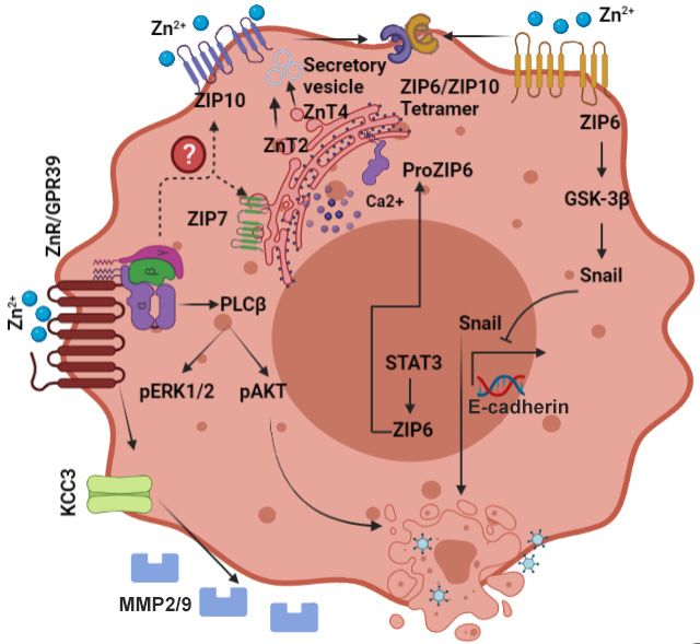
remains unclear (Figure 2).Biomedicines 2021,2021,
Biomedicines 9, x9,FOR
1204 PEER REVIEW 6 of 17
Figure
Figure 2. 2. Scheme
Scheme describing
describing the activation
the activation of signalingof signaling
pathways pathways
involved involved
in breast cancer pro-in breas
gression and metastasis. Zn 2+ dyshomeostasis results in activation of multiple pathways that affect
sion and metastasis. Zn dyshomeostasis results in activation of multiple pathway
2+
cell morphology, migration, and proliferation. Note specifically ZIP10 and ZIP6 that interact to
morphology, migration, and proliferation. Note specifically ZIP10 and ZIP6 that int
regulate epithelial mesenchymal transition, and ZnR/GPR39 that induces membrane protrusions
epithelial mesenchymal transition, and ZnR/GPR39 that induces membrane p
and MMP2/9 release. Arrows depict established pathways, while dotted lines and question marks
MMP2/9
are release. for
putative pathways Arrows
regulationdepict established pathways, while dotted lines and que
and interaction.
putative pathways for regulation and interaction.
ZnT2
5.1.Studies
ZnT2 intended to show how Zn2+ accumulation is regulated in tumor cells have
largely focused on ZnT2, which has a well-described2+role in the normal mammary gland
Studies intended to show how Zn accumulation is regulated in tu
(see Section 3). In one such study, ZnT2 was overexpressed in an ER+ breast cancer cell
largely
line (T47D)focused on ZnT2,
where it appeared whichvesicular
to enhance has a well-described
Zn2+ levels in theserole in the
cells [118]. normal m
Knock-
2+
(see of
down Section 3). In one
this transporter suchcytosolic,
increased study, i.e.,
ZnT2 was
“free” Zn overexpressed
, as well as autophagicin an cellER+ bre
death, suggesting that, by accumulating Zn2+ in vesicles/reducing cytosolic Zn 2+ , ZnT2
line (T47D) where it appeared to enhance vesicular Zn2+ levels in these cell
reduces cell death and enhances survival of the malignant cells [118]. In addition, increased
down
ZnT2 of thislevels
expression transporter increased
in an ER breast cytosolic,
cancer cell i.e., “free”
model induced lysosomalZnZn 2+,2+as well as a
accu-
death, suggesting that, by accumulating Zn in vesicles/reducing the
mulation and reduced MMP-2 activity, leading to a decrease
2+ in invasive properties of cytosolic
cells [119]. When breast cancer tumor biopsies were studied, ZnT2 overexpression was
duces cell death and enhances survival of the malignant cells [118]. In addi
demonstrated in luminal (ER+) breast tumors compared to its level in basal (ER-) tumors,
ZnT2
and expression
corresponding levels
cell line in an
models ERIndeed,
[103]. breast in cancer cell model
MDA-MB-231 inducedbasal
cells, representing lysosoma
2+
lationoverexpression
tumors, and reduced of MMP-2 activity,
ZnT2 increased leading
vesicular to a decrease
Zn accumulation in invasive
and decreased their proper
invasiveness [103]. It will be interesting to monitor whether the well-described mutations
[119]. When breast cancer tumor biopsies were studied, ZnT2 overexpressio
in ZnT2, which are associated with low levels of Zn2+ secretion during lactation, are also
strated
linked in luminal
to specific (ER+)subtypes.
breast cancer breast tumors
In addition,compared
dimerization toofits
ZnTlevel in basal (ER
transporters
corresponding cell line models [103]. Indeed, in MDA-MB-231
was suggested to provide anther pathway for their localization and functional cells, repr
regula-
tion [120]. Changes in the ZnT2 expression pattern in breast cancer cells may provide a
tumors, overexpression 2+ of ZnT2 increased vesicular Zn accumulation 2+
key to modulating Zn homeostasis in this tumor. However, it should be noted that such
their invasiveness [103]. It will be interesting to monitor whether the well-
tations in ZnT2, which are associated with low levels of Zn2+ secretion du
are also linked to specific breast cancer subtypes. In addition, dimerization
porters was suggested to provide anther pathway for their localizationBiomedicines 2021, 9, 1204 7 of 17
changes may affect expression patterns of other ZnT family members, and this should be
carefully addressed.
6. ZIP Family and Breast Cancer
The LIV-1 subfamily of ZIP proteins was initially characterized as estrogen-sensitive
proteins in breast cancer cells (Figure 2), and specifically associated with ER+ breast cancer,
and with metastatic spread of breast cancer tumors [121,122]. Members of the LIV-1 family
include ZIP4–8 and ZIP10, all of whom contain a histidine-rich sequence on transmembrane
domain 5 that is associated with their Zn2+ transport activity [123,124].
6.1. ZIP6
The ZIP6 protein, suggested to mediate cytoplasmic Zn2+ uptake, was first identified
in ER+ breast tumors and is considered as an indicator of this type of cancer [125–127].
Expression of ZIP6 is observed primarily in metastases to lymph nodes [128]. Interestingly,
ZIP6 is a downstream target of the transcription factor, i.e., signal transducer and activator
of transcription 3 (STAT3), which is involved in epithelial to mesenchymal transition (EMT)
during development of zebrafish embryos [129]. In breast cancer cells, activation of ZIP6
occurs by its N-terminal cleavage, which induces its translocation to the plasma membrane,
triggering cytoplasmic Zn2+ influx. This “free” Zn2+ rise then activates nuclear localization
of the transcription factor, Snail, likely by inhibition of glycogen synthase kinase, GSK-
3β, and by reduced expression of the junctional protein, E-cadherin [130]. Repression of
E-cadherin expression reduces cell adhesion and induces EMT, a hallmark of invasive
cancer. Indeed, silencing ZIP6 in HeLa cervical cancer cells led to reduced cell invasion
and dysregulation of the Snail pathway [131].
6.2. ZIP7
ZIP7 is localized to the endoplasmic reticulum and is responsible for Zn2+ release
from intracellular stores [132]. This transporter is regulated by phosphorylation of casein
kinase 2 which induces an increase in cellular Zn2+ and tyrosine kinase activation [133].
Aberrant signaling triggered by ZIP7 in hormone-resistant breast cancer cells and changes
in its expression level in breast tumor tissues have strongly linked this transporter to breast
cancer development [134,135]. ZIP7 induces a cytoplasmic Zn2+ rise that activates major
signaling cascades such as c-SRC, EGFR, and MAPK, and thereby enhances invasion of
these cells [135]. Numerous therapeutic approaches are aimed to attenuate tyrosine kinase
pathways that play a major role in cancer progression [136,137]. In this way, ZIP7 can
serve as a target for regulation of tyrosine kinase pathways. Moreover, protein kinase
B/AKT that is constitutively activated in breast cancer cells [138] is also directly activated
by ZIP7-mediated Zn2+ release in hormone-resistant cells [139]. In a tamoxifen-resistant
MCF-7-derived breast cancer cell model (TamR), increased expression of ZIP7 leads to a
more aggressive phenotype than the original MCF-7 cells [135]. Removal of ZIP7 from
these TamR cells inhibits EGFR and IGF-1R [135], both of which are known to trigger
growth of these cells [140]. Significantly, analysis of breast cancer biopsies showed that
ZIP7 was positively linked with the proliferation marker Ki67, and was increased in breast
cancer samples with metastases to lymph nodes [122,132].
6.3. ZIP10
Expression of ZIP10 mRNA is increased in breast cancer cell lines, while its reduction
(by knockdown) attenuated migration of these cells [141]. Corresponding elevated levels
of ZIP10 mRNA were detected in lymph node metastases of breast cancer biopsies [141].
Several Zn2+ transporters have been shown to act as heterodimers, and their interaction
is associated with synergistic function. Among these, most prominent is the interaction
between ZIP10 and ZIP6 [142]. Heterodimerization of ZIP6 and ZIP10 synergistically
enhanced glucose-dependent breast cancer cell migration [143], which could be associated
with increased breast cancer mortality in diabetic patients [144]. Knockdown of ZIP10Biomedicines 2021, 9, 1204 8 of 17
results in morphological changes in breast cancer cells, which are followed with loss
of adhesion and enhanced proliferation of the cells [142]. An elegant recent study has
shown that surface expression of ZIP10 and ZIP6 heterodimers is upregulated during
mitosis [145]. The Zn2+ influx mediated by these transporters drives serine phosphorylation
of STAT3 by an unknown mechanism, which then mediates microtubule re-organization
and mitosis [145].
7. The Zinc Sensing Receptor ZnR/GPR39 and Cancer
The selective Zn2+ -sensing Gq-protein-coupled receptor (ZnR/GPR39) acts as a link
between extracellular Zn2+ and intracellular pathways that regulate cell proliferation and
survival [30,146]. The ZnR/GPR39 is a member of the rhodopsin-like family, positioned on
chromosome 2, q21.2 that encodes two splice variants: GPR39-1a, a full-length receptor of
435 amino acids via two exons 52 kilo Dalton (kDa) in size, and GPR39-1b, which is encoded
by the first exon alone (1-285), resulting in a 32 kDa protein that is non-functional [147].
The first exon contains five transmembrane domains (TM 1–5) and the second contains
two TM domains (TM 6–7). It was shown that the full-length protein is activated by
extracellular Zn2+ and mediates metabotropic Ca2+ signaling via the IP3 pathway in
colonocytes, keratinocytes, and neurons [62,113,116,148,149]. Extracellular Zn2+ interacts
with ZnR/GPR39 and initiates cell signaling events that trigger release of intracellular
Ca2+ [60,61,150].
Downstream to the Ca2+ signaling, ZnR/GPR39 activates MAPK via extracellular
signal-regulated kinase (ERK1/2), and the phosphoinositide-3 (PI3)-kinase pathway [148],
both closely linked to cell growth and proliferation [151]. Indeed, cell proliferation and
migration, mediated by ZnR/GPR39 in a Zn2+ -dependent manner, accelerated wound
closure in HaCaT keratinocytes [113]. A similar effect on colon epithelial cells was observed
in a model of colitis, where the recovery of colon tissue in mice lacking ZnR/GPR39 was
impaired [152]. However, BrdU staining indicated that baseline proliferation rates were not
significantly different between wildtype and ZnR/GPR39 knockout colon epithelial cells.
In prostate cancer cells, ZnR/GPR39 was also associated with upregulation of ERK1/2 and
PI3K phosphorylation, and enhanced cell growth [149]. Interestingly, cytoplasmic Zn2+
levels in the prostate are high [97], and expression of the ZnR/GPR39 in non-cancer cells
was very low, likely due to desensitization of the receptor. In prostate cancer, however,
when the Zn2+ concentration is decreased [96,97], ZnR/GPR39 may be re-sensitized, such
that extracellular Zn2+ binding to it will activate Ca2+ signaling, leading to proliferation.
Further evidence for the role of ZnR/GPR39 in cancer comes from a study of esophageal
squamous cell carcinoma (ESCC) shows that overexpression of GPR39 is associated with
lymph node metastasis [153]. In contrast, silencing of GPR39 significantly reduced mobility
of the ESCC cells. Recently, our lab has determined that ZnR/GPR39 is present in breast
cancer cells, and upon activation by extracellular Zn2+ , increases cell proliferation [154].
Furthermore, consistent with a role for ZnR/GPR39 in breast cancer progression, we
demonstrated an increase in expression of the receptor in biopsies from higher grade
mammary tumors [154].
More evidence of a role for ZnR/GPR39 activation in cancer progression comes from
work showing that it regulates several important ion transporters thought to play a role
in cell survival and proliferation. In neurons and colonocytes, activation of ZnR/GPR39
enhanced H+ transport by the Na+ /H+ exchanger (NHE) [116,155]. ZnR/GPR39 upregula-
tion of NHE was also seen in colon epithelial cells from wild type, but not ZnR/GPR39
knockout mice [156]. Furthermore, inhibition of PI3K pathways in HT29 colonocytes, which
exhibit ZnR/GPR39 signaling, resulted in reversal of the effect of Zn2+ on NHE activity and
attenuation of the recovery from acidification [116]. In keratinocytes, upregulation of NHE
by the ZnR/GPR39 signaling cascade resulted in more rapid epithelial wound repair [113].
Upregulation of NHE activity via ZnR/GPR39 signaling was also observed in mouse hip-
pocampal neurons, leading to recovery of intercellular pH [155], which helps to promote
neuronal survival [157]. It has been hypothesized that upregulation of NHE activity mayBiomedicines 2021, 9, 1204 9 of 17
lead to tumor cell proliferation by enhancing aerobic glycolysis [158]. Moreover, elevated
NHE activity is correlated with increased cellular pH and decreased extracellular pH.
Lowering extracellular pH in tumors is a known trigger for metastasis [159]. In addition
to NHE1 stimulation of Na+ /H+ exchange, it also acts as a scaffold protein by recruiting
cytoskeletal linker proteins, PI3K and AKT, thereby triggering cell growth [160,161]. It
is thought that this may actually be the mechanism mediating the effects of Zn2+ and
ZnR/GPR39 in cell proliferation and migration.
In hippocampal CA3 neurons, ZnR/GPR39 increases surface expression and activation
of K+ /Cl− -cotransport (KCC) isoform 2 [162,163]. KCC2 has a role in regulating the Cl−
gradient and, thereby, GABAA currents in neurons [164]. Similar ZnR/GPR39-dependent
upregulation of KCC1 was demonstrated in colonocytes [165] and resulted in enhanced Cl−
absorption and decreased water loss in a diarrhea model. Other KCC family members are
essential for regulating epithelial cell volume, a function linked to morphological changes
underlying formation of plasma membrane protrusions involved in metastasis as well as
cell proliferation [166]. Studies in breast cancer cells show that KCC3 is upregulated by
ZnR/GPR39 in the ER+ cell lines [167]. A recent study reports that ZnR/GPR39 activation
in breast cancer cells leads to activation of K+ /Cl- cotransporter KCC3, which may locally
affect cell volume resulting in formation of protrusions [53]. In addition, ZnR/GPR39
triggers release of matrix metalloproteases, MMP-2 and MMP-9 [53], which degrade the
extracellular matrix and facilitate migration. This pathway is essential for enhancing
invasion of breast cancer cells through Matrigel and scratch closure. These results indicate
that ZnR/GPR39 may be a novel therapeutic target for controlling breast tumor growth
and progression.
8. Emerging Targets for Breast Cancer Treatment
Development of the normal breast is mostly regulated by estrogen acting through
the ER [168], thereby controlling a variety of functions, including cell proliferation, angio-
genesis, and apoptosis [66]. The ER signaling is used by breast cancer cells and, in the
initial stages of the disease, serves as a major, estrogen-driven, survival pathway [168–170].
Therefore, expression of ER is used as a biomarker to guide therapy in breast cancer [171].
As a first line, ER-expressing (positive) breast cancer patients are generally treated with
antihormones such as tamoxifen [169,172]. Electron microscope analysis showed that
tamoxifen treatment of breast cancer cells induced apoptosis or autophagy, with some
cells displaying signs of both [173,174]. While this treatment is initially highly effective in
attenuating growth, resistance of tumors to tamoxifen gradually develops usually in all
patients, leading to intermittence and re-appearance of the disease [175,176]. Tamoxifen re-
sistance may occur through modification of different signaling pathways involving growth
factors such as EGF, IGF receptors, and the HER2 tyrosine kinase receptor [77,170,172].
Even though detection and treatment of metastatic breast cancer have advanced in recent
years, the mortality caused by this disease remains high, due principally to such therapy-
resistant breast cancer cells [169,177,178]. Therapy-resistant breast cancer can be intrinsic,
which accounts for approximately 15–20% of ER-positive breast cancer, or acquired, which
accounts for an additional 25–30%, and involves the loss of the hormonal effects [179,180].
A more aggressive type of breast cancer is characterized by the absence of all hormone
receptors and is referred to as triple negative breast cancer, or TNBC. This type of tumor is
defined by negative expression of ER, progesterone receptor (PR), and human epidermal
growth factor receptor-2 (HER2) proteins [181]. TNBC has a higher rate of frequency and
shorter survival in the metastatic setting compared to other subtypes of breast cancer.
The TNBC cells show a large heterogeneity, and have been divided into several subtypes,
presenting unique molecular signatures. Many of these involve mutations in P53 or BRCA
genes [171]. Recent and novel therapeutic approaches are aimed at specific checkpoints
in the cell cycle, but a lack of biomarkers in TNBC means that chemotherapy remains the
first line of treatment. The emergence of immune therapy has led to the targeting of the
highly expressed programmed death-ligand 1 (PD-L1) checkpoint protein and to the largeBiomedicines 2021, 9, 1204 10 of 17
numbers of lymphocytes in a subset of TNBC tumors [182]. Identification of novel targets
on TNBC tumor cells that can be modulated to reduce proliferation and invasiveness is
an as-yet-unmet clinical need. Previous reports suggested specific proteins, such as ER,
P53, or of more general targets including GPCRs, which are overexpressed in cancer cells
compared to normal tissue [183,184]. As such, ZnR/GPR39, which was shown previously
to be overexpressed in breast cancer tissue [154] may prove to be a convenient and effective
target. In ER breast cancer cell lines, ZnR/GPR39 activation increased MAPK and PI3K
phosphorylation [154]. Thus, ZnR/GPR39 may provide an alternative trigger to MAPK and
PI3K/mTOR pathway in ER cells, thereby increasing their cell growth. In agreement with
this, increased expression of ZnR/GPR39 was associated with more aggressive phenotypes
in breast cancer biopsies [154]. Considering the well-known ability of the pharmaceutical
industry to identify modulators of GPCRs, future identification of specific agonists or
antagonists that interact with ZnR/GPR39 seems assured, and may well prove an im-
portant therapy in and of itself, or in combination with immunotherapy. Such molecular
modulators are likely to be at least useful as adjunct therapies given the sheer number
of proteins modulated by Zn2+ , among them are the Zn2+ transporters discussed above.
Thus, a modulator of ZnR/GPR39 that will not trigger other Zn2+ -dependent pathways is
of clear importance.
9. Summary and Conclusions
Zinc is an essential ion, required for cell function and proliferation. Less appreciated
is its role in promoting oncogenesis and in metastasis. The important role of this ion in
itself and of the transporters that are involved in maintaining cellular Zn2+ homeostasis
suggest that they may provide novel targets for cancer therapies. In addition, our work
suggests that ZnR/GPR39 can activate critical compensatory pathways following the loss of
hormone regulation of mammary cell growth. Thus, ZnR/GPR39 represents a highly viable
target for new molecular or immune approaches to hormone-resistant cancers and TNBC.
Author Contributions: M.C. Conceptualization, writing; M.H. Conceptualization, writing and
editing. All authors have read and agreed to the published version of the manuscript.
Funding: This work was supported by the Israeli Science Foundation (ISF # 812/20 to M.H.).
Acknowledgments: We thank Ze’ev Silverman for fruitful discussions and thoughtful editing of
the manuscript.
Conflicts of Interest: The authors declare no conflict of interest.
References
1. Gibson, R.S.; King, J.C.; Lowe, N. A Review of Dietary Zinc Recommendations. Food Nutr. Bull. 2016, 37, 443–460. [CrossRef]
2. Sandstead, H.H.; Freeland-Graves, J.H. Dietary phytate, zinc and hidden zinc deficiency. J. Trace Elem. Med. Biol. 2014, 28, 414–417.
[CrossRef]
3. King, J.C.; Brown, K.H.; Gibson, R.S.; Krebs, N.F.; Lowe, N.M.; Siekmann, J.H.; Raiten, D.J. Biomarkers of Nutrition for
Development (BOND)-Zinc Review. J. Nutr. 2015, 146, 858S–885S. [CrossRef] [PubMed]
4. Wessells, K.R.; Brown, K.H. Estimating the global prevalence of zinc deficiency: Results based on zinc availability in national
food supplies and the prevalence of stunting. PLoS ONE 2012, 7, e50568. [CrossRef]
5. Hesse, S. Zinc Deficiency. In Nutrition and Health in a Developing World, 3rd ed.; de Pee, S., Taren, D., Bloem, M., Eds.; Humana
Press: Cham, Switzerland, 2017; pp. 265–285.
6. Prasad, A.S. Discovery of human zinc deficiency: 50 years later. J. Trace Elem. Med. Biol. 2012, 26, 66–69. [CrossRef]
7. Prasad, A.S. Clinical manifestations of zinc deficiency. Annu. Rev. Nutr. 1985, 5, 341–363. [CrossRef]
8. Dufner-Beattie, J.; Weaver, B.P.; Geiser, J.; Bilgen, M.; Larson, M.; Xu, W.; Andrews, G.K. The mouse acrodermatitis enteropathica
gene Slc39a4 (Zip4) is essential for early development and heterozygosity causes hypersensitivity to zinc deficiency. Hum. Mol.
Genet. 2007, 16, 1391–1399. [CrossRef] [PubMed]
9. Neldner, K.H.; Hambidge, K.M.; Walravens, P.A. Acrodermatitis enteropathica. Int. J. Derm. 1978, 17, 380–387. [CrossRef]
[PubMed]
10. Neldner, K.H.; Hambidge, K.M. Zinc therapy of acrodermatitis enteropathica. N. Engl. J. Med. 1975, 292, 879–882. [CrossRef]
[PubMed]Biomedicines 2021, 9, 1204 11 of 17
11. Brown, K.H.; Peerson, J.M.; Rivera, J.; Allen, L.H. Effect of supplemental zinc on the growth and serum zinc concentrations
of prepubertal children: A meta-analysis of randomized controlled trials. Am. J. Clin. Nutr. 2002, 75, 1062–1071. [CrossRef]
[PubMed]
12. Ackland, M.L.; Michalczyk, A.A. Zinc and infant nutrition. Arch. Biochem. Biophys. 2016, 611, 51–57. [CrossRef] [PubMed]
13. Prasad, A.S. Zinc deficiency. Bmj 2003, 326, 409–410. [CrossRef]
14. Krebs, N.F.; Hambidge, K.M. Zinc requirements and zinc intakes of breast-fed infants. Am. J. Clin. Nutr. 1986, 43, 288–292.
[CrossRef]
15. Murphy, J.F.; Gray, O.P.; Rendall, J.R.; Hann, S. Zinc deficiency: A problem with preterm breast milk. Early. Hum. Dev. 1985, 10,
303–307. [CrossRef]
16. Kuramoto, Y.; Igarashi, Y.; Tagami, H. Acquired zinc deficiency in breast-fed infants. Semin. Derm. 1991, 10, 309–312.
17. Lasry, I.; Seo, Y.A.; Ityel, H.; Shalva, N.; Pode-Shakked, B.; Glaser, F.; Berman, B.; Berezovsky, I.; Goncearenco, A.; Klar, A.; et al.
A dominant negative heterozygous G87R mutation in the zinc transporter, ZnT-2 (SLC30A2), results in transient neonatal zinc
deficiency. J. Biol. Chem. 2012, 287, 29348–29361. [CrossRef] [PubMed]
18. Zimmerman, A.W.; Hambidge, K.M.; Lepow, M.L.; Greenberg, R.D.; Stover, M.L.; Casey, C.E. Acrodermatitis in breast-fed
premature infants: Evidence for a defect of mammary zinc secretion. Pediatrics 1982, 69, 176–183.
19. Prasad, A.S. Zinc deficiency in human subjects. Prog. Clin. Biol. Res. 1983, 129, 1–33. [PubMed]
20. Leone, N.; Courbon, D.; Ducimetiere, P.; Zureik, M. Zinc, copper, and magnesium and risks for all-cause, cancer, and cardiovascu-
lar mortality. Epidemiology 2006, 17, 308–314. [CrossRef]
21. Levenson, C.W.; Somers, R.C. Nutritionally regulated biomarkers for breast cancer. Nutr. Rev. 2008, 66, 163–166. [CrossRef]
[PubMed]
22. Vallee, B.L.; Falchuk, K.H. The biochemical basis of zinc physiology. Physiol. Rev. 1993, 73, 79–118. [CrossRef]
23. Corniola, R.S.; Tassabehji, N.M.; Hare, J.; Sharma, G.; Levenson, C.W. Zinc deficiency impairs neuronal precursor cell proliferation
and induces apoptosis via p53-mediated mechanisms. Brain Res. 2008, 1237, 52–61. [CrossRef]
24. Wong, S.H.; Zhao, Y.; Schoene, N.W.; Han, C.T.; Shih, R.S.; Lei, K.Y. Zinc deficiency depresses p21 gene expression: Inhibition of
cell cycle progression is independent of the decrease in p21 protein level in HepG2 cells. Am. J. Physiol. Cell Physiol. 2007, 292,
C2175–C2184. [CrossRef]
25. MacDonald, R.S. The role of zinc in growth and cell proliferation. J. Nutr. 2000, 130, 1500S–1508S. [CrossRef]
26. Sharif, R.; Thomas, P.; Zalewski, P.; Graham, R.D.; Fenech, M. The effect of zinc sulphate and zinc carnosine on genome stability
and cytotoxicity in the WIL2-NS human lymphoblastoid cell line. Mutat. Res. 2011, 720, 22–33. [CrossRef] [PubMed]
27. Fukada, T.; Yamasaki, S.; Nishida, K.; Murakami, M.; Hirano, T. Zinc homeostasis and signaling in health and diseases: Zinc
signaling. J. Biol. Inorg. Chem. 2011, 16, 1123–1134. [CrossRef] [PubMed]
28. Kelleher, S.L.; McCormick, N.H.; Velasquez, V.; Lopez, V. Zinc in specialized secretory tissues: Roles in the pancreas, prostate,
and mammary gland. Adv. Nutr. 2011, 2, 101–111. [CrossRef] [PubMed]
29. Murakami, M.; Hirano, T. Intracellular zinc homeostasis and zinc signaling. Cancer Sci. 2008, 99, 1515–1522. [CrossRef]
30. Hershfinkel, M. The Zinc Sensing Receptor, ZnR/GPR39, in Health and Disease. Int. J. Mol. Sci. 2018, 19, 439. [CrossRef]
31. Maret, W. Zinc in Cellular Regulation: The Nature and Significance of “Zinc Signals”. Int. J. Mol. Sci. 2017, 18, 2285. [CrossRef]
32. Yamasaki, S.; Sakata-Sogawa, K.; Hasegawa, A.; Suzuki, T.; Kabu, K.; Sato, E.; Kurosaki, T.; Yamashita, S.; Tokunaga, M.; Nishida,
K.; et al. Zinc is a novel intracellular second messenger. J. Cell Biol. 2007, 177, 637–645. [CrossRef] [PubMed]
33. Maret, W.; Krezel, A. Cellular zinc and redox buffering capacity of metallothionein/thionein in health and disease. Mol. Med.
2007, 13, 371–375. [CrossRef]
34. Kashiv, Y.; Austin, J.R.; Lai, B.; Rose, V.; Vogt, S.; El-Muayed, M. Imaging trace element distributions in single organelles and
subcellular features. Sci. Rep. 2016, 6, 21437. [CrossRef]
35. Lu, Q.; Haragopal, H.; Slepchenko, K.G.; Stork, C.; Li, Y.V. Intracellular zinc distribution in mitochondria, ER and the Golgi
apparatus. Int. J. Physiol. Pathophysiol. Pharm. 2016, 8, 35–43.
36. Sensi, S.L.; Ton-That, D.; Weiss, J.H.; Rothe, A.; Gee, K.R. A new mitochondrial fluorescent zinc sensor. Cell Calcium. 2003, 34,
281–284. [CrossRef]
37. Kambe, T.; Taylor, K.M.; Fu, D. Zinc transporters and their functional integration in mammalian cells. J. Biol. Chem. 2021,
296, 100320. [CrossRef] [PubMed]
38. Maret, W. Metallothionein redox biology in the cytoprotective and cytotoxic functions of zinc. Exp. Gerontol. 2008, 43, 363–369.
[CrossRef] [PubMed]
39. Hara, T.; Takeda, T.A.; Takagishi, T.; Fukue, K.; Kambe, T.; Fukada, T. Physiological roles of zinc transporters: Molecular and
genetic importance in zinc homeostasis. J. Physiol. Sci. 2017, 67, 283–301. [CrossRef] [PubMed]
40. Alam, S.; Kelleher, S.L. Cellular mechanisms of zinc dysregulation: A perspective on zinc homeostasis as an etiological factor in
the development and progression of breast cancer. Nutrients 2012, 4, 875–903. [CrossRef]
41. Knoch, M.E.; Hartnett, K.A.; Hara, H.; Kandler, K.; Aizenman, E. Microglia induce neurotoxicity via intraneuronal Zn2+ release
and a K+ current surge. Glia 2008, 56, 89–96. [CrossRef]
42. McCord, M.C.; Aizenman, E. Convergent Ca2+ and Zn2+ signaling regulates apoptotic Kv2.1 K+ currents. Proc. Natl. Acad. Sci.
USA 2013, 110, 13988–13993. [CrossRef]Biomedicines 2021, 9, 1204 12 of 17
43. Taylor, K.M.; Hiscox, S.; Nicholson, R.I.; Hogstrand, C.; Kille, P. Protein kinase CK2 triggers cytosolic zinc signaling pathways by
phosphorylation of zinc channel ZIP7. Sci. Signal. 2012, 5, ra11. [CrossRef] [PubMed]
44. Samet, J.M.; Dewar, B.J.; Wu, W.; Graves, L.M. Mechanisms of Zn2+ -induced signal initiation through the epidermal growth factor
receptor. Toxicol. Appl. Pharm. 2003, 191, 86–93. [CrossRef]
45. Haase, H.; Maret, W. Intracellular zinc fluctuations modulate protein tyrosine phosphatase activity in insulin/insulin-like growth
factor-1 signaling. Exp. Cell Res. 2003, 291, 289–298. [CrossRef]
46. Kim, Y.M.; Reed, W.; Wu, W.; Bromberg, P.A.; Graves, L.M.; Samet, J.M. Zn2+ -induced IL-8 expression involves AP-1, JNK, and
ERK activities in human airway epithelial cells. Am. J. Physiol. Lung Cell Mol. Physiol. 2006, 290, L1028–L1035. [CrossRef]
47. Bellomo, E.; Birla Singh, K.; Massarotti, A.; Hogstrand, C.; Maret, W. The metal face of protein tyrosine phosphatase 1B. Coord.
Chem. Rev. 2016, 327–328, 70–83. [CrossRef]
48. Podany, A.B.; Wright, J.; Lamendella, R.; Soybel, D.I.; Kelleher, S.L. ZnT2-Mediated Zinc Import Into Paneth Cell Granules
Is Necessary for Coordinated Secretion and Paneth Cell Function in Mice. Cell Mol. Gastroenterol. Hepatol. 2016, 2, 369–383.
[CrossRef]
49. Hennigar, S.R.; Kelleher, S.L. Zinc networks: The cell-specific compartmentalization of zinc for specialized functions. Biol. Chem.
2012, 393, 565–578. [CrossRef]
50. Cole, T.B.; Wenzel, H.J.; Kafer, K.E.; Schwartzkroin, P.A.; Palmiter, R.D. Elimination of zinc from synaptic vesicles in the intact
mouse brain by disruption of the ZnT3 gene. Proc. Natl. Acad. Sci. USA 1999, 96, 1716–1721. [CrossRef] [PubMed]
51. Nakashima-Kaneda, K.; Matsuda, A.; Mizuguchi, H.; Sasaki-Sakamoto, T.; Saito, H.; Ra, C.; Okayama, Y. Regulation of IgE-
dependent zinc release from human mast cells. Int. Arch. Allergy Immunol. 2013, 161 (Suppl. S2), 44–51. [CrossRef]
52. Gee, K.R.; Zhou, Z.L.; Qian, W.J.; Kennedy, R. Detection and imaging of zinc secretion from pancreatic beta-cells using a new
fluorescent zinc indicator. J. Am. Chem. Soc. 2002, 124, 776–778. [CrossRef]
53. Chakraborty, M.; Asraf, H.; Sekler, I.; Hershfinkel, M. ZnR/GPR39 controls cell migration by orchestrating recruitment of KCC3
into protrusions, re-organization of actin and activation of MMP. Cell Calcium. 2021, 94, 102330. [CrossRef]
54. Gore, A.; Moran, A.; Hershfinkel, M.; Sekler, I. Inhibitory mechanism of store-operated Ca2+ channels by zinc. J. Biol. Chem. 2004,
279, 11106–11111. [CrossRef]
55. Krall, R.F.; Tzounopoulos, T.; Aizenman, E. The Function and Regulation of Zinc in the Brain. Neuroscience 2021, 457, 235–258.
[CrossRef] [PubMed]
56. Wu, W.; Silbajoris, R.A.; Whang, Y.E.; Graves, L.M.; Bromberg, P.A.; Samet, J.M. p38 and EGF receptor kinase-mediated activation
of the phosphatidylinositol 3-kinase/Akt pathway is required for Zn2+ -induced cyclooxygenase-2 expression. Am. J. Physiol.
Lung Cell Mol. Physiol. 2005, 289, L883–L889. [CrossRef] [PubMed]
57. Samet, J.M.; Graves, L.M.; Quay, J.; Dailey, L.A.; Devlin, R.B.; Ghio, A.J.; Wu, W.; Bromberg, P.A.; Reed, W. Activation of MAPKs
in human bronchial epithelial cells exposed to metals. Am. J. Physiol. 1998, 275, L551–L558. [CrossRef]
58. Hansson, A. Extracellular zinc ions induces mitogen-activated protein kinase activity and protein tyrosine phosphorylation in
bombesin-sensitive Swiss 3T3 fibroblasts. Arch. Biochem. Biophys. 1996, 328, 233–238. [CrossRef]
59. Ryu, J.M.; Lee, M.Y.; Yun, S.P.; Han, H.J. Zinc chloride stimulates DNA synthesis of mouse embryonic stem cells: Involvement of
PI3K/Akt, MAPKs, and mTOR. J. Cell Physiol. 2009, 218, 558–567. [CrossRef] [PubMed]
60. Hershfinkel, M.; Moran, A.; Grossman, N.; Sekler, I. A zinc-sensing receptor triggers the release of intracellular Ca2+ and regulates
ion transport. Proc. Natl. Acad. Sci. USA 2001, 98, 11749–11754. [CrossRef] [PubMed]
61. Yasuda, S.; Miyazaki, T.; Munechika, K.; Yamashita, M.; Ikeda, Y.; Kamizono, A. Isolation of Zn2+ as an endogenous agonist of
GPR39 from fetal bovine serum. J. Recept. Signal. Transduct. Res. 2007, 27, 235–246. [CrossRef] [PubMed]
62. Besser, L.; Chorin, E.; Sekler, I.; Silverman, W.F.; Atkin, S.; Russell, J.T.; Hershfinkel, M. Synaptically released zinc triggers
metabotropic signaling via a zinc-sensing receptor in the hippocampus. J. Neurosci. 2009, 29, 2890–2901. [CrossRef]
63. Bostanci, Z.; Mack Jr, R.P.; Lee, S.; Soybel, D.I.; Kelleher, S.L. Paradoxical zinc toxicity and oxidative stress in the mammary gland
during marginal dietary zinc deficiency. Reprod. Toxicol. 2015, 54, 84–92. [CrossRef] [PubMed]
64. Mutch, P.B.; Hurley, L.S. Mammary gland function and development: Effect of zinc deficiency in rat. Am. J. Physiol. 1980, 238,
E26–E31. [CrossRef]
65. Dempsey, C.; McCormick, N.H.; Croxford, T.P.; Seo, Y.A.; Grider, A.; Kelleher, S.L. Marginal maternal zinc deficiency in lactating
mice reduces secretory capacity and alters milk composition. J. Nutr. 2012, 142, 655–660. [CrossRef]
66. Rusidzé, M.; Adlanmérini, M.; Chantalat, E.; Raymond-Letron, I.; Cayre, S.; Arnal, J.F.; Deugnier, M.A.; Lenfant, F. Estrogen
receptor-α signaling in post-natal mammary development and breast cancers. Cell Mol. Life Sci. 2021, 78, 5681–5705. [CrossRef]
67. McCormick, N.H.; Hennigar, S.R.; Kiselyov, K.; Kelleher, S.L. The biology of zinc transport in mammary epithelial cells:
Implications for mammary gland development, lactation, and involution. J. Mammary. Gland. Biol. Neoplasia. 2014, 19, 59–71.
[CrossRef]
68. Lee, S.; Kelleher, S.L. Molecular regulation of lactation: The complex and requisite roles for zinc. Arch. Biochem. Biophys. 2016, 611,
86–92. [CrossRef]
69. McCormick, N.H.; Lee, S.; Hennigar, S.R.; Kelleher, S.L. ZnT4 (SLC30A4)-null (“lethal milk”) mice have defects in mammary
gland secretion and hallmarks of precocious involution during lactation. Am. J. Physiol. Regul. Integr. Comp. Physiol. 2016, 310,
R33–R40. [CrossRef] [PubMed]Biomedicines 2021, 9, 1204 13 of 17
70. Hennigar, S.R.; Seo, Y.A.; Sharma, S.; Soybel, D.I.; Kelleher, S.L. ZnT2 is a critical mediator of lysosomal-mediated cell death
during early mammary gland involution. Sci. Rep. 2015, 5, 8033. [CrossRef] [PubMed]
71. Kelleher, S.L.; Seo, Y.A.; Lopez, V. Mammary gland zinc metabolism: Regulation and dysregulation. Genes. Nutr. 2009, 4, 83–94.
[CrossRef]
72. Golan, Y.; Kambe, T.; Assaraf, Y.G. The role of the zinc transporter SLC30A2/ZnT2 in transient neonatal zinc deficiency.
Metallomics 2017, 9, 1352–1366. [CrossRef]
73. Bhatnagar, S.; Taneja, S. Zinc and cognitive development. Br. J. Nutr. 2001, 85 (Suppl. S2), S139–S145. [CrossRef]
74. Krebs, N.F.; Miller, L.V.; Hambidge, K.M. Zinc deficiency in infants and children: A review of its complex and synergistic
interactions. Paediatr. Int. Child. Health 2014, 34, 279–288. [CrossRef] [PubMed]
75. Sian, L.; Mingyan, X.; Miller, L.V.; Tong, L.; Krebs, N.F.; Hambidge, K.M. Zinc absorption and intestinal losses of endogenous zinc
in young Chinese women with marginal zinc intakes. Am. J. Clin. Nutr. 1996, 63, 348–353. [CrossRef] [PubMed]
76. Krebs, N.F. Zinc transfer to the breastfed infant. J. Mammary. Gland. Biol. Neoplasia. 1999, 4, 259–268. [CrossRef] [PubMed]
77. Ortega, R.M.; Andrés, P.; Martínez, R.M.; López-Sobaler, A.M.; Quintas, M.E. Zinc levels in maternal milk: The influence of
nutritional status with respect to zinc during the third trimester of pregnancy. Eur. J. Clin. Nutr. 1997, 51, 253–258. [CrossRef]
[PubMed]
78. Dufner-Beattie, J.; Huang, Z.L.; Geiser, J.; Xu, W.; Andrews, G.K. Mouse ZIP1 and ZIP3 genes together are essential for adaptation
to dietary zinc deficiency during pregnancy. Genesis 2006, 44, 239–251. [CrossRef] [PubMed]
79. Kelleher, S.L.; Lopez, V.; Lönnerdal, B.; Dufner-Beattie, J.; Andrews, G.K. Zip3 (Slc39a3) functions in zinc reuptake from the
alveolar lumen in lactating mammary gland. Am. J. Physiol. Regul. Integr. Comp. Physiol. 2009, 297, R194–R201. [CrossRef]
[PubMed]
80. Kelleher, S.L.; Velasquez, V.; Croxford, T.P.; McCormick, N.H.; Lopez, V.; MacDavid, J. Mapping the zinc-transporting system in
mammary cells: Molecular analysis reveals a phenotype-dependent zinc-transporting network during lactation. J. Cell Physiol.
2012, 227, 1761–1770. [CrossRef] [PubMed]
81. Lee, S.; Hennigar, S.R.; Alam, S.; Nishida, K.; Kelleher, S.L. Essential Role for Zinc Transporter 2 (ZnT2)-mediated Zinc Transport
in Mammary Gland Development and Function during Lactation. J. Biol. Chem. 2015, 290, 13064–13078. [CrossRef]
82. Chowanadisai, W.; Lönnerdal, B.; Kelleher, S.L. Identification of a mutation in SLC30A2 (ZnT-2) in women with low milk zinc
concentration that results in transient neonatal zinc deficiency. J. Biol. Chem. 2006, 281, 39699–39707. [CrossRef]
83. Itsumura, N.; Kibihara, Y.; Fukue, K.; Miyata, A.; Fukushima, K.; Tamagawa-Mineoka, R.; Katoh, N.; Nishito, Y.; Ishida, R.; Narita,
H.; et al. Novel mutations in SLC30A2 involved in the pathogenesis of transient neonatal zinc deficiency. Pediatr. Res. 2016, 80,
586–594. [CrossRef]
84. Lee, S.; Rivera, O.C.; Kelleher, S.L. Zinc transporter 2 interacts with vacuolar ATPase and is required for polarization, vesicle
acidification, and secretion in mammary epithelial cells. J. Biol. Chem. 2017, 292, 21598–21613. [CrossRef]
85. Qian, L.; Lopez, V.; Seo, Y.A.; Kelleher, S.L. Prolactin regulates ZNT2 expression through the JAK2/STAT5 signaling pathway in
mammary cells. Am. J. Physiol. Cell Physiol. 2009, 297, C369–C377. [CrossRef]
86. Guo, L.; Lichten, L.A.; Ryu, M.S.; Liuzzi, J.P.; Wang, F.; Cousins, R.J. STAT5-glucocorticoid receptor interaction and MTF-1 regulate
the expression of ZnT2 (Slc30a2) in pancreatic acinar cells. Proc. Natl. Acad. Sci. USA 2010, 107, 2818–2823. [CrossRef] [PubMed]
87. Hennigar, S.R.; Kelleher, S.L. TNFα Post-Translationally Targets ZnT2 to Accumulate Zinc in Lysosomes. J. Cell Physiol. 2015, 230,
2345–2350. [CrossRef]
88. Rivera, O.C.; Hennigar, S.R.; Kelleher, S.L. ZnT2 is critical for lysosome acidification and biogenesis during mammary gland
involution. Am. J. Physiol. Regul. Integr. Comp. Physiol. 2018, 315, R323–R335. [CrossRef]
89. Fukunaka, A.; Suzuki, T.; Kurokawa, Y.; Yamazaki, T.; Fujiwara, N.; Ishihara, K.; Migaki, H.; Okumura, K.; Masuda, S.; Yamaguchi-
Iwai, Y.; et al. Demonstration and characterization of the heterodimerization of ZnT5 and ZnT6 in the early secretory pathway.
J. Biol. Chem. 2009, 284, 30798–30806. [CrossRef] [PubMed]
90. Fukunaka, A.; Kurokawa, Y.; Teranishi, F.; Sekler, I.; Oda, K.; Ackland, M.L.; Faundez, V.; Hiromura, M.; Masuda, S.; Nagao, M.;
et al. Tissue nonspecific alkaline phosphatase is activated via a two-step mechanism by zinc transport complexes in the early
secretory pathway. J. Biol. Chem. 2011, 286, 16363–16373. [CrossRef] [PubMed]
91. Kumar, L.; Michalczyk, A.; McKay, J.; Ford, D.; Kambe, T.; Hudek, L.; Varigios, G.; Taylor, P.E.; Ackland, M.L. Altered expression
of two zinc transporters, SLC30A5 and SLC30A6, underlies a mammary gland disorder of reduced zinc secretion into milk. Genes.
Nutr. 2015, 10, 487. [CrossRef] [PubMed]
92. Prasad, A.S. Zinc: An overview. Nutrition 1995, 11, 93–99. [PubMed]
93. Maares, M.; Haase, H. Zinc and immunity: An essential interrelation. Arch. Biochem. Biophys. 2016, 611, 58–65. [CrossRef]
94. Kim, B.; Lee, W.W. Regulatory Role of Zinc in Immune Cell Signaling. Mol. Cells 2021, 44, 335–341. [CrossRef]
95. Maywald, M.; Wang, F.; Rink, L. Zinc supplementation plays a crucial role in T helper 9 differentiation in allogeneic immune
reactions and non-activated T cells. J. Trace Elem. Med. Biol. 2018, 50, 482–488. [CrossRef] [PubMed]
96. Huang, L.; Kirschke, C.P.; Zhang, Y. Decreased intracellular zinc in human tumorigenic prostate epithelial cells: A possible role in
prostate cancer progression. Cancer Cell Int. 2006, 6, 10. [CrossRef]
97. Costello, L.C.; Franklin, R.B. A comprehensive review of the role of zinc in normal prostate function and metabolism; and its
implications in prostate cancer. Arch. Biochem. Biophys. 2016, 611, 100–112. [CrossRef] [PubMed]You can also read