A new transgene mouse model using an extravesicular EGFP tag enables affinity isolation of cell specific extracellular vesicles
←
→
Page content transcription
If your browser does not render page correctly, please read the page content below
www.nature.com/scientificreports
OPEN A new transgene mouse model
using an extravesicular EGFP
tag enables affinity isolation
of cell‑specific extracellular vesicles
Mikkel Ø. Nørgård1, Lasse B. Steffensen1, Didde R. Hansen1, Ernst‑Martin Füchtbauer2,
Morten B. Engelund3,4, Henrik Dimke1,5, Ditte C. Andersen6,7 & Per Svenningsen1*
The in vivo function of cell-derived extracellular vesicles (EVs) is challenging to establish since cell-
specific EVs are difficult to isolate and differentiate. We, therefore, created an EV reporter using
truncated CD9 to display enhanced green fluorescent protein (EGFP) on the EV surface. CD9truc-
EGFP expression in cells did not affect EV size and concentration but enabled co-precipitation of EV
markers TSG101 and ALIX from the cell-conditioned medium by anti-GFP immunoprecipitation. We
then created a transgenic mouse where CD9truc-EGFP was inserted in the inverse orientation and
double-floxed, ensuring irreversible Cre recombinase-dependent EV reporter expression. We crossed
the EV reporter mice with mice expressing Cre ubiquitously (CMV-Cre), in cardiomyocytes (αMHC-
MerCreMer) and renal tubular epithelial cells (Pax8-Cre), respectively. The CD9truc-EGFP positive mice
showed Cre-dependent EGFP expression, and plasma CD9truc-EGFP EVs were immunoprecipitated
only from CD9truc-EGFP positive CD9truc-EGFPxCMV-Cre and CD9truc-EGFPxαMHC-Cre mice, but
not in CD9truc-EGFPxPax8-Cre and CD9truc-EGFP negative mice. In urine samples, CD9truc-EGFP EVs
were detected by immunoprecipitation only in CD9truc-EGFP positive CD9truc-EGFPxCMV-Cre and
CD9truc-EGFPxPax8-Cre mice, but not CD9truc-EGFPxαMHC-Cre and CD9truc-EGFP negative mice.
In conclusion, our EV reporter mouse model enables Cre-dependent EV labeling, providing a new
approach to studying cell-specific EVs in vivo and gaining a unique insight into their physiological and
pathophysiological function.
Extracellular vesicles (EVs) are nanosized membrane-bound vesicles that may function as mediators of cell–cell
communication by transfer of cellular proteins, lipids, and nucleic a cids1. The presence of EVs in biological
fluids, such as urine and plasma, has also gained clinical interest since it facilitates monitoring physiological
and pathophysiological processes using minimally invasive techniques. While EVs potentially enable the non-
invasive interrogation of cells buried deep within an organism, the cell-specific isolation and tracking of EVs
in vivo remain challenging since EVs are secreted from almost every cell types2–9. This hampers our ability to
investigate the role of EVs in a physiologically relevant context.
EVs are a heterogeneous group of cell-derived vesicles, and the two main forms are exosomes and microvesi-
cles. Their biogenesis differs: microvesicles directly bud off the plasma membrane, while exosomes are of endo-
somal origin and accumulate in intraluminal vesicles (ILVs) in multivesicular bodies (MVBs). EVs are, however,
enriched in tetraspanins, such as CD63 and C D910–12, which are transmembrane proteins widely distributed in
the plasma membrane13. The tetraspanins are considered valid markers of E Vs14 and have been widely used for
the isolation and tracking of EVs.
The tetraspanins have intracellular N- and C-terminals that enable genetic fusion of fluorescent reporter
proteins and luciferase15–19. This strategy allows for tissue-specific signals exclusively from EVs and thereby
1
Department of Molecular Medicine, Cardiovascular and Renal Research, University of Southern Denmark,
J.B. Winsloews vej 21.3, 5000 Odense C, Denmark. 2Department of Molecular Biology and Genetics, Aarhus
University, Aarhus C, Denmark. 3Department of Clinical Genetics, Odense University Hospital, Odense C,
Denmark. 4Clinical Genome Center, University of Southern Denmark, Odense C, Denmark. 5Department of
Nephrology, Odense University Hospital, Odense C, Denmark. 6DCA‑Lab, Department of Clinical Biochemistry and
Pharmacology, Odense University Hospital, Odense C, Denmark. 7Department of Clinical Research, University of
Southern Denmark, Odense C, Denmark. *email: psvenningsen@health.sdu.dk
Scientific Reports | (2022) 12:496 | https://doi.org/10.1038/s41598-021-04512-0 1
Vol.:(0123456789)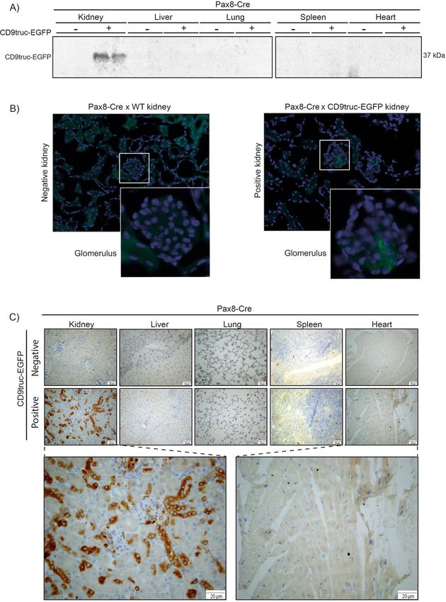
www.nature.com/scientificreports/

          Scientific Reports |   (2022) 12:496 |   https://doi.org/10.1038/s41598-021-04512-0   2

Vol:.(1234567890)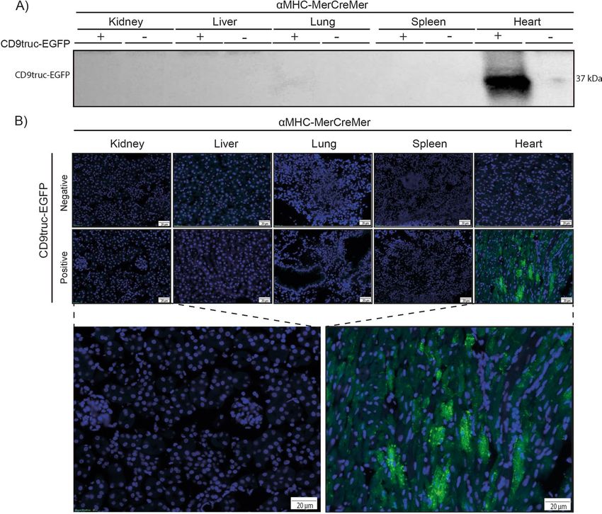
www.nature.com/scientificreports/
◂Figure 1. Expression of EGFP and associated EV markers M1-CD9truc-EGFP cells and their conditioned
medium. (A) Illustration of our EV reporter gene and protein. The CD9truc-EGFP coding sequence is driven by
the CMV promoter and encodes a fusion protein consisting of the first 3 transmembrane domains of CD9 and
EGFP, enabling genetic labeling and EV surface display of EGFP. (B) M1 cells stable transfected with CD9truc-
EGFP are green fluorescent in contrast to non-tansfected M1 cells. Nuclei (blue) and GFP (green). n = 3, ×200
magnification (C) Immunofluorescence labeling of CD9 in M1-CD9truc-EGFP cells using Anti-hCD9 AB
shows colocalization between CD9 (red), EGFP (green) and nuclei (blue). ×200 magnification (n = 3) (D) M1
cells stable transfected with CD9truc-EGFP expressing a band reacting with an anti-GFP antibody in cells
and PEG-precipitated conditioned medium. Actin and Lamin A/C were detected in cell lysates only, and EVs
markers ALIX and Flotillin were detected in conditioned medium from transfected and non-transfected M1
cells (n = 3). Full-length blots are shown in Supplementary Figure S2A-F. (E) Tunable resistive pulse sensing on
the conditioned medium from non-transfected M1 cells and stable transfected CD9truc-EGFP cells indicated
that the size distribution of EVs is not affected the expression reporter proteins (n = 3). (F) Size-exclusion
chromatography fractions of CD9truc-EGFP cell-conditioned medium showed full-length CD9truc-EGFP
in fractions 1–3, while shedded EGFP was present in fractions 10–12 (n = 3). Full-length blots are shown in
Supplementary Figure S2G. Below the western blot, the protein concentration in each fraction is shown (G)
Anti-GFP immunoprecipitation of cells conditioned medium co-isolates CD9truc-EGFP and EV markers ALIX
and TSG101 only in M1 cells stable transfected with CD9truc-EGFP (n = 3). Full-length blots are shown in
Supplementary Figure S3A-C.
overcome major limitations of previous approaches using radioisotopes, fluorescent dyes, and magnetic con-
jugated nanoparticles conjugated to lipophilic reagents to label EVs7–9. The lipophilic reagents may be released
from the EVs, resulting in the distribution of non-EV-associated fluorescent signal12,13. Furthermore, infusing
labeled EVs generated in vitro into mice may not be at physiologically relevant concentrations. While N and C
terminal fusion of reporter proteins to the tetraspanins enables tracking of cell-specific EVs, these reporter pro-
teins do not allow for affinity isolation of the labeled EVs from biological fluids in that the tetraspanin terminals
are located inside EVs12,13.
Tetraspanins, however, can be used to display fluorescent proteins on the EV surface17, and we, therefore,
hypothesized that the fusion of EGFP to the C-terminal of a truncated form of the tetraspanin CD9, devoid of
the large extracellular domain and the last transmembrane domain, would create fluorescence-labeled EVs with
affinity tags. Furthermore, flanking the inverted EV reporter with loxP sites20 would establish a genetic switch
that enabled Cre recombinase-dependent EV reporter expression in vivo and allow easy and reliable tracking
and isolation of cell-specific EVs. Using this novel transgenic mouse model enables the assimilation of new
knowledge regarding EVs’ physiological and pathophysiological functions.
Results
Fusion of EGFP to CD9 enable affinity isolation of EVs. We designed an EV reporter protein by
fusion of mouse CD9 truncated after the first 117 amino acids (the third transmembrane domain) to EGFP
(Fig. 1A). The predicted molecular weight of the fusion protein is 41.5 kDa. Stably transfected epithelial M1
cells–termed M1-CD9truc-EGFP cells-showed green fluorescent intracellular vesicles co-localized with CD9
(Fig. 1B and C). Transfected fluorescent markers for the endoplasmic reticulum (mCherry-ER), early endosome
marker Rab5 (mRFP-Rab5), and the tetraspanin CD81 (mTagBFP2-CD81) showed co-localization with EGFP
in CD9truc-EGFP cells (Supplemental Figure S1A). Western blotting revealed EGFP expression restricted to
transfected cells (Fig. 1D, the full-length blot is shown in Supplemental Figure S2A). While polyethylene gly-
col (PEG) precipitated cell-conditioned medium from M1 and M1-CD9truc-EGFP cells revealed enriched EV
markers ALIX and flotillin, and not β-actin or nuclear Lamin A/C, EGFP was detected only in cell-conditioned
medium from M1-CD9truc-EGFP cells (Fig. 1D, the full-length blots are shown in Supplemental Figure S2B-F).
We noted that without the addition of protease inhibitors to the cell-conditioned medium, EGFP was present
as two bands: one at ~ 37 kDa, slightly below the expected molecular weight of the fusion protein, and one
at ~ 27 kDa, indicating partial proteolytic shedding of EGFP (Supplemental Figure S2B). Conditioned medium
from M1 and M1-CD9truc-EGFP cells showed similar extracellular particle concentration and size distribution
(Fig. 1E). Size-exclusion chromatography (SEC) of the cell-conditioned medium from M1-CD9truc-EGFP cells
showed that full-length CD9truc-EGFP band was only present in the fractions 1–3 with a low protein content,
while shedded EGFP was present in fractions 10–12 characterized by a higher protein content (Fig. 1F, the full-
length blot is shown in Supplemental Figure S2G). This indicates that full-length CD9truc-EGFP is associated
with EVs. Consistent with this, EGFP immunoprecipitation from cell-conditioned medium isolated EGFP and
EV markers TSG101 and ALIX only from M1-CD9truc-EGFP cells (Fig. 1G, the full-length blot is shown in
Supplemental Figure S3A-C). Thus, EVs from CD9truc-EGFP expressing cells can be isolated using GFP immu-
noprecipitation.
Cre recombinase‑dependent CD9truc‑EGFP expression. To obtain cell-specific CD9truc-EGFP
expression, we inverted the coding sequence of EV reporter protein CD9truc-EGFP and flanked it by double
loxP sites, allowing for CD9truc-EGFP expression driven by a CAG promoter only in Cre recombinase express-
ing cells (Fig. 2A). HEK293T cells were transiently transfected with CD9truc-EGFP, which only was expressed
and yielded green fluorescent cells when co-transfected with Cre recombinase (Fig. 2B). CD9truc-EGFP expres-
sion was observed in Cre recombinase co-transfected cells and their corresponding condition medium (Fig. 2C,
the full-length blot is shown in Supplemental Figure S3D and E). Next, we used our CD9truc-EGFP plasmid
Scientific Reports | (2022) 12:496 | https://doi.org/10.1038/s41598-021-04512-0 3
Vol.:(0123456789)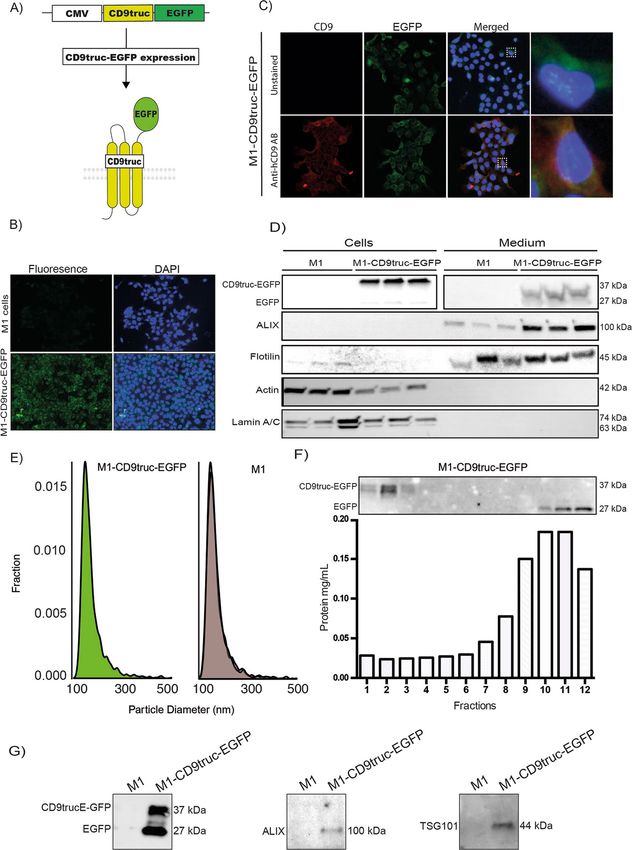
www.nature.com/scientificreports/

                                           Figure 2.  Cre recombinase-dependent expression of CD9truc-EGFP. (A) Our EV reporter CD9truc-EGFP
                                           was inverted and flanked by double lox sites. Upon Cre recombinase expression, the CD9truc-EGFP is inverted
                                           and yields CD9truc-EGFP expression driven by the CAG promoter. (B) HEK293T cells transiently transfected
                                           with double-floxed and inverted CD9truc-EGFP only express EGFP when co-transfected with Cre recombinase
                                           (n = 3). ×200 magnification (C) HEK293T cells co-transfected with double-floxed and inverted CD9truc-
                                           EGFP expressed CD9truc-EGFP in cells and conditioned medium (n = 3. Full-length blots are shown in
                                           Supplementary Figures S3D and E. (D) The double-floxed and inverted CD9truc-EGFP construct are inserted
                                           into chromosome 4 of the EVRep mouse, and mice homo- (Tg/Tg) and heterozygous (Tg/WT) can be identified
                                           by PCR. The full-length gel is shown in Supplementary Figure S3F.

                                           to generate transgenic mice through pro-nuclear injection of the linearized construct. We selected a founder
                                           mouse that showed specific and substantial expression of EGFP in Cre-expressing cardiomyocytes when crossed
                                           with the tamoxifen-treated αMHC-MerCreMer mouse (Supplemental Figure S1B). After backcrossing onto a
                                           C57Bl/6 background, we isolated genomic DNA from the liver of a transgene CD9truc-EGFP mouse. Nanop-
                                           ore sequencing revealed three reads (approximately 30 kb) containing mouse genomic sequence and CD9truc-
                                           EGFP insert sequence. The alignment of the sequenced reads was compatible with an integration of the insert in
                                           the mouse genome at approximately chromosomal location 4:99.620.973 (GRCm38.p6). The insert was present
                                           in two copies immediately adjacent to each other in opposite directions. The insert does not disrupt any known
                                           mouse genes at the location where it is embedded. The area of insertion is, however, an annotated constrained

          Scientific Reports |   (2022) 12:496 |               https://doi.org/10.1038/s41598-021-04512-0                                              4

Vol:.(1234567890)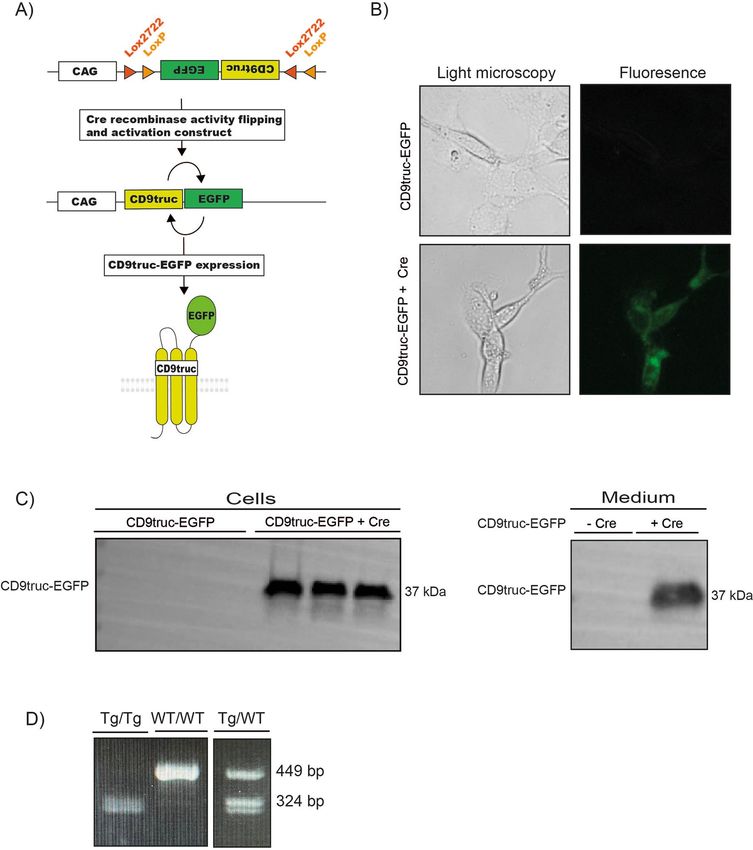
www.nature.com/scientificreports/
Sequence Annealing temperature °C Function BP
TGCACCTCAA
PS0819 60 Genotyping of CD9-EGFP forward 449/324
ACACTCAAGC
TCTTCCTGGA Genotyping of CD9-EGFP reverse 449 bp product in wildtype
PS0820 60 449
ACACAGCTCA and heterozygotes
AGCCAGATTT Genotyping of CD9-EGFP 324 bp product in CD9 homo- and
PS0821 60 324
TTCCTCCTCTC heterozygotes (with PS0819)
Table 1. Primer sequences for genotyping.
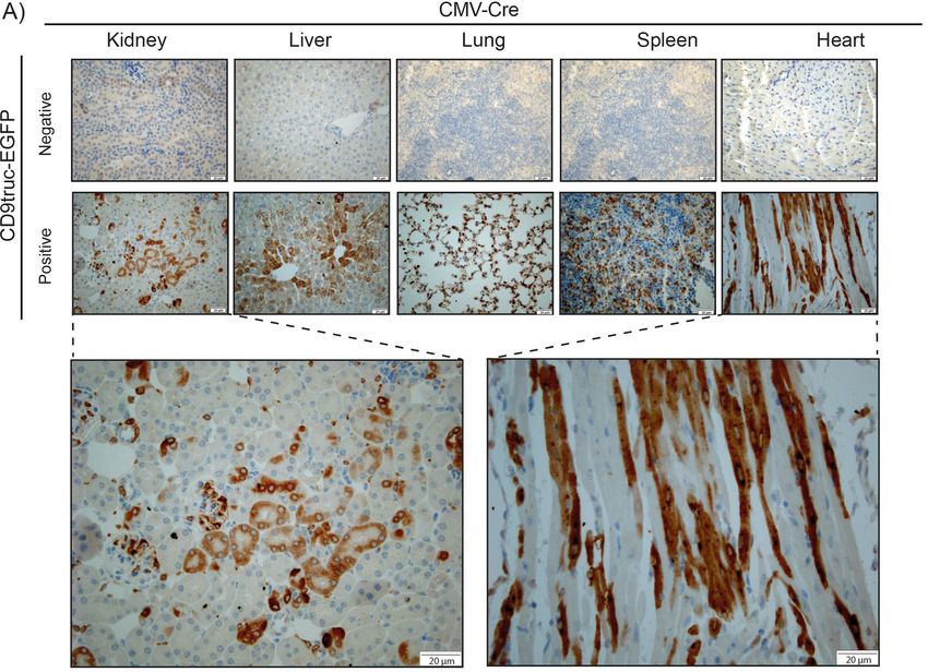
Figure 3. Expression of EGFP in tissue from CD9truc-EGFP positive CD9truc-EGFPxCMV-Cre mice. (A) In
contrast to the CD9truc-EGFP negative CD9truc-EGFPxCMV-Cre mice, EGFP expression is detected by anti-
GFP (brown) immunohistochemical staining of paraffin-embedded kidneys, liver, lung, spleens, and hearts of
CD9truc-EGFP positive CD9truc-EGFPxCMV-Cre mice. Scalebar 20 µm. (n = 2).
conserved region between multiple eutherian mammals. We designed primers for genotyping (Table 1) that
produced PCR products of 449 bp in wild-type mice and 324 bp in homozygotes (Fig. 2D, the full-length gel is
shown in Supplemental Figure S3F), enabling us to identify transgenic mice and distinguish heterozygotes from
homozygotes.
Tissue‑specific EGFP signal in different Cre‑positive transgenic mice. We next used established
transgenic Cre recombinase mice to enable tissue-specific activation of the EV reporter protein CD9truc-EGFP.
Initially, we crossed the transgene CD9truc-EGFP mice with CMV-Cre mice21 expressing Cre recombinase
under the control of the ubiquitously active human cytomegalovirus (CMV) promoter. CD9truc-EGFP posi-
tive CD9truc-EGFPxCMV-Cre mice showed EGFP expression in kidney, liver, lung spleen, and heart by anti-
GFP immunohistochemistry (IHC) (Fig. 3A). EGFP was undetectable in any of these tissues in CD9truc-EGFP
negative littermates (Fig. 3A). To show cell-specific expression of our CD9truc-EGFP construct, we crossed our
transgene CD9truc-EGFP mice with mice harboring tamoxifen-inducible cardiomyocyte-specific Cre activity
Scientific Reports | (2022) 12:496 | https://doi.org/10.1038/s41598-021-04512-0 5
Vol.:(0123456789)www.nature.com/scientificreports/
Figure 4. Expression of EGFP in cardiomyocytes from tamoxifen-treated CD9truc-EGFP positive CD9truc-
EGFPxαMHC-MerCreMer mice. (A) Tissue homogenates display CD9truc-EGFP expression only in the heart
from tamoxifen-treated CD9truc-EGFP positive CD9truc-EGFPxαMHC-Cre mice. The full-length blot is shown
in Supplementary Figure S4A. (B) Tissue sections only show EGFP expression (green) in cardiomyocytes from
tamoxifen-treated CD9truc-EGFP positive CD9truc-EGFPxαMHC-Cre. Nuclei (blue). Scalebar 20 µm. (n = 4).
(αMHC-MerCreMer)22 and Pax8-Cre mice expressing Cre recombinase kidney tubular epithelium23. In CD9truc-
EGFP positive, tamoxifen-treated CD9truc-EGFPxαMHC-MerCreMer mice, EGFP was explicitly detected in the
heart by western blotting and direct fluorescence microscopy (Fig. 4A-B, the full-length blot is shown in Sup-
plemental Figure S4A). EGFP was undetectable in kidneys, liver, lung, and spleen in tamoxifen-treated CD9truc-
EGFP positive and negative CD9truc-EGFPxαMHC-Cre mice and the hearts of tamoxifen-treated CD9truc-EGFP
negative CD9truc-EGFPxαMHC-Cre littermates (Fig. 4A). Likewise, we found EGFP expression specifically in
the kidney of CD9truc-EGFP positive CD9truc-EGFPxPax8-Cre mice using western blotting (Fig. 5A, the full-
length blot is shown in Supplemental Figure S4B). By fluorescence microscopy, however, green fluorescence was
not directly detectable in the kidney epithelium of kidney cryo-sections, and only low EGFP signals were associ-
ated with glomeruli (Fig. 5B). The preparation of cryo-sections involved exposure of the tissue to large osmotic
gradients, and we tested whether epithelial EGFP expression was present in paraffin-embedded formalin-fixed
kidneys. Using an anti-EGFP antibody, we detected kidney-specific EGFP expression in CD9truc-EGFP posi-
tive but not negative, CD9truc-EGFPxPax8-Cre mice (Fig. 5C). Thus, these data demonstrate that our transgene
CD9truc-EGFP mouse, which we term EVRep, allows for cell-specific expression of CD9truc-EGFP.
Isolation of EGFP‑positive extracellular vesicles from plasma and urine. Since EVs have been
detected in most body fluids, we next used plasma and urine samples from CD9truc-EGFP positive and nega-
tive CD9truc-EGFPxCMV-Cre, tamoxifen-treated CD9truc-EGFPxαMHC-MerCreMer, and CD9truc-EGFPx-
Pax8-Cre mice to determine the cell-specific contribution of EVs to plasma and urine fluid compartments. EVs
were precipitated and isolated from plasma using anti-GFP nanobody conjugated beads. CD9truc-EGFP was
Scientific Reports | (2022) 12:496 | https://doi.org/10.1038/s41598-021-04512-0 6
Vol:.(1234567890)www.nature.com/scientificreports/
Figure 5. Expression of EGFP in kidney epithelial cells from CD9truc-EGFP positive CD9truc-EGFPxPax8-Cre
mice. (A) Tissue homogenates from CD9truc-EGFP positive CD9truc-EGFPxPax8-Cre mice display CD9truc-
EGFP expression in kidneys. Full-length blots are shown in Supplementary Figure S4B. (B) Direct fluorescence
imaging of kidneys only shows EGFP expression (green) in glomerular cells of CD9truc-EGFP positive
CD9truc-EGFPxPax8-Cre mice. Nuclei (blue). Scalebar 20 µm. (n = 4). (C) Immunohistochemical staining of
paraffin-embedded kidneys, liver, lung, spleen, and heart from CD9truc-EGFPxPax8-Cre mice, EGFP expression
is detected by anti-GFP labeling (brown) in epithelial cells in the kidney from only CD9truc-EGFP positive
CD9truc-EGFPxPax8-Cre but not CD9truc-EGFP negative littermates. Scalebar 20 µm. (n = 2).
Scientific Reports | (2022) 12:496 | https://doi.org/10.1038/s41598-021-04512-0 7
Vol.:(0123456789)www.nature.com/scientificreports/
Figure 6. Isolation of EVs from plasma and urine by anti-GFP precipitation of plasma and urine samples
from CD9truc-EGFP transgene mice. (A) CD9truc-EGFP was only isolated from CD9truc-EGFP positive
CD9truc-EGFPxCMV-Cre and tamoxifen-treated CD9truc-EGFPxαMHC but not CD9truc-EGFPxPax8-Cre
mice or CD9truc-EGFP negative littermates. The EV marker ALIX was co-isolated with CD9truc-EGFP in
plasma samples from CD9truc-EGFP positive CD9truc-EGFPxαMHC-Cre only. Full-length blots are shown in
Supplementary Figure S5A and B. (B) CD9truc-EGFP was only detected by anti-GFP immunoprecipitation of
urine samples from CD9truc-EGFP positive CD9truc-EGFPxCMV-Cre and CD9truc-EGFPxPax8-Cre mice, but
not tamoxifen-treated CD9truc-EGFPxαMHC-Cre or CD9truc-EGFP negative littermates. The EV marker CD81
co-precipitated with CD9truc-EGFP only in urine samples from CD9truc-EGFP positive CD9truc-EGFPxPax8-
Cre mice. Full-length blots are shown in Supplementary Figures S5C and D.
detected in plasma from CD9truc-EGFP positive CD9truc-EGFPxCMV-Cre and tamoxifen-treated CD9truc-
EGFPxαMHC-Cre mice, but not from CD9truc-EGFPxPax8-Cre and CD9truc-EGFP negative littermates
(Fig. 6A, the full-length blot is shown in Supplemental Figure S5A). The EV marker ALIX co-precipitated with
CD9truc-EGFP only in plasma from tamoxifen-treated CD9truc-EGFP positive CD9truc-EGFPxαMHC-Cre
mice (Fig. 6A, the full-length blot is shown in Supplemental Figure S5B). In urine samples, CD9truc-EGFP was
observed in CD9truc-EGFP positive CD9truc-EGFPxCMV-Cre, and CD9truc-EGFPxPax8-Cre mice, whereas
tamoxifen-treated CD9truc-EGFPxαMHC-Cre urine samples were devoid of detectable CD9truc-EGFP (Fig. 6B,
the full-length blot is shown in Supplemental Figure S5C). The EV marker CD81 co-precipitated with CD9truc-
EGFP only in urine samples from CD9truc-EGFP positive CD9truc-EGFPxPax8-Cre (Fig. 6B, the full-length blot
is shown in Supplemental Figure S5D). Together, these data suggest that the EVRep mouse allows for the isola-
tion of cell-specific EVs from biological fluids.
Discussion
Using a genetically encoded CD9truc-EGFP fusion protein that displays EGFP on EVs combined with a Cre-
dependent switch, we have created a transgenic mouse enabling EV detection at the cellular level as well as rapid
and selective isolation of cell-specific EVs from plasma and urine. This EVRep mouse model is thus the first
genetic EV reporter model that allows easy tracking and isolation of EVs in vivo in mice. Therefore, we believe
that this novel tool can be utilized to generate novel insights into EV biology.
Cell-to-cell EV communication has already been demonstrated in several settings. In vitro, the transport of
miRNAs in EVs act as a neuron-to-astrocyte communication pathway in the central nervous system24, and stem
cell-derived EVs appear to target bone marrow and peripheral sites25. In vivo, intravascular administration of
mesenchymal stem cell-derived EVs (MSC-EV) labeled with PKH26 dye closely resembles the positive effect of
MSCs on postischemic recovery after acute tubular injury26. Another example includes intravenous injection
of endothelial progenitor cell-derived EVs labeled with PKH26 dye isolated from cell-conditioned medium
that protects from complement-mediated mesangial injury after experimental anti-Thy1.1 glomerulonephritis27.
Furthermore, similar positive effects have been obtained in the myocardium where intravenous administration
Scientific Reports | (2022) 12:496 | https://doi.org/10.1038/s41598-021-04512-0 8
Vol:.(1234567890)www.nature.com/scientificreports/
of EVs isolated from plasma protected against Ischemia–Reperfusion I njury28. Altogether, this suggests that EVs
retain regenerative abilities. However, for most EV studies, such injections cause a supra-physiological concentra-
tion of EVs from a single cell type, and the naturally occurring in vivo concentrations might not be sufficient to
cause similar effects. Furthermore, important paracrine and autocrine effects might be altered or unobserved with
this approach and affect the in vivo distribution and the biological function of the EVs. Importantly, injected EVs
often accumulate in the liver, spleen, and lungs regardless of the origin26,27,29. We did not observe CD9truc-EGFP
accumulation in these organs under normal physiological circumstances. These observations emphasize some
of the hurdles with injections of labeled E Vs30,31, which may be overcome using the EVRep mouse developed to
enable a robust EV fate mapping.
EVs isolated from biological fluid provide access to cell-derived lipids, RNAs, and proteins; however, the
representation of EVs from different cell types in, for example, urine and plasma is not well described. Consist-
ent with a previous study32, we found that EVs from cardiomyocytes were readily detectable in plasma samples.
Intravascular injection of labeled EVs isolated from serum have been detected in u rine33; however, we did not
detect cardiomyocyte-derived EVs in urine, indicating that EVs are not freely filtered across the glomerular
filtration barrier in the kidney, probably as a result of their size and negative c harge34–36. On the other hand, we
detected kidney epithelial EVs in urine. This is consistent with our previous findings using proteomic database
analysis showing that 99.96% of urinary EV-associated proteins are likely to originate from the kidney, the
urinary tract epithelium, and the male reproductive tract in h umans37. It should be noted that, for example,
the CD9truc-EGFPxPax8-Cre mice only showed CD9truc-EGFP in some of the tubular epithelial cells; thus,
methods with higher sensitivity than western blotting may be able to detect cardiomyocyte and renal tubular
epithelial-derived EVs in plasma and urine. Nonetheless, the distribution of EVs appears to be fluid compartment
restricted, and EVs isolated from different fluid compartments, e.g., plasma versus urine, may only represent a
subset of cell types in the body.
We found that kidney epithelial-derived EV signal was significantly affected by preparation of frozen kidney
cross-sections before fluorescence microscopy, while cardiomyocyte EV tissue abundance was less affected.
Western blotting suggested that CD9truc-EGFP was abundantly expressed in CD9truc-EGFP positive CD9truc-
EGFPxPax8-Cre mice, but in frozen kidney sections, the EGFP was only slightly visible in the glomerulus of
kidneys by directed fluorescence microscopy. The reason for this discrepancy is not known, but renal epithelial
cells are, however, highly water p ermeable38, and we speculate that rapid osmotic changes (e.g., 25% sucrose)
imposed during tissue preparation are involved. Recently, it was shown that the handling and preparation of
tissue samples significantly affect tissue EV abundance through release to, e.g., washing buffers39. Our observa-
tions agree with this and suggest that careful and specific tissue preparation is essential when analyzing tissue
EV levels and intercellular communication.
Similar to our approach, three other studies have used genetic labeling of E Vs15,18,40 to track the faith of endog-
enous EVs. While their use of an intravesicular localization of the reporter proteins prevents affinity isolation,
our approach shares the limitation that there is a risk that only a subpopulation of EVs is labeled. Notably, the
number of identified EV subtypes is continuously growing, and there is no consensus on which protein markers
represent the different populations. Thus, we cannot exclude that genetic labeling using CD9 may add a bias in
the downstream analysis, as it may only represent EV fractions. Moreover, we used a truncated version of CD9
in which the large extracellular loop was deleted. The large extracellular loop mediates many lateral CD9 interac-
tions, and its deletion might reduce potential adverse effects of its overexpression; however, the CD9truc-EGFP
may not show the exact same behavior as wild-type CD9. Nonetheless, the ease at which cell-specific EVs can
be isolated using the EVRep mouse will benefit a more comprehensive characterization and identification of EV
subtypes and enable other cell-specific EV markers to be identified.
In summary, our novel transgenic EVRep mouse allows easy in vivo tracking and isolation of EVs and can
be used to elucidate EV biology and their further use.
Methods
EV track data. We have submitted all relevant data of our experiments to the EV-TRACK knowledgebase
(EV-TRACK ID: EV210297)41.
Plasmids. The genes encoding truncated CD9 fused to EGFP, and the doubled-floxed and inverted ver-
sion were synthesized and cloned into plasmid pcDNA3.1 by BioCat GMBH. The coding sequence is shown
in Supplemental Figure S1C. The double-floxed, inverted CD9truc-EGFP from the pcDNA3.1 plasmid was
amplified by PCR and ligated into pCAG-Cre (a gift from Connie Cepko; Addgene plasmid # 13775) digested
with EcoRI and NotI (New England Biolabs). All constructs were verified by Sanger sequencing at Euro-
fins Genomics. mRFP-Rab5 was a gift from Ari Helenius (Addgene plasmid # 14437; http://n2t.net/addge
ne:14437; RRID:Addgene_14437), mCherry-ER (Addgene plasmid # 55041; http://n2t.net/addgene:55041;
RRID:Addgene_55041) and mTagBFP2-CD81-10 (Addgene plasmid # 55281; http://n2t.net/addgene:55281;
RRID:Addgene_55281) were gifts from Michael Davidson.
Cell cultures. Mouse epithelial M1 (ATCC, Virginia, United States) cells were cultured as previously
escribed42. For analysis of cell-conditioned medium, PC1 serum-free medium (Lonza, No. 344018, Walkers-
d
ville, MD, USA) was used. The M1 cells with stable CD9truc-EGFP expression were generated by transfection
with pcDNA3.1 CD9truc-EGFP using Metafectene Pro (Biontex, München, Germany) and selection with G418
(InvivoGen). HEK293T (ATCC, Virginia, United States) cells were maintained in Dulbecco’s Modified Eagle
Medium, F-12 Nutrient Mixture (Gibco, Sigma Aldrich, Denmark) containing 10% fetal bovine serum (FBS,
Scientific Reports | (2022) 12:496 | https://doi.org/10.1038/s41598-021-04512-0 9
Vol.:(0123456789)www.nature.com/scientificreports/
Fisher Scientific, DK) and 1% Penicillin–Streptomycin in a 5% CO2 humidified incubator at 37 °C. Harvesting
of cells and medium were performed when cells reached 80% confluence in 25 c m2-flasks.
Animal models. We linearized pCAG-DIO-CD9truc-EGFP with HindIII and SalI (New England Biolabs)
and gel extracted (New England Biolabs) the 3649-bp fragment containing the transgene. The purified fragment
was used to generate transgenic mice with genomic integration of the CAG-promotor driven double-floxed
inverted CD9truc-EGFP by pro-nucleus injection43. The αMHC-MerCreMer strain (JAX stock #005650, The
Jackson Laboratory, Bar Harbor, ME, USA) expresses inducible Cre in cardiomyocytes upon tamoxifen treat-
ment, and it is maintained by homozygous b reeding22. The CMV-Cre (JAX stock #006054), The Jackson Labo-
ratory, Bar Harbor, ME, USA) expresses Cre in all tissues and is maintained by homozygous b reeding21. The
B6.129P2(Cg)-Pax8tm1.1(cre)Mbu/J (JAX stock #028196), The Jackson Laboratory, Bar Harbor, ME, USA) strain
referred to as Pax8-Cre expresses Cre recombinase in known Pax8 expression domains including the developing
thyroid gland, inner ear, kidney, and mid-hindbrain r egion23. The animal experiment was performed in accord-
ance with Danish Law and approved by the Danish Animal Experimentation Council (#2019-15-0201-01644
and 2019-15-0202-00052). The study was performed in accordance with ARRIVE guidelines.
Sample collection. 7–9 weeks old mice were transferred individually to metabolic cages (12:12 h light–
dark cycle, 28 ± 1 °C) for three days with free access to water and regular rodent diet (LabDiet® 5001, Forth
Worth, TX, USA). After 24 h acclimation, urine samples were collected on days 2 and 3. Subsequently, mice were
used for perfusion fixation or direct organ harvest, as described below. Urine was stored at − 80 °C and a protease
inhibitor (1:1000, P8340, Protease Inhibitor Cocktail, Sigma Aldrich, Denmark) were added when thawed. Mice
used for immunohistochemistry and fluorescent microscopy were anesthetized by i.p. injection with 10 mg/kg
Xylazine (Rompun, Bayer Healthcare, Shawnee Mission, KS) and 50 mg/kg Ketamine (Ketalar, Pfizer, Sandwich,
Kent, UK). Blood samples were taken by cardiac puncture through the apex before mice were flushed with
1 × PBS and fixed with 4% paraformaldehyde by retrograde perfusion via the left ventricle. Subsequently, mice
were fixed for an additional 6 h in paraformaldehyde and transferred to 1 × PBS. Mice used for western blotting
were likewise anesthetized, blood samples were taken, and relevant organs were removed and snap-frozen in
liquid nitrogen.
Immunohistochemistry and fluorescence microscopy. Staining was performed on 2 μm paraffin-
embedded tissue samples deparaffined in xylene and rehydrated in decreasing ethanol solutions (99–70%) fol-
lowed by target retrieval in heated TEG buffer (1 mmol/L Tris, 0.5 mM EGTA, pH 9.0). After cooling, slides were
exposed to a 50 mM NH4Cl and 0.3% H2O2 solution for 10 min, washed in 1 × PBS, and incubated in 1 × PBS
with 0.3% Triton for 30 min at room temperature. Next, the slides were incubated with primary antibody (Anti-
GFP) diluted in 1 × PBS with 0.3% Triton × 100 at 4 °C overnight.
Next, slides were incubated for 30 min at room temperature, washed in 1 × PBS + 0.05% tween five times, and
incubated with secondary HRP antibody in PBS + 0.05% tween for 1 h at room temperature. Slides were then
washed in PBS, and HRP was visualized by DAB-staining (3,3’-diaminobenzidine). Lastly, slides were stained
in hematoxylin mounted with Aquatex (Merck KGaA, Darmstadt, Germany).
For fluorescence microscopy, snap-frozen organs were removed from mice and placed in 1 × PBS with 0.05%
acid and 25% sucrose overnight. Next, organs were placed in 1 × PBS with 0.05% acid and 50% sucrose for 2 h,
embedded in OCT tissue freezing medium and 5 μm cross-sectioned on a Cryostat (Leica CM3050 S, Leica
Biosystems, USA). Cross-sections were placed in 1 × TBS for 10 min, dried, and one drop of Slowfade™ Gold
antifade reagent with DAPI (Invitrogen, Thermo Fisher Scientific, Eugene, OR USA) was added before mounting.
All tissue and cell samples were obtained using an Olympus BX51 Fluorescence microscope.
Isolation of extracellular vesicles from cells and medium. 48 h before, the cell medium was changed
to PC1 serum-free medium (Lonza, No. 344018, Walkersville, MD, USA). Cells were lysed in RIPA Lysis buffer
for 1 h at 4 °C on an orbital shaker. Next, 25 cm cell scrapers were used to release cells from the flask into the
RIPA buffer. The solution was then centrifugated at 13,000g at 4 °C for 10 min, and the supernatant was stored
in new tubes at − 80 °C. The medium was transferred to 15 mL tubes and centrifugated for 10 min at 5000 g. The
supernatant was then transferred to new tubes and stored at − 80 °C. To isolate EVs, the thawed medium was
centrifugated for 15 min at 5000 g at 4 °C. The supernatant was mixed with an equal amount of freshly made
ExtraPEG44 (16% PEG-6000, Sigma Aldrich, 1 M NaCl, Milli-Q water) and left in a Multi-Rotator at 4 °C over-
night. Next, samples were centrifugated for 15 min at 5000 g at 4 °C, and the supernatant was discarded. The
remaining pellet was resuspended in 100 μl × 1 RIPA buffer and stored at − 20 °C until further use.
Immunofluorescence of cells. Cells were seeded on coverslips in multiple 12-wells plates (Biocoat Cell
environments, Poly-D-Lysine Cellware) and given 24 h to attach to the coverslips. In the experiments with
transfection of fluorescent organelle markers, the cells were transfected the day after seeding on coverslips using
Metafectene Pro (Biontex, München, Germany) and grown for 24 h. Next, cells were fixed in 4% paraformalde-
hyde for 10 min and rinsed 2 times in 1 × PBS with 1 mM M gCl2 and 0.1 mM C aCl2, followed by permeabiliza-
tion with 0.3% Triton in 1 × PBS for 15 min and another wash in 1 × PBS with 1 mM M gCl2 and 0.1 mM C aCl2.
Cells where then incubated in 300 μl primary antibody (Table 2) at 4 °C overnight washed 3 times in 1xPBS and
incubated with 300 μl secondary antibody for 1 h at room temperature. Afterward, cells were incubated with
4’,6-diamidino-2-phenylindole (D9542-10MG DAPI, Sigma Aldrich, Denmark) to stain DNA followed by 5
washes in 1 × PBS. Lastly, coverslips containing cells were mounted with fluorescent mounting media (DAKO,
Carpinteria, CA, USA) on coverslips. ImageJ, version 2.0.0-RC-43/1.10e) was used to analyze pictures.
Scientific Reports | (2022) 12:496 | https://doi.org/10.1038/s41598-021-04512-0 10
Vol:.(1234567890)www.nature.com/scientificreports/
Target protein Company Catalog # Dilution
Immunocytochemistry
Primary antibody
Anti-hCD9 RD systems MAB1880 1:50
Secondary antibody
Anti-mouse AlexaFlour 568 Abcam, UK 1:500
Western blot
Primary antibodies
GFP Nordic BioSite, SE GTX113617 1:1000
ALIX Cell Signaling Technology, NL 2171 1:1000
Flotilin Abcam, UK Ab13493 1:1000
ß-Actin Abcam, UK Ab8227 1:10,000
CD81 Santa Cruz Biotechnology, USA E1817 1:1000
TSG-101 Abcam, UK Ab30871 1:1000
Immunohistochemistry Primary antibodies
Anti-GFP Abcam, UK Ab6673 1:200
Sencondary antibodies
Polyclonal Goat Anti-Mouse immunoglobulins/HRP DAKO, DK P0447
Polyclonal Goat Anti-Rabbit immunoglobulins/HRP DAKO, DK P0448
Polyclonal Rabbit Anti-Goat immunoglobulins/HRP DAKO, DK P0449
Table 2. Primary and secondary antibodies.
Immunoblotting. Samples were mixed with LDS sample buffer (NuPAGE, Invitrogen, Thermo Fischer Sci-
entific, Van Allen Way, Carlsbad, USA) and sample reducing agent (NuPAGE, Invitrogen, Thermo Fischer Scien-
tific, Van Allen Way, Carlsbad, USA) and heated for 10 min before they ran on a gel. For cell and tissue fractions,
10 μg was loaded per well. The protein was then transferred to a membrane activated in 99% Ethanol. Afterward,
the membrane was blocked for 30 min in 5% skimmed milk and incubated overnight with primary antibodies at
4 °C (Table 2). Following, membranes were washed 3 times in Tris-Buffered saline with Tween-20 (TBST, 20 mM
Tris-base, 137 nM NaCl, 0.05% Tween-20 (Merck), pH 7.6) and incubated with secondary antibody (Table 2) for
1 h at room temperature. Next, membranes were washed 3 times in TBS-T, and proteins were visualized by ECL
plus a Molecular Imager (ChemiDoc XRS + , BIO-RAD) with Image Lab software (BIO-RAD).
Immunoprecipitation of EGFP associated EVs. EVs containing EGFP were precipitated from 200 μl
plasma and 750 μl urine with extraPEG as described above, except that the pellet was resuspended in 100 μl
1 × PBS to keep EVs intact. EVs immunoprecipitated using ChromoTek GFP-trap Magnetic Beads (Chromotek
GmbH, Germany). 25 μl beads were added to a 1.5 ml tube and rinsed with 1000 μl ice-cold 1 × PBS. Next,
plasma or urine was added to the equilibrated beads, and tubes were placed in a Multi-Rotator (Grant-bio, PTR-
35) at 4 °C for 1 h. Samples were when placed back in the magnet and allowed to attach before the supernatant
was discarded. Subsequently, beads were resuspended in 1 mL ice-cold 1 × PBS-T, and the samples were placed
back in the magnet. The supernatant was discarded after 3 min when the beads were attached to the magnet, and
this step was repeated 4 times.
Tunable resistive pulse sensing. A qNano platform with a Nanopore NP150 (Izon Science, Oxford, UK)
and polystyrene calibration beads CPC200 (Izon Science), Oxford, UK) were used for calibration of relative
particle size and speed. 35 μl cell media was loaded, and analyses were performed according to manufactory
instructions.
Size exclusion chromatography. A qEVoriginal/35 nm SMART column (Izon Science, Oxford, UK)
with optimal separation size between 35–350 nm was used to isolate EVs from 0.5 ml from M1-CD9-EGFP
medium according to manufacturer’s instructions. 0.5 mL fractions were collected, and western blotting was
used to verify the presence of EGFP. To verify that proteins mainly were eluted in the last fractions, proteins
concentrations were measured in each fraction with DC Protein Assay (Biorad, California, U. S.) according to
the manufacturer’s guidelines.
Nanopore sequencing. Mouse liver from a CD9truc-EGFP positive EVRep mouse was homogenized, and
DNA was extracted using the Nanobind Tissue Big DNA Kit (Circulomics, Baltimore, MD, USA) according
to the manufacturer’s instructions. The DNA was prepared for Oxford Nanopore sequencing using the Liga-
tion Sequencing Kit (Oxford Nanopore Technologies, Oxford, UK). The prepared library was then sequenced
using one flow cell on the Oxford Nanopore PromethION sequencing platform. Base-calling was performed
with MinKNOW software, and FASTQ files were aligned to a custom mouse genome (GRCm38.p6) containing
Scientific Reports | (2022) 12:496 | https://doi.org/10.1038/s41598-021-04512-0 11
Vol.:(0123456789)www.nature.com/scientificreports/
the CD9truc-EGFP insert and accessory sequences as an extra chromosome. Alignment was performed using
Minimap245. Data were visualized using IGV ver2.8.1 (Broad Institute), Genome Ribbon46, and NCBI BLAST.
Received: 31 August 2021; Accepted: 21 December 2021
References
1. Yáñez-Mó, M. et al. Biological properties of extracellular vesicles and their physiological functions. J. Extracell. Vesicles 4, 1–60
(2015).
2. Akers, J. C. et al. miR-21 in the extracellular vesicles (EVs) of cerebrospinal fluid (CSF): A platform for glioblastoma biomarker
development. PLoS ONE 8, e78115 (2013).
3. Goto, T. et al. An elevated expression of serum exosomal microRNA-191, −21, −451a of pancreatic neoplasm is considered to be
efficient diagnostic marker. BMC Cancer 18, 116 (2018).
4. Pisitkun, T., Shen, R. F. & Knepper, M. A. Identification and proteomic profiling of exosomes in human urine. Proc. Natl. Acad.
Sci. U. S. A. 101, 13368–13373 (2004).
5. Vojtech, L. et al. Exosomes in human semen carry a distinctive repertoire of small non-coding RNAs with potential regulatory
functions. Nucleic Acids Res. 42, 7290–7304 (2014).
6. Zlotogorski-Hurvitz, A. et al. Human saliva-derived exosomes: Comparing methods of isolation. J. Histochem. Cytochem. 63,
181–189 (2015).
7. Grigor’eva, A. E. et al. Exosomes in tears of healthy individuals: Isolation, identification, and characterization. Biochem. Suppl. Ser.
B Biomed. Chem. 10, 165–172 (2016).
8. Yoon, S. B. & Chang, J. H. Extracellular vesicles in bile: A game changer in the diagnosis of indeterminate biliary stenoses?. Hepa-
toBiliary Surg. Nutr. 6, 408–410 (2017).
9. Yamamoto, H. Detection of DNA methylation of gastric juice-derived exosomes in gastric cancer. Integr. Mol. Med. 1, 105 (2014).
10. Van Niel, G., Porto-Carreiro, I., Simoes, S. & Raposo, G. Exosomes: A common pathway for a specialized function. J. Biochem.
140, 13–21 (2006).
11. Escola, J. M. et al. Selective enrichment of tetraspan proteins on the internal vesicles of multivesicular endosomes and on exosomes
secreted by human B-lymphocytes. J. Biol. Chem. 273, 20121–20127 (1998).
12. Umeda, R. et al. Structural insights into tetraspanin CD9 function. Nat. Commun. 11, 7 (2020).
13. Andreu, Z. & Yáñez-Mó, M. Tetraspanins in extracellular vesicle formation and function. Front. Immunol. 5, 442 (2014).
14. Bobrie, A., Colombo, M., Krumeich, S., Raposp, G. & Clotilde, T. Diverse subpopulations of vesicles secreted by different intracel-
lular mechanisms are present in exosome preparations obtained by differential ultracentrifugation. J. Extracell. vesicles 1, 18397
(2012).
15. Luo, W. et al. Spatial and temporal tracking of cardiac exosomes in mouse using a nano-luciferase-CD63 fusion protein. Commun.
Biol. 3, 1–9 (2020).
16. Sung, B. H., Ketova, T., Hoshino, D., Zijlstra, A. & Weaver, A. M. Directional cell movement through tissues is controlled by exo-
some secretion. Nat. Commun. 6, 1–14 (2015).
17. Sung, B. H. et al. A live cell reporter of exosome secretion and uptake reveals pathfinding behavior of migrating cells. Nat. Com-
mun. 11, 1–15 (2020).
18. Frederik Verweij, A. J. et al. Live tracking of inter-organ communication by endogenous exosomes in vivo. Dev. Cell 48, 573-589.
e4 (2019).
19. Yoshimura, A. et al. The Sox2 promoter-driven CD63-GFP transgenic rat model allows tracking of neural stem cell-derived
extracellular vesicles. Dis. Model. Mech. 11, 028779 (2018).
20. Schnütgen, F. et al. A directional strategy for monitoring Cre-mediated recombination at the cellular level in the mouse. Nat.
Biotechnol. 21, 562–565 (2003).
21. Schwenk, F., Baron, U. & Rajewsky, K. cre-transgenic mouse strain for the ubiquitous deletion of loxP-flanked gene segments
including deletion in germ cells. Nucleic Acids Res. 23, 5080–5081 (1995).
22. Sohal, D. S. et al. Temporally regulated and tissue-specific gene manipulations in the adult and embryonic heart using a tamoxifen-
inducible Cre protein. Circ. Res. 89, 20–25 (2001).
23. Bouchard, M., Souabni, A., Mandler, M., Neubüser, A. & Busslinger, M. Nephric lineage specification by Pax2 and Pax8. Genes
Dev. 16, 2958–2970 (2002).
24. Morel, L. et al. Neuronal exosomal mirna-dependent translational regulation of astroglial glutamate transporter glt1. J. Biol. Chem.
288, 7105–7116 (2013).
25. Salvucci, O. et al. MicroRNA126 contributes to granulocyte colony-stimulating factor-induced hematopoietic progenitor cell
mobilization by reducing the expression of vascular cell adhesion molecule 1. Haematologica 97, 818–826 (2012).
26. Bruno, S. et al. Mesenchymal stem cell-derived microvesicles protect against acute tubular injury. J. Am. Soc. Nephrol. 20, 1053–1067
(2009).
27. Cantaluppi, V. et al. Endothelial progenitor cell-derived extracellular vesicles protect from complement-mediated mesangial injury
in experimental anti-Thy1.1 glomerulonephritis. Nephrol. Dial. Transpl. 30, 410–422 (2015).
28. Vicencio, J. M. et al. Plasma exosomes protect the myocardium from ischemia-reperfusion injury. J. Am. Coll. Cardiol. 65, 1525–
1536 (2015).
29. Choi, H. & Lee, D. S. Illuminating the physiology of extracellular vesicles. Stem Cell Res. Ther. 7, 1 (2016).
30. Wiklander, O. P. B. et al. Extracellular vesicle in vivo biodistribution is determined by cell source, route of administration and
targeting. J. Extracell. Vesicles 4, 1–13 (2015).
31. Li, P. et al. PKH26 can transfer to host cells in vitro and vivo. Stem Cells Dev. 22, 340–344 (2013).
32. Sluijter, J. P. G. et al. Extracellular vesicles in diagnostics and therapy of the ischaemic heart: Position Paper from the Working
Group on Cellular Biology of the Heart of the European Society of Cardiology. Cardiovasc. Res. 114, 19–34 (2018).
33. Cheng, Y. et al. A translational study of urine miRNAs in acute myocardial infarction. J. Mol. Cell. Cardiol. 53, 668–676 (2012).
34. Midekessa, G. et al. Zeta potential of extracellular vesicles: Toward understanding the attributes that determine colloidal stability.
ACS Omega 5, 16701–16710 (2020).
35. Rodewald, R. & Karnovsky, M. J. Porous substructure of the glomerular slit diaphragm in the rat and mouse. J. Cell Biol. 60, 423–433
(1974).
36. Costanzo, L. S. & Preceded by: Costanzo, L. S. Physiology. (Elsevier, 2015).
37. Svenningsen, P., Sabaratnam, R. & Jensen, B. L. Urinary extracellular vesicles: Origin, role as intercellular messengers and biomark-
ers; efficient sorting and potential treatment options. Acta Physiol. 228, e13346 (2020).
38. Verkman, A. S. Mechanisms and regulation of water permeability in renal epithelia. Am. J. Physiol. Cell Physiol. 257, C837–C850
(1989).
Scientific Reports | (2022) 12:496 | https://doi.org/10.1038/s41598-021-04512-0 12
Vol:.(1234567890)www.nature.com/scientificreports/
39. Gupta, M. P. et al. Non-reversible tissue fixation retains extracellular vesicles for in situ imaging. Nat. Methods 16, 1269–1273
(2019).
40. Zomer, A., Steenbeek, S. C., Maynard, C. & Van Rheenen, J. Studying extracellular vesicle transfer by a Cre-loxP method. Nat.
Protoc. 11, 87–101 (2016).
41. Van Deun, J. et al. EV-TRACK: Transparent reporting and centralizing knowledge in extracellular vesicle research. Nat. Methods
14, 228–232 (2017).
42. Svenningsen, P., Granhoj, J. & Dimke, H. Proteolytic activation of the epithelial sodium channel: Role of pro-protein convertases
and prostasin. FASEB J. 34, 1–1 (2020).
43. Hansen, L. A. & Füchtbauer, E. M. Genome editing in mice. In Methods in Molecular Biology vol. 1961, 249–254 (Humana Press
Inc., 2019).
44. Rider, M. A., Hurwitz, S. N. & Meckes, D. G. ExtraPEG: A polyethylene glycol-based method for enrichment of extracellular
vesicles. Sci. Rep. 6, 1–14 (2016).
45. Heng, L. Minimap2: Pairwise alignment for nucleotide sequences. Bioinformatics 34, 3094–3100 (2018).
46. Nattestad, M., Aboukhalil, R., Chin, C.-S. & Schatz, M. C. Ribbon: Intuitive visualization for complex genomic variation. Bioin-
formatics 37, 413–415 (2021).
Acknowledgements
We thank Lene B. Andersen, Inger Nissen, Charlotte Nielsen, Natasha Stanic, Seham Bouziri, Kristoffer Rosen-
strand, and Mohamed A. Ahmed for excellent technical assistance. The work was supported by research grants
from A. P. Møller Foundation (#20-L-0112 to MØN), Nyreforeningen (to MØN), Novo Nordisk Foundation
(#NNF19OC0058222 to PS), the Carlsberg Foundation (#CF-180404 to PS), and Innovation Fund Denmark
(#9122-00058B to PS).
Author contributions
Study conception and design: M.Ø.N., D.C.A., and P.S. Acquisition of data: M.Ø.N., D.R.H., and M.B.E. Analysis
and interpretation of data: M.Ø.N., D.R.H., L.B.S., and P.S. Article draft: M.Ø.N. and P.S. Review for important
intellectual content: L.B.S., D.R.H., E.M.F., M.B.E., H.D., D.C.A. All authors have reviewed and approved the
manuscript.
Competing interests
The authors declare no competing interests.
Additional information
Supplementary Information The online version contains supplementary material available at https://doi.org/
10.1038/s41598-021-04512-0.
Correspondence and requests for materials should be addressed to P.S.
Reprints and permissions information is available at www.nature.com/reprints.
Publisher’s note Springer Nature remains neutral with regard to jurisdictional claims in published maps and
institutional affiliations.
Open Access This article is licensed under a Creative Commons Attribution 4.0 International
License, which permits use, sharing, adaptation, distribution and reproduction in any medium or
format, as long as you give appropriate credit to the original author(s) and the source, provide a link to the
Creative Commons licence, and indicate if changes were made. The images or other third party material in this
article are included in the article’s Creative Commons licence, unless indicated otherwise in a credit line to the
material. If material is not included in the article’s Creative Commons licence and your intended use is not
permitted by statutory regulation or exceeds the permitted use, you will need to obtain permission directly from
the copyright holder. To view a copy of this licence, visit http://creativecommons.org/licenses/by/4.0/.
© The Author(s) 2022
Scientific Reports | (2022) 12:496 | https://doi.org/10.1038/s41598-021-04512-0 13
Vol.:(0123456789)You can also read