Abstract - Semantic Scholar
←
→
Page content transcription
If your browser does not render page correctly, please read the page content below
Open Access Case
                              Report                                                DOI: 10.7759/cureus.16280

                                              Giant Coronary Aneurysm Causing Ostial
                                              Occlusion of Coronary Artery by Mass Effect: A
                                              Case Report
                                              Vanlalmalsawmdawngliana Fanai 1 , Amit Malviya 1 , Animesh Mishra 1 , Donboklang Lynser 2 , Tony Ete 1

                                              1. Cardiology, North Eastern Indira Gandhi Regional Institute of Health and Medical Sciences, Shillong, IND 2.
                                              Radiology, North Eastern Indira Gandhi Regional Institute of Health and Medical Sciences, Shillong, IND

                                              Corresponding author: Amit Malviya, dramit_malviya@rediffmail.com

                                              Abstract
                                              Coronary atherosclerosis can rarely lead to complications like giant coronary aneurysm (GCA), and acute
                                              myocardial infarction (AMI) due to thrombosis in the GCA is even rarer. Multimodality imaging is preferred
                                              over relying solely on selective coronary angiogram in such cases due to the limitations of invasive coronary
                                              angiogram in visualizing thrombosed aneurysms. We report a rare case of a patient with ST-elevation
                                              myocardial infarction caused by ostial occlusion of a right coronary artery (RCA) due to mass effect created
                                              by thrombosis in a GCA, thereby highlighting a mechanism of AMI that has not been previously described in
                                              GCA. Multimodality imaging led to the correct diagnosis and detection of the underlying mechanism, which
                                              had been completely missed by invasive coronary angiography (ICA). We also discuss the utility of
                                              multimodality imaging in such cases.

                                              Categories: Cardiology
                                              Keywords: coronary anomaly, giant coronary artery aneurysm, invasive coronary angiography, acute myocardial
                                              infarction, diagnostic methods, thrombosis, management, computed tomography, cardiac imaging, mechanism

                                              Introduction
                                              Aneurysmal dilatation of coronary artery and giant coronary artery aneurysm (GCA) are rare findings during
                                              angiography [1,2]. There is no clear definition of GCA, but in the context of Kawasaki disease, large or giant
                                              aneurysms are defined as those with an internal lumen diameter >8 mm [3], and some experts consider
                                              atherosclerotic aneurysms >2 cm as GCA [4]. Although invasive coronary angiography (ICA) is the gold
                                              standard imaging technique in such cases, it may fail to detect coronary artery aneurysm (CAA) in the
                                              presence of luminal thrombi. In this report, we present a case of GCA involving left anterior descending
                                              artery (LAD) and right coronary artery (RCA) presenting as acute myocardial infarction (AMI) due to the
                                              occlusion of RCA ostium caused by the mass effect of thrombosed GCA of RCA. In this case, multimodality
                                              imaging led to the correct diagnosis and the detection of the underlying mechanism, which had been
                                              completely missed by ICA. To the best of our knowledge, GCA causing AMI due to mass effect has not been
                                              previously reported in the literature.
Review began 06/28/2021
Review ended 06/30/2021
Published 07/09/2021
                                              Case Presentation
© Copyright 2021
Fanai et al. This is an open access article
                                              A 60-year-old male non-smoker, non-diabetic, and normotensive patient was referred to our center with a
distributed under the terms of the            history of retrosternal chest pain and dyspnea [New York Heart Association (NYHA) class IV] for two days.
Creative Commons Attribution License          The patient had a history of exertional dyspnea and angina (NYHA class II) for the past year, but he was
CC-BY 4.0., which permits unrestricted        neither on any sort of medication and nor had consulted any physician for the same. The patient denied any
use, distribution, and reproduction in any
                                              past history of fever, joint pain, limb claudication, trauma to the chest. He was a farmer by occupation, and
medium, provided the original author and
source are credited.
                                              prior to this visit, there was no history of any medication or procedure done on him. On physical
                                              examination, his blood pressure was 100/70 mmHg, pulse was 100/minute, and regular in rhythm. The rest
                                              of the cardiovascular examination was unremarkable. An electrocardiogram at admission showed sinus
                                              rhythm with ST elevation in inferior leads (lead III/aVF) along with reciprocal ST depression in lead I, aVL,
                                              and V5-V6 (Figure 1A). The cardiac biomarkers were significantly elevated. Initial imaging with chest X-ray
                                              revealed cardiomegaly with enlarged right heart border (Figure 1B), and 2D-echocardiogram demonstrated
                                              inferior wall hypokinesia with mildly reduced left ventricular systolic fraction and a well-circumscribed
                                              cystic mass adjacent to the right ventricle (Figures 1C, 1D). The routine biochemistry findings are provided
                                              in Table 1.

                              How to cite this article
                              Fanai V, Malviya A, Mishra A, et al. (July 09, 2021) Giant Coronary Aneurysm Causing Ostial Occlusion of Coronary Artery by Mass Effect: A Case
                              Report. Cureus 13(7): e16280. DOI 10.7759/cureus.16280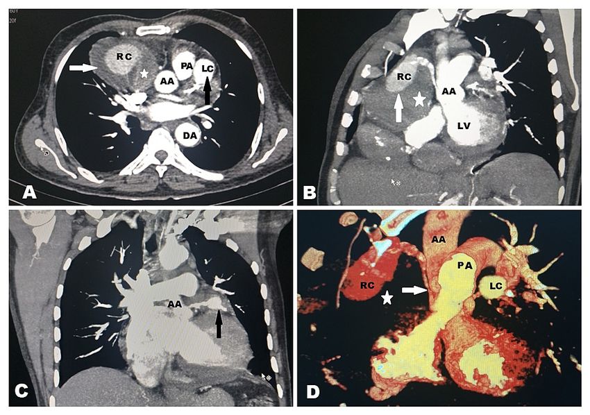
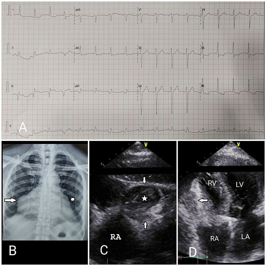
FIGURE 1: Noninvasive diagnostic images
                                                    A: Electrocardiogram showing sinus rhythm with ST elevation in lead III and aVF along with reciprocal ST
                                                    depression in lead I, aVL, and V5-V6. B: Posteroanterior view of chest X-ray showing cardiomegaly with a
                                                    focal bulge from the right (long white arrow) and left cardiac contours (short white arrow) consistent with
                                                    aneurysms from the right and left coronary arteries respectively. C: Echocardiographic image: modified
                                                    subcoastal view demonstrating a well-circumscribed cystic mass measuring 74 x 60 mm (white arrow) with
                                                    heterogeneous echogenic content (white star). D: Echocardiographic image: apical four-chamber view
                                                    showing extra-cardiac mass (white arrow) compressing RV

                                                    RA: right atrium; RV: right ventricle; LA: left atrium; LV: left ventricle

2021 Fanai et al. Cureus 13(7): e16280. DOI 10.7759/cureus.16280                                                                                                  2 of 7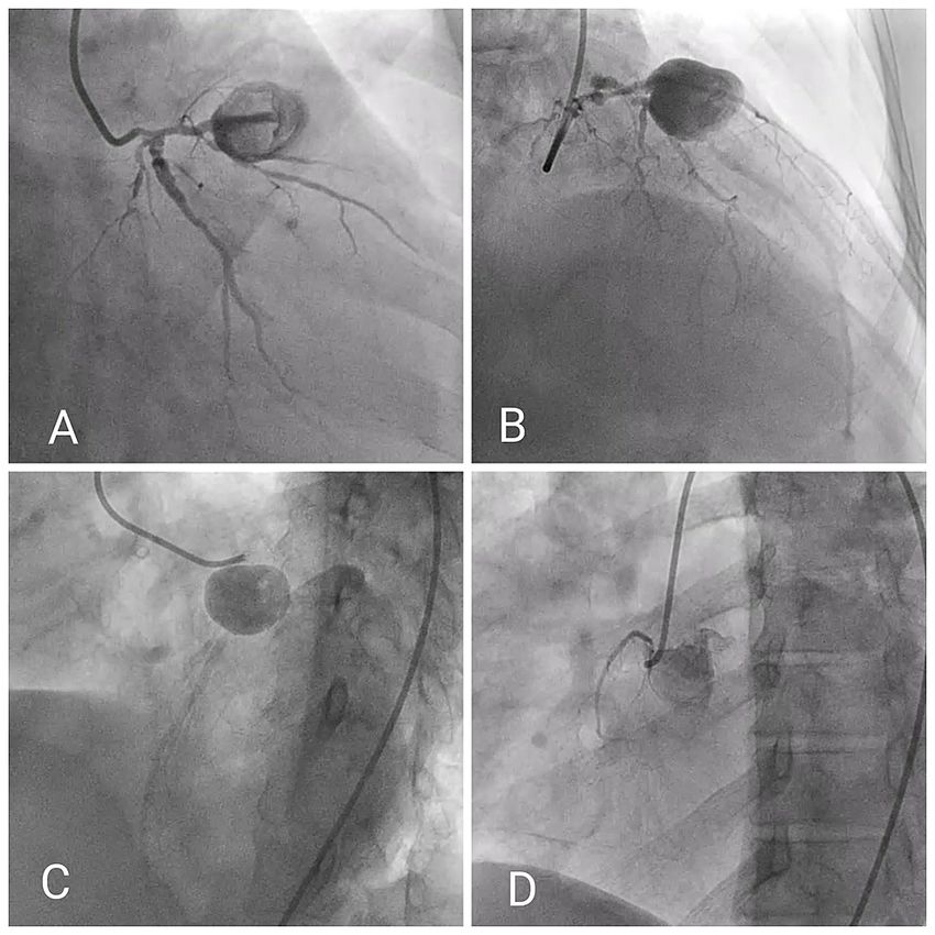
Investigation                                                                      Value

               Hemoglobin (g/dl)                                                                  13.6

               Total leukocyte count (per microliter)                                             6700

               Platelets (per microliter)                                                         270 x 103

               Random blood sugar (mg/dl)                                                         98

               Urea (mg/dl)                                                                       50

               Creatinine (mg/dl)                                                                 1.1

               Uric acid (mg/dl)                                                                  6.6

               Sodium (mmol/L)                                                                    138

               Potassium (mmol/L)                                                                 4.34

               Triglyceride (mg/dl)                                                               86

               Total cholesterol (mg/dl)                                                          86

               High-density lipoprotein (mg/dl)                                                   16.5

               Low-density lipoprotein (mg/dl)                                                    69.6

               Total bilirubin (mg/dl)                                                            0.7

               Aspartate aminotransferase (Unit/L)                                                105

               Alanine aminotransferase (Unit/L)                                                  50

               Creatine kinase-MB (Unit/L)                                                        154

               High-sensitivity troponin I (pg/ml)                                                1315 (normal range: 0-17.5)

               HBsAg                                                                              Nonreactive

               Anti-HCV IgM                                                                       Nonreactive

               HIV 1 and 2                                                                        Nonreactive

               VDRL test                                                                          Nonreactive

               Blood culture                                                                      Sterile

               C-reactive protein (mg/L)                                                          7

              TABLE 1: Routine biochemistry at admission
              HBsAg: hepatitis B surface antigen; HCV: hepatitis C virus; IgM: immunoglobulin M; HIV: human immunodeficiency virus; VDRL: venereal disease
              research laboratory

                                                  Angiogram of the left coronary system (Figures 2A-2D; Videos 1, 2) showed significant left main stenosis
                                                  with a GCA arising from proximal LAD (Figures 2A-2C) with Thrombolysis in Myocardial Infarction (TIMI)
                                                  grade II flow distal to the aneurysm. Diffuse significant lesions in the proximal left circumflex artery and its
                                                  major branches were also observed.

2021 Fanai et al. Cureus 13(7): e16280. DOI 10.7759/cureus.16280                                                                                                     3 of 7
FIGURE 2: Angiogram of coronary arteries
A: RAO caudal view showing significant stenosis of left main coronary artery and GCA at proximal LAD artery
with freely flowing contrast material within the aneurysm. B: RAO cranial view showing GCA at proximal LAD
artery with TIMI flow II distal to the aneurysm. C: LAO cranial view showing similar lesion as described above
with TIMI flow II distal to the aneurysm in LAD. D: LAO view of nonselective contrast injection at the aortic
root demonstrating total occlusion of ostial-proximal RCA and conus branch arising from a separate ostium
GCA: giant coronary aneurysm; LAD: left anterior descending; LCX: left circumflex artery; LAO: left anterior
oblique; RAO: right anterior oblique; TIMI: Thrombolysis in Myocardial Infarction
VIDEO 1: RAO cranial view of coronary angiogram demonstrating freely
flowing contrast material within GCA at proximal LAD artery and TIMI
grade II flow distal to the aneurysm
GCA: giant coronary aneurysm; LAD: left anterior descending; RAO: right anterior oblique; TIMI: Thrombolysis
in Myocardial Infarction
View video here: https://youtu.be/eB_wvfHl-OM
2021 Fanai et al. Cureus 13(7): e16280. DOI 10.7759/cureus.16280 4 of 7VIDEO 2: LAO caudal view of coronary angiogram demonstrating
significant stenosis involving left main coronary artery and proximal
LCX artery
LAO: left anterior oblique; LCX: left circumflex artery
View video here: https://youtu.be/3OMRBceGJJc
The ostium of RCA could not be engaged selectively despite several attempts, and a nonselective angiogram
revealed occlusion of RCA from the ostium with mild disease in the conal branch of RCA arising from a
separate ostium (Figure 2D, Video 3).
VIDEO 3: Nonselective coronary angiogram at aortic root demonstrating
occlusion of RCA from the ostium with mild disease in the conal branch
of RCA arising from a separate ostium
RCA: right coronary artery
View video here: https://youtu.be/BKSqDVO1jgw
Subsequently, contrast-enhanced CT (CECT) of the thorax and coronary CT revealed a partially thrombosed
giant aneurysm (80 x 60 mm) arising from the proximal RCA (Figure 3), compressing the proximal segment
of RCA against the aortic wall and causing occlusion from the ostium and a giant saccular coronary
aneurysm (30 x 30 mm) arising from the proximal segment of LAD. No other abnormality was noted in
the aorta and other major vessels, nor any signs of vasculitis were detected.
2021 Fanai et al. Cureus 13(7): e16280. DOI 10.7759/cureus.16280 5 of 7FIGURE 3: CECT of the thorax showing aneurysms arising from both
right coronary artery and left anterior descending artery
A: CECT in axial sections showing aneurysm (80 x 60 mm) (arrows) arising from the RCA (RC) with mural
thrombus (star) and left LAD (30 x 30 mm) (LC) without mural thrombus (black arrow). Note the ascending
aorta (AA), descending aorta (DA), and main pulmonary artery (PA). B: Maximum intensity projection (MIP)
CECT in the oblique coronal plane showing the RCA arising from ascending aorta (AA) with aneurysm (RC)
with mural thrombus (star). Note the ascending aorta (AA) and left ventricle (LV). C: MIP CECT in the coronal
section showing the LAD arising from the ascending aorta (AA) with aneurysm (black arrow). D. Volumetric
reconstructed CT scan showing the aneurysm from the RCA (RC) and LAD (LC). Note the mass effect on the
proximal RCA (white arrow) at its origin from the ascending aorta (AA) due to aneurysm with mural thrombus
component (white star)
CECT: contrast-enhanced computed tomography: RCA: right coronary artery; LAD: left anterior descending
The patient was counseled on the need for surgical intervention for his treatment, but he opted for medical
management only. He was treated with guideline-directed medical therapy including anticoagulation and
was discharged in a hemodynamically stable condition. The follow-up data was not available at the time of
writing this case report.
Discussion
Giant aneurysms are very uncommon and are found in only 0.02-2% of the general population [5]. The
majority of GCA in adults is attributed to atherosclerosis. Other causes of GCA include congenital
malformation, Takayasu's arteritis (3
cm because of the risk of rupture [10]. The Coronary Artery Aneurysm Registry (CAAR) [12], the largest
2021 Fanai et al. Cureus 13(7): e16280. DOI 10.7759/cureus.16280 6 of 7multicentre registry (including >1500 patients) of such cases, found the mortality and major adverse cardiac
events (MACE) rates to be 15.3% and 31%, respectively, in such cohorts, and hence timely treatment is of
utmost importance.
The main aim of the current case report is to highlight the importance of multimodality imaging in the
diagnosis and characterization of GCA. Although selective invasive coronary angiogram remains the gold
standard to diagnose GCA, an initial noninvasive test may provide a clue to its diagnosis by the detection of
mass-like appearance on chest X-ray and echocardiogram. Moreover, the major limitation of ICA is the
failure to detect aneurysms in the presence of intraluminal thrombi and its relationship to the surrounding
structures, as it is a luminogram only [8]. In our case, thrombosis of RCA aneurysm led to compression of the
proximal segment of RCA due to mass effect causing AMI. This was missed by ICA, which gave an impression
of ostial occlusion. Also, the RCA aneurysm was not visualized on ICA.
Conclusions
GCAs are very uncommon and are found in only 0.02-2% of the general population. AMI caused by thrombus
formation is an uncommon complication of GCA. We described a case of AMI due to compression of a
coronary artery due to mass effect, which has not been described before. Noninvasive tests may provide a
clue to the diagnosis of GCA, and relying solely on invasive selective coronary angiogram may fail to detect
thrombosed GCA; hence, it is prudent to incorporate multimodality cardiac imaging when GCA is
encountered. Besides providing information about the morphology of aneurysm, MSCT-CA also helps to
capture complex anatomy and detect intraluminal thrombi.
Additional Information
Disclosures
Human subjects: Consent was obtained or waived by all participants in this study. Conflicts of interest: In
compliance with the ICMJE uniform disclosure form, all authors declare the following: Payment/services
info: All authors have declared that no financial support was received from any organization for the
submitted work. Financial relationships: All authors have declared that they have no financial
relationships at present or within the previous three years with any organizations that might have an
interest in the submitted work. Other relationships: All authors have declared that there are no other
relationships or activities that could appear to have influenced the submitted work.
References
1. Malviya A, Jha PK, Mishra A: Isolated coronary artery ectasia: Clinical, angiographic, and follow up
characteristics. Indian Heart J. 2017, 69:619-23. 10.1016/j.ihj.2016.12.017
2. Syed M, Lesch M: Coronary artery aneurysm: a review . Prog Cardiovasc Dis. 1997, 40:77-84. 10.1016/s0033-
0620(97)80024-2
3. McCrindle BW, Rowley AH, Newburger JW, et al.: Diagnosis, treatment, and long-term management of
Kawasaki disease: a scientific statement for health professionals from the American Heart Association.
Circulation. 2017, 135:e927-99. 10.1161/CIR.0000000000000484
4. Cao H, Ye L, Chan P, Fan H, Liu Z: Giant coronary artery aneurysm with fistula to the pulmonary artery
complicated by frequent ventricular premature contractions: a case report. Medicine (Baltimore). 2015,
94:e530. 10.1097/MD.0000000000000530
5. Li D, Wu Q, Sun L, et al.: Surgical treatment of giant coronary artery aneurysm. J Thorac Cardiovasc Surg.
2005, 130:817-21. 10.1016/j.jtcvs.2005.04.004
6. Pham V, Hemptinne Q, Grinda JM, Duboc D, Varenne O, Picard F: Giant coronary aneurysms, from diagnosis
to treatment: a literature review. Arch Cardiovasc Dis. 2020, 113:59-69. 10.1016/j.acvd.2019.10.008
7. Keyser A, Hilker MK, Husser O, Diez C, Schmid C: Giant coronary aneurysms exceeding 5 cm in size . Interact
Cardiovasc Thorac Surg. 2012, 15:33-6. 10.1093/icvts/ivs111
8. Hayashida S, Yagi T, Suzuki Y, Tachibana E: Usefulness of multimodality cardiac imaging in a patient with
ST elevation myocardial infarction caused by two giant coronary artery aneurysms. BMJ Case Rep. 2019,
12:e229995. 10.1136/bcr-2019-229995
9. Forte E, Aiello M, Inglese M, et al.: Coronary artery aneurysms detected by computed tomography coronary
angiography. Eur Heart J Cardiovasc Imaging. 2017, 18:1229-35. 10.1093/ehjci/jew218
10. Kanamaru H, Sato Y, Takayama T, Ayusawa M, Karasawa K, Sumitomo N, Harada K: Assessment of coronary
artery abnormalities by multislice spiral computed tomography in adolescents and young adults with
Kawasaki disease. Am J Cardiol. 2005, 95:522-5. 10.1016/j.amjcard.2004.10.011
11. Whittaker A, Wilkinson JR: Incidental finding of a giant coronary artery aneurysm of the left anterior
descending artery. Heart. 2013, 99:1138-9. 10.1136/heartjnl-2012-303307
12. Núñez-Gil IJ, Cerrato E, Bollati M, et al.: Coronary artery aneurysms, insights from the international
coronary artery aneurysm registry (CAAR). Int J Cardiol. 2020, 299:49-55. 10.1016/j.ijcard.2019.05.067
2021 Fanai et al. Cureus 13(7): e16280. DOI 10.7759/cureus.16280 7 of 7You can also read