Autologous adipose-derived mesenchymal stem cells and hydroxyapatite for bone defect in rabbits
←
→
Page content transcription
If your browser does not render page correctly, please read the page content below
Original Paper                                                      Veterinarni Medicina, 67, 2022 (01): 38–45

                                                                      https://doi.org/10.17221/85/2020-VETMED

Autologous adipose-derived mesenchymal stem cells
and hydroxyapatite for bone defect in rabbits
Guilherme Galhardo Franco1, Bruno Watanabe Minto1,
Livia de Paula Coelho1, Patricia Furtado Malard2,
Elizabeth Regina Carvalho1, Fernando Yoiti Kitamura Kawamoto1,
Brenda Mendonca de Alcantara1*, Luis Gustavo Gosuen Goncalves Dias1
1
  Department of Clinical and Veterinary Surgery, Faculty of Agrarian and Veterinary Sciences,
  São Paulo State University (UNESP), Jaboticabal, SP – Brazil
2
  BioCell, Brasília, DF – Brazil
*Corresponding author: brealcantara@yahoo.com.br

Citation: Franco GG, Minto BW, Coelho LP, Malard PF, Carvalho ER, Kawamoto FYK, Alcantara BM, Dias LGGG (2022):
Autologous adipose-derived mesenchymal stem cells and hydroxyapatite for bone defect in rabbits. Vet Med-Czech 67,
38–45.

Abstract: This study aims to evaluate the effect of autologous adipose-derived mesenchymal stem cells (AAD-MSC),
with and without synthetic absorbable hydroxyapatite (HAP-91), on the bone regeneration in rabbits. Thirty-four
female white New Zealand rabbits were submitted to a 10 mm distal diaphyseal radius ostectomy, divided into
3 experimental groups according to the treatment established. The bone gap was filled with 0.15 ml of a 0.9% saline
solution containing two million AAD-MSC (G1), or AAD-MSC associated with HAP-91 (G2). The control group
(CG) received only 0.15 ml of the 0.9% saline solution. Radiographs were made post-operatively, and after 15, 30,
45 and 90 days. Fifty percent of the samples were submitted to a histological examination at 45 days and the remain-
ing ones at 90 days post-operatively. Radiographically, the periosteal reaction, bone callus volume and bone bridge
quality were superior in G2 (P < 0.05). Histologically, the bone repair was faster and more efficient in G1 at 45 days
(P < 0.05). In conclusion, AAD-MSC improved the regeneration on the experimentally induced bone defects in rab-
bits; however, the use of hydroxyapatite requires caution given the granulomatous reaction produced in the species.

Keywords: adipose derived MSC; cell transplantation; fracture healing; Oryctolagus cuniculus

  An appropriate intervention is crucial to poten-          enchymal stem cells (MSC) have emerged as im-
tiate the physiological repair mechanisms in bone           portant aids in veterinary medicine (Voga et al.
fractures, particularly when there is a critical fail-      2020). These cells are easily isolated and grown
ure or bone integration is expected to be challeng-         in cultures, show multipotentiality, immunomodu-
ing, such as substantial bone loss due to trauma            latory and paracrine effects, besides their migration
or tumour resection, non-union or delayed union,            capacity (Naji et al. 2019).
arthroplasties, and patients with systemic comor-             According to tissue engineering concepts, many
bidities (Ho-Shui-Ling et al. 2018).                        tissues and organs can be regenerated using MSC
  Innovations and improvements involving bioma-             sown in three-dimensional scaffolds (Li et al. 2019).
terials and cellular therapy used for fracture man-         The study conducted by de Girolamo et al. (2011)
agement have been used to optimise bone unions              demonstrated that a hydroxyapatite disk seeded
(Venkatesan et al. 2016). Following this reality, mes-      with rabbit autologous adipose-derived mesen-

38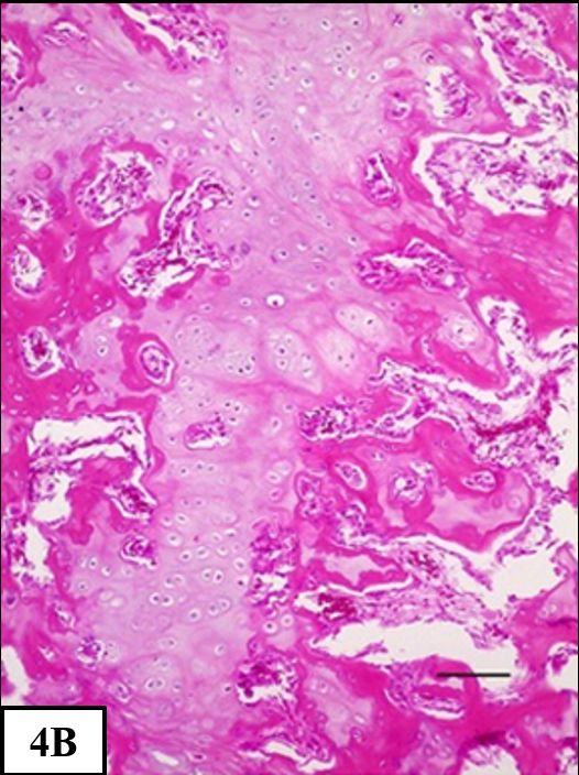
Veterinarni Medicina, 67, 2022 (01): 38–45                                                   Original Paper

https://doi.org/10.17221/85/2020-VETMED

chymal stem cells (AAD-MSC) can be a promising           (FBS; GIBCO, Big Cabin, OK, USA). The mixture
treatment for critical bone defects. However, more       was centrifuged at 290 × g for 10 min and the cell
clinical trials are needed to evaluate the useful-       button was washed in 5 ml of Dulbecco’s modified
ness of these cells in veterinary orthopaedics. The      eagle medium (DMEM; Sigma-Aldrich, St. Louis,
aim of this study was to investigate the use of AAD-     MO, USA) with 10% FBS, the washed cells were
MSC, with and without synthetic absorbable hy-           centrifuged at 290 × g for 5 minutes. The washing
droxyapatite (HAP-91), in the bone regeneration          process was repeated three times. Then, the cell
in rabbits.                                              viability was analysed using the trypan blue percent
                                                         counting method.
                                                           After being isolated, MSC were grown in T25 cul-
MATERIAL AND METHODS                                     ture flasks with DMEM supplemented with 10%
                                                         FBS and incubated in a humid environment at 37 °C
Animals and experimental design                          and in a 5% CO2 atmosphere. The cell culture me-
                                                         dium was changed every 2 days until 80% conflu-
  All the procedures described below were in com-        ence was reached. Once the desired confluence was
pliance with the institutional Animal Care and Use       reached, the cells were trypsinised and transferred
Committee (Protocol No. 007168/15).                      to T75 culture bottles. Again, the culture medi-
  Thirty-four female white New Zealand rab-              um was changed every 2 days until 80% confluence
bits (Oryctolagus cuniculus), 160–170 days old,          was reached. Then the cells were trypsinised and
3.5–4.5 kg, were included in the experiment and          frozen in straws, at a concentration of one million
randomly divided into three groups (G1: n = 13;          cells per straw. The cells were cryopreserved using
G2: n = 10; CG: n = 11), according to the treatment      a cell freezing medium composed of 80% DMEM,
established.                                             10% dimethyl sulfoxide (DMSO; Sigma-Aldrich,
                                                         St Louis, MO, USA) and 10% FBS, and kept frozen
                                                         in liquid nitrogen until use.
Mesenchymal stem cells harvesting                          As a standard procedure for the quality con-
                                                         trol of the mesenchymal stem cell culture, a sam-
  The G1 and G2 animals were premedicated with           ple of the cells was induced to differentiate into
morphine (2 mg/kg), midazolam (2 mg/kg), and             the bone tissue using a StemPro ® Osteogenesis
ketamine (25 mg/kg) (intramuscularly, i.m.). The         Differentiation Kit (Invitrogen, Waltham, MA,
anaesthetic induction and maintenance were un-           USA) in a 12-well plate, with a half change every
dertaken with isoflurane in 100% oxygen, through         two days for approximately 25 days. After differ-
a face mask.                                             entiation, the plate was stained with Alizarin Red
  Initially, 10 g to 25 g of adipose tissue was har-     (Sigma Aldrich, St Louis, MO, USA) to detect the
vested from the fatty sac in the interscapular area      bone matrix through calcium red staining. The
and stored for 2 min in a tube with 50 ml of a sterile   cells were characterised by flow cytometry with
cell washing solution. It was conditioned in a sec-      image quantification and identification of the mo-
ond washing tube for three minutes, and placed           lecular markers. Molecular characterisation tests
in a third tube containing the transport medium.         for the MSC were performed as determined by the
  At the laboratory, the samples were washed             International Society for Cell Therapy (Dominici
three times with a phosphate buffered saline (PBS;       et al. 2006). The surface markers positive for the
Reprodux, Itapira, SP, Brazil), chopped into small       tested MSCs were mouse anti-human CD29-
pieces and placed in contact with a collagenase          RD1 (Beckman Counter, Brea, CA, USA), mouse
0.075% concentration in PBS with a low Ca and            anti-equine CD44-FITC (AbD Serotec, Raleigh,
Mg and hyaluronidase 0.1% (1 mg/ml) solution             NC, USA), caprine anti-canine CD90 primary
so that enzymatic digestion could be carried out.        (Washington State University, Pullman, WA, USA)
For the digestion, the tissue that was in contact        and mouse IgM anti-caprine conjugated AF594
with the digestive solution was incubated at 37 °C       (secondary) (Thermo Fisher Scientific, Waltham,
for 40 min, being homogenised every 5 minutes.           MA, USA). The negative surface marker for the
The enzymatic inactivation was performed with            tested MSCs was mouse anti-human CD34-FITC
PBS supplemented with 10% Faetal Bovine Serum            (Invitrogen, Waltham, MA, USA). These markers

                                                                                                          39
Original Paper                                                Veterinarni Medicina, 67, 2022 (01): 38–45

                                                               https://doi.org/10.17221/85/2020-VETMED

were evaluated by the immunophenotyping method        mAs = 3.2), immediately post-operatively and after
on an Amnis® Imaging Flow Cytometer (Luminex,         15, 30, 45 and 90 days.
Austin, TX, USA). The MSC showed high capacity          Their evaluations were double-blinded made
for adhesion to plastic, phenotypic characteristics   by three different evaluators. The periosteal reac-
typical of MSC and were attached to the bottom        tion, bone callus formation, and bone bridge qual-
of the bottle with a fusiform shape when reaching     ity between fragments were the semi-quantitative
80% confluence. The results of the immunophe-         parameters measured, based on An et al. (1999)
notyping showed that 90% of the cells expressed       and Ozturk et al. (2008).
the three surface undifferentiation markers CD29,
CD44 and CD90 and had low expression of the neg-
ative marker CD34. Still, 96% showed expression       Histological evaluation
of the pluripotency transcription factor SOX2 and
92% of OCT3/4.                                          Randomly, at 45 days, 50% of the animals in each
                                                      group (16 animals) were euthanised and the re-
                                                      maining animals were euthanised at 90 days post-
Surgical induction of segmental bone defect           operatively.
                                                        The radius and ulna were fixed in formaldehyde
  Using the same anaesthetic protocol, a 2.5 cm       for 48 h and decalcified in 7% nitric, daily, for ap-
craniomedial skin incision was made at the end        proximately 2 to 3 days. These were embedded
of the right radius and a 10 mm long ostectomy        in paraffin and cut into a 5 mm microtome, and
was made in the radial diaphysis, 15 mm above the     sequentially stained with haematoxylin and eosin
radiocarpal joint, using an oscillatory saw, in all   and Masson trichrome.
the animals.                                            Under optic microscopy, the neovascularisation,
  In group 1 (G1), two million of AAD-MSC resus-      inflammatory infiltrate, formation of fibrous tis-
pended in 0.15 ml of a 0.9% sterile saline solution   sue, formation of cartilaginous tissue, formation
was applied percutaneously into the bone defect.      of bone tissue, quality of the union and forma-
In group 2 (G2), the defect was filled with HAP-91®   tion of a bone bridge were quantified using scores
(JHS Biomateriais, Sabará, MG, Brazil) followed       (Table 1). The bone architecture and morphology
by the MSC application in the same way. In the con-   of the new bone formation were described.
trol group (CG), 0.15 ml of the 0.9% sterile saline
solution was injected percutaneously. The muscular
and subcutaneous tissue were closed with a contin-    Statistical analysis
uous pattern, and the skin was closed with a simple
interrupted suture, using 4.0 nylon.                    The statistical analyses were performed using
                                                      a commercially available statistical software (Prism
                                                      Windows v5.0; GraphPad Software, CA, USA). The
Clinical evaluation                                   Shapiro-Wilk test was used to investigate the nor-
                                                      mal distribution of the data.
  The animals were observed daily, and the surgi-       Comparisons between the groups and times were
cal wound was checked for oedema or secretions        accomplished by the Kruskal-Wallis test, due to the
on the 3rd and 7th post-operative days, when palpa-   non-parametric data. The statistical significance
tion of the affected limb was performed to identify   was defined as P ≤ 0.05.
pain and a lameness observation was also under-
taken. Scores were attributed to these parameters     Table 1. Scale for the semi-quantititative evaluation
according to Table 1.                                 of the clinical and histological parameters

                                                      Score            Characteristic of the parameter
                                                      1                            absent
Radiographic evaluation
                                                      2                             mild
  Radiographs were made in mediolateral and cra-      3                          moderate
niocaudal projections (mA = 100, KV = 40 and          4                           severe

40
Veterinarni Medicina, 67, 2022 (01): 38–45 Original Paper
https://doi.org/10.17221/85/2020-VETMED
RESULTS At this moment, the lowest oedema score (1.7)
(P < 0.05) was recorded in G1, against 2.7 and 2.5
Regarding the clinical evaluation, the lowest mean in CG and G2, respectively.
score for the pain on palpation (1.6) and for the By the 7th day, all the animals were using the af-
lameness (3.1) of the affected limb was seen in G1 fected limb.
on the 3rd day post-operatively, although this dif- About the radiographic evaluation, the perios-
ference was not statistically significant (P > 0.05). teal reaction was more evident in G2 at 15, 30 and
45 days. At 90 days, the periosteal reaction in G2
Periosteal reaction was similar to G1 and superior to CG (P < 0.05)
3
(Figure 1). The bone callus volume was higher
2.5 in G2 in all of the evaluation time points (Figure 2)
2 and from 30 days, it was higher in G1 than in CG
Mean scores
1.5
CG (P < 0.05). Considering the bone bridge quality, G2
G1
G2 was superior to G1 and G1 was superior to CG at all
1 the evaluation moments (Figures 3–6).
0.5 In the histopathological analysis, at 45 days, 75%
0
of the animals in CG had no bone bridge, and the
Post- 15 days 30 days 45 days 90 days bone defect was predominantly filled by fibrous
operatory
or fibrocartilaginous tissues. Moreover, mild neo-
Figure 1. Development of the mean scores of the radio- vascularisation was present in the bone defect
graphic evaluations of the periosteal reaction in all the region, and there was no inflammatory infiltrate.
groups At 90 days, 80% of the animals in CG showed non-
Bone callus volume union, the bone defect was filled with fibrocarti-
3 laginous tissue and the medullary canal was thin
2.5 and closed (Figure 7A). Only 22% of the rabbits
from CG showed bone healing.
2
Mean scores
CG At 45 days, 100% of the rabbits in G1 showed
1.5 G1 bone healing and a bone bridge with an intense
G2
1 formation of the periosteal trabecular bone, and
0.5 (A) (B) (C) (D) (E)
0
Post- 15 days 30 days 45 days 90 days
operatory
Figure 2. Development of the mean scores of the radio-
graphic evaluation of the bone callus volume in all
the groups
Bone bridge quality
3
2.5
2
Mean scores
1.5 CG
CG 10
G1
1 G2 Figure 4. Radiographic images in the craniocaudal pro-
0.5 jection of the bone reparation process in animal num-
ber 10 of the control group (CG)
0
Post- 15 days 30 days 45 days 90 days (A) Immediate post-operatory, the segmental bone defect
operatory at the distal diaphysis of radius (arrow). (B) 15 days post-
Figure 3. Development of the mean scores of the radio- operatively. (C) 30 days post-operatively. (D) 45 days post-op-
graphic evaluation of the bone bridge quality in all eratively. (E) 90 days post-operatively. The repair process
the groups was insufficient, resulting in a non-union
41Original Paper Veterinarni Medicina, 67, 2022 (01): 38–45
https://doi.org/10.17221/85/2020-VETMED
(A) (B) (C) (D) (A) (B) (C) (D) (E)
G1 10 G2 3
Figure 5. Radiographic images in the craniocaudal pro- Figure 6. Radiographic images in the craniocaudal pro-
jection of the bone healing process in animal number 10 jection of the bone healing process in animal number 3
of the mesenchymal stem cells group (G1) of the mesenchymal stem cells and hydroxyapatite
(A) Radiography at immediately post-operatively of the seg- group (G2)
mental defect (arrow). (B) 15 days post-operatively, intense (A) Radiography immediately post-operatively of the seg-
periosteal reaction at the segmental defect at the distal mental defect at the distal diaphysis of the radius filled
diaphysis of the radius (arrow). (C) 30 days post-operatively, with hydroxyapatite (arrow). (B) 15 days post-operatively,
the bone callus completely filled the bone defect (arrow). moderate periosteal reaction (arrow). (C) 30 days post-
(D) 45 days post-operatively, the defect completely filled operatively, intense periosteal reaction (arrow). (D) 45 days
by a bone callus (arrow). Note the intense periosteal reac- post-operatively, intense periosteal reaction and initial
tion at the extremity of the radius, adjacent to the bone remodelling (arrow). (E) 90 days post-operatively, the bone
defect defect is filled by a bone callus (arrow)
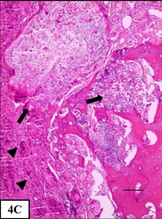
Figure 7
(A) (B) (C)
Figure 7. Histological analysis of the bone segmental defect at the distal diaphysis of radius on a rabbit of each group,
at 90 days
(A) Control group: Prominent bone proliferation and complete non-union [haematoxylin and eosin (H&E), 400 µm]. (B)
G1: Detail of the cartilaginous and bone formation (H&E, 400 µm). (C) G2: Hydroxyapatite deposits (arrow) surrounded
by an intense granulomatous inflammation containing multinucleated giant cells (H&E, 100 µm)
reestablishment of the cortex. At 90 days, 71.4% union quality (Figure 7B). The neovascularisation
of the rabbits in G1 had maximum scores on the was discrete, and there was no inflammatory infil-
histological evaluation of the bone bridge and trate in any animal.
42Veterinarni Medicina, 67, 2022 (01): 38–45 Original Paper
https://doi.org/10.17221/85/2020-VETMED
In G2, 60% of animals had maximum scores ularly in the initial evaluations, so the radiographic
on the histological evaluation of the bone bridge scores may have been overestimated in this group.
and union quality at 45 days, and 100% at 90 days. Synthetic hydroxyapatite is a plastic biomaterial
A periosteal and endosteal intense granulomatous that acts like a scaffold to the neovascularisation,
inflammation and HAP-91 deposits surrounded cellular proliferation, fibrovascular growth, osteoid
by giant cells were present (Figure 7C). The fibrous formation and growth of mineralised bone (Pereira-
tissue formation was moderate to intense in all the Junior et al. 2013). It has osteoconductive proper-
animals from G2, and was statistically significantly ties (Ramesh et al. 2018), and can be combined
superior to that in CG or G1 (P < 0.05). with MSC to promote osteogenic induction (Chi
The treated animals showed better healing et al. 2020). In this study, hydroxyapatite showed
of the bone defect in comparison with that in CG. the characteristics of a good scaffold, promoting
Although the animals in G2 had higher medium an intense periosteal reaction in association with
scores for the bone tissue formation at 45 and AAD-MSC. Nevertheless, the rabbits in G2 devel-
90 days than those in the other groups (P < 0.05), oped an intense inflammatory infiltrate, with foamy
the animals in G1 exhibited a better and more ef- macrophages and giant cells developing around the
fective bone bridge formation and better quality hydroxyapatite deposits, impairing the bone heal-
of the union at 45 days (P < 0.05). The G1 animals ing process. Rabbits have a high tendency towards
showed intense formation of the periosteal trabecu- excessive granulation tissue against foreign materi-
lar bone tissue, decreased cartilaginous tissue and als, which could explain the reaction seen in our
better structural and morphological organisation study (Rich 2002).
at 45 days than the G2 animals. G1 showed the lowest score of all the groups for
the post-operative oedema, probably due to the an-
ti-inflammatory effects of MSC. PGE2, one of the
DISCUSSION mainly soluble factors secreted by MSC, is a central
mediator of the immunomodulatory response over
Many studies have demonstrated promising re- other immune cells. Among its actions, it has anti-
sults for improvements in bone healing using cel- proliferative effects on T lymphocytes and NKs
lular therapy associated with biomaterials (Tajima (natural killer cells), and modulates the release
et al. 2015; Venkatesan et al. 2016; Volkov et al. of inflammatory substances from dendritic cells
2020). Our results provide strong evidence of the and macrophages (Martinet et al. 2009). However,
beneficial effects of MSC in bone neoformation we hypothesise that the oedema reduction was not
in segmental defects in the distal radial diaphysis. seen in G2 due to granulomatous reaction induced
The radiographic and histological evaluations in- by HAP-91.
dicate that the action of MSC, alone or associated One study described the beneficial effects of ad-
with HAP-91, allowed 31% of the G1 rabbits and ipose-derived MSC on the neovascularisation
20% of the G2 animals to complete bone healing, of skin lesions, secondary to the secretion of angio-
in a critical size defect. Moreover, the rabbits in the genic cytokines [vascular endothelial growth fac-
G1 and G2 that failed to show complete healing, tor (VEGF) and hepatocyte growth factor (HGF)]
still had a periosteal reaction and trabecular bone (Nauta et al. 2013). Moreover, many studies report
formation clearly superior than those in CG. the formation of vessels promoted by VEGF dur-
Radiographically, the healing in G2 was superior, ing bone healing and remodelling (Stegen et al.
while the histological healing parameters of the 2015). In spite of the potential benefits, there was
bone tissue formation, bone bridge formation and no statistical difference in the neovascularisation
quality of the union were better in G1 at 45 days. between the groups. It is possible that the histologi-
Additionally, G1 showed faster bone healing in com- cal analysis was not able to quantify the neovascu-
parison to G2, in which there was a high score larisation due to the low density of blood vessels
of fibrous tissue formation interspersed with bone and discreet coverage of soft tissues on the distal
tissue. The radiographic evaluation of G2 was dif- radial diaphysis. Immunohistochemistry would
ficult due to the radiopacity of the hydroxyapatite, be a more appropriate method for this purpose.
which made it difficult to differentiate the pellets In this study, the used AAD-MSCs were isolated
from the periosteal reaction or a bone callus, partic- from the adipose tissue of each animal in a prior
43Original Paper Veterinarni Medicina, 67, 2022 (01): 38–45
https://doi.org/10.17221/85/2020-VETMED
procedure, featuring an autologous cell therapy; de Girolamo L, Arrigoni E, Stanco D, Lopa S, Di Gianca-
however heterologous MSCs have been used in vet- millo A, Addis A, Borgonovo S, Dellavia C, Domeneghini C,
erinary practice as they are rapidly available, in a fast Brini AT. Role of autologous rabbit adipose-derived stem
and safe way, and can be stored in a cell bank frozen cells in the early phases of the repairing process of criti-
in liquid nitrogen. The MSCs express major class I cal bone defects. J Orthop Res. 2011 Jan;29(1):100-8.
histocompatibility complex (MHC I) molecules, Dominici M, Le Blanc K, Mueller I, Slaper-Cortenbach I,
but do not express class II histocompatibility com- Marini FC, Krause DS, Deans RJ, Keating A, Prockop DJ,
plexes (MHC II), thus, they are unable to produce Horwitz EM. Minimal criteria for defining multipotent
alloreactivity in mammals (Aggarwal and Pittenger mesenchymal stromal cells. The International Society for
2005), which is important because it prevents the Cellular Therapy position statement. Cytotherapy. 2006
cell’s rejection. Jan 1;8(4):315-7.
A limitation of the present study was the spe- Ho-Shui-Ling A, Bolander J, Rustom LE, Johnson AW,
cies included, since clinically, the target species are Luyten FP, Picart C. Bone regeneration strategies: Engi-
dogs, cats and even humans. neered scaffolds, bioactive molecules and stem cells cur-
Meanwhile, the rabbit is a widely used experi- rent stage and future perspectives. Biomaterials. 2018 Oct;
mental animal model for the study of bone patho- 180:143-62.
physiology and bone healing processes (Pearce Li H, Shen S, Fu H, Wang Z, Li X, Sui X, Yuan M, Liu S,
et al. 2007). Wang G, Guo Q. Immunomodulatory functions of mes-
Further studies are necessary to elucidate the ac- enchymal stem cells in tissue engineering. Stem Cells Int.
tions of MSC, especially about the stimuli that lead 2019 Jan 13;2019:1-18.
to their differentiation, which could allow the better Martinet L, Fleury-Cappellesso S, Gadelorge M, Dietrich G,
use of these cells in tissue regeneration. Bourin P, Fournie JJ, Poupot R. A regulatory cross-talk
In conclusion, the use of AAD-MSC is beneficial between Vγ9Vδ2 T lymphocytes and mesenchymal stem
to bone healing in critical size segmental bone de- cells. Eur J Immunol. 2009 Mar;39(3):752-62.
fects in the distal radial diaphysis in rabbits. The Naji A, Eitoku M, Favier B, Deschaseaux F, Rouas-Freiss N,
association of MSC with HAP-91 produced an in- Suganuma N. Biological functions of mesenchymal stem
tense granulomatous reaction in this species that cells and clinical implications. Cell Mol Life Sci. 2019
delayed the healing process, although G2 was the Sep;76(17):3323-48.
only group where there were no cases of non-union Nauta A, Seidel C, Deveza L, Montoro D, Grova M, Ko SH,
and more homogeneous results were observed be- Hyun J, Gurtner GC, Longaker MT, Yang F. Adipose-
tween the animals. derived stromal cells overexpressing vascular endothelial
growth factor accelerate mouse excisional wound healing.
Mol Ther. 2013 Feb;21(2):445-55.
Conflict of interest Ozturk A, Ilman AA, Saglam H, Yalcinkaya U, Aykut S, Ak-
goz S, Ozkan Y, Yanik K, Kivcak B, Yalcin N, Ozdemir
The authors declare no conflict of interest. RM. The effects of phytoestrogens on fracture healing:
Experimental research in New Zealand white rabbits.
Ulus Travma Acil Cerrahi Derg. 2008 Jan;14(1):21-7.
REFERENCES Pearce AI, Richards RG, Milz S, Schneider E, Pearce SG.
Animal models for implant biomaterial research in bone:
Aggarwal S, Pittenger MF. Human mesenchymal stem cells A review. Eur Cell Mater. 2007 Mar 2;13:1-10.
modulate allogeneic immune cell responses. Blood. 2005 Pereira-Junior OCM, Rahal SC, Lima-Neto JF, Landim-
Feb 15;105(4):1815-22. Alvarenga FC, Monteiro FOB. In vitro evaluation of three
An YH, Friedman RJ, Draughn RA. Animal models of bone different biomaterials as scaffolds for canine mesenchy-
fracture or osteotomy. In: An YH, Friedman RJ, editors. mal stem cells. Acta Cir Bras. 2013 May;28(5):353-60.
Animal models in orthopaedic research. Boca Raton: CRC Ramesh N, Moratti SC, Dias GJ. Hydroxyapatite-polymer
Press; 1999. p. 197-217. biocomposites for bone regeneration: A review of current
Chi H, Chen G, He Y, Chen G, Tu H, Liu X, Yan J, Wang X. trends. J Biomed Mater Res B Appl Biomater. 2018
3D-HA scaffold functionalized by extracellular matrix Jul;106(5):2046-57.
of stem cells protomotes bone repair. Int J Nanomedicine. Rich GA. Rabbit orthopedic surgery. Vet Clin North Am
2020 Aug 6;15:5825-38. Exot Anim Pract. 2002 Jan 1;5(1):157-68.
44Veterinarni Medicina, 67, 2022 (01): 38–45 Original Paper
https://doi.org/10.17221/85/2020-VETMED
Stegen S, van Gastel N, Carmeliet G. Bringing new life Voga M, Adamic N, Vengust M, Majdic G. Stem cells in vet-
to damaged bone: The importance of angiogenesis in bone erinary medicine – Current state and treatment options.
repair and regeneration. Bone. 2015 Jan;70:19-27. Front Vet Sci. 2020 May 29;7:1-20.
Tajima S, Tobita M, Orbay H, Hyakusoku H, Mizuno H. Volkov AV, Muraev AA, Zharkova II, Voinova VV, Akoulina
Direct and indirect effects of a combination of adipose- EA, Zhuikov VA, Khaydapova DD, Chesnokova DV, Men-
derived stem cells and platelet-rich plasma on bone re- shikh KA, Dundun AA, Makhina TK, Bonartseva GA,
generation. Tissue Eng Part A. 2015 Mar;21(5-6):895- Asfarov TF, Stamboliev IA, Gazhva YV, Ryabova VM,
905. Zlatev LH, Ivanov SY, Bonartsev AP. Poly(3-hydroxybu-
Venkatesan J, Lowe B, Anil S, Kim SK, Shim MS. Combina- tyrate)/hydroxyapatite/alginate scaffolds seeded with
tion of nano-hydro xyapatite with stem cells for bone mesenchymal stem cells enhance the regeneration
tissue engineering. J Nanosci Nanotechno. 2016 Sep;16(9): of critical-sized bone defect. Mater Sci Eng C. 2020 Sep;
8881-94. 114:1-14.
Received: April 9, 2020
Accepted: August 2, 2021
45You can also read