Changes in glaucoma in dogs and cats
←
→
Page content transcription
If your browser does not render page correctly, please read the page content below
E3S Web of Conferences 273, 02028 (2021)                                   https://doi.org/10.1051/e3sconf/202127302028
INTERAGROMASH 2021

         Glaucoma       incidence.    Morphofunctional
         changes in glaucoma in dogs and cats
         Maria Oboeva1*, Sergey Kartashov1, Anna Fomina1, and Alexandr Butenkov2
         1
          Federal State Budget Educational Institution of Higher Education “Don State Technical University”,
         344000, Rostov-on-Don, Russian Federation
         2
          VITA Veterinary Clinic, Rostov-on-Don, Russian Federation

                        Abstract. The article analyzes the data of case histories of dogs and cats
                        for the period from September 2018 to the end of December 2020 with
                        increased intraocular pressure in the Rostov region. Changes in eye
                        structures, causes of glaucoma, research methods, problems of diagnosis
                        and treatment of diseases are described. Particular attention is paid to the
                        consideration of the complications of pathology, as well as the methods of
                        their prevention.

         1 Introduction
         Glaucoma – group of heavy progressive neurodegenerative diseases, characterized by
         increased intraocular pressure, damage to the retina and optic nerve [1,2,3]. Is one of the
         leading causes of loss of visual function in dogs and cats [2]. The disease can be both
         primary and secondary, developing because of other pathologies. In addition, the scientific
         literature describes cases of retinal detachment and the development of glaucoma after
         phacoemulsification in dogs [4,5]. These postoperative complications are most often
         recorded in the Cocker Spaniel (11.7%), Boston Terrier (10.5%), Labrador Retriever
         (6.5%), Bichon Frize (6.5%), Shih Tzu (5.8%) and Jack Russell Terrier (5.8%) [5].
             In humane medicine, changes in the structures of the eye, detected at increased
         intraocular pressure, have been studied in sufficient detail, descriptions of the fundus and
         early markers of the disease are given [6,7,8]. However, literary sources lack data on the
         manifestations of pathology in small domestic animals. According to these data, the clinical
         diagnosis in cats is made much less frequently than in dogs. A rare occurrence - 0.3% of the
         total number of diseases in cats, is most likely associated with a small number of examined
         by an ophthalmologist. In more than half of cases, neoplasia and about a quarter of anterior
         uveitis are described as the cause of glaucoma in cats [9].
             The aim of the current study was to study the prevalence of ophthalmic pathologies
         among the total number of patients, as well as to determine the proportion of glaucoma
         among diseases of the organ of vision and the auxiliary apparatus of the eye in the Rostov
         region. In addition, we analyzed the complaints that the owners of such animals applied to

         *
             Corresponding author: drama94@mail.ru

   © The Authors, published by EDP Sciences. This is an open access article distributed under the terms of the Creative
   Commons Attribution License 4.0 (http://creativecommons.org/licenses/by/4.0/).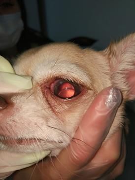
E3S Web of Conferences 273, 02028 (2021) https://doi.org/10.1051/e3sconf/202127302028
INTERAGROMASH 2021
the clinic, morphofunctional changes detected by a specialist during examination, etiology,
breed predisposition. Since this pathology is not an endemic disease, the analysis of
incident statistics using the example of one clinic can characterize the state of the problem
as a whole.
2 Materials and methods
The study was carried out at the Vita veterinary clinic in Rostov-on-Don. In addition to the
owners' self-referral to the clinic, patients for ophthalmological examination were sent from
third-party clinics and other branches of the Rostov region. The Viasna database was used
to analyze the case histories. Clinical cases were selected by the keywords "glaucoma" and
"glaucomatous uveitis" among the total mass of patients for the period from September
2018 to the end of December 2020. To make a diagnosis, all animals underwent a clinical
and ophthalmological examination, a series of sequential measurements of intraocular
pressure (IOP) with a TonoVet tonometer. In the process of tonometry, the position of the
patient with a straight head, the absence of pressure on the neck and head area by the
assistant, and the correct application of six touches with the probe to the middle of the
cornea are important (the angle between them should be 90º). There were 5 such
measurements on each eye, the results were recorded and the arithmetic mean value was
chosen. The study of the anterior segment of the eye and the auxiliary apparatus was
performed using a binocular loupe and a ShinNipponXL1 slit lamp. The assessment of
visual function was based on the threat test and the maze test. Comorbidities were detected
by assessing tear production with test strips Tearstrips (Contacare), fluorescin (Fluostrips
test strips (Contacare)) and LissamineGreen (Contacare) lissamine tests. During the initial
examination and the entire observation period, chromatic pupillomotor reactions,
ultrasound examination of the eye were also assessed (MindrayDC-n6). Ophthalmoscopy
was performed using an OptomedSmartscope m-5 fundus camera and a
PanOpticWelchAllyn direct ophthalmoscope.
3 Results
During the study period, 164,973 patients with various diseases applied to the clinic, as well
as for preventive care, including medical examination and vaccination, sent animals for
research from third-party clinics. Of these, 413 were at the veterinary ophthalmologist with
complaints of pathology of the organ of vision or on a routine examination. Increased IOP
was noted in 35 subjects. Thus, the incidence of increased IOP was 8.47% of the total
number of visits (only primary appointments were taken into account). 60% with
ophthalmic hypertension were cats (21 cases) and 40% dogs (14 cases).
The most frequent complaints during treatment were lacrimation, blepharospasm,
changes in the transparency of the cornea, periodically "reddening" eyes, depression of the
animal. In the terminal stage, the owners treated with an enlargement of the animal's
eyeball in size (Fig. 1), blindness in bilateral lesions. In 11.4%, increased IOP values were
revealed during a routine examination, allowing to identify pathology at an early stage and
intervene in time. At the same time, the owners did not notice any signs of pathology.When
the animals were examined by an ophthalmologist, in addition to the above signs,
symptoms of anterior uveitis were detected - opacification of the intraocular fluid (IHF),
precipitates on the corneal endothelium, eversion of the pigment layer of the iris and
chorioretinitis, rupture of descemet's membrane and penetration of the intraocular
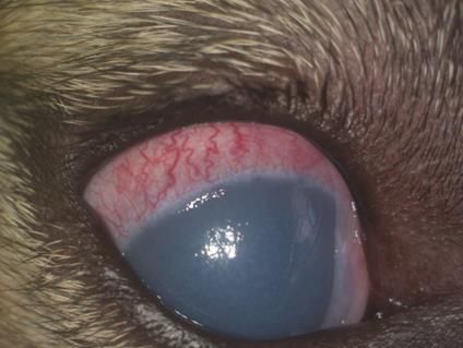
membrane and in the corneal stroma - Gaab's stripes (Fig. 2), congestive episcleral vessels
(Fig. 3). Clinical signs in terms of frequency of occurrence are presented in Table 1.
2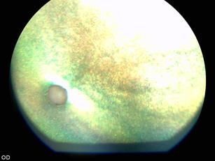
E3S Web of Conferences 273, 02028 (2021)              https://doi.org/10.1051/e3sconf/202127302028
INTERAGROMASH 2021

         Fig. 1. Buphthalmus (right eye).

         Fig. 2. Gaab stripes.

         Fig. 3. Congestive episcleral vessels.

                                                  3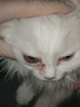
E3S Web of Conferences 273, 02028 (2021)                                https://doi.org/10.1051/e3sconf/202127302028
INTERAGROMASH 2021

                                 Table 1. Frequency of occurrence of clinical signs.
                                               dogs                                    cats
                                                                                                 % of all
                                                    % of all clinical
             clinical signs     Number of cases                         Number of cases       clinical cases
                                                     cases in dogs
                                                                                                  in cats
           fundus vascular
                                       1                  2,8                   4                 11,4
              attenuation
          optic disc atrophy
                                       1                  2,8                   3                  8,5
                (ODA)
          retinal detachment           3                  8,5                   5                 14,3
               blindness               4                  11,4                  5                 14,3
           lens subluxation            1                  2,8                   3                 8,5
             lens luxation             1                  2,8                   -
             buphthalmus               4                  11,4                  5                 14,3
           hemophthalmos               3                  8,5                   3                 8,5
            chorioretinitis            -                   -                    3                 8,5
              Gaab stripes             3                  8,5                   1                 2,8
               synechiae               2                  5,7                   2                 5,7
                cataract               4                  11,4                  4                 11,4
              congestive
                                       3                  8,5                   1                  2,8
           episcleral vessels

          endothelial corneal
                                       2                  5,7                   2                  5,7
                edema
                corneal
                                       1                  2,8                   -                   -
            vascularization
                 Total                 14                                      21

             It was possible to establish the cause of the increased intraocular pressure only in 14
         patients. In other cases, it was not possible to objectively determine the root cause due to
         various factors. Some of the animals (8.5%) were picked up by volunteers on the street and
         taken to the clinic for examination. Other reasons for the unknown anamnesis were: free-
         range keeping of animals and lack of information from the owners, initial referral to an
         ophthalmologist already in the terminal stage of the disease, unwillingness of the owners to
         examine and treat their animal for various reasons.
             The most numerous group in dogs and cats was post-traumatic glaucoma in 42% of
         cases. Injuries to the head and, in particular, the eyeball and periocular tissues are one of the
         most common reasons for contacting a veterinary ophthalmologist. These include
         penetrating wounds of the cornea, bitten wounds for the periorbital region, closed injuries
         of the eyeball as a result of autoinjuries, falls from a height or blows with blunt objects.
         Penetrating wounds with a cat's claw, branches and other sharp objects often cause damage
         to intraocular structures with rupture of the lens capsule, damage to the iris and, as a result,
         the formation of anterior and posterior synechia. In blunt trauma, the causes of increased
         intraocular pressure may be hemorrhages in the anterior chamber with a block of the
         iridocorneal angle, hemophthalmus, partial or complete separation of the lens from the zinn
         ligaments.
             The second most common group was postuveal glaucoma as an emerging complication
         of anterior uveitis. In some cases, such a problem arose against the background of the
         development of acute or exacerbation of chronic iridocyclitis, and in others it was a distant
         result of post-inflammation of the choroid. In 3 cases in cats with glaucoma, in addition to
         anterior uveitis, we observed signs of chorioretinitis. All were subsequently confirmed to

                                                         4
E3S Web of Conferences 273, 02028 (2021)                              https://doi.org/10.1051/e3sconf/202127302028
INTERAGROMASH 2021

         have diseases such as leukemia, immunodeficiency, coronavirus infection or toxoplasmosis
         in various combinations.
             In 8.5% of the subjects, acute ophthalmic hypertension was recorded, which was
         stopped by medication, after which it was possible to gradually discontinue the drugs under
         the control of tonometry. Subsequently, no clinical manifestations were identified. Further
         observation of the animals from 3 to 18 months did not lead to the detection of a relapse.
             Phakotopic glaucoma was observed in two cases in Chinese Crested and Chihuahua
         dogs. In the first case, a primary dislocation of the lens of the eye is likely, since there is a
         breed predisposition to this pathology. Secondary glaucoma developed due to block of the
         iridocorneal angle, and as a result of late seeking help in this dog, we observed endothelial
         corneal edema and subtotal retinal detachment detected by ultrasound. However, the
         luxation of the lens into the anterior chamber is possible in this animal as a consequence of
         the development of cataracts, since examination with a slit lamp in the second eye revealed
         initial cataract changes. The intraocular pressure was within the normal range. The dog's
         owner was advised to enucleate the eye with buphthalmus and carry out
         phacoemulsification on the other side, without waiting for complications. In the case of the
         Chihuahua, the initial examination revealed signs of anterior uveita-unilateral (deep scleral
         hyperemia according to the results of the test with irifrin, slight eversion of the pigment
         layer of the iris). At the same time, IOP values were borderline - 10 mm Hg. A therapeutic
         treatment was prescribed and re-admission after 3 days. Despite the recommendations, the
         owner only asked for help after 2 weeks. On examination, we observed subluxation of the
         lens in the PT (Fig. 4), increased IOP - 54 mm Hg. It was recommended to remove the lens,
         but the further fate of the patient is unknown to us.

         Fig. 4. Lens subluxation in CT in Chihuahua.
             Two dogs developed glaucoma as a consequence of overripe cataracts. One 24-year-old
         cat had neoplastic glaucoma that underwent exenteration.
             The distribution of the frequency of occurrence of increased IOP among breeds is
         presented in Table 2.

                                                        5
E3S Web of Conferences 273, 02028 (2021) https://doi.org/10.1051/e3sconf/202127302028
INTERAGROMASH 2021
Table 2. Frequency of occurrence by breed.
dogs cats
Numb
Number of
breed % breed er of %
cases
cases
Shih tzu 1 7 Scottish lop-eared 1 4,8
Chinese Crested 2 14 Angora 2 9,5
Yorkshire Terrier 1 7 Oriental 1 4,8
American staffordshire terrier 1 7 Siamese 1 4,8
Chihuahua 3 21 British 1 4,8
Dachshund 1 7 Mestizo 15 71
French Bulldog 1 7
Tibetan terrier 1 7
Jack Russell Terrier 1 14
Mestizo 2 7
Total 14 100 21 100
3.1 Ophthalmoscopy
Examination of the fundus (FD) is an integral part of an ophthalmological examination,
including for a patient with increased intraocular pressure, whenever possible. Preserved
transparency of the media and contraindications to drug mydriasis allowed ophthalmoscopy
to be performed in all patients only with the help of a fundus camera. In the case of intact
transparency and patients who came with mydriasis, it was additionally possible to examine
the fundus of the eye with a direct ophthalmoscope. Characteristic changes at an early stage
were attenuation of the fundus vessels of varying severity, expansion of the excavation of
the optic nerve head. At more advanced stages, in most animals, we noted atrophy of the
optic nerve disc with a characteristic pale gray color, the absence of peripheral retinal
vessels and thinning of the central ones, and in some animals, the complete disappearance
of retinal vessels (Fig. 5). The study revealed signs of chorioretinitis in 3 cats, retinal
detachment (local or subtotal, total) in two cats and two dogs. In other cases, the diagnosis
of retinal detachment was made after an ultrasound scan of the eye, since ophthalmoscopy
was impossible due to the opacity of the eye media for various reasons.
Fig. 5. The fundus of a blind cat with glaucoma. Complete absence of retinal vessels, optic disc
atrophy.
3.2 Ultrasound examination of the eye
This study is a minimally invasive and safe method for diagnosing ophthalmic pathologies.
And although ultrasound of the eye was performed in all patients who consulted an
6E3S Web of Conferences 273, 02028 (2021) https://doi.org/10.1051/e3sconf/202127302028
INTERAGROMASH 2021
ophthalmologist, it played a special role in violation of the transparency of the eye media
(edema and opacity of the cornea, mature cataract, hemophthalmus, etc.), making it
possible to assess the thickness of the cornea, the size and contents of the anterior chamber
of the eye (ACE), position, size and transparency of the lens, the contents of the vitreous
body (VB), the state of the retina, the anterior-posterior size of the eyeball. In patients with
end-stage glaucoma, we observed an increase in the size of the eye, an increase in the
anterior chamber. A frequent finding was partial, subtotal (with attachment at the optic
nerve head and dentate line), and total retinal detachment. In traumatic damage to the
eyeball, changes in echogenicity were visualized in hemophthalmos (hyphema,
hemorrhages in the ST), and ruptures of the anterior lens capsule. Lens dislocation, in
addition to examination, was also confirmed using the ultrasound method (Fig. 6).
Fig. 6. Ultrasound examination of a dog's eye with lucation of the cataract lens in the PCH, increased
PCH, corneal edema and buphthalmos.
3.3 Visual function
In 26% of the subjects, visual function was irretrievably lost due to late seeking help and
the development of serious complications. At the same time, in about half of the cases, the
owners did not suspect decreased vision or blindness in their pet (only cases with bilateral
pathology or with a single preserved eye were taken into account). Basically, this fact was
recorded among cats, since they were well oriented in their usual home environment,
especially if the decrease in visual function progressed gradually. Some owners only
noticed dilated pupils, but some did not attach importance to this fact either. In the
advanced stages of the disease, we observed no positive effect on the therapy, and in some
cases we immediately recommended the owners to enucleate or exenterate.
4 Discussion
Among the studied group of dogs, no breed predisposition was revealed, which may be
associated with a small sample. The vast majority of ophthalmic hypertensive cats were
mestizo males. This fact, in our opinion, is associated with the fact that among this group
there are more often cats with free-range content, not castrated. According to statistics, it is
these individuals who are more likely to receive injuries, including the eyeball, and the
prevalence of infections among the population is also higher. Such factors can lead to
iridocyclitis, damage to intraocular structures, complicate the closure of drainage tracts,
pupillary block and cause ophthalmic hypertension. In addition, a specialist is often
consulted several days, weeks, or even months after the injury. such animals often
7E3S Web of Conferences 273, 02028 (2021) https://doi.org/10.1051/e3sconf/202127302028
INTERAGROMASH 2021
disappear from home for a long time. Another reason for late visits is the inattentive
attitude of the owners to their animals, the lack of annual preventive examinations by the
veterinarian, the unwillingness of people to examine the pets even with obvious signs of
pathology. In such cases, it can be difficult to help the animal preserve its vision, to
determine the root cause and prognosis. A high percentage of cases with an unknown
history and unknown fate, even after the diagnosis and clarification to the owner about the
problem and the methods of treatment, shows their irresponsible attitude.
A serious problem, according to which iridocyclitis is complicated by glaucoma in the
long-term period, is the inaccessibility of ophthalmological care in many districts of the
Rostov region. Most clinics do not have the availability of specialized equipment and
narrow-profile specialists, thus leading to diagnostic errors. An examination with the naked
eye is often insufficient to detect initial anterior uveitis. In addition to measuring intraocular
pressure, patients require biomicroscopy, ophthalmoscopy, ultrasound, and, if necessary,
additional research methods. Due to inflammation of the ciliary body and hypotension, the
obtained tonometry results can be interpreted incorrectly. In addition, in such a situation,
the IOP values can be borderline and fluctuate during the day, thus, without arousing
suspicion from the doctor.
To assess the effectiveness of the prescribed therapy, it is imperative to monitor IOP
according to an individual plan. Short intervals between tonometry have to be done at first,
while the frequency of use of certain drugs is being determined, the need to add new ones is
being considered. The lack of effect on drug treatment is an indication for surgical
intervention. In an acute attack of glaucoma, it is often necessary to resort to invasive
methods of lowering IOP as an emergency measure, since high ophthalmotonus always
leads to the development of complications. An acute attack of ophthalmic hypertension
requires immediate medical intervention, careful monitoring of the patient in the future and
timely correction of IOP. High IOP values within 24-48 hours lead to irreversible loss of
vision.
A high percentage of owners who do not notice the blindness of their pet proves that in
a familiar environment, animals may not demonstrate a decrease in visual function, even in
its complete absence. To establish this fact, a specialist examination and special tests and
studies are required.
5 Output
This study showed that glaucoma is a common disease among animals in the Rostov region,
but at the moment it is not possible to identify the breed predisposition. An analysis of the
statistics of clinical signs made it possible to understand what manifestations of the disease
the owners pay attention to and what the doctor finds during a thorough examination.
Thanks to the study, it became clear that the most common secondary ophthalmic
hypertension occurs in the Rostov region as a result of traumatic injury and a complication
of anterior uveitis. Based on the incidence of complications of glaucoma, it can be
concluded that more often the owners of the patients apply already at the later stages of the
disease. This fact becomes a serious problem in understanding the etiological factor, makes
it difficult, and sometimes does not give the opportunity to help the animal. Preventive
examinations help in many cases to suspect a problem, to make a diagnosis at an early stage
and thereby increase the chances of successful treatment.
8E3S Web of Conferences 273, 02028 (2021) https://doi.org/10.1051/e3sconf/202127302028
INTERAGROMASH 2021
References
1. G. J. McLellan, P. E. Miller, Vet. Ophthalmol., 14, 15–29 (2011), doi: 10.1111/j.1463-
5224.2011.00912.x.
2. G. Jutley, S. M. Luk, M. H. Dehabadi, et al., Neurodegener Dis. Manag., 7, 157-172
(2017)
3. R. W. Nickells, G. R. Howell, I. Soto, S. W. John, Annu. Rev. Neurosci., 35, 153–179,
(2012), [PubMed] [Google Scholar]
4. B. C. Foote, S. L. Pederson, A. Welihozkiy, et al., Vet. Ophthalmol., 21, 240-248,
(2018)
5. E. M. Scott, D. W. Esson, K. J. Fritz, et al., Vet. Ophthalmol., 16(Suppl 1), 64-72,
(2013)
6. R. N. Weinreb, T. Aung, F. A. Medeiros, J. Am. Med. Assoc., 311, 1901-1911 (2014)
7. J. Dietze, K. Blair, S. J. Havens, Glaucoma (StatPearls Publishing, 2020)
8. A. K. Schuster, C. Erb, E. M. Hoffmann, T. Dietlein, and N. Pfeiffer, DtschArztebl
Int., 117(13), 225–234 (2020)
9. G. J. McLellan, L. B. C. Teixeira, Veterinary Clinics of North America: Small Animal
Practice, 45(6), 1307–1333 (2015)
10. T. Hasegawa, M. Kawata, M. Ota, J. Vet. Med. Sci. 77, 1625-1631 (2016)
11. A. M. Komaromy, S. M. Petersen-Jones, Vet. Clin. North Am. Small Anim. Pract., 45,
1159-1182 (2015)
12. Y. C. Tham, X. Li, T. Y. Wong, et al., Ophthalmology, 121, 2081-2090 (2014)
13. C. Cook, P. Foster, Can. J. Ophthalmol., 47(3), 223-6 (2012)
14. C. Wright, M. A. Tawfik, M. Waisbourd, L. J. Katz, Acta Ophthalmol., 94(3), 217-25
(2016), [PubMed]
15. F. Maggio, Top Companion Anim. Med., 30(3), 86-96 (2015), doi:
10.1053/j.tcam.2015.07.011
9You can also read