Characterisation of peri-implantation endometrial Treg and identification of an altered phenotype in recurrent pregnancy loss
←
→
Page content transcription
If your browser does not render page correctly, please read the page content below

www.nature.com/mi

ARTICLE                 OPEN

Characterisation of peri-implantation endometrial Treg
and identification of an altered phenotype in recurrent
pregnancy loss
Ingrid Granne1, Mengni Shen1, Helena Rodriguez-Caro1, Gurmeher Chadha1, Elizabeth O’Donnell1, Jan J. Brosens2,3,
                                                             ✉
Siobhan Quenby2,3, Tim Child1,4 and Jennifer H. Southcombe 1

© The Author(s) 2021

    Recurrent Pregnancy Loss (RPL) affects 2–4% of couples, and with increasing numbers of pregnancy losses the risk of miscarrying a
    euploid pregnancy is increased, suggesting RPL is a pathology distinct from sporadic miscarriage that is due largely to lethal
    embryonic aneuploidy. There are a number of conditions associated with RPL including unspecified “immune” pathologies; one of
    the strongest candidates for dysregulation remains T regulatory cells as depletion in the very early stages of pregnancy in mice
    leads to pregnancy loss. Human endometrial Treg and conventional CD4T cells were isolated during the peri-implantation period of
    the menstrual cycle in normal women. We identified an endometrial Treg transcriptomic signature and validated an enhanced
    regulatory phenotype compared to peripheral blood Treg. Parous women had an altered endometrial Treg transcriptome
    compared to nulliparity, indicating acquired immune memory of pregnancy within the Treg population, by comparison endometrial
    conventional CD4T cells were not altered. We compared primary and secondary RPL to nulliparous or parous controls respectively.
    Both RPL subgroups displayed differentially expressed Treg gene transcriptomes compared to controls. We found increased cell
    surface S1PR1 and decreased TIGIT protein expression by Treg in primary RPL, confirming the presence of altered Treg in the peri-
    implantation RPL endometrium.

    Mucosal Immunology (2022) 15:120–129; https://doi.org/10.1038/s41385-021-00451-1

INTRODUCTION                                                                             NK cell densities are associated with RPL,5 and CD8T cells are
The female reproductive tract (FRT) is a mucosal barrier tissue                       also known to be phenotypically altered.7 The CD4T cell
and like other mucosa, immune cells infiltrate the stromal layers                      compartment (consisting of Th1, Th2, Th17 and Treg defined
adjacent to the epithelial surface. Throughout the hormonally                         by cytokine production) has not been extensively studied. Of
controlled menstrual cycle host immunity provides protection                          specific interest are the Treg cells, which in mice have been
from pathogens that may enter the FRT, however when an                                show to prime tolerance, as depletion during early stages of
embryo implants into the endometrium, the immune system is                            pregnancy during embryonic implantation leads to pregnancy
temporarily modified to permit attachment and invasion of                              loss.8 Murine Treg cells are also pivotal to tolerance of paternal
trophoblast cells from the developing conceptus. At the time of                       alloantigen.9 The transcription factor FOXP3 confers regulatory
embryonic implantation NK, T, B and macrophage cells are                              lineage commitment to Tregs, multiple mechanisms of action
present in the human endometrium.1 Sporadic pregnancy loss is                         are known such as high CD25 expression which consumes IL-2
most commonly caused by embryonic aneuploidy, but with                                preventing effector T cell functions, CTLA4 driven inhibition of
increasing numbers of pregnancy losses the risk of losing a                           antigen presenting cells, and inhibitory cytokine production (IL-
euploid pregnancy is increased. Recurrent Pregnancy Loss (RPL)                        10, TGF-beta and IL-35).10 Decreased numbers of FOXP3+ cells
is the consecutive loss of two or more pregnancies before                             are found in endometrium of patients with RPL compared to
24 weeks’ gestation,2,3 and can affect nulliparous (primary RPL)                      fertile controls,11 however in humans FOXP3 can also be
or parous women (secondary RPL). RPL is partly associated with                        expressed by activated T cells and other cells12 and the
genetic variation,4 uterine anomalies, endocrine dysfunction,                         phenotype and functional nature of these cells has not been
parental balanced chromosomal translocation and specific                               clearly elucidated.
maternal autoantibodies,5 however these associated clinical                              In this study human endometrium was collected during the
factors are identified in fewer than 50% of cases. The maternal                        peri-implantation period of the menstrual cycle, from normal
immune system is implicated in some cases of recurrent                                fertile women and from women with RPL, with the aim of
pregnancy loss.6                                                                      identifying if Treg are defective in tolerance mechanisms in

1
 Nuffield Department of Women’s and Reproductive Health, L3 Women’s Centre, John Radcliffe Hospital, University of Oxford, Oxford, UK. 2Division of Biomedical Sciences,
Warwick Medical School, University of Warwick, Coventry CV2 2DX, UK. 3Tommy’s National Centre for Miscarriage Research, University Hospitals Coventry & Warwickshire NHS
Trust, Coventry CV2 2DX, UK. 4Oxford Fertility, The Fertility Partnership, Oxford OX4 2HW, UK. ✉email: Jen.southcombe@wrh.ox.ac.uk

Received: 19 April 2021 Revised: 31 August 2021 Accepted: 31 August 2021
Published online: 22 September 2021
I. Granne et al.
                                                                                                                                                            121
                              a

                                                                                                                                   p
                                                                                                                                   –
                                                                                                                                       –   –

                              b                            c                  d                             e
1234567890();,:

                              f                                               g

                                                                              h

                  women with RPL, leading to pregnancy loss. The methods applied         multiparous women to determine if there is a memory to
                  in this study are detailed in Fig. 1a. The primary aim of this study   pregnancy in one or both of these adaptive immune cell
                  was to characterize the phenotype and transcriptional signature of     populations, and (3) against peripheral blood Tregs to determine
                  endometrial Tregs by comparing their profile (1) against conven-        the tissue specific signature of endometrial Treg. In addition,
                  tional CD4T cells in the endometrium to identify the lineage           because we hypothesized that endometrial Tregs would be
                  specific phenotype of Treg, (2) between primiparous versus              defective in tolerance mechanisms in women with RPL, we

                  Mucosal Immunology (2022) 15:120 – 129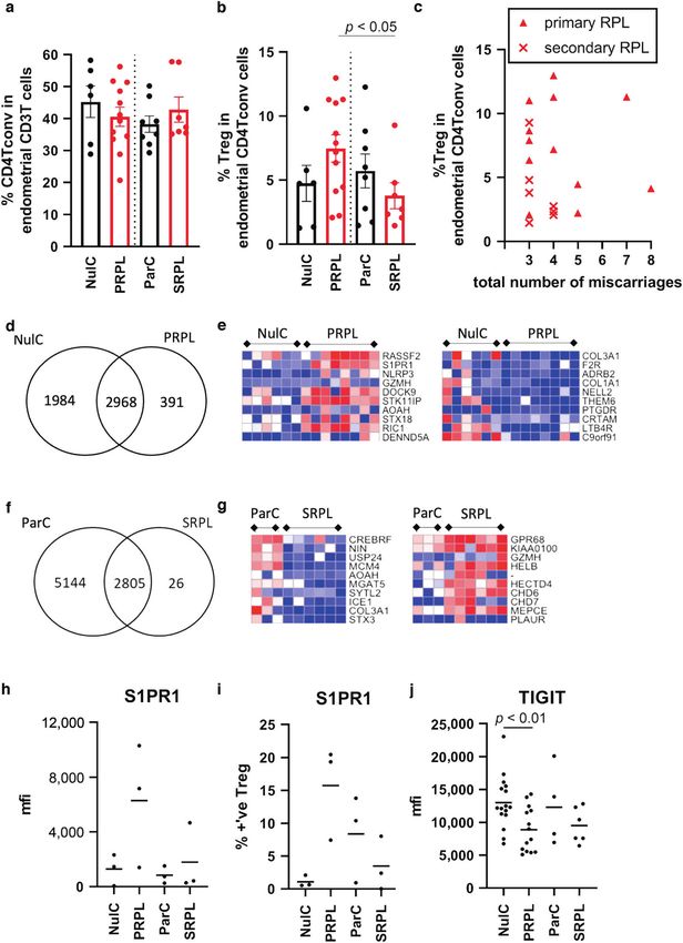
I. Granne et al.
122
      Fig. 1 CD4T cells in the human endometrium. a Workflow of methods applied in this study, patients are recruited and single cell
      preparations of endometrium or PBMC are prepared for either (top panel) flow cytometric analysis of Treg and Tconv cells in human
      endometrium: FSC-A vs. SSC-A and FSC-H determine lymphocyte singlet cells, ZAlo/CD45+/CD3+/CD4+ (total CD4T cells) are subpopulated
      CD25hi/CD127lo/FOXP3+ (Treg) or CD25−/FOXP3− (Tconv), (bottom panel) basic description of method for RNA sequencing. b Flow cytometric
      analysis of endometrial tissue single cell preparations from proven fertile women (n = 15) showing the proportion of total CD4T cells in the T
      cell population in the endometrium, and (c) the proportion of Treg in the total CD4T cell population. d % CD45RO expression by endometrial
      (E) (n = 12) or peripheral blood (PB) (n = 3) Tconv or Treg; bars = median + IQR. e Principal Component Analysis (PCA) of RNASeq data from
      peripheral blood Treg (PB-Tr) and endometrial Treg (E-Treg) or endometrial Tconv (E-Tconv) from nulliparous (Nul) or parous controls (Par),
      primary RPL (PRPL) or secondary (SRPL). f Volcano plot depicting differential gene expression (DESeq2) between endometrial Tconv and Treg,
      top 25 differentially expressed genes are labelled, (g) heatmap of top 30 upregulated and downregulated genes, red = high, blue = low
      expression, arrows show genes chose for further analysis in (h) using flow cytometry showing mean fluorescence intensity of intracellular
      FOXP3 and HELIOS (n = 15), and cell surface CTLA4 and TIGIT (n = 15), CCR8 (n = 8), CD39 (n = 12) in Tconv (left/grey bars) and Treg (right/blue
      bars), dots are individual samples and bars = mean +/− S.E.M.

      compared the transcriptional and protein expression profiles of
      endometrial Tregs in controls versus RPL.

      RESULTS
      The endometrial CD4-T cell compartment in normal
      physiology
      Endometrial biopsies were taken during the mid luteal phase of
      the menstrual cycle, when the tissue is receptive to embryo
      implantation. Single cell suspensions were obtained and flow
      cytometry performed to identify the total CD4T cell population
      (CD45/CD3/CD4), and the Treg subpopulation (CD25hi/CD127lo/
      FoxP3+), a representative example is shown in Fig. 1a. We first
      analysed this compartment in fertile women (nulliparous and
      parous combined). CD4T cells comprise 43.0 ± 9.5% of the total
      CD3 positive T cell population, which is a lower proportion than
      the equivalent compartment in the peripheral blood, Fig. 1b. Treg
      were 5.5 ± 3.5% of the total CD4T cell compartment in the human
      endometrium, this is similar to the proportion of Treg in the CD4T
      compartment in the peripheral blood, Fig. 1c. A higher proportion         Fig. 2 Parous women have an altered Treg, but not Tconv,
      of CD4T cells in the endometrium are CD45RO+ memory cells                 transcriptome. a VENN diagram of uniquely expressed or shared
      (Tconv 92.6% versus Treg 97.9%) compared to those in the                  genes within the Treg (a) or Tconv (b) endometrial subpopulations
      circulation (Fig. 1d), indicating a tissue specific enrichment of          between nulliparous and parous control women. c Heatmaps
      memory cells warranting further analysis.                                 depicting the top 10 most differentially expressed genes between
                                                                                nulliparous and parous women.
      Phenotypic analysis of endometrial Treg and Tconv cells
      We isolated Treg cells (CD3+, CD4+, CD25+, CD127lo cells) or
      Tconv (CD3+, CD4+, depleted of Treg) from endometrial tissue              Treg have higher expression of the regulatory molecules CTLA4,
      biopsy digests, from fertile women (nulliparous (n = 7) or parous         TIGIT, TNFSRF9 and the chemokine CCR8. Along with the classical
      controls (n = 3)). In addition we isolated cells from women with          master Treg transcription factor FOXP3 gene, the endometrial Treg
      primary (n = 8) or secondary (n = 7) recurrent pregnancy loss             express greater levels of the transcription factor genes BATF, IKZF2
      (PRPL or SRPL respectively), and Treg were isolated from 5                (IKAROS) and IKZF4 (HELIOS). We confirmed endometrial Treg
      peripheral blood samples from RPL patients. RNA-sequencing was            express significantly higher protein levels of the transcription
      performed and principal component analysis revealed that, as              factors FOXP3 and HELIOS, regulatory molecules CTLA4, TIGIT and
      expected, endometrial conv-CD4T cells and endometrial Treg cells          CD39 and also the chemokine CCR8, than CD4-Tconv, Fig. 1h.
      have distinct signatures, Fig. 1e. In addition, peripheral blood Treg     Together these data demonstrate the distinct phenotypes of
      are separated from the endometrial Treg cluster (Fig. 1e)                 endometrial Tregs and CD4-Tconv cells.
      indicating an endometrial specific Treg signature which will be
      discussed in more detail later.                                           Comparison of endometrial Treg and Tconv cells in
         Differential gene expression between endometrial Tconv and             nulliparous versus multiparous women
      Treg was investigated, depicted by Volcano plot, Fig. 1f. The gene        Endometrial NK cells are known to be modified by exposure to full
      for the classical Treg signature protein IL2RA (CD25) was the most        term pregnancy with specific subpopulations persisting after birth
      significantly differentially expressed gene, along with other well         and enhancing placentation in subsequent pregnancies,13 there-
      described Treg genes such as FOXP3, TIGIT and IKZF2 (HELIOS). One         fore we explored if parous women also have an altered
      of the highest differentially expressed genes in endometrial Tconv        transcriptomic endometrial Treg or Tconv profile. Endometrial
      compared to Treg counterparts was EOMES, encoding a transcrip-            Treg or Tconv from controls were segregated into groups of
      tion factor involved with CD4T cell differentiation. The most             nulliparous (n = 6) or parous women (n = 3), Treg from parous
      abundantly expressed, significantly differentially expressed genes         women had a large number of uniquely expressed genes
      from endometrial Treg cells and Tconv derived from nulliparous            compared to those from nulliparous women (38.8 versus 1.8%,
      control women are shown in Fig. 1g. Tconv are enriched in genes           Fig. 2a) indicating that exposure to term pregnancy alters the Treg
      encoding cytokines (IFNγ), chemokines (e.g., CCL5 and CCL4),              transcriptome. In contrast, the majority of genes expressed in
      chemokine receptors (e.g., CCR7), cytotoxic factors (GZMA and             Tconv overlapped (90.7%), with a modest 870 (7.9%) uniquely
      GZMH) and the anti-inflammatory protein AnnexinA1. In contrast,            expressed genes in the parous group, Fig. 2b. Differential

                                                                                                              Mucosal Immunology (2022) 15:120 – 129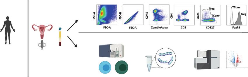
I. Granne et al.
                                                                                                                                              123
expression analysis yielded 419 upregulated and 801 down-             analysis identified 657 upregulated and 716 downregulated
regulated genes in Treg from parous women, heatmaps of the top        genes, the top 10 up- and down-regulated genes are displayed
10 are shown in Fig. 2c, the highest upregulated gene in the Treg     as heatmaps in Fig. 4e.
from parous women was IFNγ. Comparing nulliparous and parous              Comparing SRPL to parous controls, few genes were unique to
Tconv differential gene expression using DESeq2 only 17 genes         SRPL compared to the parous controls, Fig. 4f, however 1466
were differentially expressed (data not shown).                       genes were lower and 773 genes higher in expression in SRPL
                                                                      when compared to parous controls, top 10 differentially
Comparison of peripheral blood and endometrial Treg cells in          expressed genes are shown in Fig. 4g. Of note, all comparisons
RPL                                                                   were also performed on Tconv, PRPL and SRPL had similar
Within a limited subset of samples with both endometrial and PB       transcriptomic profiles to their controls, with 45 and 79 total
available, we asked whether the Treg transcriptional profile           differentially expressed genes between groups respectively
differed in the two compartments. The peripheral blood Treg           (data not shown).
were very similar with only 45 genes found to be differentially           The second highest upregulated gene in PRPL was S1PR1;
expressed between the PRPL (n = 3) and SRPL (n = 2) groups,           Sphingosine-1-Phosphate receptor-1 is a G-Protein coupled
(DeSeq2 as detailed in “Methods”, FDR-corrected p < 0.01; data not    receptor involved with lymphocyte activation, migration and
shown). Next we compared peripheral blood Treg cells (n = 5) and      trafficking.14 We analysed mRNA expression of this factor, along
endometrial Treg cells (n = 5) from RPL patients to determine the     with other factors identified by DESeq2 between groups and also
endometrial tissue specific Treg signature, a volcano plot of          known Treg factors identified as core signature genes, such as
differentially expressed genes with the 25 most significantly          transcription factors, regulatory molecules, tissue residency/
altered genes is shown in Fig. 3a. We found that endometrial Treg     chemoattraction/exit molecules,15 between all Treg and Tconv
have 169 downregulated and 571 upregulated genes compared to          cell subsets, Supplementary Fig. 2. Endometrial Tconv have higher
peripheral blood Treg (p < 0.01 padj and >3,
I. Granne et al.
124

      Fig. 3 Endometrial and peripheral blood Treg have alternate characteristics. a Transcriptomic analysis of Treg derived from endometrium
      (n = 5) versus peripheral blood (PB) (n = 5) from RPL patients, volcano plot depicting differential gene expression (DESeq2) between
      endometrial and PB Treg showing top 25 differentially expressed genes, (b) heatmap of top 30 upregulated (right panel) and downregulated
      (left panel) genes in the endometrium versus PB, red = high, blue = low expression, P = PRL (n = 3) and S = SRPL (n = 2) for endometrium (E)
      and peripheral blood (PB) Treg, (c) Flow cytometric analysis of matched peripheral blood (red bars) and endometrium (grey bars) Treg from
      patients with RPL, the mean fluorescence intensity (mfi) of FOXP3, HELIOS, CTLA4, TIGIT, IL18R (n = 10), CXCR6, ICOS (n = 3), LAG, PD1, CD39
      and TIM3 (n = 4) was determined (bars = mean ± SEM; dots = individual cell sample).

      to the endometrium, which help facilitate embryo implantation               We targeted key T cell transcription factors (Supplementary
      through tissue remodelling.18 Endometrial Treg, had enriched             Fig. 2) and found that Treg were also high for GATA, BLIMP1,
      regulatory molecules such as CTLA4, TIGIT and CD39, which were           STAT3. The Tconv population contained cells expressing Tbet,
      also validated via flow cytometry. CCR8 expression was highly             GATA and also some RORC, the Tconv population are therefore
      enriched in Treg, the CCL1-CCR8 axis is known to potentiate              as expected a likely combination of Th1, Th2 and Th17 cells.
      Treg suppressive function19 which is in line with the phenotype          In addition, Treg expression of IKZF2 (HELIOS) was also detected;
      noted here.                                                              HELIOS, along with Neuropilin, has been suggested by some

                                                                                                           Mucosal Immunology (2022) 15:120 – 129
I. Granne et al.
125
Fig. 4 Treg characterisation in primary and secondary RPL reveals an altered transcriptome. a Tconv and (b) Treg were enumerated by
flow cytometry in nulliparous and parous controls (NulC (n = 6) and ParC (n = 8)—black bars) and primary and secondary RPL (PRPL (n = 12)
and SRPL (n = 7)—red bars), (c) the Treg proportion of the CD4 Tconv population versus the number of miscarriage in PRPL (triangles) and
SRPL (crosses). Treg gene expression signatures were compared analysing uniquely expressed and shared genes, displayed as VENN diagrams,
and top 10 differentially upregulated and downregulated expressed genes, displayed as heatmaps, between (d, e) NulC and PRPL Treg or (f, g)
ParC and SRPL, respectively. Flow cytometric analysis of cell surface (h, i) S1PR1 (n = 3 each group) and (j) TIGIT (n = 16, 14, 4, 6 in order)
between RPL and control patients showing mean fluorescent intensity (mfi) (h and j) or the % positive Treg cells (i) detected by flow
cytometry. Bars = mean value.
Mucosal Immunology (2022) 15:120 – 129I. Granne et al.
126
researchers to be expressed by peripheral Treg (pTreg) as women to SRPL. Using flow cytometry to quantify the proportions
opposed to thymic derived Treg, however controversy exists as of Tconv in the T cell population and Treg in the Tconv
to their true value as such markers,20 here we find low populations, we found no significant differences in the cell
endometrial Treg gene transcripts for Neuropilin and higher proportions between groups (Fig. 4b). Treg/CD4 Tconv cell
HELIOS with variable protein levels. proportions in human endometrium have not been previously
The endometrial specific tissue Treg signature has not yet been clearly defined,32 there is limited evidence of increased FOXP3+
described, but it is to be expected as Tregs have non-lymphoid cells (immunohistochemical analysis) in the endometrium of RPL
tissue specific local adaptations.15 CXCR6 was highly expressed patients,33 but Tconv and Treg proportions and populations have
by endometrial Treg, this has previously been shown to be mainly been studied in in the peripheral blood or decidua
upregulated in human colon Treg.15 ICOS (inducible T-cell obtained after miscarriage. Fertile women have expanded
costimulator) was also highly upregulated in endometrial Treg, peripheral blood CD4+CD25+FOXP3+ Treg in the late follicular
in vitro ICOS+ Treg produce abundant IL-1021 and ICOS phase, this expansion is not seen in RPL which also have reduced
expression is closely linked to Blimp1 expression in Treg located functional capacity to inhibit proliferation34,35 and inhibit NK cell
in mucosal sites22 as we have detected in these endometrial Treg. cytotoxicity.36 In general, CD4T cells numbers are lower in RPL
Immune-checkpoint molecules are a key target for immunother- decidua than controls37 and in normal early pregnancy Tconv
apy, upon ligation inhibitory signals limit immunity;23 family were not clonally expanded.38 CD4+CD25bright cells were a higher
members include cytotoxic T-lymphocyte-associated protein 4 proportion of the CD4T cell compartment in early pregnancy
(CTLA-4), T-cell immunoreceptor with immunoglobulin and ITIM decidua obtained after induced abortion (21.84 ± 2.92) than after
domains (TIGIT), programmed cell death protein 1 (PD-1), T-cell sporadic miscarriage (7.14 ± 1.85),39 and Treg numbers are
immunoglobulin and mucin-domain containing-3 (Tim-3) and reportedly reduced in decidua from RPL patients compared to
lymphocyte-activation gene 3 (LAG-3), all of which have been normal first trimester pregnancy.35,40–42 Here we find that the
studied here. Of note the PD-1/PD-L1 pathway is critically numbers of cells present are similar prior to pregnancy, in RPL and
important in murine pregnancy as blockade causes foetal controls, therefore the observed reduction in Treg numbers in
resorption, due to an imbalance in Th17/Treg cells which is decidua in women suffering miscarriage is likely a consequence of
diminished with Treg cell adoptive transfer.24 The phenotype failure to recruit or expand Treg populations upon establishment
identified here indicated endometrial Treg have enhanced of pregnancy.
expression levels of several immune checkpoint molecules, PRPL Treg had altered Rassf2, a novel tumour suppressor gene
indicating endometrial Treg cells have differing phenotypic with associated K-Ras pro-apoptotic effector functions and the
functions to those found in the blood. second highest differentially expressed gene was S1PR1, Fig. 4.
This is the first description of the CD4T and Treg compartment S1PR1 has well established roles in T cell trafficking out of
in human endometrium comparing parity and controls. Treg have lymphoid tissue, it’s role for T cell egress from mucosal tissues is
fundamental importance for materno-foetal tolerance: CNS1 less well defined.14 Mice with S1PR1 deficient Treg develop
dependent extrathymic differentiation of Treg occurred concur- autoimmunity, Treg display an activated phenotype prone to
rently with placental evolution,25 and in mice Tregs expand during apoptosis.43 The functional role of endometrial S1PR1 Treg
pregnancy forming a memory response that is recalled in expression remains to be determined, and if the expression
subsequent pregnancy.26 Previous studies on decidua from term indicates a subset of Treg in PRPL which can exit the tissue. In
placenta show that Tregs undergo clonal expansion throughout addition, we studied key Treg immune checkpoint molecules,
pregnancy, of note this cannot be detected in the peripheral and found that endometrial Treg from PRPL had significantly
blood Treg,27 but peripheral blood Treg numbers increase in lower TIGIT protein expression levels that corresponding
human pregnancies as gestation progresses. Our observation that controls. This is the first observation of altered Treg populations
RPL Treg have significant phenotypic differences between PB and in RPL, phenotypic changes indicate reduced inhibitory capacity
endometrium, suggests Treg tissue specificity, this was also in PRPL. Whether patients with PRPL would benefit from
observed by a study analysing the T cell receptor beta variable therapies enhancing tolerance through this pathway merits
(TRBV) repertoire that found most variation in the Treg population further investigation.
between cells derived from the blood or decidua.28 Of note, we
were unable to obtain peripheral blood samples from control
patients due to ethical constraints, further analysis of Treg from METHODS
peripheral blood between controls and RPL is required in future Tissue collection and processing
studies. IFNγ was identified as the most upregulated gene in The study was approved by the Oxford Research Ethics Committee C
endometrial Treg from parous women compared to nulliparity. (ref:08/H0606/94). All participants gave written informed consent in
Interestingly, parous women have been shown to harbour a accordance with the Helsinki Declaration of 1975.
subpopulation of NKG2Chi dNK cells thought to be produced All women wereI. Granne et al.
127
Table 1. Details of patients that provided endometrial and peripheral blood samples for RNAseq. Data are (Mean | S.D.)
(Mean | S.D.) Endometrium Blood
Nulliparous control Parous control Primary RPL Secondary RPL PB RPL
(n = 7) NulC (n = 3) ParC (n = 8) PRPL (n = 7) SRPL (n = 5)
Age 33.3 | 4.1 35.7 | 2.1 35.3 | 2.9 36.7 | 3.7 36.2 | 4.1
Live Birth Rate 0|0 1|0 0|0 1|0 0.4 | 0.5
Miscarriages 0|0 0.3| 0.5 4.75 | 2.6 4.9 | 2.2 3.8 | 1.8
excluded. Control samples for RNAseq or matched control to RPL flow mapped to Ensemble Homo Sapiens cDNA database release 94. 1 NulC
cytometry experiments were from patients who are proven fertile; they and 1 SRPL Treg sample did not pass QC and were excluded from
either have had a previous live birth but were undergoing IVF for male analysis.
factor infertility (Parous Controls; ParC), or had a live birth in the IVF cycle
following endometrial biopsy (Nulliparous Controls; NulC).
Endometrial samples were obtained using an Endocell disposable Data analysis
endometrial cell sampler (Wallach Surgical devices, CT, USA) and digested Data analysis was performed using R (version 3.5.3) and RStudio,
using 1X Liberase (Roche Life Sciences) as previously described.7 Peripheral differential gene expression analysis was performed using DESeq2.46
blood was collected into sodium heparin anti-coagulant (10 U/ml) and Heatmaps were generated using p < 0.01 padj and >3, 10 and calculated using Bioinformatics & Evolutionary Genomics tool
RNA Sequencing patient sample information hosted by the University of Ghent, Belgium (http://bioinformatics.psb.
Matched blood and endometrial samples (n = 5) were taken from women ugent.be/webtools/Venn/)
with an average age of 36.2 ± 4.1 years (Mean, S.D.), 2 were nulliparous
(Primary RPL) and 3 were parous (Secondary RPL), 4 patients had
experienced 3 miscarriages and one patient had 7 prior miscarriages. Flow cytometry phenotyping
Table 1 provides details of patients that provided endometrial samples for All flow cytometry reagents were from Biolegend unless otherwise stated.
RNAseq. Samples were incubated with Zombie-Aqua Fixable Viability kit for 15 min
in the dark, then cells washed in PBS/2% FCS and antibodies towards cell
surface markers were added for 20 min at 4 °C in the dark. Antibodies used
Cell Sorting for RNAseq and RNA extraction were CD4-FITC(OKT4), CD8a-PE(HIT8a), CD8-PeCy7 (RPA-T8), CD3-PeCy5
Frozen endometrial cell digests or isolated PBMC were thawed into HS- (UCHT1), CD56-PeCy7(BD Biosciences; B159), CD16-APCCy7(BD Biosciences;
media (RPMI1640 supplemented with 10% FCS, 1% Human Serum, 3G8), CD45-AlexaFluor700 or CD45-APC (HI30),CD45RO-APC-Cy7(UCHL1),
glutamine and pen/strep) at 37 °C and centrifuged at 300 × g. Endometrial CD127-AlexaFluor647(A019D5), PD-1(CD279)-APC(EH12.2H7), CXCR6-
cells were resuspended in HS-Media and transferred to a 25 cm2 flask for PECy7 (K041E5), CD25-PE/Dazzle594 (M-A251), CCR8(CD198)-PE (L263G8),
30 minutes at 37 °C/5% CO2, then supernatants containing non-adherent CD3-APC/Fire750 (SK7), CD45- AlexaFluor700 (HI30), CD127-BV711
cells were further transferred to a fresh flask for an additional 30 minutes (A019D5), ICOS-BV650 (C392.4A), CTLA-4(CD152)-BV786 (BD Biosciences;
incubation, for stromal and epithelial cell depletion. Supernatants were BNI3), TIGIT-BV421, (A15153G), ENTPD1(CD39)-BV421 (BD Biosciences;
then removed and transferred onto a lymphoprep layer with centrifuga- TU66), LAG-3(CD223)-PE (113C65), TIM-3(CD366)-BV785 (F38-2E2), IL18R-
tion at 800 × g for 20 min. Cells in the interface were washed in PBS and PE (H44), S1PR1-eFluor550 (eBiosciences; SW4GYPP). Afterwards, cells
prepared for cell sorting on an Aria III (BD Biosciences). Briefly, both PBMC were washed in PBS/2% FCS and intranuclear antibody staining was
or endometrial cells were stained with Zombie AquaTM Live/Dead Viability performed using ‘True-Nuclear Transcription Factor Buffer Set’ (Biolegend),
dye then incubated with antibodies (CD3-APC Fire750, CD45-Briliant Violet cells were fixed for 1 h then transferred into PBS/2% FCS overnight, then
(BV)650, CD4-FITC, CD25-PE, CD8-BV711, CD19-PeCy7, CD127-APC), all permabilisation performed before addition of antibodies FOXP3-
reagents were from Biolegend unless otherwise stated. AlexaFluor647 (259D), IKZF2(HELIOS)-PeCy7 (22F6) and additional CTLA-4
CD4T cells were isolated as singlet cells (FSC-H vs FSC-A), Zombie Aqua-, antibody for total cellular CTLA-4 measurement, for 45 min incubation.
CD45+, CD3+, CD4+, CD8−, CD19-, CD25lo cells and Treg were Zombie Data was acquired using an LSR-II flow cytometer (BD Biosciences) and
Aqua−, CD45+, CD3+, CD4+, CD8−, CD19−, CD25+ and CD127lo. data analysed using FlowJo software (Tree Star Inc.), fluorescence Minus
Immediately after sorting, cells were pelleted by centrifugation and One (FMO) controls established gating strategies. Graphs were plotted and
resuspended in RLT (Qiagen) and RNA was extracted using QIAGEN RNeasy statistics generated in GraphPad Prism 8.4.3.
Micro Kits according to the manufacturer’s protocol. RNA quantity and
quality were accessed on Agilent 4200 TapeStation System.
Sorted cell populations (mean ± 1.S.D.) were 2555 ± 2254 for endome- REFERENCES
trial Treg, 36373 ± 37819 for endometrial CD4T cells and 34450 ± 14531 1. Wira, C. R., Rodriguez-Garcia, M. & Patel, M. V. The role of sex hormones in
blood Treg. No significant differences were detected within sorted cell immune protection of the female reproductive tract. Nat. Rev. Immunol. 15,
population counts between nulliparous controls, parous controls, primary 217–230 (2015).
RPL and secondary RPL. 2. Recurrent Pregnancy Loss: Guideline of the European Society of Human Repro-
duction and Embryology. ESHRE Early Pregnancy Guidline Development Group.
Library preparation and RNA sequencing [Available from: https://www.eshre.eu/Guidelines-and-Legal/Guidelines/
Samples were processed following Smart-Seq 2 protocols,45 the library Recurrent-pregnancy-loss.aspx.
was prepared using a Standard Nextera Illumina Library Prep kit using a 3. Practice Committee of American Society for Reproductive M. Definitions of
unique dual- index strategy to barcode cDNA fragments. DNA samples infertility and recurrent pregnancy loss: a committee opinion. Fertil. Steril. 99, 63
were amplified for 16 cycles and the cDNA quantities assessed using a (2013).
2100 Bioanalyzer (Agilent, USA) with Quant-iT PicoGreen (Thermo Fisher, 4. Laisk, T. et al. The genetic architecture of sporadic and multiple consecutive
USA). Clustering and sequencing was carried out by Novogene Co., Ltd. miscarriage. Nat. Commun. 11, 5980 (2020).
Briefly, clustering of the index-coded samples was performed on a cBot 5. Dimitriadis, E., Menkhorst, E., Saito, S., Kutteh, W. H. & Brosens, J. J. Recurrent
Cluster Generation System using PE Cluster Kit cBot-HS (Illumina) and pregnancy loss. Nat. Rev. Dis. Prim. 6, 98 (2020).
paired end RNA sequencing was performed using the Novoseq 6000 6. Wang, N. F., Kolte, A. M., Larsen, E. C., Nielsen, H. S. & Christiansen, O. B. Immu-
platform, with 125 bp/150 bp read length and ~25 million reads per nologic abnormalities, treatments, and recurrent pregnancy loss: what is real and
sample. Quality control was performed by Novogene Co., Ltd and reads what is not? Clin. Obstet. Gynecol. 59, 509–523 (2016).
Mucosal Immunology (2022) 15:120 – 129I. Granne et al.
128
7. Southcombe, J. H. et al. An altered endometrial CD8 tissue resident memory T cell 36. Zhu, L. et al. Foxp3 TSDR Hypermethylation is correlated with decreased tregs in
population in recurrent miscarriage. Sci. Rep. 7, 41335 (2017). patients with unexplained recurrent spontaneous abortion. Reprod. Sci. 28,
8. Zenclussen, A. C. et al. Abnormal T-cell reactivity against paternal antigens in 470–478 (2021).
spontaneous abortion: adoptive transfer of pregnancy-induced CD4+CD25+ T 37. Windsperger, K. et al. Densities of decidual high endothelial venules correlate
regulatory cells prevents fetal rejection in a murine abortion model. Am. J. Pathol. with T-cell influx in healthy pregnancies and idiopathic recurrent pregnancy
166, 811–822 (2005). losses. Hum. Reprod. 35, 2467–2477 (2020).
9. Aluvihare, V. R., Kallikourdis, M. & Betz, A. G. Regulatory T cells mediate maternal 38. Vento-Tormo, R. et al. Single-cell reconstruction of the early maternal-fetal
tolerance to the fetus. Nat. Immunol. 5, 266–271 (2004). interface in humans. Nature 563, 347–353 (2018).
10. Sakaguchi, S. et al. Regulatory T cells and human disease. Annu Rev. Immunol. 38, 39. Sasaki, Y. et al. Decidual and peripheral blood CD4+CD25+ regulatory T cells in
541–566 (2020). early pregnancy subjects and spontaneous abortion cases. Mol. Hum. Reprod. 10,
11. Galgani, M. et al. Regulatory T cells, inflammation, and endoplasmic 347–353 (2004).
reticulum stress in women with defective endometrial receptivity. Fertil. Steril. 40. Mei, S., Tan, J., Chen, H., Chen, Y. & Zhang, J. Changes of CD4+CD25high reg-
103, 1579–1586.e1 (2015). ulatory T cells and FOXP3 expression in unexplained recurrent spontaneous
12. Devaud, C., Darcy, P. K. & Kershaw, M. H. Foxp3 expression in T regulatory cells abortion patients. Fertil. Steril. 94, 2244–2247 (2010).
and other cell lineages. Cancer Immunol. Immunother. 63, 869–876 (2014). 41. Yang, H. et al. Proportional change of CD4+CD25+ regulatory T cells in decidua
13. Gamliel, M. et al. Trained memory of human uterine NK cells enhances their and peripheral blood in unexplained recurrent spontaneous abortion patients.
function in subsequent pregnancies. Immunity 48, 951–962.e5 (2018). Fertil. Steril. 89, 656–661 (2008).
14. Baeyens, A. A. L. & Schwab, S. R. Finding a way out: S1P signaling and immune 42. Qian, J. et al. Distinct pattern of Th17/Treg cells in pregnant women with a history
cell migration. Annu Rev. Immunol. 38, 759–784 (2020). of unexplained recurrent spontaneous abortion. Biosci. Trends 12, 157–167
15. Miragaia, R. J. et al. Single-cell transcriptomics of regulatory T cells reveals tra- (2018).
jectories of tissue adaptation. Immunity 50, 493–504.e7 (2019). 43. Eken, A. et al. S1P1 deletion differentially affects TH17 and regulatory T cells. Sci.
16. Thome, J. J. et al. Early-life compartmentalization of human T cell differentiation and Rep. 7, 12905 (2017).
regulatory function in mucosal and lymphoid tissues. Nat. Med. 22, 72–77 (2016). 44. Regan L., Backos M. & Rai R. Recurrent Miscarriage, Investigation and Treatment
17. Dejean, A. S., Joulia, E. & Walzer, T. The role of Eomes in human CD4 T cell of Couples (Green-top Guideline No. 17) (2011) [Available from: https://www.
differentiation: a question of context. Eur. J. Immunol. 49, 38–41 (2019). rcog.org.uk/en/guidelines-research-services/guidelines/gtg17/.
18. Zenclussen, A. C. & Hammerling, G. J. Cellular regulation of the uterine micro- 45. Picelli, S. et al. Full-length RNA-seq from single cells using Smart-seq2. Nat. Protoc.
environment that enables embryo implantation. Front Immunol. 6, 321 (2015). 9, 171–181 (2014).
19. Barsheshet, Y. et al. CCR8(+)FOXp3(+) Treg cells as master drivers of immune 46. Love, M. I., Huber, W. & Anders, S. Moderated estimation of fold change and
regulation. Proc. Natl Acad. Sci. USA 114, 6086–6091 (2017). dispersion for RNA-seq data with DESeq2. Genome Biol. 15, 550 (2014).
20. Lee, W. & Lee, G. R. Transcriptional regulation and development of regulatory 47. Subramanian, A. et al. Gene set enrichment analysis: a knowledge-based
T cells. Exp. Mol. Med 50, e456 (2018). approach for interpreting genome-wide expression profiles. Proc. Natl. Acad. Sci.
21. Li, D. Y. & Xiong, X. Z. ICOS(+) Tregs: a functional subset of Tregs in immune USA 102, 15545–15550 (2005).
diseases. Front Immunol. 11, 2104 (2020). 48. Otasek, D., Morris, J. H., Boucas, J., Pico, A. R. & Demchak, B. Cytoscape auto-
22. Cretney, E. et al. The transcription factors Blimp-1 and IRF4 jointly control the mation: empowering workflow-based network analysis. Genome Biol. 20, 185
differentiation and function of effector regulatory T cells. Nat. Immunol. 12, (2019).
304–311 (2011).
23. Zhang, Y. H. & Sun, H. X. Immune checkpoint molecules in pregnancy: focus on
regulatory T cells. Eur. J. Immunol. 50, 160–169 (2020).
24. D’Addio, F. et al. The link between the PDL1 costimulatory pathway and Th17 in
ACKNOWLEDGEMENTS
We would like to thank the participants in this study, and the staff at Oxford Fertility,
fetomaternal tolerance. J. Immunol. 187, 4530–4541 (2011).
the John Radcliffe Hospital and University Hospitals Coventry and Warwickshire and
25. Samstein, R. M., Josefowicz, S. Z., Arvey, A., Treuting, P. M. & Rudensky, A. Y.
Warwick Medical School who have assisted with the collections of samples. Dr Helen
Extrathymic generation of regulatory T cells in placental mammals mitigates
Ferry, NDM University of Oxford assisted with flow cytometric sorting and Dr Neil
maternal-fetal conflict. Cell 150, 29–38 (2012).
Ashley, WIMM University of Oxford assisted with the SmartSeq2 and library
26. Rowe, J. H., Ertelt, J. M., Aguilera, M. N., Farrar, M. A. & Way, S. S. Foxp3(+)
preparation. Funding was from the IVI Foundation FINOX.
regulatory T cell expansion required for sustaining pregnancy compromises host
defense against prenatal bacterial pathogens. Cell Host Microbe 10, 54–64 (2011).
27. Tsuda, S. et al. Clonally expanded decidual effector regulatory T cells increase in
late gestation of normal pregnancy, but not in preeclampsia, in humans. Front AUTHOR CONTRIBUTIONS
Immunol. 9, 1934 (2018). J.S. and I.G. conceived of the study. Data was generated and analysed by J.S., M.S.,
28. Neller, M. A. et al. Multivariate analysis using high definition flow cytometry H.R.C., G.C., G.M., E.O’.D. and E.D.. J.B., S.Q., T.C. and I.G. provided clinical expertise to
reveals distinct T cell repertoires between the fetal-maternal interface and the the study design. All authors contributed to refinement of the study protocol and
peripheral blood. Front Immunol. 5, 33 (2014). approved the final manuscript.
29. Erlebacher, A. Mechanisms of T cell tolerance towards the allogeneic fetus. Nat.
Rev. Immunol. 13, 23–33 (2013).
30. Tilburgs, T. et al. Evidence for a selective migration of fetus-specific
CD4+CD25bright regulatory T cells from the peripheral blood to the decidua COMPETING INTERESTS
in human pregnancy. J. Immunol. 180, 5737–5745 (2008). The authors declare no competing interests.
31. Salvany-Celades, M. et al. Three Types of Functional Regulatory T Cells Control T Cell
Responses at the Human Maternal-Fetal Interface. Cell Rep. 27, 2537–2547.e5 (2019).
32. Ticconi, C., Pietropolli, A., Di Simone, N., Piccione, E. & Fazleabas, A. Endometrial ADDITIONAL INFORMATION
immune dysfunction in recurrent pregnancy loss. Int. J. Mol. Sci. 20, 5332 (2019). Supplementary information The online version contains supplementary material
33. Lyzikova, Y. A., Zinovkin, D. A. & Pranjol, M. Z. I. Increase in FoxP3, CD56 available at https://doi.org/10.1038/s41385-021-00451-1.
immune cells and decrease in glands PGRMC1 expression in the endometrium
are associated with recurrent miscarriages. Eur. J. Obstet. Gynecol. Reprod. Biol. Correspondence and requests for materials should be addressed to Jennifer H.
245, 121–126 (2020). Southcombe.
34. Arruvito, L., Sanz, M., Banham, A. H. & Fainboim, L. Expansion of CD4+CD25+ and
FOXP3+ regulatory T cells during the follicular phase of the menstrual cycle: Reprints and permission information is available at http://www.nature.com/
implications for human reproduction. J. Immunol. 178, 2572–2578 (2007). reprints
35. Bao, S. H. et al. Decidual CD4+CD25+CD127dim/- regulatory T cells in patients
with unexplained recurrent spontaneous miscarriage. Eur. J. Obstet. Gynecol. Publisher’s note Springer Nature remains neutral with regard to jurisdictional claims
Reprod. Biol. 155, 94–98 (2011). in published maps and institutional affiliations.
Mucosal Immunology (2022) 15:120 – 129I. Granne et al.
129
Open Access This article is licensed under a Creative Commons
Attribution 4.0 International License, which permits use, sharing,
adaptation, distribution and reproduction in any medium or format, as long as you give
appropriate credit to the original author(s) and the source, provide a link to the Creative
Commons license, and indicate if changes were made. The images or other third party
material in this article are included in the article’s Creative Commons license, unless
indicated otherwise in a credit line to the material. If material is not included in the
article’s Creative Commons license and your intended use is not permitted by statutory
regulation or exceeds the permitted use, you will need to obtain permission directly
from the copyright holder. To view a copy of this license, visit http://creativecommons.
org/licenses/by/4.0/.
© The Author(s) 2021
Mucosal Immunology (2022) 15:120 – 129You can also read