COVID-19 MRNA VACCINE INDUCED ANTIBODY RESPONSES AGAINST THREE SARS-COV-2 VARIANTS
←
→
Page content transcription
If your browser does not render page correctly, please read the page content below
ARTICLE
                  https://doi.org/10.1038/s41467-021-24285-4                   OPEN

                  COVID-19 mRNA vaccine induced antibody
                  responses against three SARS-CoV-2 variants
                  Pinja Jalkanen 1 ✉, Pekka Kolehmainen 1, Hanni K. Häkkinen2, Moona Huttunen 1, Paula A. Tähtinen3,
                  Rickard Lundberg1, Sari Maljanen1, Arttu Reinholm1, Sisko Tauriainen 1, Sari H. Pakkanen2, Iris Levonen2,
                  Arttu Nousiainen2, Taru Miller2, Hanna Välimaa2,4, Lauri Ivaska 3, Arja Pasternack5, Rauno Naves5,
                  Olli Ritvos5, Pamela Österlund 6, Suvi Kuivanen4, Teemu Smura4, Jussi Hepojoki 4, Olli Vapalahti4,
                  Johanna Lempainen1,3, Laura Kakkola1,8, Anu Kantele2,8 & Ilkka Julkunen 1,7,8 ✉
1234567890():,;

                  As SARS-CoV-2 has been circulating for over a year, dozens of vaccine candidates are under
                  development or in clinical use. The BNT162b2 mRNA COVID-19 vaccine induces spike
                  protein-specific neutralizing antibodies associated with protective immunity. The emergence
                  of the B.1.1.7 and B.1.351 variants has raised concerns of reduced vaccine efficacy and
                  increased re-infection rates. Here we show, that after the second dose, the sera of
                  BNT162b2-vaccinated health care workers (n = 180) effectively neutralize the SARS-CoV-2
                  variant with the D614G substitution and the B.1.1.7 variant, whereas the neutralization of the
                  B.1.351 variant is five-fold reduced. Despite the reduction, 92% of the seronegative vaccinees
                  have a neutralization titre of >20 for the B.1.351 variant indicating some protection. The
                  vaccinees’ neutralization titres exceeded those of recovered non-hospitalized COVID-19
                  patients. Our work provides evidence that the second dose of the BNT162b2 vaccine induces
                  cross-neutralization of at least some of the circulating SARS-CoV-2 variants.

                  1 Institute of Biomedicine, University of Turku, Turku, Finland. 2 Department of Infectious Diseases, Meilahti Vaccination Research Center, MeVac, Helsinki

                  University Hospital and University of Helsinki, Helsinki, Finland. 3 Department of Paediatrics and Adolescent Medicine, Turku University Hospital and
                  University of Turku, Turku, Finland. 4 Department of Virology, University of Helsinki, Helsinki, Finland. 5 Department of Physiology, University of Helsinki,
                  Helsinki, Finland. 6 Finnish Institute for Health and Welfare, Helsinki, Finland. 7 Clinical Microbiology, Turku University Hospital, Turku, Finland. 8These authors
                  jointly supervised: Laura Kakkola, Anu Kantele, Ilkka Julkunen. ✉email: pinja.r.jalkanen@utu.fi; ilkka.julkunen@utu.fi

                  NATURE COMMUNICATIONS | (2021)12:3991 | https://doi.org/10.1038/s41467-021-24285-4 | www.nature.com/naturecommunications                                          1
ARTICLE NATURE COMMUNICATIONS | https://doi.org/10.1038/s41467-021-24285-4
T
he emergence and spread of the severe acute respiratory vaccination on 28 December 2020, and the last vaccinations for
syndrome coronavirus 2 (SARS-CoV-2) has caused a those HCWs in this study were given on 12 February 2021. Serum
pandemic with over 3.8 million deaths1 and rapid devel- samples from 50 non-hospitalized, recovered COVID-19 patients
opment of multiple vaccine candidates2. SARS-CoV-2 infection from 2020 were also included in the analysis. The generation of
elicits antibodies against spike protein (S) and nucleoprotein anti-S1 IgG, IgA, IgM, and total Ig antibodies after vaccination
(N)3–5, of which, on the basis of virus challenge studies in ani- was analyzed with enzyme immunoassay (EIA). The original
mals, the spike protein-specific antibodies are neutralizing and optical density values in the assay were converted to EIA units to
associated with protective immunity6,7. In addition, recent studies minimize inter-assay variation (Fig. 1A). To verify the EIA results
of COVID-19 patients and vaccinees indicate that previous and to study the rate of SARS-CoV-2 infections after vaccination,
infections and vaccinations are related to a decreased rate of sera were also analyzed with N protein-specific EIA (Fig. 1B).
SARS-CoV-2 infections8–10. Although the persistence of vaccine- Before vaccination (0 day sampling) 11/180 (6%) had anti-S1
induced antibodies is still not known, infection-induced neu- IgG antibodies (Supplementary Fig. 1A) indicating that these
tralizing antibodies have remained detectable for at least six individuals had undergone a previous SARS-CoV-2 infection.
months after symptom onset11. Five of these anti-S1 positive participants had also anti-N IgG
Currently, European Medicines Agency (EMA) has authorized antibodies. Already after the first dose of the vaccine (3 weeks),
four vaccines to be used in European Union: two mRNA vaccines vaccinees with prior SARS-CoV-2 infection showed clearly
(BNT162b2/Comirnaty by Pfizer-BioNTech and mRNA-1273 by increased levels of anti-S1 IgG antibodies (geometric mean 99).
Moderna) and two adenoviral vector-based vaccines (ChAdOx1-S After the second dose of the vaccine (6 weeks), all vaccinees with
by AstraZeneca-Oxford and COVID-19 Vaccine Janssen by prior SARS-CoV-2 infection had very high levels of anti-S1 IgG
Janssen Biologics B.V. and Janssen Pharmaceutica NV)12. All four antibodies (geometric mean 109) (Supplementary Fig. 1A).
vaccines aim to generate spike protein-specific antibodies and all Three weeks after the first dose of BNT162b2, vaccinees
have been shown to induce anti-S IgG antibodies with neu- without prior SARS-CoV-2 infection (169/180, 94%) developed
tralizing activity against the first pandemic SARS-CoV-2 Wuhan varying levels of anti-S1 IgG antibodies (geometric mean 47), and
Hu-1 variant and the currently circulating D614G variants13–15. moderate levels of anti-S1 IgA and IgM antibodies (Fig. 1A).
The recent emergence of SARS-CoV-2 variants of concern, such Total Ig levels for S1 ranged from 1 to 98 EIA units. The EIA
as B.1.1.7 first identified in the United Kingdom16 and B.1.351 levels for anti-N antibodies among the vaccinees remained the
first identified in South Africa17, has raised concerns about same as before the vaccination (Fig. 1B, Table 1) indicating the
increased virus transmissibility and reduced vaccine efficacy. absence of SARS-CoV-2 infections in this study group. Anti-N
These two variants of concern are defined by eight to ten amino IgG antibody levels were higher in sera of non-hospitalized
acid changes or deletions in the spike protein to which vaccine- COVID-19 patients (geometric mean 17) than of vaccinees
induced antibodies are targeted17–20. Both of these variants are (geometric mean 2) (Fig. 1B, Table 1). However, already after the
now transmitted in several countries (https://cov-lineages.org/ first vaccination dose, the geometric mean of anti-S1 IgG and
global_report.html). Initial studies reported that antibodies pro- anti-S1 total Ig antibodies of vaccinees exceeded those of
duced in response to vaccination and natural infection neutralize convalescent-phase COVID-19 patients, 47 and 37 vs. 20 and
the B.1.1.7 variant19,21, whereas neutralization of the B.1.351 is 23, respectively (Fig. 1A, Table 1).
reduced 8–13-fold18,22,23. However, it is still unclear whether the Six weeks after the first vaccine dose (3 weeks after the second
B.1.351 variant can escape from humoral and cell-mediated vaccine dose) all participants, despite the initial response after the
immunity. first vaccine dose, elicited high levels of anti-S1 IgG antibodies
Here, we characterize the BNT162b2 vaccine-induced antibody together with a modest increase in anti-S1 IgA and IgM antibody
responses among a sequential serum sample cohort of 180 Finnish levels (Fig. 1A). Based on anti-N antibodies, only one person was
healthcare workers who, belonging to the group vaccinated first in infected with SARS-CoV-2 during the 6 weeks: the participant
Finland, received two doses of COVID-19 vaccine with three weeks was anti-N and anti-S1 IgG negative at 0-day sampling, and anti-
interval. SARS-CoV-2 S1-specific IgG, IgA, and IgM antibody N and anti-S1 IgG positive at 3-week sampling. The negative
responses and neutralization titres for three SARS-CoV-2 variants control antigen signals were close to the background values
were determined. We show that two-dose immunization yields high (Supplementary Fig. 2). Anti-S1 IgG antibody and total anti-S1 Ig
levels of anti-S1 IgG antibodies in 100% of vaccinees. The second levels induced by two doses of BNT162b2 vaccine were clearly
vaccine dose induces antibodies for efficient neutralization of higher than the anti-S1 IgG levels measured from the
D614G and B.1.1.7. variants, whereas the neutralization titres for convalescent-phase patient sera, geometric means being 107
B.1.351 are lower. and 86 vs. 20 and 23, respectively.
Results
Characterization of SARS-CoV-2 isolates. To analyze the neu-
Study subjects. The vaccinee group comprised 180 volunteers
tralization capacity of the vaccinees’ sera, we isolated for micro-
(115 from Turku University Hospital, TUH and 65 from Helsinki
neutralization tests four virus variants circulating in Finland: D614G
University Hospital, HUH), aged 20–65 years (mean age 43 and
variants FIN-25 (spring 2020) representing B.1 lineage and SR121
median 41); 149/180 (83%) were females (age 20–65 years) and
(autumn 2020) representing B.1.463 lineage, a variant of concern
31/180 (17%) were males (age 22–60 years). The group of
85HEL representing B.1.1.7 lineage and a variant of concern HEL12-
recovered COVID-19 patients comprised 50 volunteers (from
102 representing B.1.351 lineage. FIN-25 isolate was passaged first in
HUH), aged 19–93 (mean 43 and median 38); 33 were females
VeroE6 cells followed by passaging in VeroE6 cells expressing
and 17 males.
transmembrane protease serine 2 (VeroE6-TMPRSS2-H10). Other
three isolates were passaged in VeroE6-TMPRSS2-H10 cells to avoid
Antibody responses against SARS-CoV-2 S1 and N proteins in the generation of mutations in the vicinity of the furin cleavage site
vaccinees and convalescent-phase patients. In order to monitor (Fig. 2A). The isolates were sequenced to compare the mutations in
the immunological responses of vaccinees, we collected sequential SR121, 85HEL (B.1.1.7), and HEL12-102 (B.1.351) variants to FIN-
serum samples (0, 3, 6 weeks) from 180 vaccinated health care 25 that represented the circulating strains in Finland until the
workers (HCWs). The first vaccinated HCWs received their first emergence of variants of concern. Sequence analysis of SARS-CoV-2
2 NATURE COMMUNICATIONS | (2021)12:3991 | https://doi.org/10.1038/s41467-021-24285-4 | www.nature.com/naturecommunicationsNATURE COMMUNICATIONS | https://doi.org/10.1038/s41467-021-24285-4 ARTICLE
anti-S1 IgG anti-S1 IgA anti-S1 IgM anti-S1 total Ig
A 150 150 100
100
100
100
EIA Units
EIA Units
EIA Units
EIA Units
50
50
50
50
0 0 0 0
0d 3wk 6wk Conv 0d 3wk 6wk Conv 0d 3wk 6wk Conv 0d 3wk 6wk Conv
B anti-N IgG anti-N IgA anti-N IgM anti-N total Ig
100 150 100
60
100
EIA Units
EIA Units
EIA Units
40
EIA Units
50 50
20 50
0 0 0 0
0d 3wk 6wk Conv 0d 3wk 6wk Conv 0d 3wk 6wk Conv 0d 3wk 6wk Conv
Fig. 1 Antibody responses against SARS-CoV-2 S1 and N proteins in BNT162b2 vaccinated health care workers and non-hospitalized recovered
COVID-19 patients. A Anti-S1 and B anti-N IgG, IgA, IgM, and total Ig antibody levels were measured with EIA. Serum samples from BNT162b2 vaccinated
initially seronegative participants (n = 169) were collected before vaccination (0d), and three (3wk) and six (6wk) weeks after the first dose of the vaccine.
All vaccinees received the second dose of the vaccine three weeks after the first dose. Convalescent phase patient samples (Conv, n = 50) were collected
14 days–6 weeks after the positive RT-qPCR test result. Data are represented as geometric means and geometric standard deviations (SD). Cut-off values
are indicated with dashed lines.
Table 1 Antibody responses in BNT162b2 vaccinated health care workers (HCW) without previous SARS-CoV-2 infection and
non-hospitalized convalescent-phase COVID-19 patients.
0d 3wk 6wk Convalescent
GM Positive GM Positive GM Positive GM Positive
(95% CI) (n/n) (95% CI) (n/n) (95% CI) (n/n) (95% CI) (n/n)
EIA Anti-S1 IgG 1 0% 47 96% 107 100% 20 62%
(1.4–1.7) (0/169) (43–52) (160/167) (105–108) (169/169) (15–28) (31/50)
Anti-S1 tot Ig 2 4% 37 96% 86 100% 23 82%
(1.7–2.2) (6/169) (33–41) (160/167) (85–88) (169/169) (17–29) (41/50)
Anti-N IgG 2 4% 2 7% 2 5% 17 66%
(1.9–2.4) (6/169) (1.9–2.5) (11/167) (1.8–2.4) (9/169) (13–23) (33/50)
MNT FIN-25 10 0% 24 63% 234 100% 55 86%
(10–10) (0/169) (21–28) (106/167) (210–261) (169/169) (42–73) (43/50)
SR121 10 0% 32 83% 275 100% 86 96%
(10–10) (0/84) (27–37) (70/84) (234–323) (86/86) (67–110) (48/50)
85HEL 10 0% 24 63% 240 100% 74 96%
(10–10) (0/169) (21–28) (106/167) (214–269) (169/169) (58–93) (48/50)
HEL12-102 10 0% 12 15% 48 92% 16 56%
(10–10) (0/169) (11–13) (25/167) (45–54) (156/169) (14–18) (28/50)
HCW serum samples (n = 169) were collected before vaccination (0d), and three (3wk) and six (6wk; three weeks after the second vaccine dose) weeks after the first vaccine dose. Geometric means
(GM), 95% confidence intervals (CI) and the number of positive samples for anti-S1 IgG and total Ig, and anti-N IgG antibodies and neutralizing antibodies is indicated. In microneutralization test (MNT)
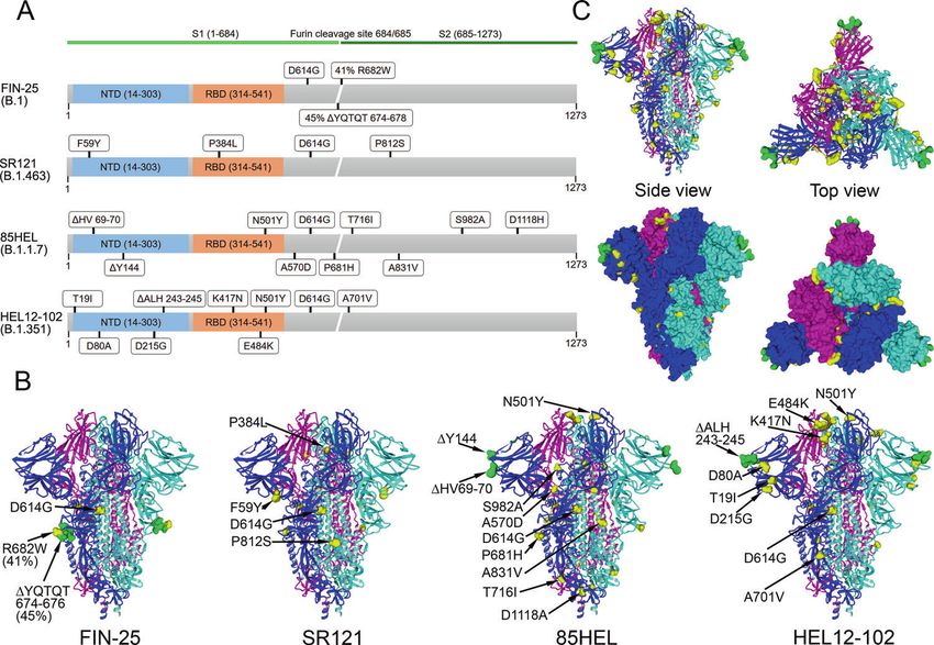
neutralization titre 20 or higher was considered positive and for calculation of geometric means a value of 10 was given for values ofARTICLE NATURE COMMUNICATIONS | https://doi.org/10.1038/s41467-021-24285-4 Fig. 2 Genetic analysis of virus variants and spike protein structure prediction. A Schematic presentation of S gene and amino acid changes in FIN-25 (B.1 lineage), SR121 (B.1.463), 85HEL (B.1.1.7), and HEL12-102 (B.1.351) virus isolates used in the present study. B Trimeric SARS-CoV-2 S protein, referred to as the spike, structure in the closed conformation (pdb: 6VXX). Amino acid substitutions (yellow) and deletions (green) as compared to the original spike structure. C Collective presentation of all amino acid changes found in virus isolates. Space-filling model indicating amino acids changes (yellow) and deletions (green) on the surface of a trimeric S protein. Side and top views are shown. The availability of the 3-dimensional structure of the SARS- GMTs further increased to 435 and 682, to 320 and 640, and to CoV-2 spike protein enabled the positioning of the amino acid 101 and 132, respectively (Supplementary Fig. 1B). These results changes into the structure of the trimeric spike protein (Fig. 2B). with this small group indicate that even one dose of the Substitutions found in FIN-25 and SR121 spike proteins localize BNT162b2 vaccine induces high MNT titres in those individuals in the stem regions of the trimeric spike protein. The who had suffered a previous COVID-19 infection. substitutions found in the spike proteins of B.1.1.7 and B.1.351 None of the vaccinees without a prior SARS-CoV-2 infection variants located both to the stem region and on the surface of the (169/180) had neutralizing antibodies before the vaccination trimeric spike protein close to the receptor-binding domain (Fig. 3A). Three weeks after the first vaccine dose, neutralizing (RBD). The three B.1.351 variant substitutions E484K, K417N, titres against all four isolates were slightly increased (GMT of 24 and N501Y are in the groove of the RBD–ACE2 interaction for FIN-25, 32 for SR121, 24 for 85HEL (B.1.1.7), and 12 for domain. In addition, both the B.1.1.7 and B.1.351 variants had HEL12-102 (B.1.351)). Six weeks after the first dose of the vaccine three amino acid deletions in the far edges of the 3-dimensional (3 weeks after the second dose), neutralizing titres were increased structure (Fig. 2B). Figure 2C shows combined amino acid to a GMT of 234 against FIN-25, 275 against SR121, 240 against changes found in the isolates used in this study indicating the 85HEL (B.1.1.7), and 48 against HEL12-102 (B.1.351) (Fig. 3A, accumulation of substitutions on multiple localizations on the Table 1). Three weeks after the first dose 37%, 17%, 37%, and 85% trimeric structure of the spike protein. The amino acid changes in of vaccinees had a neutralization titre 0.8, p < 0.0001) (Fig. 4). MNT titres for FIN-25 and HEL-12- increased geometric mean titres (GMT) of 35, 31, and 16 against 102 (B.1.351) correlated relatively well and highly significantly (r = FIN-25, 85HEL (B.1.1.7), and HEL12-102 (B.1.351) virus isolate, 0.74, p < 0.0001), as did the two variants of concern, 85HEL respectively. Following the first and the second vaccination, the (B.1.1.7) and HEL-12-102 (B.1.351) (r = 0.75, p < 0.0001). 4 NATURE COMMUNICATIONS | (2021)12:3991 | https://doi.org/10.1038/s41467-021-24285-4 | www.nature.com/naturecommunications
NATURE COMMUNICATIONS | https://doi.org/10.1038/s41467-021-24285-4                                                                           ARTICLE

Fig. 3 Neutralization of B.1.1.7 and B.1.351 variants by BNT162b2 vaccinees’ sera and COVID-19 patient sera. A Neutralization titres of initially
seronegative vaccinees (n = 169) for D614G variants FIN-25 and SR121, and 85HEL (B.1.1.7) and HEL12-102 (B.1.351) variants before (0d), three (3wk), and
six weeks (6wk) after the first dose of BNT162b2 vaccine and neutralization titres of convalescent sera of non-hospitalized patients (Conv, n = 50). Values
above the groups indicate geometric mean titres (GMTs) and data are shown as geometric means and geometric SDs. Neutralization titres
ARTICLE                                                                   NATURE COMMUNICATIONS | https://doi.org/10.1038/s41467-021-24285-4

Fig. 4 Correlation of MNT titres against SARS-CoV-2 isolates. MNT titres of BNT162b2 vaccinees (initially seronegative, n = 169) against FIN-25 were
compared with MNT titres against 85HEL (B.1.1.7) and HEL12-102 (B.1.351) variants including 0d, 3wk, and 6wk samples. Comparison between two D614G
virus isolates, FIN-25 and SR121, was done with sera from 86 BNT162b2 vaccinees. Correlation co-efficient (r) was calculated with Pearsons correlation test
and two-tailed p-values < 0.05 were considered significant. Each dot may represent multiple samples.

   To analyze the effect of age and gender on the antibody                     Discussion
responses, the vaccinees were divided into age and gender groups,              The emergence of the COVID-19 pandemic in early 2020
and the S1 IgG EIA and MNT results were compared between the                   prompted a rapid development of various types of vaccines such
groups (Fig. 5A and B). After the first vaccine dose, anti-S1 IgG               as mRNA encoding SARS-CoV-2 spike protein, viral vector-
antibody levels and neutralization titres were significantly lower in           based (e.g. adenovirus), inactivated virus, virus-like particle, and
the older age group (55–65 years) compared to younger age groups               recombinant protein vaccines. Once the European Union had
(20–34 and 35–44 years) (Fig. 5A). However, after the second                   made agreements with a number of vaccine producers, mass
vaccine dose, the neutralization titres were similar between the age           immunization was started in Finland at the end of December
groups (GMT 257, 268, 200, and 206 in age groups of 20–34,                     2020, first with the mRNA-based Pfizer-BioNTech vaccine and
35–44, 45–54, and 55–65 years, respectively) (Fig. 5A). We also                somewhat later the Moderna mRNA and AstraZeneca
compared gender-related antibody responses even though male                    adenovirus-based vaccines12. Vaccination of health care profes-
vaccinees were underrepresented, comprising only 17% (29/169) of               sionals within a national vaccination programme in Finland
the vaccinees. After the second dose, female vaccinees had slightly            enabled us to start, independent of pharmaceutical companies, a
higher neutralization titres than males (p = 0.0412), although the             follow-up study of vaccine-induced immunity. In the present
anti-S1 IgG antibody levels remained at the same level (Fig. 5B).              report, we show that two-dose vaccination with the BNT162b2
                                                                               mRNA COVID-19 vaccine induces very high antibody levels
EIA values correlate with MNT titres. Neutralization tests with                against viral spike protein and high titres of neutralizing anti-
live SARS-CoV-2 viruses are very time-consuming, and at the                    bodies. The vaccine induced good cross-reactivity to D614G and
moment the assay requires BSL-3 laboratory conditions, whereas                 B.1.1.7 variants in all vaccinees and, albeit reduced levels,
EIA and other similar colorimetric/fluorometric antibody assays                 detectable neutralizing antibodies to B.1.351 variant in 92% of the
are faster and user-friendlier. To assess whether EIA values are               vaccinees.
associated with MNT titres, anti-S1 IgG and total anti-S1 Ig were                 EIA is a rapid and sensitive method to analyze immune
compared to neutralization titres against FIN-25 (Fig. 6, Sup-                 responses against vaccine antigens or different viral proteins in
plementary Fig. 3). Both anti-S1 IgG and total anti-S1 Ig EIA                  response to infection. The method is easily quantitative and
measurements correlated very well with MNT titres (r > 0.9, p <                suitable for analyzing different immunoglobulin classes. In this
0.0001) suggesting that EIA, especially IgG EIA, using spike                   study, we observed that practically all seronegative health care
protein as an antigen can be a useful method to determine                      workers (20–65 years of age) responded to the first BNT162b2
COVID-19 immunity.                                                             vaccine dose and an increase in spike protein-specific antibody

6                  NATURE COMMUNICATIONS | (2021)12:3991 | https://doi.org/10.1038/s41467-021-24285-4 | www.nature.com/naturecommunications
NATURE COMMUNICATIONS | https://doi.org/10.1038/s41467-021-24285-4 ARTICLE Fig. 5 Antibody responses against SARS-CoV-2 S1 protein and neutralization of FIN-25 by age and gender. A BNT162b2 vaccinated health care workers (initially seronegative, n = 169) were divided into four age groups. Age specific differences of anti-S1 IgG antibody levels and neutralization titres against FIN-25 virus isolate were analyzed. Sera was collected three weeks (3w) and six weeks (6wk) after the first vaccine dose. Differences between age groups were tested with two-tailed Mann–Whitney U test. Two-tailed p-values < 0.05 were considered significant. Exact p-values were **=0.0078, ***=0.0007, and ****
ARTICLE                                                                   NATURE COMMUNICATIONS | https://doi.org/10.1038/s41467-021-24285-4

Fig. 6 Correlation of anti-S1 antibody levels with SARS-CoV-2 neutralization titres. Anti-S1 IgG and total Ig antibody levels were determined with EIA
and neutralization titres of BNT162b2 vaccinated health care workers (initially seronegative, n = 169) against FIN-25 virus isolate were obtained with
microneutralization test (MNT). All sequential serum samples (0d, 3wk, and 6wk) were included in the analysis. Spearman’s rank correlation coefficient (r)
is indicated.

antibody levels than those measured in patients29,30. Remarkably,              data accumulating from COVID-19 vaccine studies strongly
the administration of two doses of the mRNA vaccine induced                    suggest that already one dose of the vaccine provides protection
very high antibody responses in 100% of the vaccinees.                         against severe COVID-19, even when neutralizing antibody levels
    The global circulation of SARS-CoV-2 and a huge number of                  cannot be detected in all vaccinees33,34. This suggests that the first
infections worldwide have led to the emergence of hundreds of                  vaccine dose may prime the individual for rapid induction of
evolutionary lineages and variants of the virus (https://cov-lineages.         protective immunity when contracting the virus in nature and
org/global_report.html). The evolutionary speed of SARS-CoV-2                  avoiding severe COVID-19. According to previous data29,35–38,
has been relatively slow, at least compared to influenza A viruses,             we found that individuals with prior SARS-CoV-2 infection
presumably due to a virus-encoded enzyme with a proof-reading                  readily responded to the first vaccine dose with high antibody
capability. Within the first 16 months of circulation, up to 30–35              levels and neutralization titres.
mutations have been identified accumulating into the viral genome.                 Humoral immune response to vaccinations has been shown to
Many of these mutations are silent or appear in places of the                  decline with age39,40. Consistently, we observed a trend of
genome that are not critical for avoiding immunity induced by                  declining immune response to the COVID-19 mRNA vaccine by
vaccination or natural infection. However, a number of variants                age. This trend was not very strong, presumably because the ages
have raised concern due to mutations accumulating particularly in              of our vaccinees ranged from 20 to 65 years, while age-dependent
the S-gene and causing changes in the immunodominant epitopes                  immunosenescence should be more pronounced in the age group
of the trimeric spike protein. Mapping the spike protein mutations             >65 years39. Another explanation might be that the BNT162b2
on variants sequenced and used in this study revealed that they                mRNA vaccine is exceptionally immunogenic and therefore,
occur outside the globular head of the trimeric spike protein. The             especially when given two doses, it enables practically all indivi-
D614G and B.1.1.7 variant viruses were readily neutralized by the              duals regardless of gender and age, to develop high antibody
vaccinees’ sera, indicating that these mutations are unlikely to               levels and neutralization titres.
impair the neutralizing antibody capacity induced by vaccination or               In summary, in the present study we show that the Pfizer-
natural infection. However, it should be noted that the neutralizing           BioNTech BNT162b2 COVID-19 mRNA vaccine is highly
titre of these sera was five-fold lower against the B.1.351 variant,            immunogenic, and particularly after two vaccine doses, all vacci-
which denotes that the amino acid changes accumulating in this                 nees showed a very high humoral immune response to D614G
variant are potentiating the escape of the virus from the humoral              variant viruses. Immunity to a recent B.1.1.7 variant was equally
immune responses. Despite this, more than 92% of the vaccinees                 good as compared to the D614G variant, whereas vaccine and
showed measurable neutralizing antibody titres against the B.1.351             SARS-CoV-2 infection induced immunity against B.1.351 variant
variant, suggesting that the spike protein encoded by Pfizer-BioN-              was reduced. Despite this, almost all vaccinees showed neutralizing
Tech’s mRNA vaccine is similar enough to also mount an immune                  antibodies against the B.1.351 variant, suggesting to provide at least
response against the B.1.351 variant.                                          some degree of protection against these variant viruses. In the
    The critical amino acid changes linked to escape from humoral              future, it will be intriguing to study the development and persis-
immunity in the B.1.351 variant appear to be K417N, E484K, and                 tence of cell-mediated immunity induced by COVID-19 vaccines.
N501Y30–32. These amino acids are situated in the grooves within               Promising data have been reported at least for the BNT162b2
the receptor-binding site of the trimeric S protein complex. There             vaccine which in preliminary studies has induced good cell-
is no three-dimensional structure presently available for the                  mediated immunity41,42. As the use of other types of SARS-CoV-2
B.1.351 variant spike protein trimer, but because of its relatively            vaccines will be increased, it is the responsibility of the scientific
radical amino acid substitutions, conformational changes in the                community and public health professionals to systematically collect
spike structure may prove substantial. Interestingly, the B.1.351              serum and cellular samples for comparative analyses of vaccine-
and B.1.1.7 variants have deletions in the tips of the globular S1             induced immunity, cross-protection, and longevity of vaccine and
domain (amino acids 243–245 and amino acids 69–70 and 244,                     natural infection-induced immunity.
respectively) which could contribute to the impaired recognition                  As a whole, all vaccines that have currently obtained market
by neutralizing antibodies.                                                    authorization in the EU show excellent protective efficacy against
    It is currently not known how high neutralizing antibody titres            severe COVID-19. Thus, it is very likely that immunogenicity
against a given virus variant are required for antibody-mediated               results similar to those presented here will be applicable to them
protection against the COVID-19. However, the clinical efficacy                 as well.

8                  NATURE COMMUNICATIONS | (2021)12:3991 | https://doi.org/10.1038/s41467-021-24285-4 | www.nature.com/naturecommunications
NATURE COMMUNICATIONS | https://doi.org/10.1038/s41467-021-24285-4 ARTICLE
Methods streptomycin. For virus propagation in VeroE6-TMPRSS2-H10 cells, D-MEM
Study participants. SARS-CoV-2 vaccinations started in Finland at the end of supplemented with 2% FBS, 2 mM L-glutamine, and penicillin/streptomycin was
December 2020 with Pfizer-BioNTech BNT162b2 mRNA (Comirnaty) vaccine. used. Supernatants containing viruses were harvested, cell debris removed with
Study participants (n = 180) were recruited among healthcare personnel of Turku centrifugation at 500×g for 5 min, and aliquots stored at −80 °C.
University Hospital (TUH, Turku, Finland) (Southwest Finland health district Fifty-percent tissue culture infective dose (TCID50) of virus stocks was
ethical permission ETMK 19/1801/2020) and Helsinki University Hospital (HUH, determined with endpoint dilution assay in VeroE6-TMPRSS2-H10 cells. Briefly,
Helsinki, Finland) (Helsinki-Uusimaa health district ethical permission HUS/1238/ 50,000 cells per well were plated on 96-well tissue culture plates (Sarstedt), and the
2020) prior to receiving an optimal regimen of two doses of BNT162b2 mRNA next day media was changed to infection media (2% FBS). Ten-fold virus dilutions
vaccine at a 3-week dosing interval as part of hospital occupational health care. in infection media were applied onto cells, and the plates were incubated for 3 days
Serum samples were collected before or on the day of the first vaccine dose (0-day at +37 °C and 5% CO2. Cells were fixed with 4% formaldehyde and stained with
sample, n = 180), 16–28 days (mean 20) after the first vaccine dose (3-week sample, crystal violet. Virus dilution resulting in 50% cell death was determined to
n = 176), and 13–33 (mean 23) days after the second vaccine dose (34–54 days represent TCID50 value of the stock virus. Virus propagations and end-point
after the first vaccine dose) (6-week sample, n = 180). At enrollment, written dilution assays were done in BSL-3 laboratory conditions.
informed consent was collected from all participants.
Convalescent phase serum samples (n = 50) were collected at HUH from Sequencing of SARS-CoV-2 isolates. For sequencing of virus stocks, the viral
patients with initial RT-qPCR confirmed home-treated COVID-19 infection RNA was extracted from supernatants using the RNeasy Mini kit (Qiagen) and
(Helsinki-Uusimaa health district ethical permission HUS/1238/2020). The reverse-transcribed to cDNA with LunaScript RT SuperMix kit (New England
patients provided written informed consent and were sampled 14 days–6 weeks Biolabs). Primer pools (Supplementary Table 1) targeting SARS-CoV-2 were
after the positive PCR test result. As negative control serum samples (n = 20) we designed using PrimalScheme tool45 and PCR was done with PhusionFlash PCR
used randomly selected diagnostic serum samples collected at TUH prior to master mix (Thermo Scientific). Sequencing libraries were prepared with NEBNext
COVID-19 pandemic43. Negative control serum samples were used for calculation ultra II FS DNA library kit (New England Biolabs) according to the manufacturer’s
of cut-off values for EIA (see below) and the samples were fully de-identified and instructions and sequenced using Illumina Miseq with v3 sequencing kit. Raw
collected primarily for epidemiological purposes and thus do not require written sequence reads were trimmed, and low quality (quality scoreARTICLE NATURE COMMUNICATIONS | https://doi.org/10.1038/s41467-021-24285-4
4. Amanat, F. et al. A serological assay to detect SARS-CoV-2 seroconversion in 35. Krammer, F. et al. Antibody responses in seropositive persons after a single
humans. Nat. Med. 26, 1033–1036 (2020). dose of SARS-CoV-2 mRNA vaccine. N. Engl. J. Med. 384, 1372–1374 (2021).
5. Wajnberg, A. et al. Robust neutralizing antibodies to SARS-CoV-2 infection 36. Saadat, S. et al. Binding and neutralization antibody titers after a single vaccine
persist for months. Science 370, 1227–1230 (2020). dose in health care workers previously infected with SARS-CoV-2. JAMA 325,
6. Deng, W. et al. Primary exposure to SARS-CoV-2 protects against reinfection 1467–1469 (2021).
in rhesus macaques. Science 369, 818–823 (2020). 37. Polack, F. P. et al. Safety and Efficacy of the BNT162b2 mRNA Covid-19
7. Alsoussi, W. B. et al. A potently neutralizing antibody protects mice against Vaccine. N. Engl. J. Med. 383, 2603–2615 (2020).
SARS-CoV-2 infection. J. Immunol. 205, 915–922 (2020). 38. Capetti, A. F. et al. Impressive boosting of anti-S1/S2 IgG production in
8. Hanrath, A. T., Payne, B. A. I. & Duncan, C. J. A. Prior SARS-CoV-2 infection COVID-19-experienced patients after the first shot of the BNT162b2 mRNA
is associated with protection against symptomatic reinfection. J. COVID-19 vaccine. Clin. Infect. Dis. ciab214 (2021).
Infect. 82, e29–e30 (2020). 39. Chen, W. H. et al. Vaccination in the elderly: an immunological perspective.
9. Hall, V. J. et al. SARS-CoV-2 infection rates of antibody-positive compared Trends Immunol. 30, 351–359 (2009).
with antibody-negative health-care workers in England: a large, multicentre, 40. Prendecki, M. et al. Effect of previous SARS-CoV-2 infection on humoral and
prospective cohort study (SIREN). Lancet 397, 1459–1469 (2021). T-cell responses to single-dose BNT162b2 vaccine. Lancet 6736, 10–12 (2021).
10. Chodick, G. et al. Assessment of Effectiveness of 1 Dose of BNT162b2 Vaccine 41. Sahin, U. et al. COVID-19 vaccine BNT162b1 elicits human antibody and
for SARS-CoV-2 Infection 13 to 24 Days After Immunization. JAMA Netw TH1 T cell responses. Nature 586, 594–599 (2020).
Open. 4, e2115985 (2021). 42. Goel, R. R. et al. Distinct antibody and memory B cell responses in SARS-
11. Pradenas, E. et al. Stable neutralizing antibody levels 6 months after mild and CoV-2 naive and recovered individuals following mRNA vaccination. Sci.
severe COVID-19 episodes. Med 2, 313–320 (2021). Immunol. 6, eabi6950 (2021).
12. EMA. COVID-19 Vaccines: Authorised. Accessed 5th June 2021. https://www. 43. Jalkanen, P. et al. A combination of N and S antigens with IgA and IgG
ema.europa.eu/en/human-regulatory/overview/public-health-threats/ measurement strengthens the accuracy of SARS-CoV-2 serodiagnostics. J.
coronavirus-disease-covid-19/treatments-vaccines/vaccines-covid-19/covid- Infect. Dis. jiab222 (2021).
19-vaccines-authorised#authorised-covid-19-vaccines-section. 44. Juuso, R. et al. A Generic, Scalable, and Rapid Time-Resolved Förster
13. Folegatti, P. M. et al. Safety and immunogenicity of the ChAdOx1 nCoV-19 Resonance Energy Transfer-Based Assay for Antigen Detection—SARS-CoV-
vaccine against SARS-CoV-2: a preliminary report of a phase 1/2, single-blind, 2 as a Proof of Concept. MBio 12, e00902–21 (2021).
randomised controlled trial. Lancet 396, 467–478 (2020). 45. Quick, J. et al. Multiplex PCR method for MinION and illumina sequencing of
14. Anderson, E. J. et al. Safety and immunogenicity of SARS-CoV-2 mRNA-1273 Zika and other virus genomes directly from clinical samples. Nat. Protoc. 12,
vaccine in older adults. N. Engl. J. Med. 383, 2427–2438 (2020). 1261–1266 (2017).
15. Walsh, E. E. et al. Safety and immunogenicity of two RNA-based Covid-19 46. Bolger, A. M., Lohse, M. & Usadel, B. Trimmomatic: a flexible trimmer for
vaccine candidates. N. Engl. J. Med. 383, 2439–2450 (2020). Illumina sequence data. Bioinformatics 30, 2114–2120 (2014).
16. Public Health England. Investigation of SARS-CoV-2 Variants of Concern in 47. Li, H. Aligning sequence reads, clone sequences and assembly contigs with
England https://www.gov.uk/government/publications/investigation-of-novel- BWA-MEM. Preprint at arXiv:1303.3997 (2013).
sars-cov-2-variant-variant-of-concern-20201201 (2021). 48. Li, H. et al. The sequence alignment/Map format and SAMtools.
17. Tegally, H. et al. Emergence and rapid spread of a new severe acute respiratory Bioinformatics 25, 2078–2079 (2009).
syndrome-related coronavirus 2 (SARS-CoV-2) lineage with multiple spike 49. Walls, A. C. et al. Structure, function, and antigenicity of the SARS-CoV-2
mutations in South Africa. Preprint at bioRxiv https://www.medrxiv.org/ spike glycoprotein. Cell 181, 281–292.e6 (2020).
content/10.1101/2020.12.21.20248640v1 (2020).
18. Zhou, D. et al. Evidence of escape of SARS-CoV-2 variant B.1.351 from
natural and vaccine induced sera. Cell 184, 2384–2361.e6 (2021). Acknowledgements
19. Shen, X. et al. SARS-CoV-2 variant B.1.1.7 is susceptible to neutralizing We thank Soili Jussila for cell maintenance, Mikael Ritvos for assistance in protein
antibodies elicited by ancestral spike vaccines. Cell Host Microbe 29, 529–539. production, and Anne Suominen, Anne-Mari Pieniniemi, and Simo Miettinen for
e3 (2021). technical assistance. I.J. was funded by Jane and Aatos Erkko Foundation (grant numbers
20. Rambaut, A. et al. Preliminary genomic characterisation of an emergent 3067-84b53 and 5360-cc2fc), the Academy of Finland (AoF; grant numbers 336410 and
SARS-CoV-2 lineage in the UK defined by a novel set of spike mutations. 337530) and Sigrid Jusélius Foundation. J.H. was funded by AoF (grant number 308613)
Virological.org (2020). A.K. was funded by AoF (grant numbers 336439 and 335527), the Finnish Medical
21. Muik, A. Neutralization of SARS-CoV-2 lineage B.1.1.7 pseudovirus by Foundation, and private donors through UH. O.V., T.S., and S.K. were funded by Jane
BNT162b2 vaccine-elicited human sera. Science 6105, 1–5 (2021). and Aatos Erkko Foundation, AoF (grant numbers 336490 to O.V.) and Helsinki Uni-
22. Cele, S., et al. Escape of SARS-CoV-2 501Y.V2 from neutralization by versity Hospital Funds (TYH2018322). P.A.T., L.I., and J.L. were funded by The Turku
convalescent plasma. Nature593, 142–146 (2021). University Hospital Research Foundation.
23. Virtanen, J. et al. Kinetics of Neutralizing Antibodies of COVID-19 Patients
Tested Using Clinical D614G, B.1.1.7, and B 1.351 Isolates in Author contributions
Microneutralization Assays. Viruses 13, 996 (2021). P.J., L.K., J.L., A.K., and I.J. designed the experiments; P.J., P.K., M.H., S.M., R.L. and L.K.
24. Plante, J. A. et al. Spike mutation D614G alters SARS-CoV-2 fitness. did microneutralization tests and analyzed the data; P.J., A.R. and S.T. did EIA tests and
Nature 592, 116–121 (2020). analyzed the data; H.K.H., S.H.P., P.A.T., I.L., A.N., T.M., H.V., L.I., J.L. and A.K.
25. Hou, Y. J. et al. SARS-CoV-2 D614G variant exhibits enhanced recruited vaccinees and patients and collected their sera and data; A.P., R.N., P.J. and
replication ex vivo and earlier transmission in vivo. Science 370, 1464–1468 O.R. produced antigens for EIA; P.Ö., S.K., J.H. and O.V. isolated and characterized virus
(2021). strains; J.H. produced VeroE6-TMPRSS2-H10 cell line; T.S. did sequencing and T.S., J.H.
26. Wibmer, C. K. et al. SARS-CoV-2 501Y. V2 escapes neutralization by South
and P.K. analyzed sequences and structures; P.J. analyzed all data sets; P.J., L.K., A.K. and
African COVID-19 donor plasma. Nat. Med. 27, 622–625 (2021).
I.J. wrote the manuscript and all co-authors contributed to the edition of the text.
27. Lynch, K. L. et al. Magnitude and kinetics of anti-severe acute respiratory
syndrome Coronavirus 2 antibody responses and their relationship to disease
severity. Clin. Infect. Dis. 72, 301–308 (2021). Competing interests
28. Grossberg, A. N. et al. A multiplex chemiluminescent immunoassay for The authors declare no competing interests.
serological profiling of COVID-19-positive symptomatic and asymptomatic
patients. Nat. Commun. 12, 740 (2021).
29. Manisty, C. et al. Correspondence antibody response to first BNT162b2 Additional information
Supplementary information The online version contains supplementary material
dose in previously SARS-CoV-2-infected individuals. Lancet 6736, 2–3
available at https://doi.org/10.1038/s41467-021-24285-4.
(2021).
30. Wang, Z. et al. mRNA vaccine-elicited antibodies to SARS-CoV-2 and
Correspondence and requests for materials should be addressed to P.J. or I.J.
circulating variants. Nature 592, 616–622 (2021).
31. Collier, D. A. et al. Sensitivity of SARS-CoV-2 B.1.1.7 to mRNA vaccine- Peer review information Nature Communications thanks Peter Kelleher and the other,
elicited antibodies. Nature 593, 136–141 (2021). anonymous, reviewer(s) for their contribution to the peer review of this work. Peer
32. Liu, Y. et al. Neutralizing activity of BNT162b2-elicited serum. N. Engl. J. Med. reviewer reports are available.
384, 1466–1468 (2021).
33. Dagan, N. et al. BNT162b2 mRNA Covid-19 vaccine in a nationwide mass
Reprints and permission information is available at http://www.nature.com/reprints
vaccination setting. N. Engl. J. Med. 384, 1412–1423 (2021).
34. Amit, S., Regev-Yochay, G., Afek, A., Kreiss, Y. & Leshem, E. Early rate
Publisher’s note Springer Nature remains neutral with regard to jurisdictional claims in
reductions of SARS-CoV-2 infection and COVID-19 in BNT162b2 vaccine
published maps and institutional affiliations.
recipients. Lancet 397, 875–877 (2021).
10 NATURE COMMUNICATIONS | (2021)12:3991 | https://doi.org/10.1038/s41467-021-24285-4 | www.nature.com/naturecommunicationsNATURE COMMUNICATIONS | https://doi.org/10.1038/s41467-021-24285-4 ARTICLE
Open Access This article is licensed under a Creative Commons
Attribution 4.0 International License, which permits use, sharing,
adaptation, distribution and reproduction in any medium or format, as long as you give
appropriate credit to the original author(s) and the source, provide a link to the Creative
Commons license, and indicate if changes were made. The images or other third party
material in this article are included in the article’s Creative Commons license, unless
indicated otherwise in a credit line to the material. If material is not included in the
article’s Creative Commons license and your intended use is not permitted by statutory
regulation or exceeds the permitted use, you will need to obtain permission directly from
the copyright holder. To view a copy of this license, visit http://creativecommons.org/
licenses/by/4.0/.
© The Author(s) 2021
NATURE COMMUNICATIONS | (2021)12:3991 | https://doi.org/10.1038/s41467-021-24285-4 | www.nature.com/naturecommunications 11You can also read