JBC Papers in Press. Published on July 11, 2019 as Manuscript RA119.007730 The latest version is at ...
←
→
Page content transcription
If your browser does not render page correctly, please read the page content below
JBC Papers in Press. Published on July 11, 2019 as Manuscript RA119.007730
         The latest version is at http://www.jbc.org/cgi/doi/10.1074/jbc.RA119.007730

    Cross-reactivity of a rice NLR immune receptor to distinct effectors from the rice blast pathogen
                          Magnaporthe oryzae provides partial disease resistance

    Freya A. Varden1, Hiromasa Saitoh2, Kae Yoshino2, Marina Franceschetti1, Sophien Kamoun3, Ryohei
                                      Terauchi4,5, Mark J. Banfield1*
1
 Department of Biological Chemistry, John Innes Centre, Norwich Research Park, NR4 7UH, Norwich,
UK. 2Laboratory of Plant Symbiotic and Parasitic Microbes, Department of Molecular Microbiology,
Faculty of Life Sciences, Tokyo University of Agriculture, Tokyo 156-8502, Japan. 3The Sainsbury
Laboratory, University of East Anglia, Norwich Research Park, NR4 7UH, Norwich, UK. 4Division of
Genomics and Breeding, Iwate Biotechnology Research Center, Iwate 024-0003, Japan. 5Laboratory of
Crop Evolution, Graduate School of Agriculture, Kyoto University, Kyoto, 606-8501, Japan.

                       Running title: Cross-reactivity of a rice NLR to blast effectors

* Correspondence to: Mark J. Banfield, Department of Biological Chemistry, John Innes Centre, Norwich
Research Park, NR4 7UH, Norwich, UK. Tel: +44 (0)1603 450742. email: mark.banfield@jic.ac.uk

                                                                                                               Downloaded from http://www.jbc.org/ by guest on November 7, 2019
Keywords: Magnaporthe oryzae, protein-protein interaction, effector, NLR, MAX, HMA, integrated
domain, plant immunity, host-pathogen interaction, X-ray crystallography

ABSTRACT                                                 to AVR-Pia at 1.9 Å resolution revealed a binding
                                                         interface different from those formed with AVR-
Unconventional integrated domains in plant               Pik effectors, suggesting plasticity in integrated
intracellular immune receptors of the nucleotide-        domain–effector interactions. The results of our
binding leucine-rich repeat (NLRs) type can              work indicate that a single NLR immune receptor
directly bind translocated effector proteins from        can bait multiple pathogen effectors via an
pathogens and thereby initiate an immune response.       integrated domain, insights that may enable
The rice (Oryza sativa) immune receptor pairs Pik-       engineering plant immune receptors with extended
1/Pik-2 and RGA5/RGA4 both use integrated                disease resistance profiles.
heavy metal–associated (HMA) domains to bind
the effectors AVR-Pik and AVR-Pia, respectively,         INTRODUCTION
from the rice blast fungal pathogen Magnaporthe
oryzae. These effectors both belong to the MAX           When plants encounter biotic stresses, they respond
effector family and share a core structural fold,        rapidly to defend themselves against attack.
despite being divergent in sequence. How                 Microbial pathogens translocate effector proteins
integrated domains in NLRs maintain specificity of       inside host cells to undermine plant immunity and
effector recognition, even of structurally similar       promote pathogen growth and proliferation (1). To
effectors, has implications for understanding plant      detect these effectors, plants have developed
immune receptor evolution and function. Here,            intracellular immune receptors, many of which are
using plant cell death and pathogenicity assays and      of the NLR (nucleotide-binding leucine-rich repeat)
protein–protein interaction analyses, we show that       class (2). The hallmark feature of NLR-mediated
the rice NLR pair Pikp-1/Pikp-2 triggers an              immunity is the hypersensitive response (HR), a
immune response leading to partial disease               programmed cell death around the site of infection
resistance towards the “mis-matched” effector            that helps to isolate and halt the spread of the
AVR-Pia in planta, and that the Pikp-HMA domain          pathogen (3).
binds AVR-Pia in vitro. We observed that the HMA         NLRs recognise effector proteins via different
domain from another Pik-1 allele, Pikm, cannot           mechanisms, including by direct or indirect binding
bind AVR-Pia, and does not trigger a plant               (4,5). Some NLRs function in pairs, with one
response. The crystal structure of Pikp-HMA bound        receptor responsible for recognising the effector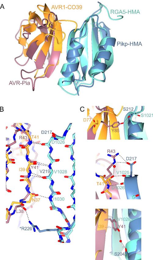
Running title: Cross-reactivity of a rice NLR to blast effectors

(referred to as the sensor), and one responsible for        interact with RGA5-HMA and this interaction is
translating the recognition into a signalling               required for triggering resistance (13,26).
response (the helper) (6). One mechanism to evolve          Despite similarities in the Pik-1/Pik-2 and
direct binding has been for NLRs to integrate an            RGA5/RGA4 systems, their mechanisms of
unconventional domain into the protein architecture         activation are different. The Pik-1/Pik-2 pair appear
(7,8), with this domain thought to be derived from          to use a cooperative mechanism, where effector
the virulence-associated host target of the effector.       recognition by the HMA in the sensor NLR Pik-1
Once integrated, these domains may adapt to                 requires the helper NLR Pik-2 to initiate signalling,
recognise effectors (and different effector alleles).       but Pik-2 cannot signal on its own. Contrastingly,
Their widespread distribution in NLRs from diverse          the RGA5/RGA4 pair functions via negative
plant species suggests this is an ancient mechanism         regulation, where recognition of the effector
for evolving effector recognition (9,10).                   through RGA5-HMA derepresses signalling by
Two paired rice NLR immune receptors are known              RGA4 (27,28). However, details of the NLR
that contain an integrated heavy metal-associated           interactions and the resultant downstream
(HMA) domain, Pik-1/Pik-2 and RGA5/RGA4. In                 signalling remain to be understood.
Pik, this domain is integrated between the coiled-          The interface between AVR-Pik effectors and the
coil (CC) and nucleotide-binding (NB-ARC)                   HMA domain of both Pikp and Pikm has been

                                                                                                                    Downloaded from http://www.jbc.org/ by guest on November 7, 2019
domains of Pik-1 (11,12), whereas in the RGA pair           extensively studied and structurally characterised
the HMA domain is found at the C-terminus of                (11,12). Recently, the structure of AVR1-CO39 in
RGA5 (13). Both these pairs of immune receptors             complex with the HMA domain of RGA5 was also
recognise effectors from the blast fungus                   elucidated (29), and revealed that the HMA/effector
Magnaporthe oryzae, a global threat to rice                 interface was substantially different compared to
production causing loss of up to a third of the total       the Pik NLR pairs. This has raised intriguing
annual harvest of this crop (14-16).                        questions concerning how structurally similar but
M. oryzae secretes a large repertoire of effector           sequence divergent HMA domains distinguish
proteins and many of these, including the                   between structurally similar but sequence divergent
structurally characterised AVR-Pizt, AVR-Pia,               pathogen effectors.
AVR-Pik, AVR1-CO39 and AVR-Pib (11,17-19),                  Here we reveal that Pikp is able to trigger partial
share a conserved structure comprising a six                disease resistance to the “mis-matched” effector
stranded β-sandwich known as the MAX                        AVR-Pia in rice, and elicits a weak cell death
(Magnaporthe Avrs and ToxB-like) fold (18,20).              response in N. benthamiana. Pikp-HMA binds
Therefore, despite being sequence-unrelated, these          AVR-Pia in vitro, at the RGA5/AVR1-CO39-like
effectors are all similar in overall shape.                 interface, rather than the Pik/AVR-Pik-like
The Pik-1/Pik-2 NLR pair recognise the M. oryzae            interface. This structural understanding of effector
effector AVR-Pik (21), and both the NLRs and                cross-reactivity in the Pik/RGA systems provides
effectors are found as allelic series in natural            insights into the evolution and function of
populations (22). Direct interaction between the            integrated HMA domains in NLRs. It also hints at
Pik-HMA domain and AVR-Pik is required for                  the potential to engineer the HMA of Pikp to
triggering an immune response to the effector (11).         respond robustly to both AVR-PikD and AVR-Pia
At the sequence level, the allelic Pikp (23) and            at the different interfaces.
Pikm (24) pair differ mainly in their polymorphic
HMA domains (12) and this underpins different               RESULTS
recognition specificities for different AVR-Pik
alleles; Pikp is only able to recognise the effector        Rice plants expressing Pikp are partially
variant AVR-PikD, whereas Pikm can recognise                resistant to Magnaporthe oryzae expressing
AVR-PikD and other additional AVR-Pik variants.             AVR-Pia
The AVR-PikC effector variant is currently                  We used a spot-inoculation assay to infect rice
unrecognised by any Pik NLR (22).                           cultivars with a pathogen strain (Sasa2)
The RGA5/RGA4 NLR pair responds to the M.                   transformed to express different effectors. As
oryzae effectors AVR-Pia (25) and AVR1-CO39                 expected, rice plants that do not express either Pik
(13). Both AVR-Pia and AVR1-CO39 physically                 or RGA NLRs (cv. Nipponbare) are susceptible to

                                                        2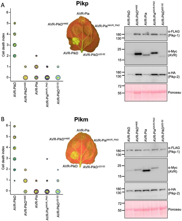
Running title: Cross-reactivity of a rice NLR to blast effectors

infection by all M. oryzae Sasa2 lines tested (clear        the weak cell death response to AVR-Pia is specific
spreading lesions away from the infection site, Fig.        for the Pikp allele.
1). Rice plants expressing Pikp (cv. K60) showed
resistance to the Sasa2 lines expressing AVR-PikD           The HMA domain of Pikp can bind AVR-Pia in
(positive control) and consistently displayed a             vitro
qualitatively reduced susceptibility (partial               Previously, a tight correlation was observed
resistance) phenotype to lines expressing AVR-Pia,          between in planta response phenotypes in N.
developing disease lesions that spread away from            benthamiana and rice, and in vitro binding between
the infection site, but are not as developed as the         Pik-HMA domains and effectors (12,18). We
negative controls. This partial resistance phenotype        therefore tested the interaction of Pikp-HMA and
was not observed in rice plants expressing Pikm             Pikm-HMA domains with AVR-Pia following
(cv. Tsuyuake), consistent with results from N.             heterologous expression and purification of these
benthamiana. Further, rice plants expressing                proteins.
RGA5/RGA4 (cv. Sasanishiki) are susceptible to              Firstly, analytical gel filtration was used to
the Sasa2 line expressing AVR-PikD, showing                 qualitatively determine whether Pik-HMA domains
these NLRs do not partially respond to this effector.       and AVR-Pia could form a complex. In isolation,
All pairwise resistance phenotypes behaved as               AVR-Pia elutes at a retention volume of 15-15.5

                                                                                                                      Downloaded from http://www.jbc.org/ by guest on November 7, 2019
expected.                                                   mls (Fig. 3A). When mixed with the Pikm-HMA
                                                            domain, no change in AVR-Pia retention was
Co-expression of Pikp/AVR-Pia in Nicotiana                  observed, consistent with the lack of response in
benthamiana elicits a weak cell death response              plants. By contrast, when mixed with the Pikp-
N. benthamiana is a well-established model system           HMA domain, AVR-Pia elutes earlier at ~12 mls
for assaying the response of rice NLRs to M. oryzae         suggesting a complex is formed, which was
effectors (11,12,28). Therefore, we used this system        confirmed by SDS-PAGE (Fig. S1). Note that Pik-
to test whether Pik NLRs would show any response            HMA domains do not sufficiently absorb UV light
to the effector AVR-Pia. When AVR-Pia was                   to give a signal in gel filtration under the conditions
transiently expressed in N. benthamiana via                 shown, but can be seen by SDS-PAGE.
agroinfiltration, along with Pikp-1 and Pikp-2, there       We then used surface plasmon resonance (SPR) to
was a weak cell death response observed, as                 measure binding affinities, as described previously
visualised by a yellowing of the tissue at the              (12). These results were expressed as a percentage
infiltration site, and fluorescence under UV light          of the theoretical maximum response (Rmax), which
(Fig. 2A). The cell death was weaker compared to            gives a relative indication of binding strength. The
AVR-PikD (positive control), but was stronger than          positive and negative controls for Pikp-HMA and
for the AVR-PikD point mutant (AVR-PikDH46E), a             Pikm-HMA binding, the effector variants AVR-
negative control that is not recognised by Pikp (11).       PikD and AVR-PikC, show strong and weak/no
To confirm that each protein was expressed,                 binding, as expected (Fig. 3B, Fig. S1, Fig. S2).
Western blot analysis of extracted leaf tissue was          Consistent with gel filtration, essentially no binding
used to assess protein accumulation (Fig. 2A).              is observed between Pikm-HMA and AVR-Pia, but
These results show that the Pikp NLRs can respond           Pikp-HMA binds AVR-Pia at ~50 % Rmax (for the
to AVR-Pia, although the response was limited               100 nM Pikp-HMA concentration), independently
compared to their ‘matched’ effector AVR-PikD.              confirming in vitro interaction and correlating with
Interestingly, when the Pikm-1/Pikm-2 pair were             in planta responses.
tested against the same effectors (AVR-PikD,
AVR-PikDH46E and AVR-Pia), there was no                     Pikp-HMA binds AVR-Pia at a different
macroscopic cell death observed to AVR-Pia in               interface to AVR-PikD
planta, despite confirmed expression of all proteins        To visualise the interface formed between Pikp-
in the leaf tissue (Fig. 2B). There was a weak              HMA and AVR-Pia, and compare it to that with
response to the AVR-PikDH46E negative control, as           AVR-Pik, we purified the complex between these
previously observed, due to differences in the              proteins and determined the structure to 1.9 Å
AVR-PikD His46 interface with Pikm-HMA                      resolution using X-ray crystallography. The details
compared with Pikp-HMA (12). This suggests that             of X-ray data collection, structure solution and

                                                        3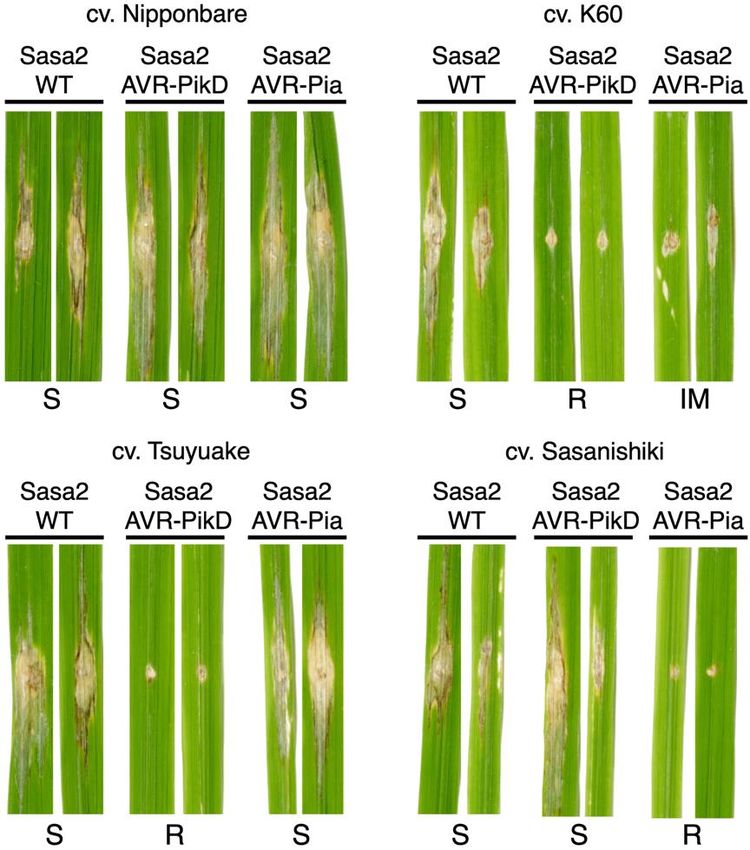
Running title: Cross-reactivity of a rice NLR to blast effectors
structure completion are given in Materials and complex, with a hydrogen bond/salt bridge
Methods, Table 1 and Fig. S3. interaction formed between AVR-PiaR43 and
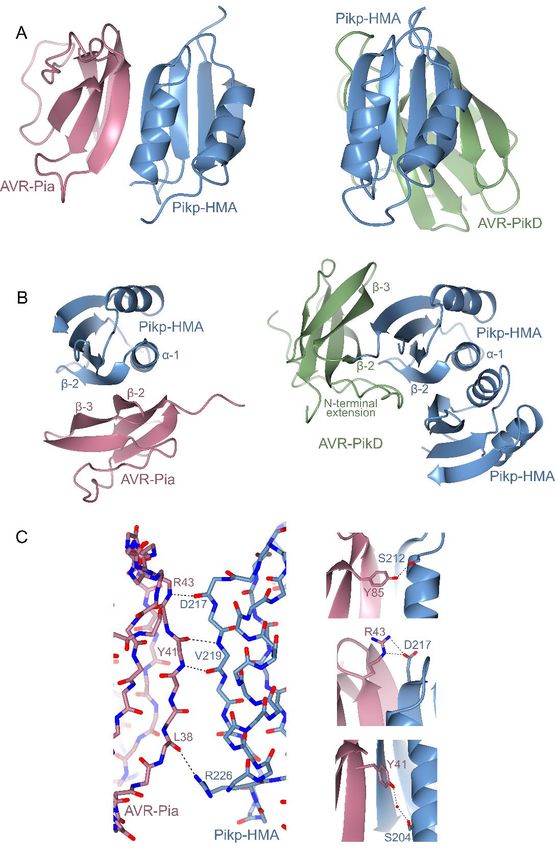
Each partner in the complex adopts a similar overall PikpD217, and the hydroxyl group on the C-terminal
fold to previously solved structures. Pikp-HMA residue of AVR-Pia, Tyr85, also forms a hydrogen
(11,12) comprises two adjacent α-helices opposite bond with PikpS212 (Fig. 4C). Finally, an indirect
a four-stranded β-sheet (Fig. 4A, 4B). Previous interaction, mediated by a water molecule, is found
structures of AVR-Pia were determined by NMR between the side chains of AVR-PiaY41 and PikpS204
spectroscopy (18,30), and the crystal structure (Fig. 4C). These limited intermolecular interactions
determined here is very similar (0.92 Å over 65 and small interface area provide an explanation for
aligned residues), comprising the six-stranded β- the weaker binding affinity seen for Pikp-HMA to
sandwich characteristic of MAX effectors (18). In AVR-Pia when compared to AVR-PikD in vitro
the crystal structure, β-5 is not well-defined and (Fig. 3B, Fig. S1, Fig. S2), and reduced responses
appears as a loop joining β-4 and β-6, but overall in planta.
the configuration of this region is similar to the
NMR ensemble. As previously observed, a Pikp recognises AVR-Pia through different
disulphide bond is formed between residues Cys25 molecular features compared to AVR-PikD
and Cys66. Despite only sharing 17 % sequence identity (Fig.
Downloaded from http://www.jbc.org/ by guest on November 7, 2019
Strikingly, although the two proteins in the complex S5), AVR-Pia and AVR-PikD both adopt the MAX
adopt essentially identical folds to their structures effector fold. However, AVR-PikD also contains an
in isolation, Pikp-HMA binds AVR-Pia at a additional N-terminal extension (comprising
completely different interface to the AVR-Pik residues Arg31 to Pro52) that partially wraps
effectors (Fig. 4A, 4B). Whilst Pikp-HMA binds around, and is held in place by, the core structure
AVR-PikD opposite the face of its β-sheet, it binds (see Fig. 4B, Fig. S5). This extension plays a key
AVR-Pia adjacent to α-1 and β-2 (Fig. 4B). In both role in the interaction of AVR-PikD and Pikp-
cases, the position of Pikp-HMA relative to the HMA, including a histidine residue (His46), which
effector allows the formation of a continuous anti- forms hydrogen bond/salt bridge interactions with
parallel β-sheet between the proteins (Fig. S4). In Ser218 and Glu230 in Pikp-HMA (11). We
the case of AVR-PikD, the β-strands from Pikp- considered that modifying the core MAX fold of
HMA form a sheet with β-strands 3-5 of AVR- AVR-Pia, to add the AVR-PikD N-terminal
PikD. For AVR-Pia, the β-strands involved are 1, 2 extension, might allow Pikp to respond more
and 6. Another striking feature is that while Pikp- strongly to the effector by switching the interaction
HMA is a dimer in the structure with AVR-PikD of the chimeric effector (AVR-PiaNAVR-PikD) to the
(11,12), it is a monomer with AVR-Pia. Indeed, ‘AVR-PikD-like’ interface of Pikp-HMA. We also
AVR-Pia occupies the same binding surface as the investigated the effect of removing the N-terminal
Pikp-HMA dimer in the Pikp-HMA/AVR-PikD extension from AVR-PikD (AVR-PikDΔ22-52).
structure, which suggests that AVR-Pia binding is After generating the appropriate constructs, they
competing with Pikp-HMA dimerization in were expressed in N. benthamiana via
solution. agroinfiltration alongside Pikp-1/Pikp-2 or Pikm-
The interface formed between Pikp-HMA and 1/Pikm-2. In these assays, neither Pikp nor Pikm
AVR-Pia covers an area of 460 Å2 (as calculated by responded to either AVR-PiaNAVR-PikD or AVR-
PISA (31)), approximately half of that seen PikDΔ22-52 (Fig. 5). Western blot analysis showed
between Pikp-HMA and AVR-PikD (986 Å2 (12)). that accumulation of AVR-PikDΔ22-52 in the leaf
Further, the interface between Pikp-HMA and tissue is low, suggesting that the N-terminal
AVR-Pia is dominated by hydrogen bonds between truncation has destabilised AVR-PikD (Fig. 5).
the peptide backbone, with the main contributions However, we confirmed the expression of AVR-
derived from Pikp-HMAD217, Pikp-HMAV219, PiaNAVR-PikD in the infiltrated leaf tissue, suggesting
AVR-PiaY41 and AVR-PiaR43 (Fig. 4C). The that the lack of cell death in this case is not due to
backbone oxygen atom of AVR-PiaL38 also forms a lack of protein accumulation (Fig. 5). It is possible
hydrogen bond with the side chain of Pikp- that AVR-PiaNAVR-PikD retains interaction at the
HMAR226. There are only limited side chain ‘AVR-Pia-like’ interface, but the presence of a
mediated interactions in the Pikp-HMA/AVR-Pia disordered N-terminal extension hinders response
4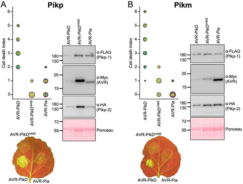
Running title: Cross-reactivity of a rice NLR to blast effectors

in the full-length protein (the N-terminus cannot           regions in the RGA5-HMA/AVR1-CO39 complex
adopt the same conformation as AVR-PikD at the              noted by Guo et al. are shared by Pikp-HMA/AVR-
‘AVR-Pia-like’ interface as this would generate a           Pia, although the nature of the residues and
steric clash, see Fig. S6).                                 interactions involved differ. At the equivalent
                                                            AVR1-CO39T41 and RGA5D1026 binding area, there
DISCUSSION                                                  is a side chain interaction between AVR-PiaR43 and
                                                            PikpD217. At an equivalent location to the second
Integrated domains in plant NLR immune receptors            binding area (AVR1-CO39I39 and RGA5V1028),
bait pathogen effectors to initiate an immune               there are AVR-PiaY41 and PikpV219 backbone
response. Understanding the specificity of effector         interactions and a water-mediated hydrogen bond
binding by these integrated domains gives                   between the side chain of AVR-PiaY41 and PikpS204.
important insights into evolution and function of           Finally, the third binding area involves a backbone
plant innate immunity. The discovery that rice blast        interaction between RGA5-HMAIle1030 and AVR1-
pathogen effectors with a common structural fold            CO39Asn37. At a similar area in the Pikp-
can be recognised by the same type of integrated            HMA/AVR-Pia interface, there is a hydrogen bond
domain in rice NLRs raises questions about                  between the backbone of AVR-PiaL38 and the side
specificity, and possible plasticity of recognition.        chain of PikpR226. The overall close similarities

                                                                                                                     Downloaded from http://www.jbc.org/ by guest on November 7, 2019
M. oryzae MAX effectors AVR-PikD and AVR1-                  between these complexes implies that this is a
CO39 are bound at different interfaces by their             biologically relevant interface, and supports
respective     NLR-encoded       HMA       domains          binding studies that AVR-Pia also interacts with
(11,12,29). Here, we investigated the interaction of        RGA5-HMA at this interface (29).
a “mis-matched” NLR integrated domain (Pikp-                While the different HMA domains of RGA5 and
HMA) and pathogen effector (AVR-Pia), to better             Pik use different interfaces to interact with their
understand how protein interfaces contribute to             cognate effectors, Pikp has the capacity to use both
signalling. Ultimately, we hope such studies will           of these for binding different effectors. Our initial
lead to improved engineering of NLRs for use in             observations in rice suggest that RGA5/RGA4
crops.                                                      cannot respond to AVR-PikD, indicating that
                                                            RGA5 might not be able to use the alternative
A single NLR integrated domain can bait                     “AVR-PikD-like”         binding    interface.    We
distinct pathogen effectors                                 hypothesise that following HMA domain
Intriguingly, while Pikp-HMA binds AVR-Pia at a             integration into their respective ancestor proteins,
different interface to AVR-PikD, it uses the same           Pik-1 and RGA5 have evolved to respond to their
interface that RGA5-HMA uses to bind AVR1-                  cognate effectors through variation both within the
CO39 (29). Therefore, a single integrated domain            HMA domains, but also within the rest of the NLR
in a plant NLR can interact with divergent effectors        architecture. The position of the HMA domain
via different surfaces. Fig. 6 shows a comparison           integration is likely critical, and may affect
between the Pikp-HMA/AVR-Pia complex and that               available HMA-binding interfaces for both the
of the published RGA5-HMA/AVR1-CO39                         effectors and intra-/inter-molecular interactions
structure (29) (HMA sequence alignments shown in            within the NLRs that support downstream
Fig. S5). Like Pikp-HMA, RGA5-HMA forms a                   signalling.
dimer in solution, and binding to the effector
competes with this, such that only an HMA                   The Pik NLR response to and interaction with
monomer is present in each complex (29). Globally,          AVR-Pia is allele-specific
the complexes are very similar, and both rely               Pikm is not able to respond to AVR-Pia, despite
heavily on peptide backbone interactions for                both Pikp and Pikm recognising the same MAX
maintaining an interaction between the HMA and              effector AVR-PikD. When the structure of the
effector. One of the most striking differences is the       Pikp-HMA/AVR-Pia complex is overlaid with
contribution of residues in the N-terminus of               Pikm-HMA (12), the overall HMA conformation is
AVR1-CO39 (Trp23 and Lys24) to the interaction,             virtually identical, but sequence diversity results in
which is not seen in the Pikp-HMA/AVR-Pia                   different side chains being presented at the
complex. However, the three important binding               predicted interaction surface. Most apparent is that

                                                        5
Running title: Cross-reactivity of a rice NLR to blast effectors
Pikp-HMAD217, which forms a hydrogen bond/salt N. benthamiana cell death assays
bridge interaction with AVR-PiaR43 (Fig. 4C, Fig. Transient in planta expression, cell death assays and
6C, Fig. S3), is replaced by a histidine residue at the confirmation of protein expression was carried out
equivalent position in Pikm-HMA. This change as described by de la Concepcion et al. (12).
may, in part, account for a reduced affinity for Briefly, Agrobacterium tumefaciens GV3101 was
AVR-Pia, although it seems unlikely to fully used to deliver T-DNA constructs into 4-week-old
account for a lack of interaction. Further N. benthamiana plants (grown at high light
experiments are required to investigate why Pikm- intensity, 22-25 oC). Pik-1, Pik-2, AVR-Pik and the
HMA does not bind AVR-Pia in vitro or Pikm P19 suppressor of silencing were mixed prior to
respond to AVR-Pia in planta. infiltration and delivered at OD600 0.4, 0.4, 0.6 and
0.1 respectively. At 5 dpi, detached leaves were
Using integrated domain cross-reactivity for imaged under UV light on the abaxial side, and
NLR engineering visually scored against a cell death index described
The cross-reactivity of Pikp for the “mis-matched” previously (11). Scores from three independent
AVR-Pia effector raises exciting possibilities repeats (comprising 10, 30, 30 internal repeats) are
around engineering Pikp to respond more robustly shown as dot plots, generated using R (34) and
to this effector, whilst maintaining AVR-PikD graphics package ggplot2 (35). The size of the
Downloaded from http://www.jbc.org/ by guest on November 7, 2019
interactions. As noted by Guo et al., the use of centre dot at each cell death value is directly
different interfaces for the effectors may allow proportional to the number of replicates in the
engineering of one surface without significantly sample with that score. All individual data points
disrupting the binding at the other (29). Such are represented as dots, coloured by independent
detailed structural knowledge paves the way repeat.
towards future NLR engineering for improved
disease resistance that may be applicable to other To confirm expression of relevant proteins, leaf
NLR/effector pairs. disks taken from representative infiltration spots
were frozen, ground and mixed with 2x w/v
EXPERIMENTAL PROCEDURES extraction buffer (25 mM Tris, pH 7.5, 150 mM
NaCl, 1 mM EDTA, 10 % v/v glycerol, 10 mM
Cloning and construct generation DTT, 2 % w/v PVPP, 0.1 % Tween®-20, 1x plant
Constructs for N. benthamiana cell death assays protease inhibitor cocktail (Sigma)). These samples
were generated by Golden Gate cloning methods were then centrifuged (20,00xg at 4 oC for 5 mins)
(32). Domesticated Pik-1 and Pik-2 NLRs were the supernatant decanted and centrifuged again for
used as described in de la Concepcion (12) and each a further 2 mins. 20 μl of sample was mixed with 8
effector construct was generated with an N-terminal μl SDS-PAGE loading dye. Following SDS-PAGE,
4xMyc tag, a Ubi10 promoter (from A. thaliana) protein samples were transferred to PVDF
and 35S terminator. (polyvinylidene difluoride) membrane using a
For in vitro studies, isolated Pikp-HMA (residues trans-blotter. Membranes were blocked with TBS-
186-263) and Pikm-HMA (residues 186-264) T (50 mM Tris-HCl, pH8.0, 150 mM NaCl, 0.1 %
domain constructs were used as described in de la Tween20) supplemented with 5 % w/v dried milk
Concepcion (12). For analytical gel filtration and powder for at least 60 mins at 4 oC. Blots were then
crystallography studies, AVR-Pia (residues 20-85) probed with relevant antibody conjugates to epitope
was cloned into the pOPINS3C vector by In-Fusion tags, -FLAG-HRP (Generon, 1:5000 dilution
cloning (33) to yield a cleavable N-terminal 6xHis- used), -Myc-HRP (Santa Cruz, 1:1000 dilution
SUMO tagged construct. For surface plasmon used) or -HA-HRP (ThermoFisher, 1:3000
resonance, effectors were amplified from dilution used), washed, and developed with
pOPINS3C and cloned into pOPINE to yield a non- LumiBlue ECL Extreme reagents (Expedeon).
cleavable C-terminal 6xHis tag in addition to the Chemiluminescence was recorded using an
SUMO tag, following the strategy used in (11). ImageQuant LAS 500 spectrophotometer (GE
Healthcare). Finally, blots were incubated with
Ponceau stain to control for protein loading.
6Running title: Cross-reactivity of a rice NLR to blast effectors
Rice pathogenicity assays HEPES pH 7.5, 150 mM NaCl. Purified protein was
M. oryzae strains Sasa2 and Sasa2 expressing AVR- concentrated by ultrafiltration and stored at -80 oC.
PikD (the transformant harboring 22p:pex31-D
(AVR-PikD allele fused with the promoter region of Expression and purification of proteins for
AVR-Pia)) used in this study are stored at the Iwate crystallisation
Biotechnology Research Center (21). To obtain To prepare the Pikp-HMA/AVR-Pia complex for
protoplasts, hyphae of Sasa2 strain were incubated crystallisation studies, separate cell cultures of
for 3 days in 200 ml of YG medium (0.5% yeast SUMO-tagged AVR-Pia and 6xHis-MBP-tagged
extract and 2% glucose, wt/vol). Protoplast Pikp-HMA were grown and harvested as described
preparation and transformation with pex22p:pex22 above. After initial protein purification and
(AVR-Pia fused with the promoter region of AVR- immediately following removal of the solubility
Pia) were performed as previously described (36) tags, both proteins were combined and
to generate Sasa2 strain expressing AVR-Pia. subsequently treated as a single sample for the final
Bialaphos-resistant transformants were selected on gel filtration purification stage.
plates with 250 µg/ml of Bialaphos (Wako Pure
Chemicals). Protein-protein interaction studies in vitro
Rice leaf blade spot inoculations were performed Analytical gel filtration and surface plasmon
Downloaded from http://www.jbc.org/ by guest on November 7, 2019
with M. oryzae strains as previously described (37). resonance experiments were carried out as
Disease lesions were scanned 14 days post- described in de la Concepcion et al. (12). For
inoculation (dpi). The assays were repeated at least analytical gel filtration, purified proteins were run
3 times with qualitatively similar results. down a Superdex™ 75 10/300 column (GE
Healthcare) at 0.5 ml/min either alone or mixed to
Expression and purification of proteins for in assess complex formation (mixtures were incubated
vitro studies on ice for 2 hours prior to experiment). Effectors
All proteins for in vitro studies were expressed from were used at 50 μM final concentration, and Pikp-
E. coli SHuffle cells (38) in auto-induction media HMA and Pikm-HMA were used at 100 μM and 50
(39). Cell cultures were grown at 30 oC for 5 hours, μM respectively, to account for dimer formation in
followed by 16 oC overnight. Proteins were purified solution. For surface plasmon resonance
as described in Maqbool et al. (11). experiments, all proteins were prepared in SPR
Briefly, cells were harvested by centrifugation and running buffer (20 mM HEPES pH 7.5, 860 mM
re-suspended in 50 mM Tris-HCl pH8.0, 500 mM NaCl, 0.1% Tween 20). C-terminal 6xHis-tagged
NaCl, 50 mM Glycine, 5% (vol/vol) glycerol, 20 effector proteins were immobilised onto an NTA
mM imidazole supplemented with EDTA-free sensor chip (GE Healthcare) loaded into a Biacore
protease inhibitor tablets (Roche). Cells were T200 system (GE Healthcare) activated with 30 μl
sonicated and, following centrifugation at 36,250xg of 0.5 mM NiCl2, and giving a response of 250 ±
for 30 min, the clarified lysate was applied to a 30. HMA protein was flowed over the immobilised
Ni2+-NTA column connected to an AKTA Xpress effector at 30 μl/min (360 sec contact time and 180
purification system (GE Healthcare). Proteins were sec dissociation time) at 4, 40 and 100 nM
step-eluted with elution buffer (50 mM Tris-HCl concentrations, considering HMA dimer formation
pH8.0, 500 mM NaCl, 50mM Glycine, 5% where appropriate. The response of a reference cell
(vol/vol) glycerol, 500 mM imidazole) and directly was subtracted for each measurement. Raw data
injected onto a Superdex 75 26/60 gel filtration was exported, % Rmax values were calculated in
column pre-equilibrated 20mM HEPES pH 7.5, 150 Microsoft Excel, and then individual % Rmax data
mM NaCl. Purification tags were removed by from three separate experiments were displayed as
overnight incubation with 3C protease (10 μg/mg box plots in R. The sensor chip was regenerated
fusion protein) followed by passing through Ni2+- between each cycle with an injection of 30 μl of 350
NTA (and for HMA domains MBP Trap HP mM EDTA.
columns (GE Healthcare)). The flow-through was
concentrated as appropriate and loaded on a
Superdex 75 26/60 gel filtration column for final
purification and buffer exchange into 20 mM
7Running title: Cross-reactivity of a rice NLR to blast effectors
Crystallisation, data collection and structure % v/v Ethylene glycol; 20 % w/v PEG 8000).
determination Crystals were frozen in liquid nitrogen and X-ray
For crystallisation, Pikp-HMA/AVR-Pia complex data were collected at the Diamond Light Source
(in a buffer of 20 mM HEPES, 150 mM NaCl, pH (Oxfordshire) on beamline DLS-i03.
7.5) was used in sitting drop vapour diffusion Crystallographic data was processed using the Xia2
experiments. Drops were set up in 96-well plates, pipeline (40) and AIMLESS (41), as implemented
composed of 0.3 μl purified protein (between 10 - in the CCP4 software suite (42). To solve the
20 mg/ml) with 0.3 μl reservoir solution, dispensed structure, a single model from the ensemble of
using the Oryx Nano crystallisation robot (Douglas AVR-Pia (PDB file 2MYW) and a monomer
Instruments). Crystals for data collection were structure of Pikp-HMA (PDB accession 5A6P)
obtained in the Morpheus® screen (Molecular were used for molecular replacement in PHASER
Dimensions), using protein at 18 mg/ml (measured (43). COOT (44) was used for manual rebuilding,
by Direct Detect® spectrometer (Merck)). The and successive rounds of manual rebuilding were
crystals were found in well D2 of the screen, and followed by rounds of refinement using REFMAC5
the conditions in this well were: 0.12 M Alcohols (45). The structure was validated using tools
(0.2 M 1,6-Hexanediol; 0.2 M 1-Butanol; 0.2 M provided in COOT, and finally assessed by
1,2-Propanediol; 0.2 M 2-Propanol; 0.2 M 1,4- MolProbity (46). All structure figures were
Downloaded from http://www.jbc.org/ by guest on November 7, 2019
Butanediol; 0.2 M 1,3-Propanediol), 0.1 M Buffer prepared using the CCP4 molecular graphics
System 1 (1.0 M imidazole; MES monohydrate program (CCP4MG) (42).
(acid), pH 6.5) and 50 % v/v Precipitant Mix 2 (40
8Running title: Cross-reactivity of a rice NLR to blast effectors
Supporting Information: Supplementary Figures 1 – 6 are shown in the Supporting Information.
Accession codes: The protein structure of the complex between Pikp-HMA and AVR-Pia, and the data
used to derive this, have been deposited at the PDB with accession number 6Q76.
Acknowledgements: We thank the Diamond Light Source (beamline i03, under proposal MX13467) for
access to X-ray data collection facilities. We also thank D. Lawson and C. Stevenson (JIC X-ray
Crystallography/Biophysical Analysis Platform) for help with X-ray data collection and SPR. This work
was supported by: Biotechnology and Biological Sciences Research Council (BBSRC, UK), grant nos.
BB/P012574, BB/M02198X; the ERC (proposal 743165), the John Innes Foundation, the Gatsby Charitable
Foundation and JSPS KAKENHI 15H05779 and 18K05657.
Conflict of interest: The authors declare that they have no conflicts of interest with the contents of this
article.
Downloaded from http://www.jbc.org/ by guest on November 7, 2019
9Running title: Cross-reactivity of a rice NLR to blast effectors
REFERENCES
1. Dodds, P. N., and Rathjen, J. P. (2010) Plant immunity: towards an integrated view of plant–
pathogen interactions. Nat. Rev. Genet. 11, 539-548
2. Jones, J. D. G., Vance, R. E., and Dangl, J. L. (2016) Intracellular innate immune surveillance
devices in plants and animals. Science 354, 1117-1125
3. Spoel, S. H., and Dong, X. (2012) How do plants achieve immunity? Defence without specialized
immune cells. Nat. Rev. Immunol. 12, 89-100
4. Cesari, S. (2018) Multiple strategies for pathogen perception by plant immune receptors. New
Phytol. 219, 17-24
5. Kourelis, J., and van der Hoorn, R. A. L. (2018) Defended to the nines: 25 years of resistance
gene cloning identifies nine mechanisms for R protein function. Plant Cell 30, 285-299
6. Bonardi, V., Tang, S., Stallmann, A., Roberts, M., Cherkis, K., and Dangl, J. L. (2011) Expanded
functions for a family of plant intracellular immune receptors beyond specific recognition of
pathogen effectors. Proc. Natl. Acad. Sci. USA 108, 16463-16468
7. Cesari, S., Bernoux, M., Moncuquet, P., Kroj, T., and Dodds, P. N. (2014) A novel conserved
mechanism for plant NLR protein pairs: the "integrated decoy" hypothesis. Front Plant Sci 5, 606
Downloaded from http://www.jbc.org/ by guest on November 7, 2019
8. Wu, C.-H., Krasileva, K., Banfield, M., Terauchi, R., and Kamoun, S. (2015) The “sensor
domains” of plant NLR proteins: more than decoys? Front Plant Sci 6, 134
9. Kroj, T., Chanclud, E., Michel‐Romiti, C., Grand, X., and Morel, J. B. (2016) Integration of
decoy domains derived from protein targets of pathogen effectors into plant immune receptors is
widespread. New Phytol. 210, 618-626
10. Sarris, P. F., Cevik, V., Dagdas, G., Jones, J. D. G., and Krasileva, K. V. (2016) Comparative
analysis of plant immune receptor architectures uncovers host proteins likely targeted by
pathogens. BMC Biol. 14, 8
11. Maqbool, A., Saitoh, H., Franceschetti, M., Stevenson, C. E., Uemura, A., Kanzaki, H., Kamoun,
S., Terauchi, R., and Banfield, M. J. (2015) Structural basis of pathogen recognition by an
integrated HMA domain in a plant NLR immune receptor. eLife 4, e08709
12. de la Concepcion, J. C., Franceschetti, M., Maqbool, A., Saitoh, H., Terauchi, R., Kamoun, S.,
and Banfield, M. J. (2018) Polymorphic residues in rice NLRs expand binding and response to
effectors of the blast pathogen. Nat Plants 4, 576-585
13. Cesari, S., Thilliez, G., Ribot, C., Chalvon, V., Michel, C., Jauneau, A., Rivas, S., Alaux, L.,
Kanzaki, H., Okuyama, Y., Morel, J.-B., Fournier, E., Tharreau, D., Terauchi, R., and Kroj, T.
(2013) The rice resistance protein pair RGA4/RGA5 recognizes the Magnaporthe oryzae
effectors AVR-Pia and AVR1-CO39 by direct binding. Plant Cell 25, 1463-1481
14. Fernandez, J., and Orth, K. (2018) Rise of a Cereal Killer: The Biology of Magnaporthe oryzae
Biotrophic Growth. Trends Microbiol. 26, 582-597
15. Fisher, M. C., Henk, D. A., Briggs, C. J., Brownstein, J. S., Madoff, L. C., McCraw, S. L., and
Gurr, S. J. (2012) Emerging fungal threats to animal, plant and ecosystem health. Nature 484, 186
16. Bebber, D. P., and Gurr, S. J. (2015) Crop-destroying fungal and oomycete pathogens challenge
food security. Fungal Genet. Biol. 74, 62-64
17. Zhang, Z. M., Zhang, X., Zhou, Z. R., Hu, H. Y., Liu, M., Zhou, B., and Zhou, J. (2013) Solution
structure of the Magnaporthe oryzae avirulence protein AvrPiz-t. J Biomol NMR 55, 219-223
18. de Guillen, K., Ortiz-Vallejo, D., Gracy, J., Fournier, E., Kroj, T., and Padilla, A. (2015)
Structure Analysis Uncovers a Highly Diverse but Structurally Conserved Effector Family in
Phytopathogenic Fungi. PLoS Path. 11, e1005228
19. Zhang, X., He, D., Zhao, Y., Cheng, X., Zhao, W., Taylor, I. A., Yang, J., Liu, J., and Peng, Y. L.
(2018) A positive-charged patch and stabilized hydrophobic core are essential for avirulence
function of AvrPib in the rice blast fungus. Plant J. 96, 133-146
10Running title: Cross-reactivity of a rice NLR to blast effectors
20. Franceschetti, M., Maqbool, A., Jiménez-Dalmaroni, M. J., Pennington, H. G., Kamoun, S., and
Banfield, M. J. (2017) Effectors of filamentous plant pathogens: commonalities amid diversity.
Microbiol. Mol. Biol. Rev. 81, e00066
21. Yoshida, K., Saitoh, H., Fujisawa, S., Kanzaki, H., Matsumura, H., Yoshida, K., Tosa, Y.,
Chuma, I., Takano, Y., Win, J., Kamoun, S., and Terauchi, R. (2009) Association genetics reveals
three novel avirulence genes from the rice blast fungal pathogen Magnaporthe oryzae. Plant Cell
21, 1573-1591
22. Kanzaki, H., Yoshida, K., Saitoh, H., Fujisaki, K., Hirabuchi, A., Alaux, L., Fournier, E.,
Tharreau, D., and Terauchi, R. (2012) Arms race co-evolution of Magnaporthe oryzae AVR-Pik
and rice Pik genes driven by their physical interactions. Plant J. 72, 894-907
23. Yuan, B., Zhai, C., Wang, W., Zeng, X., Xu, X., Hu, H., Lin, F., Wang, L., and Pan, Q. (2011)
The Pik-p resistance to Magnaporthe oryzae in rice is mediated by a pair of closely linked CC-
NBS-LRR genes. Theor Appl Genet 122, 1017-1028
24. Ashikawa, I., Hayashi, N., Yamane, H., Kanamori, H., Wu, J., Matsumoto, T., Ono, K., and
Yano, M. (2008) Two Adjacent Nucleotide-Binding Site–Leucine-Rich Repeat Class Genes Are
Required to Confer Pikm-Specific Rice Blast Resistance. Genetics 180, 2267-2276
25. Okuyama, Y., Kanzaki, H., Abe, A., Yoshida, K., Tamiru, M., Saitoh, H., Fujibe, T., Matsumura,
Downloaded from http://www.jbc.org/ by guest on November 7, 2019
H., Shenton, M., Galam, D. C., Undan, J., Ito, A., Sone, T., and Terauchi, R. (2011) A
multifaceted genomics approach allows the isolation of the rice Pia-blast resistance gene
consisting of two adjacent NBS-LRR protein genes. Plant J. 66, 467-479
26. Ortiz, D., de Guillen, K., Cesari, S., Chalvon, V., Gracy, J., Padilla, A., and Kroj, T. (2017)
Recognition of the Magnaporthe oryzae effector AVR-Pia by the decoy domain of the rice NLR
immune receptor RGA5. Plant Cell 29, 156-168
27. Bialas, A., Zess, E. K., De la Concepcion, J. C., Franceschetti, M., Pennington, H. G., Yoshida,
K., Upson, J. L., Chanclud, E., Wu, C. H., Langner, T., Maqbool, A., Varden, F. A., Derevnina,
L., Belhaj, K., Fujisaki, K., Saitoh, H., Terauchi, R., Banfield, M. J., and Kamoun, S. (2018)
Lessons in Effector and NLR Biology of Plant-Microbe Systems. Mol. Plant-Microbe Interact.
31, 34-45
28. Cesari, S., Kanzaki, H., Fujiwara, T., Bernoux, M., Chalvon, V., Kawano, Y., Shimamoto, K.,
Dodds, P., Terauchi, R., and Kroj, T. (2014) The NB-LRR proteins RGA4 and RGA5 interact
functionally and physically to confer disease resistance. EMBO J. 33, 1941-1959
29. Guo, L., Cesari, S., de Guillen, K., Chalvon, V., Mammri, L., Ma, M., Meusnier, I., Bonnot, F.,
Padilla, A., Peng, Y. L., Liu, J., and Kroj, T. (2018) Specific recognition of two MAX effectors
by integrated HMA domains in plant immune receptors involves distinct binding surfaces. Proc.
Natl. Acad. Sci. USA 115, 11637-11642
30. Ose, T., Oikawa, A., Nakamura, Y., Maenaka, K., Higuchi, Y., Satoh, Y., Fujiwara, S., Demura,
M., Sone, T., and Kamiya, M. (2015) Solution structure of an avirulence protein, AVR-Pia, from
Magnaporthe oryzae. J Biomol NMR 63, 229-235
31. Krissinel, E. (2015) Stock-based detection of protein oligomeric states in jsPISA. Nucleic Acids
Res. 43, 314-319
32. Engler, C., Kandzia, R., and Marillonnet, S. (2008) A One Pot, One Step, Precision Cloning
Method with High Throughput Capability. PLoS One 3, e3647
33. Berrow, N. S., Alderton, D., Sainsbury, S., Nettleship, J., Assenberg, R., Rahman, N., Stuart, D.
I., and Owens, R. J. (2007) A versatile ligation-independent cloning method suitable for high-
throughput expression screening applications. Nucleic Acids Res. 35, e45
34. Team, R. D. C. (2008) R: A Language and Environment for Statistical Computing, R Foundation
for Statistical Computing, Vienna
35. Wickham, H. (2016) ggplot2: Elegant Graphics for Data Analysis, Springer-Verlag, New York
36. Takano, Y., Komeda, K., Kojima, K., and Okuno, T. (2001) Proper regulation of cyclic AMP-
dependent protein kinase is required for growth, conidiation, and appressorium function in the
anthracnose fungus Colletotrichum lagenarium. Mol. Plant-Microbe Interact. 14, 1149-1157
11Running title: Cross-reactivity of a rice NLR to blast effectors
37. Kanzaki, H., Nirasawa, S., Saitoh, H., Ito, M., Nishihara, M., Terauchi, R., and Nakamura, I.
(2002) Overexpression of the wasabi defensin gene confers enhanced resistance to blast fungus
(Magnaporthe grisea) in transgenic rice. Theor Appl Genet 105, 809-814
38. Lobstein, J., Emrich, C. A., Jeans, C., Faulkner, M., Riggs, P., and Berkmen, M. (2012) SHuffle,
a novel Escherichia coli protein expression strain capable of correctly folding disulfide bonded
proteins in its cytoplasm. Microb Cell Fact 11, 56
39. Studier, F. W. (2005) Protein production by auto-induction in high density shaking cultures.
Protein Expression Purif. 41, 207-234
40. Winter, G. (2010) xia2: an expert system for macromolecular crystallography data reduction. J
Appl Crystallogr 43, 186-190
41. Evans, P. R., and Murshudov, G. N. (2013) How good are my data and what is the resolution?
Acta Crystallogr D Biol Crystallogr 69, 1204-1214
42. Winn, M. D., Ballard, C. C., Cowtan, K. D., Dodson, E. J., Emsley, P., Evans, P. R., Keegan, R.
M., Krissinel, E. B., Leslie, A. G. W., McCoy, A., McNicholas, S. J., Murshudov, G. N., Pannu,
N. S., Potterton, E. A., Powell, H. R., Read, R. J., Vagin, A., and Wilson, K. S. (2011) Overview
of the CCP4 suite and current developments. Acta Crystallogr D Biol Crystallogr 67, 235-242
43. McCoy, A. J., Grosse-Kunstleve, R. W., Adams, P. D., Winn, M. D., Storoni, L. C., and Read, R.
Downloaded from http://www.jbc.org/ by guest on November 7, 2019
J. (2007) Phaser crystallographic software. J Appl Crystallogr 40, 658-674
44. Emsley, P., Lohkamp, B., Scott, W. G., and Cowtan, K. (2010) Features and development of
Coot. Acta Crystallogr D Biol Crystallogr 66, 486-501
45. Murshudov, G. N., Skubak, P., Lebedev, A. A., Pannu, N. S., Steiner, R. A., Nicholls, R. A.,
Winn, M. D., Long, F., and Vagin, A. A. (2011) REFMAC5 for the refinement of
macromolecular crystal structures. Acta Crystallogr D Biol Crystallogr 67, 355-367
46. Chen, V. B., Arendall, W. B., 3rd, Headd, J. J., Keedy, D. A., Immormino, R. M., Kapral, G. J.,
Murray, L. W., Richardson, J. S., and Richardson, D. C. (2010) MolProbity: all-atom structure
validation for macromolecular crystallography. Acta Crystallogr D Biol Crystallogr 66, 12-21
12Running title: Cross-reactivity of a rice NLR to blast effectors
TABLE
Table 1: X-ray data collection and refinement statistics for Pikp-HMA/AVR-Pia.
Data collection statistics
Wavelength (Å) 0.9763
Space group P22121
Cell dimensions
a, b, c (Å) 34.84, 53.44, 117.81
α, β, γ (◦) 90.00, 90.00, 90.00
Resolution (Å)* 48.67-1.90 (1.94-1.90)
Rmerge (%)# 5.7 (122.9)
Mean I/I# 19.7 (2.4)
Completeness (%)# 100 (100)
Unique reflections# 18107 (1151)
Redundancy# 12.6 (13.3)
Downloaded from http://www.jbc.org/ by guest on November 7, 2019
CC(1/2) (%)# 99.9 (80.9)
Refinement and model statistics
Resolution (Å) 48.72-1.90 (1.95-1.90)
Rwork/Rfree (%)^ 20.3/24.5 (35.8/41.8)
No. atoms
Protein 2113
Water 89
Average B-factors (Å2)
Protein 54.1
Water 58.1
R.m.s deviations^
Bond lengths (Å) 0.0117
Bond angles (º) 1.501
Ramachandran plot (%)**
Favoured 98.5
Allowed 1.5
Outliers 0
MolProbity Score 1.52 (95th percentile)
*The highest resolution shell is shown in parenthesis.
#
As calculated by Aimless, ^As calculated by Refmac5, **As calculated by MolProbity
13Running title: Cross-reactivity of a rice NLR to blast effectors
FIGURES
Downloaded from http://www.jbc.org/ by guest on November 7, 2019
Figure 1. Pikp confers partial resistance to M. oryzae expressing AVR-Pia. Images of rice leaves
following spot-inoculation assays of Sasa2 M. oryzae strain expressing no effectors (WT), AVR-PikD or
AVR-Pia. Strains were inoculated onto rice cultivars containing either Pikp-1/Pikp-2 (cv. K60), Pikm-
1/Pikm-2 (cv. Tsuyuake), RGA5/RGA4 (cv. Sasanishiki) or none of the above (cv. Nipponbare). S =
Susceptible, R = Resistant, IM = Intermediate are all qualitative phenotype descriptors based on
observations. Leaf samples were harvested 10 days post inoculation. The assays were repeated at least 3
times with similar results.
14Running title: Cross-reactivity of a rice NLR to blast effectors
Downloaded from http://www.jbc.org/ by guest on November 7, 2019
Figure 2. Pikp, but not Pikm, responds weakly to AVR-Pia when transiently expressed in N.
benthamiana. N. benthamiana leaves were visually scored for macroscopic cell death 5 days post
infiltration using the previously published scoring scale (11) from 0 to 6. Representative leaf image shows
cell death as autofluorescence under UV light (note: data not used for dot plot). Dot plots each show 70
repeats of the cell death assay (10, 30, 30 technical repeats over 3 independent experiments). The size of
the centre dot at each cell death value is directly proportional to the number of replicates in the sample with
that score. All individual data points are represented as dots, coloured by independent repeat. Western blots
show protein accumulation following transient expression in N. benthamiana 5 days post agroinfiltration,
and are representative of three biological repeats (the amount of protein in the Pik-1/Pik-2/AVR-PikD
samples appears lower (as indicated in the Ponceau image for total loading) due to greater cell death in this
sample, limiting protein accumulation). A) Pikp-1/Pikp-2 transiently expressed with AVR-PikD, AVR-
PikDH46E and AVR-Pia. B) Pikm-1/Pikm-2 transiently expressed with AVR-PikD, AVR-PikDH46E and
AVR-Pia.
15Running title: Cross-reactivity of a rice NLR to blast effectors
Downloaded from http://www.jbc.org/ by guest on November 7, 2019
Figure 3. Pikp-HMA, but not Pikm-HMA, binds AVR-Pia in vitro. A) Analytical gel filtration traces
assessing complex formation of Pikp-HMA (top panel) and Pikm-HMA (bottom panel) with AVR-Pia.
Elution volumes for AVR-Pia alone (pink) and when mixed with Pikp-HMA (blue) and Pikm-HMA (gold)
are labelled. Earlier elution indicates a larger molecular mass. The void volume of the column is 7.4 mls.
SDS-PAGE analysis of eluent at the relevant volumes is shown in Fig. S1. Absorbance observed is only
due to the effectors, as Pik-HMA domains do not absorb light at the wavelength measured. The interaction
between Pik-HMAs and AVR-PikD was previously shown (11) (12). B) Surface plasmon resonance data
showing Rmax (%) (the percentage of theoretical maximum response for HMA binding to immobilised
effector) for Pikp-HMA (left panel) and Pikm-HMA (right panel) at 100 nM concentration binding to AVR-
PikD, AVR-PikC or AVR-Pia. Based on previously published data (12), binding was assumed to be 2:1
for Pikp-HMA with AVR-PikD and AVR-PikC, and 1:1 for all other interactions. Box plots show data for
three repeats carried out in triplicate, where data points for each repeat are shown as a different shape. Note
that only 8 data points are shown for Pikp-HMA with the negative control AVR-PikC, due to poor effector
capture in a single run. Equivalent data for 40 nM and 4 nM HMA concentrations are shown in Fig. S1,
Fig. S2.
16Running title: Cross-reactivity of a rice NLR to blast effectors
Downloaded from http://www.jbc.org/ by guest on November 7, 2019
Figure 4. The structural basis of Pikp-HMA interaction with AVR-Pia. A) Schematic diagram of the
structure of Pikp-HMA in complex with AVR-Pia refined to 1.9 Å resolution by X-ray crystallography
(left), compared to the structure of Pikp-HMA in complex with AVR-PikD (PDB 6G10, right, only a Pikp-
HMA monomer displayed here). AVR-Pia is shown in pink, AVR-PikD in green and Pikp-HMA in blue.
The Pikp-HMA monomer is shown in the same orientation for both structures. B) An alternative view
(rotated ~90○ horizontally and vertically) of the Pikp-HMA/AVR-Pia and Pikp-HMA/AVR-PikD structures
shown in A, with secondary structure features labelled (Pikp-HMA dimer structure shown in this view). C)
17Running title: Cross-reactivity of a rice NLR to blast effectors
Details of the interface between Pikp-HMA and AVR-Pia, showing interactions at the peptide backbone
(left), and selected side-chain interactions (right). Dotted lines show hydrogen bonds, red spheres represent
water molecules. Carbons are coloured according to the protein (Pikp-HMA in blue, AVR-Pia in pink) with
oxygen atoms shown in red and nitrogen in dark blue. Labels show the single letter amino acid code with
position in the peptide chain. Bond distances for hydrogen bonds shown are 2.80 Å, 3.05 Å, 2.81 Å, 3.06
Å (left panel, top to bottom), and 2.87 Å (right panel, top), 3.00 Å/2.86 Å (right panel, middle), and 2.66
Å/3.05 Å (right panel, bottom).
Downloaded from http://www.jbc.org/ by guest on November 7, 2019
18Running title: Cross-reactivity of a rice NLR to blast effectors
Downloaded from http://www.jbc.org/ by guest on November 7, 2019
Figure 5. Modifying AVR-Pia with the N-terminal extension of AVR-PikD does not affect the Pik
NLR response. N. benthamiana leaves were visually scored for cell death 5 days post infiltration using the
previously published scoring scale (11) from 0 to 6. Representative leaf image shows cell death as
autofluorescence under UV light. Dot plots each show 70 repeats of the cell death assay (10, 30, 30 technical
repeats over 3 independent experiments). The size of the centre dot at each cell death value is directly
proportional to the number of replicates in the sample with that score. All individual data points are
represented as dots, coloured by independent repeat. Western blots show protein accumulation following
transient expression in N. benthamiana 5 days post agroinfiltration, and are representative of three
19Running title: Cross-reactivity of a rice NLR to blast effectors
biological repeats (the amount of protein in the Pik-1/Pik-2/AVR-PikD samples appears lower (as indicated
in the Ponceau image for total loading) due to greater cell death in this sample, limiting protein
accumulation). A) Pikp-1/Pikp-2 transiently expressed with AVR-PikD, AVR-PikDH46E, AVR-Pia, AVR-
PiaNAVR-PikD and AVR-PikDΔ22-52. B) Pikm-1/Pikm-2 transiently expressed with AVR-PikD, AVR-PikDH46E,
AVR-Pia, AVR-PiaNAVR-PikD and AVR-PikDΔ22-52. The data shown for AVR-PikD, AVR-PikDH46E and
AVR-Pia is the same as shown in Fig. 2, to give direct comparison (all of this data was acquired within the
same experimental repeats).
Downloaded from http://www.jbc.org/ by guest on November 7, 2019
20Running title: Cross-reactivity of a rice NLR to blast effectors
Downloaded from http://www.jbc.org/ by guest on November 7, 2019
Figure 6. Structural comparison of Pikp-HMA/AVR-Pia and RGA5-HMA/AVR1-CO39 complexes.
Overlays of Pikp-HMA/AVR-Pia with RGA5-HMA/AVR1-CO39 (PDB 5ZNG), superposed on the HMA
domain (RMSD 0.81 Å over 73 residues). AVR-Pia is shown in pink, Pikp-HMA in blue, AVR1-CO39 in
orange and RGA5-HMA in turquoise. A) Cartoon ribbon structure to represent overall structures. B) Details
of interactions between the peptide backbones at the interface. Dotted lines show hydrogen bonds, carbons
are coloured according to the chain with oxygen atoms shown in red and nitrogen in dark blue. Labels show
the single letter amino acid code (coloured according to protein) with position in the peptide chain. ‘*’
Indicates a side chain, rather than backbone interaction. C) Further details of important interactions at the
interfaces. Red spheres represent water molecules.
21Cross-reactivity of a rice NLR immune receptor to distinct effectors from the rice
blast pathogen Magnaporthe oryzae provides partial disease resistance
Freya A Varden, Hiromasa Saitoh, Kae Yoshino, Marina Franceschetti, Sophien Kamoun,
Ryohei Terauchi and Mark J. Banfield
J. Biol. Chem. published online July 11, 2019
Access the most updated version of this article at doi: 10.1074/jbc.RA119.007730
Alerts:
• When this article is cited
• When a correction for this article is posted
Click here to choose from all of JBC's e-mail alerts
Downloaded from http://www.jbc.org/ by guest on November 7, 2019You can also read