Crystal structures of N-terminally truncated telomerase reverse transcriptase from fungi
←
→
Page content transcription
If your browser does not render page correctly, please read the page content below
4768–4781 Nucleic Acids Research, 2021, Vol. 49, No. 8                                                                        Published online 15 April 2021
doi: 10.1093/nar/gkab261

Crystal structures of N-terminally truncated
telomerase reverse transcriptase from fungi‡
Liu-Tao Zhai1,† , Stephane Rety 2,† , Wei-Fei Chen1,† , Ze-Yu Song1 , Daniel Auguin3 ,
Bo Sun 4 , Shuo-Xing Dou5,6 and Xu-Guang Xi 1,7,*
1
 State Key Laboratory of Crop Stress Biology in Arid Areas, College of Life Sciences, Northwest A&F University,
Yangling, Shaanxi 712100, China, 2 University Lyon, ENS de Lyon, University Claude Bernard, CNRS UMR 5239,
INSERM U1210, LBMC, 46 Allée d’Italie Site Jacques Monod, F-69007 Lyon, France, 3 Laboratoire de Biologie des
Ligneux et des Grandes Cultures (LBLGC), Université d’Orléans, INRA, USC1328, 45067 Orléans; Structural Motility,

                                                                                                                                                                  Downloaded from https://academic.oup.com/nar/article/49/8/4768/6226674 by guest on 22 December 2021
Institut Curie, CNRS, UMR 144 Paris, France, 4 School of Life Science and Technology, ShanghaiTech University,
Shanghai 201210, China, 5 Beijing National Laboratory for Condensed Matter Physics and CAS Key Laboratory of
Soft Matter Physics, Institute of Physics, Chinese Academy of Sciences, Beijing 100190, China, 6 School of Physical
Sciences, University of Chinese Academy of Sciences, Beijing 100049, China and 7 Laboratoire de Biologie et de
Pharmacologie Appliquée (LBPA), UMR 8113 CNRS, Institut D’Alembert, École Normale Supérieure Paris-Saclay,
Université Paris-Saclay, 4, Avenue des Sciences, 91190 Gif sur Yvette, France

Received October 20, 2020; Revised March 28, 2021; Editorial Decision March 29, 2021; Accepted April 05, 2021

ABSTRACT                                                                              chanics of fungal TERTs, which show common TERT
                                                                                      characteristics, but also display species-specific fea-
Telomerase plays critical roles in cellular aging, in
                                                                                      tures.
the emergence and/or development of cancer, and
in the capacity for stem-cell renewal, consists of
a catalytic telomerase reverse transcriptase (TERT)                                   INTRODUCTION
and a template-encoding RNA (TER). TERs from di-                                      Telomerase is a specialized ribonucleoprotein that prevents
verse organisms contain two conserved structural                                      chromosome shortening during replication by synthesizing
elements: the template-pseudoknot (T-PK) and a he-                                    telomeric DNA repeats from an RNA template de novo (1).
lical three-way junction (TWJ). Species-specific fea-                                 Below a critical length threshold, telomere shortening ul-
tures of the structure and function of telomerase                                     timately leads to telomere fusions and cell senescence (2).
make obtaining a more in-depth understanding of the                                   However, aberrant activation of telomerase is deleterious,
molecular mechanism of telomerase particularly im-                                    providing a mechanism for 90% of human cancers to bypass
portant. Here, we report the first structural studies of                              the tumor-suppressing activity of telomere shortening (3).
N-terminally truncated TERTs from Candida albicans                                    Thus, a more thorough understanding of telomerase regu-
and Candida tropicalis in apo form and complexed                                      lation may provide not only a molecular basis of cancer pro-
                                                                                      gression, but also a way to manipulate telomerase activity as
with their respective TWJs in several conformations.
                                                                                      a potential therapeutic option. The telomerase holoenzyme
We found that Candida TERT proteins perform only                                      contains accessory proteins that are important for cell sta-
one round of telomere addition in the presence or ab-                                 bility and/or activity (4–6); the catalytic core of telomerase
sence of PK/TWJ and display standard reverse tran-                                    consists of telomerase reverse transcriptase (TERT) and
scriptase activity. The C-terminal domain adopts at                                   template-encoding RNA (TER) (7,8). Human and other
least two extreme conformations and undergoes con-                                    eukaryotic TERTs share conserved functional domains in-
formational interconversion, which regulates the cat-                                 cluding the telomerase essential N-terminal domain (TEN),
alytic activity. Most importantly, we identified a con-                               telomerase RNA binding domain (TRBD), the reverse tran-
served tertiary structural motif, called the U-motif,                                 scriptase domain (RT) and the C-terminal extension (CTE)
which interacts with the reverse transcriptase do-                                    (Figure 1A) (9). Although the TERT protein is highly con-
main and is crucial for catalytic activity. Together                                  served across different species, TER varies dramatically in
                                                                                      size, primary sequence, secondary structure and biogenesis
these results shed new light on the structure and me-
                                                                                      pathway (10). Phylogenetic and functional studies have re-

* To   whom correspondence should be addressed. Tel: +33 01 4740 7754; Fax: +33 01 4740 7754; Email: xxi01@ens-cachan.fr
†
 The authors wish it to be known that, in their opinion, the first three authors should be regarded as Joint First Authors.
‡
  This paper is dedicated in memory of Dr Wei-Fei Chen, who passed away in July 2019.

C The Author(s) 2021. Published by Oxford University Press on behalf of Nucleic Acids Research.
This is an Open Access article distributed under the terms of the Creative Commons Attribution-NonCommercial License
(http://creativecommons.org/licenses/by-nc/4.0/), which permits non-commercial re-use, distribution, and reproduction in any medium, provided the original work
is properly cited. For commercial re-use, please contact journals.permissions@oup.com
Nucleic Acids Research, 2021, Vol. 49, No. 8 4769

                                                                                                                                                           Downloaded from https://academic.oup.com/nar/article/49/8/4768/6226674 by guest on 22 December 2021
Figure 1. Characterization of the reverse transcriptase activity of Candida TERT. (A) Schematic presentation of the conserved domains/motifs of the TERT
protein from yeast to humans. (B) Telomerase activity assays performed from reconstituted CaTERTFL -TERmin. Assays were performed as described in
Methods with the telomeric DNA substrate Ca11 (5 -ATGTACGGATGT-3 ) in the presence or absence of the TERT, TER, or dNTPs (0.4 mM), as
indicated in the figure. Numbers (+1, +3, +5, +7 and +10, etc.) indicate the number of nucleotides incorporated by telomerase at the indicated band.
Primer is Ca11. (C) In vitro telomerase activity assay performed with the wild type (CaTERTFL ) and mutants (CaTERTFL/D509A , CaTERTFL/D667A ,
CaTERTFL/D668A ) in which the putative active-site amino-acid residues were replaced by alanine. The assay was performed with the primer Ca10 (5 -
GATGTACGGATG-3 ) and CaTERmin. (D) Primer alignment with various sites of CaTERmin. The red marked nucleotide is template region, the
arrowheads below the sequence indicate 3 -end positions of products shown in (E). (E) Recombinant telomerase exhibited varying processivity for primers
with different 3 ends in vitro. Numbers above the gel correspond to the primers shown in (D), while the arrowheads above the lanes indicate the 3 -end
position of the product, corresponding to (D). Positions +2, +4, +6, +8 and +10 are indicated. (F) CaTERTFL telomerase activity is independent of the
PK and TWJ. The assays were performed with the primer Ca10 under the same experimental conditions, in the presence or absence of various fragments
of TER, as indicated.

vealed that all TERs contain two conserved structural ele-                    ies have suggested that the telomerase CTE may also un-
ments: the catalytically essential template-pseudoknot core                   dergo changes from an open to a closed conformation
domain (T-PK) and a stem-terminus element called the                          during the catalytic cycle (18). For example, the study
three-way junction (TWJ) (11). Despite much experimental                      of the dynamics of TERT and TER during catalysis us-
effort, the detailed molecular mechanism of telomerase en-                    ing single-molecule Förster resonance energy transfer (sm-
zymatic activity regulation and the structural details of the                 FRET) (19), TERT modelling (14) and based on the struc-
interactions between the different domains and the other                      ture of TERT in complexes with a BIBR1532 telomerase
components of the complex (RT, TRBD, TER and telom-                           inhibitor (20) have suggested that CTE may undergo a con-
eric DNA) are still not fully understood (12). The absence                    formational change. Electron microscopy images of human
of a high-resolution, experimentally determined structure                     TERT have also suggested the existence of open and closed
for the assembled telomerase core catalytic complex is a se-                  conformations (21).
rious impediment to elucidating the molecular mechanism                          The studies of species-specific features of telomerase
of telomerase action.                                                         structure and function can help understand the mechanis-
   The past years have seen exciting progress in determining                  tic conservation or divergence of particular pathways and
telomerase holoenzyme architecture and the structural ba-                     interactions from structural conservation/divergence. Fun-
sis of telomerase activity (5,6,13–16). Cryo-EM structures                    gal telomerase has been extensively studied in budding yeast
of the human telomerase holoenzyme and that of the ciliate                    (Saccharomyces cerevisiae and Kluyveromyces lactis) (22–
Tetrahymena have not only given the first glimpse into dif-                   24), fission yeast (Schizosaccharomyces pombe) (25), Can-
ferences in subunit composition and architecture between                      dida species (26–28), and Neurospora crassa (29), particu-
two very distant species, but have also provided an interest-                 larly their TER sequences, secondary structures and func-
ing initial model which reveals common, conserved struc-                      tions. The fungal telomere repeat base composition and
tural features (6,13). The recently refined Cryo-EM struc-                    length, binding factors and telomerase components are ex-
ture at 4.8 Å for Tetrahymena has revealed additional struc-                 ceptionally diverse and distinct from those found in other
tural motifs that play an important role in telomerase activ-                 eukaryotes (10,30). A unique feature of telomerase, which
ity regulation (16).                                                          distinguishes it from other RTs, is its ability to repetitively
   Because C-terminal ‘thumb’ domains of reverse tran-                        reverse transcribe its relatively short RNA template after a
scriptase undergo significant conformational changes that                     single primer-binding event, a process known as repeat ad-
are strictly required for polymerase activity (17), some stud-                dition processivity (RAP) (31). However, unlike the core en-
4770 Nucleic Acids Research, 2021, Vol. 49, No. 8
zymes from humans and ciliates, some fungal telomerases purified following the similar procedure as described
appear to be naturally non-processive in repeat addition above.
(22–24,32,33). One study suggested that yeast telomerase is
processive for a single round of replication, and its telom- RNA preparation
eric repeat addition in cells is accomplished by multiple cy-
The secondary structure of the full-length of TERs were
cles of distributive action (22), indicating that fungal yeast
calculated with RNAfold package and the template re-
telomerase may possess fundamentally distinct structural
gion, the TWJ structure and the pseudoknot structure were
and mechanistic features (33). Very little is known about
constrained and co-varying motifs were searched using
the fungal telomerase structure (12), because fungal telom-
CMfinder as Gunisova et al. (28).
erase, as well as other telomerases, have been particularly
The genes corresponding to the different-length TER
refractory to structural studies due to their naturally low
fragments were either cloned into the pGEM 9Zf vector
abundance, their complexity and to the difficulty in obtain-
(Promega) or amplified by PCR as in vitro transcription
ing stable recombinant proteins.
Downloaded from https://academic.oup.com/nar/article/49/8/4768/6226674 by guest on 22 December 2021
templates. RNA was produced using T7 RNA polymerase
We have overcome the various difficulties in fungal re-
for 4 h at 37◦ C. The optimum concentration of MgCl2 for
combinant TERT expression and structural analysis, and
each given RNA was individually determined, and ranged
obtained the first crystal structure of the N-terminally trun-
between 20 and 30 mM. RNA was purified on a Mono-
cated fungal TERT complexed with part of the TER from
Q column with a linear 100–1000 mM NaCl gradient in a
two Candida species in high resolution. The reconstituted
buffer containing 20 mM HEPES–KOH, pH 7.5, 100 mM
telomerases can perform one round of telomere addition
NaCl and 5 mM MgCl2 and was concentrated to 5 mg/ml.
and can act as a standard reverse transcriptase in isolation.
The constructs with CtTWJ and CaTWJ were optimized to
We solved several structures of N-terminally truncated Can-
stabilize the P5 end and improve transcription efficiency by
dida TERT alone and in complex with the TWJ and we
the addition of one/two G–C base pairs, respectively.
observed a significant conformational change in the CTE
domain, in a closed conformation. Most importantly, our
structures reveal a previously unidentified structural motif In vitro reconstitution of telomerase
(the U-motif), whose unique structure may serve to sense Full-length or modified (truncated) TERT and the appro-
and transfer the template boundary definition signal to the priate TER fragment (TERmin, T-PK and/or TWJ) were
active site. mixed at a 1:1.2 ratio and incubated in 50 l reconstitution
buffer (final concentrations: 10 mM HEPES–KOH, pH 8.0,
100 mM NaCl, 25% glycerol, 1 mM MgCl2 , 3 mM KCl,
MATERIALS AND METHODS 0.1 mM phenylmethylsulfonyl fluoride, 1 mM DTT and 10
units/l RNasin) at room temperature for 45 min and then
Protein preparation placed at 33◦ C to produce recombinant telomerase, typi-
The detailed protocols for CtTERT and CaTERT pro- cally between 10–20 M.
teins expression and purification have been published un-
der Chinese Patent with number of 202011599625.6. Briefly, Telomerase activity assay
the genes corresponding to the full-length CtTERT and
The direct primer-extension assay was used to detect telom-
CaTERT were cloned into the protein expression vector
erase activity. The primer-extension assay was initiated by
pET15b-SUMO (Takara) and transformed into the Es-
the addition of 0.2–1 M 12-nt DNA primer labelled at
cherichia coli strain BL21 (DE3) (Invitrogen), respectively.
the 5 -end with 6-FAM into 25 l reaction buffer (40 mM
When the culture reached early stationary phase (OD600 =
Tris–HCl, pH 8.0, 2 mM MgCl2 , 1 mM spermidine, 100
0.5–0.6) at 37◦ C, 0.3 mM IPTG was added and the protein
mM NaCl, 20 mM KCl, 1 mM DTT, 0.2 mM dTTP,
was overexpressed for 14 h at 18◦ C. Cells were harvested and
dATP, dCTP, and dGTP and 2 mM Mg(AC)2 ) contain-
pellets were resuspended in lysis buffer (20 mM Tris–HCl,
ing ∼0.5 M reconstituted recombinant telomerase. The
pH 7.8, 500 mM NaCl, 5 mM Imidazole and 5% glycerol)
reactions were incubated at 30◦ C for 40 min and stopped
and lysed using an ultra-high pressure cell disrupter (JN-
by the addition of 300 l proteinase K buffer (100 mM
BIO). After centrifugation, the supernatants were loaded
Tris–HCl, pH 7.4, 150 mM NaCl, 12.5 mM EDTA, 1%
onto a Ni-NTA column (QIAGEN), then the SUMO fu-
SDS and 400 ng/l proteinase K) and then extracted with
sion protein was eluted with elution buffer (20 mM Tris–
phenol/chloroform followed by ethanol precipitation. Ex-
HCl, pH 7.8, 500 mM NaCl, 500 mM Imidazole and 5%
tension products were resolved on a 15% polyacrylamide
glycerol). The SUMO tag was cleaved with SUMO protease
(19:1)/7 M urea denaturing gel. The gel was exposed using
at 4◦ C overnight, and the protein was further purified by
ChemiDoc XRS+ (Bio-Rad).
cation-exchange chromatography (HiTrap SP, GE Health-
care). The protein was finally purified by gel-filtration
Crystallization
chromatography on HiLoad Superdex 200 pre-equilibrated
with 20 mM Tris–HCl, pH 7.8, 10% glycerol, 500 mM Purified apo proteins of CtTERT178–879 and CtTERT158–745
NaCl. After the final step, the purified proteins were were concentrated to 8 mg/ml for crystallization. Puri-
about 95% pure as determined by SDS-PAGE and stored fied CtTERT158–879 was mixed with the CtTWJ at a mo-
at –80◦ C. All mutant, truncated, SeMet-CtTERT178–879 lar ratio of 1:1.3 and incubated on ice for 1 h to ob-
and SeMet-CtTERT158–879 proteins were expressed and tain a stable RNA–protein complex. Then, the complex
Nucleic Acids Research, 2021, Vol. 49, No. 8 4771
(CtTERT158–879 -TWJ) was purified by gel filtration chro- Data collection, phasing and refinement
matography over a Superdex 200 10/300 GL column in a Datasets of CtTERT178–879/366C-774C were collected on the
buffer containing 250 mM NaCl, 20 mM Tris–HCl, pH Proxima 2A beamline at the SOLEIL Synchrotron (France)
7.8, 4 mM MgCl2 and 1 mM DTT. The elution peak, cor- and the rest of the diffraction data were collected on the
responding to the ribonucleoprotein, was collected, and BL17U1 and BL19U1 beamlines at the SSRF synchrotron
concentrated to 8 mg/ml for crystallization. The sitting- (Shanghai, China) and processed using the XDS package
drop vapor-diffusion method was performed at 18◦ C for (34). Crystals of CtTERT178–879 , SeMet-CtTERT178–879 and
crystallization. The apo protein (CtTERT178–879 ) was crys- CtTERT178–879/366C-774C belong to space group P41 21 2. The
tallized with a buffer containing 0.2 M sodium citrate remaining crystals of CtTERT, including CtTERT158–745 ,
tribasic dihydrate, 0.1 M Bis-Tris propane (pH 6.1), and CtTERT158–879 -TWJ and SeMet-CtTERT158–879 -TWJ are
15% PEG3350. The crystal of the CTE-truncated protein all in the C2221 space groups. The CtTERT178–879 struc-
(CtTERT158–745 ) was grown in 60% tacsimate (pH 7.0).
ture was solved by SAD using 2.46 Å data collected at the
Crystals of the CtTERT158–879 -TWJ complex were grown
Downloaded from https://academic.oup.com/nar/article/49/8/4768/6226674 by guest on 22 December 2021
selenium peak wavelength. Phasing was performed using
in 0.2 M sodium citrate tribasic dihydrate, 0.1 M Bis–Tris
AutoSHARP software (35), with Se sites primarily found
propane (pH 6.1), and 13% PEG2000-MME as precipi-
using ShelXD (36). After solvent flattening, the figure of
tant. All crystals grew to full size within five days. Then,
merit was 0.70 and most of the residues could be built au-
crystals were collected into a cryo-protectant solution and
tomatically with Buccaneer. The model was then manu-
flash frozen in liquid nitrogen. The cryo-protectant so-
ally built with Coot and refined with Phenix (37,38). The
lution of CtTERT178–879 contained 0.2 M sodium citrate
CtTERT158–745 and CtTERT178–879/366C-774C structures were
tribasic dihydrate, 0.1 M Bis–Tris propane (pH 6.1), 25%
solved by Molecular Replacement with Phaser using this
PEG3350 and 15% glycerol. The crystal of CtTERT158–745
model (39). The CtTERT158–879 -TWJ structure was deter-
was cryo-protected in 60% tacsimate (pH 7.0). Crystals of
CtTERT158–879 -TWJ were harvested into cryo-protectant mined using the MR-SAD module of Phaser with 2.85 Å
solution containing 0.2 M sodium citrate tribasic dihy- data collected at the selenium peak wavelength. The RNA
drate, 0.1 M Bis-Tris propane (pH 6.1), 28% PEG2000- structure was built manually and refined using ERRASER
MME, and 15% glycerol. The growth conditions and cryo- (40).
protectant solutions of crystals of SeMet-CtTERT178–879 , The CaTERT177–867 -TWJ structure was determined us-
SeMet-CtTERT158–879 -TWJ and CtTERT178–879/366C-774C ing the MR-SAD module of Phaser with 3 Å data col-
were the same as those used for their native or wild type lected at the selenium peak wavelength. The RNA structure
crystals. was built manually and refined using ERRASER (40). The
Purified CaTERT177–867 or CaTERT95–867 were mixed CaTERT95–867 -TWJ structure was solved by Molecular Re-
with the CaTWJ at a molar ratio of 1:1.3 and incubated placement with Phaser using this model (39). Cell param-
on ice for 1 h to obtain a stable RNA-protein complex. eters and data collection statistics are reported in Supple-
Then, the complex (CaTERT177–867 -TWJ or CaTERT95–867 - mentary Table S1.
TWJ) was purified by gel filtration chromatography over
a Superdex 200 10/300 GL column in a buffer contain- Dynamic light scattering (DLS) assay
ing 300 mM NaCl, 20 mM Tris–HCl, pH 7.8, 4 mM DLS measurements were performed at 20◦ C using a Dy-
MgCl2 and 1 mM DTT. The elution peak, corresponding to naPro NanoStar instrument (Wyatt Technology Corpora-
the ribonucleoprotein, was collected and concentrated to 8 tion) with a thermostat cell holder. The protein concentra-
mg/ml for crystallization. Crystals of CaTERT177–867 -TWJ tion was 5–10 M in DLS buffer (500 mM NaCl, 20 mM
and CaTERT95–867 -TWJ were grown by sitting-drop vapor- Tris–HCl, pH 7.8, 5% glycerol). The measurement data was
diffusion at 18◦ C. Crystals of the CaTERT177–867 -TWJ com- recorded every 5 s with a period of 50 s and analyzed with
plex were grown in 0.2 M sodium citrate tribasic dihydrate, the Dynamics version 7.0 software by using regularization
0.1 M Bis–Tris propane (pH 7.5) and 17% PEG2000-MME arithmetic (Wyatt Technology) as described previously (41).
as precipitant. Crystals of the CaTERT95–867 -TWJ complex
were grown in 0.1 M Buffer System 1 (1 M imidazole, 1 M
Analytical size exclusion chromatography (SEC)
MES monohydrate), pH 6.5, 0.1 M CA (0.2 M sodium for-
mate, 0.2 M ammonium acetate, 0.2 M sodium citrate triba- SEC measurements were performed using a Superdex 200
sic dihydrate; 0.2 M sodium potassium tartrate tetrahydrate, 10/300 GL (24 ml) column at 20◦ C, which was equilibrated
and 0.2 M sodium oxamate), and 30% GOL P4K (40% with S200 buffer (500 mM NaCl, 20 mM Tris–HCl, pH 7.8,
glycerol and 20% PEG4000) as precipitant. All crystals grew 4 mM MgCl2 ) at a flow rate of 0.3 ml/min. Approximately
to full size within five days. Then crystals were collected into 100 g purified protein was loaded onto the column and
cryo-protectant solution and flash frozen in liquid nitrogen. eluted in the S200 buffer at same rate and monitored the
The cryo-protectant solution for CaTERT177–867 -TWJ con- ultraviolet absorption at 280 nm. The calibration graph of
tained 0.2 M sodium citrate tribasic dihydrate, 0.1 M Bis– log RS versus Kav was constructed using a calibration kit
Tris propane (pH 7.5), 25% PEG2000-MME, and 15% glyc- from Sigma: cytochrome c (12.4 kDa), carbonic anhydrase
erol. The crystal of CaTERT95–867 -TWJ was cryo-protected (29 kDa), albumin (67 kDa), phosphorylase b (97.4 kDa),
with 0.1 M Buffer System 1, pH 6.5, 0.1 M CA, and 30% and thyroglobulin (669 kDa). Assuming similar shape fac-
GOL P4K. The growth conditions and cryo-protectant so- tors, the corresponding molecular weights of the Candida
lutions of crystals of SeMet-CaTERT177–867 -TWJ were the TERT were calculated with the plot calibration of log Mw
same as those used for their native type crystals. versus Kav.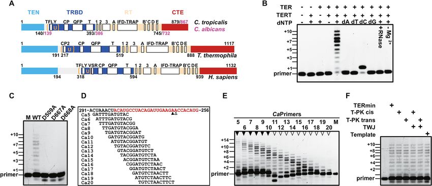
4772 Nucleic Acids Research, 2021, Vol. 49, No. 8
Small-angle X-ray scattering (SAXS) in the cryoEM structure of TtTERT holoenzyme and
DNA (5 -AGGATGTCACGATCATTGG-3 ) and RNA
All samples were buffer exchanged into 20 mM HEPES–
(5 -UACACCAAUGAUC-3 ) were modeled based on this
KOH, pH 7.8, 250 mM NaCl, 4 mM MgCl2 and 5% glyc-
structure (PDB: 6D6V). After manual inspection for
erol and concentrated to 10 mg/ml. Data were collected at
clashes, the CtTERT open structure with DNA substrate
beamlines BL19U2 (SSRF, China) and SWING (SOLEIL
and RNA template was energy minimized with Gromacs
synchrotron, France) in HPLC mode. For BL19U2 data,
(47). From this structure, complex of closed CtTERT struc-
∼40–60 l of sample were injected into the size-exclusion
ture with DNA and RNA was modelled. CTE was moved
chromatography column Superdex 200 10/300 (GE Health-
in 1000 steps from open to closed position and each inter-
care) attached on an HPLC system at a flow rate of 0.3
mediate structure minimized. DNA and RNA were mov-
ml/min and SAXS data were collected using a Pilatus 1M
ing during this process, leading to the final CtTERT closed
detector (Dectris) at a rate of 1 frame per second (2500
DNA/RNA model structure. Molecular dynamics calcula-
frames for total elution). For SWING data, ∼40–60 l of
tion was done from CtTERT open structure with DNA and
sample were injected into the size-exclusion chromatogra-
Downloaded from https://academic.oup.com/nar/article/49/8/4768/6226674 by guest on 22 December 2021
RNA using CHARMM36 forcefield during 0.5 s in peri-
phy column Superdex 200 Increase 5/150 GL (GE Health-
odic boundary box with TIP3 solvent at 298 K and 1 bar.
care) attached on an Agilent 1100 HPLC system at a flow
Molecular dynamics trajectory was analyzed with the tools
rate of 0.3 ml/min and SAXS data were collected using a
provided by Gromacs.
Eiger 4M detector (Dectris) at a rate of 1 frame per second
(560 frames for total elution) (42). The sample to detector
RESULTS
distance covers the range of scattering vectors q from 0.001
to 0.5 Å−1 . 2D data were processed and reduced with either Telomerase activity from Candida is PK/TWJ-independent
BioXtas RAW for data from BL19U2 or Foxtrot software and non-processive in telomeric repeat addition
(Xenocs) for data from SWING (43). Further data process-
Recombinant telomerase proteins from C. albicans
ing, evaluation and calculation were done with programs of
(CaTERT) and C. tropicalis (CtTERT) were highly purified
ATSAS 2.8 software package (44). Radius of gyration (Rg)
and reconstituted with their core TER (TERmin), which
was analyzed using the Guinier approximation with low an-
were mostly composed of two ubiquitous structural do-
gle data (q.Rg < 1.3) using PRIMUS. Ab initio modeling
mains: the template–proximal pseudoknot (T-PK) and a
was done with program DAMMIF or MONSA for protein-
template–distal stem–loop moiety that consists of the P5,
RNA complexes. Fifty independent models were aligned,
P6 and P6.1 stems connected in a three-way junction (TWJ)
filtered and averaged with DAMAVER to provide the final
(Supplementary Figure S1) (28). The telomerase activity of
ab initio envelope, which quality is estimated by averaged
the assembled ribonucleoprotein (CaTERTFL− TERmin)
normalized spatial discrepancies (NSD). Full-atom mod-
depended on the presence of the cognate template and
els were generated with MODELLER (see Molecular mod-
dNTPs (Figure 1B). Pretreatment of the complex with
eling section). Rigid-body modeling of structures for ori-
RNase, omission of Mg2+ from the reaction buffer or
entation and position of individual domains in the struc-
replacement of one of the three conserved active site
ture modes were done with SASREF and SREFLEX pro-
residues (D509, D667 or D668) of CaTERTFL completely
grams. Further flexible modeling was done with DADI-
eliminated activity (Figure 1B-C), indicating that the
MODO (45). The comparison of experimental scattering in-
reconstituted telomerases possess intrinsic telomerase
tensities and calculated scattering intensities from the model
activity. Recombinant TERTs of both Candida species were
are computed using program CRYSOL (v2.8.3).
able to perform one round of processive telomere addition,
but without repeat addition processivity, as demonstrated
Isothermal titration calorimetry (ITC) by assays performed with a set of primers designed to align
Binding between TERT and TWJ was determined by Nano with the template region of the TERmin at different reg-
ITC (TA Instruments) at 20◦ C. TERT and wild type or mu- istration sites (Figure 1D and E). Furthermore, although
tant TWJ were dialyzed in reaction buffer containing 20 the TERTs were assembled with isolated template or linked
mM Tris–HCl, pH 7.8, 250 mM NaCl and 4 mM MgCl2 . with the PK and/or TWJ in cis or in trans, the determined
The titration was performed by injecting 10 l TERT (50 activities were identical to those with TERmin (Figure
M) into the TWJ solution (10 M) 24 times. The binding 1F). CtTERT showed essentially the same properties as
stoichiometry and dissociation constants were calculated by CaTERT (Supplementary Figure S2A–C).
concatenating the three runs. Binding parameters were de- Thus, the above results revealed at least two distinct char-
termined from the fit. acteristics of Candida TERTs. First, although it is widely
recognized that a unique property of telomerase as a reverse
transcriptase is its ability to make consecutive copies of
Molecular modelling
telomere repeats, the Candida telomeres are nonprocessive
Since the crystal structures have some unresolved frag- in terms of telomere repeat addition. This may be an intrin-
ments, missing parts were predicted with Rosetta ab ini- sic property of Candida TERTs and is consistent with previ-
tio protocol and full-atom models were further gener- ous observations that yeast telomerase add only one telom-
ated with Modeller (46). CtTERT open structure was eric repeat to a DNA oligonucleotide primer (22–24,32,33).
modelled from the CtTERT closed crystal structure Further investigations are required to identify whether these
and CaTERT open crystal structure as reference for TERTs have evolved a multiple-cycle distributive mecha-
CTE position. CtTERT open structure was superimposed nism to handle the unusually long telomeric sequence (23–Nucleic Acids Research, 2021, Vol. 49, No. 8 4773
26 nt) (28). Secondly, it has been documented that at least ure S4). The r.m.s.d. values of the CtRT domain calcu-
two conserved structural motifs in TER (T-PK and TWJ) lated from TcTERT and TtTERT over 224 C␣ ranged from
are essential for stimulatory functions in vertebrates, the fil- 3.63 to 4.92 Å (Supplementary Figure S5). The CTE do-
amentous fungus N. crassa and the fission yeast S. pombe main superimposed on those of TcTERT and TtTERT
(10,29). However, budding yeast (S. cerevisiae) TERT has over 128 C␣ yielded an r.m.s.d. of 3.67 and 3.92 Å, re-
exceptionally evolved to not rely on the TWJ for enzy- spectively (Supplementary Figure S6). Noteworthily, the
matic function (48). Surprisingly, we found that Candida individual domains of TERT from different species are
TERT replicates the telomeric sequence in vitro with just its generally structurally conserved, and there are no spe-
RNA template, raising the possibility that Candida TERT cific regions except for the lack of TRAP-motif in the
acts as a conventional reverse transcriptase with a non- TcRT domain (Supplementary Figure S5) (16,50). More-
telomerase RNA template. To confirm this point, DNA over, the TRAP-motif from the RT domain was only par-
primer extension assays were performed with three addi- tially visible by electron density and fragments 552–564
tional RNAs that were non-telomeric templates and/or ran- and 578–582 were not defined for CaTERT177–867 , nor
Downloaded from https://academic.oup.com/nar/article/49/8/4768/6226674 by guest on 22 December 2021
dom RNA templates with differences in base composition were fragments 568–593 for CtTERT178–879 (Supplementary
and length (Supplementary Figure S3). As shown in Sup- Figure S5A–B).
plementary Figure S3, Candida TERTs efficiently replicate The TWJ has been proposed to be necessary for verte-
the ‘input’ RNA templates, indicating that Candida TERTs brate telomerase activity and potentially interacts with the
indeed function as a typical reverse transcriptase. Although CTE domain (14,51–53). In contrast to the previously de-
this finding is striking, it is far from unprecedented. A simi- termined structure of the TWJ complexed with an isolated
lar phenomenon has been observed in TERT from T. casta- TRBD domain (14), our TERT-TWJ complex structures
neum in which the TERT bound to an RNA–DNA hair- were solved with the catalytic core telomerases of Candida,
pin resembling the putative RNA-templating region and revealing TWJ binding and its spatial configuration in the
telomeric DNA extended three bases in the presence of an context of the N-terminally truncated TERT molecule (Fig-
non-telomerase RNA template (49). The mechanistic basis ure 2B, D–E). Candida TWJs adopt similar folding and spa-
through which fungal TERT has evolved less dependence on tial conformation as the OlTWJ, except that CtTWJ ap-
RNA structures for catalytic activity remains to be clarified. pears as a Y-form due to a 12 base-pair-long double strand
It is noteworthy that fungal telomerase ribonucleoprotein in its stem (P5) (Figure 3A). Although, P6 and P5 are main-
structure and organization is more complex than most other tained by coaxial stacking as a contiguous helix, as observed
telomerases (12). Candida TERs are unusually long both in in the OlTRBD-CR4/5 complex (Figure 3B–C), the previ-
the whole sequence and in the template region, which can ously described two-base triple helix at the P5–P6 junction
be as long as 25–28 bp, but less G-rich (28). These peculiari- in the OlTWJ was absent in Candida TWJ (Supplementary
ties in TER structure may provide evolutionary pressure for Figure S7A–C) (14), indicating that such RNA base stack-
TERT to behave more like a conventional reverse transcrip- ing is not necessary for maintaining the three-branch junc-
tase. tion conformation of the TWJ in fungi. The global geome-
try of the intermolecular interactions between the TWJ and
TRBD are similar to those shown for OlTRBD-CR4/5 (14).
The overall structures of the TERT-TWJ complexes from
For example, the triangular protrusion of TRBD is wedged
Candida species are conserved
into the cleft composed of P6 and P6.1 (Figure 2B, D–E)
We explored the structural basis of the observed functional and the amino acids involved in the TWJ interactions are
properties by carrying out structural studies on TERTs highly conserved. Mutations of the highly conserved ade-
from Candida. Although full-length TERT is not easily nine base at the invariant P6–6.1 junction (A1323 in CtTWJ
crystallized, N-terminally truncated TERT from C. trop- or A1291 in CaTWJ) hindered TWJ folding, maintaining
icalis in apo form (CtTERT178–879 ) and complexed with its conformation and thus TERT binding (Supplementary
its TWJ (CtTERT158–879 -TWJ), as well as TERT from C. Figure S7D–E).
albicans complexed with its TWJ (CaTERT177–867 -TWJ, Although the interactions between P6.1 and the CTE
CaTERT95–867 -TWJ), were crystallized and diffracted to domain in CtTERT158–879 -TWJ cannot be precisely deter-
2.47, 2.85, 2.98 and 3.46 Å, respectively (Figure 2A–C mined because of poor electron density for the stem-loop
and D–F, respectively, Supplementary Table S1). Consis- of P6.1, the structure of CaTERT177–867 complexed with the
tent with the high levels of sequence conservation between TWJ provided accurate information on the recognition of
CaTERT and CtTERT (with 44% identity and 81.05% the CTE domain by P6.1 (Figure 3D–F). Stem P6.1 consists
similarity, Supplementary Figure S2D), folding of the in- of eight base-pairs, adopting a double-helical conforma-
dividual domains, including TRBD, RT domain and the tion in the A-form, essentially composed of the canonical
CTE domain, was very similar between the two Candida Watson-Crick helix with two wobble base-pairs (Figure 3D-
TERTs, with a root mean square deviation (r.m.s.d.) of F). Major positively charged residues from the CTE domain
0.782–0.958 Å (Supplementary Figures S4–S6). In con- of helix ␣22 and those from the loop interact with helix ␣24,
trast, structural alignment across the available TERT struc- and helix ␣25 mainly interacts with the sugar-phosphate
tures for either the full-length T. castaneum (TcTERT, PDB backbone of the minor groove of stem P6.1 (Figure 3D-F).
3DU6) (50) and T. thermophila (TtTERT, PDB 6D6V) Our crystal structures obtained with apo-TERT and those
(16) or isolated domain O. latipes (OlTERT, PDB 4O26) complexed with the TWJ from CaTERT and CtTERT shed
(14) showed an r.m.s.d. of 2.98, 3.51 and 3.77 Å over new light on the mechanism underlying the recognition of
160 C␣, respectively, with CtTRBD (Supplementary Fig- the CTE domain by the TWJ.4774 Nucleic Acids Research, 2021, Vol. 49, No. 8
Downloaded from https://academic.oup.com/nar/article/49/8/4768/6226674 by guest on 22 December 2021
Figure 2. Structures of Candida TERT proteins in apo and complexed with the TWJ. (A) Overview of the organization of the apo CtTERT178–879 domain.
The RNA-binding domain (TRBD) is shown in blue, the reverse transcriptase domain (RT) domain is in wheat, and the C-terminal domain (CTE) in
red. (B) Structural view of CtTERT158–879 complexed with the CtTWJ. The TWJ is shown in green and the U-motif is orange. (C) and (F) Cut-away
views of CtTERT178–879 (C) and CaTERT177–867 (F) are shown, respectively. (D) and (E) Structural view of the CaTERT177–867 -TWJ complex (D) and
CaTERT95–867 -TWJ (E), respectively.
The CTE domain assumes two extreme conformations in Thus, the CTE conformation constrained by extensive and
crystals exquisite interactions is unlikely to be due to the crys-
tal packing. Second, to further probe the configuration of
Although the overall structures of TERT and/or TWJ are
CTE in solution, small-angle X-ray scattering (SAXS) cou-
conserved, there was a substantial difference in the spa-
pled with size-exclusion chromatography (SEC) was used
tial orientation of the CTE domain between CaTERT177–867
to determine the spatial conformations of the CTE in the
and CtTERT178–879 . The previously determined struc-
context of the full-length TERT (CtTERTFL ) and the N-
tures of TcTERT and TtTERT show that the conserved
terminal domain (TEN) truncated form (CtTERT178–879 ).
TRBD, RT, and CTE domains create a ring-shaped cav-
Two models corresponding respectively to CtTERTFL and
ity in which the RNA-DNA helix appears to be docked
CtTERT178–879 were established from ab initio SAXS en-
(5,49,50). However, although the conserved domains in the
velops followed by rigid body refinement of our crystal
CaTERT177–867 -TWJ structure form a ring-like shape, as
structure or/and homologous TEN structure from H. poly-
observed in TtTERT, the CTE domain of CtTERT178–879
morpha TERT (54). The full-length (CtTERTFL ) and TEN-
collapses by 108◦ relative to that of CaTERT177–867 and oc-
truncated TERT (CtTERT178–879 ) are well accommodated
cludes the cavity, making the CTE domain perpendicular to
with the corresponding SAXS envelops, in which the CTE
the RT domain and vertically parallel to the TRDB (Figure
has always occupied the central position of the cavity (Sup-
2A, C). Such spatial orientation of the CtCTE is surprising
plementary Figure S8E-F). Finally, in fact, crystals grew
and raises the question of whether the observed CTE orien-
from the same protein (CtTERT178–879 ) under multiple con-
tation is an intrinsic structural property in solution or just
ditions, and the resolved structures were the same. Alto-
an artifact of crystal packing. First, close inspection of our
gether, these findings and arguments indicate that the col-
crystal structure revealed that the spatial configuration of
lapsed conformation of CTE is an intrinsic structural prop-
CTE is determined by a network of polar contacts between
erty.
TRBD and RT (Supplementary Figures S8A, C–D and
Overlaying the apo-CtTERT178–879 and TWJ-bound
S9C). It is particularly evident that when the molecule as-
CtTERT158–879 structures showed that TWJ binding does
sumes the rotational configuration of 45◦ , the CTE appears
not turn the CTE domain to the equivalent spatial posi-
to be sitting on a sofa which is composed of TRBD and RT
tion as in TcTERT to form a ring-like conformation, but
as the main body and the T-motif and IFD-TRAP motif
rather the CTE domain is pushed away by 29.5◦ in the for-
as the armrest on both sides (Supplementary Figure S8B).Nucleic Acids Research, 2021, Vol. 49, No. 8 4775
Downloaded from https://academic.oup.com/nar/article/49/8/4768/6226674 by guest on 22 December 2021
Figure 3. Structural view of the TWJ solved from the TERT-TWJ complexes. (A) Superposition of the crystal structures of CtTWJ and CaTWJ with that
of OlTWJ (Ol-CR4/5, PDB: 4O26). CtTWJ is shown in cyan, CaTWJ in green, and Ol-CR4/5 in orange. (B, C) Secondary structures of CtTWJ and
CaTWJ in which the bases involved in the three way junction (P5-P6-P6.1) were enlarged as the crystal structure presentation. (D) Detailed structural view
of the interactions between CTE and P6.1 of CaTWJ. (E) RNA backbone–mediated interactions between the TWJ and CTE domain of CaTERT. The
CTE domain is shown as an electrostatic potential surface in which the amino acids interacting with the RNA sugar-phosphate backbone are indicated.
(F) Schematic diagram of the TWJ-CTE interactions from CaTERT. Stems P6.1 of the TWJ are shown in green and their interacting CTE residues are
shown in red. Hydrogen bonds are represented by purple dashed lines.
ward direction (Supplementary Figure S9A). In addition, bile rigid body, improved significantly from 1.36 to 1.07 and
TWJ binding disrupts the amino-acid interactions between 2.23 to 1.15 for CaTERT177–867 and CtTERT158–879 , respec-
the CTE domain and both the T-motif and TRAP-motif, tively (Supplementary Figure S10A–F and Supplementary
maintaining the spatial position of the CTE domain relative Tables S2–S3). Overall, the combined structural and SAXS
to TRAP-motif and the T-motif in apo-TERT (Supplemen- evidence suggests that the open conformation of the CTE
tary Figure S9B-D). domain in CaTERT and the closed conformation of the
CTE domain in CtTERT represent two extreme conforma-
tions as the CTE domain freely rotates in solution.
The flexible CTE domain plays a regulatory role
We further speculated that if the CTE domain does rotate
We determined the spatial conformation of the CTE freely, then the conformation of the TRBD and RT domains
domain in solution using SEC, dynamic light scatter- should be independent of the presence of the CTE domain.
ing (DLS), and SEC-SAXS. Characterization of the full- To confirm this hypothesis, we crystallized the CTE-domain
length and/or N-terminally truncated TERTs (CaTERT deleted mutant CtTERT158–745 (CtTERTCTE ), in which the
and CtTERT) in their apo-form with SEC and DLS showed C-terminal residues 746–879 were entirely deleted. Super-
these proteins to be monomeric in solution, in concordance imposition of the crystal structures of CtTERT158–879 and
with the crystal structures (Supplementary Figure S10G). CtTERTCTE over the entire TRBD and RT domain (475
SEC-SAXS was further used to correlate the conformation C␣) yielded an r.m.s.d. value of 0.911 Å (Figure 4A), show-
of the entire molecules, particularly the spatial configura- ing that the folding and spatial positions of TRBD and the
tions of the CTE domain in solution, with those in the crys- RT domain are independent of the CTE domain. We next
tallographic structures (Supplementary Tables S2–3). The sequentially replaced three pairs of residues at the interface
SAXS data obtained with the same apo-TERTs used for between the CTE and RT domains with cysteine residues
the crystal-structure determinations (CaTERT177–867 and to restrain CTE in its closed conformation through disul-
CtTERT158–879 ) could be fitted with the rigid-body models, fide bonds to better understand the functional impact of
in which the CTE domain assumes either an open or closed the CTE domain on telomerase activity (Figure 4B). Bio-
conformation (Supplementary Figure S10A-F and Supple- chemical and crystal structure analysis of the three mutants
mentary Tables S2–S3). However, the 2 values calculated showed only CtTERTR366C/S774C to form the expected disul-
from the crystal structure, leaving the CTE domain as a mo- fide bond (Figure 4B and C). Activity assays performed4776 Nucleic Acids Research, 2021, Vol. 49, No. 8
Downloaded from https://academic.oup.com/nar/article/49/8/4768/6226674 by guest on 22 December 2021
Figure 4. Structural and functional analysis of CTE domain. (A) Structural superposition of apo-CtTERT158–745 (gray) and CtTERT158–879 complexed
with TWJ indicates that the TRBD and RT domains can overlap completely. (B) Presentation of the replaced three pairs of residues with cysteine residue to
constrain CTE in closed conformation. (C) Electron density map of the disulfide bonds between C774-C366 of CtTERT178–879/R366C/S774C . (D) Telomerase
activity of the wild type and mutant CtTERT, as indicated. The experiments were initiated by adding 2 M in vitro-reconstituted TERT-TERmin complex
in the presence of 1 M telomeric DNA primer (5 -TTAGTGTAAGGA-3 ). (E, F) Telomerase assays with the full-length wild type (CtTERTFL and
CaTERTFL ), CTE-truncated (CtTERT1–745 and CaTERT1–735 ). The assays were performed as indicated in (D), but with different telomeric DNA primers,
as indicated above the gel.
with the wild type and mutants including individual mu- CTE domain and finally separated (Supplementary Figure
tations (CtTERTFL/R366C and CtTERTRFL/S774C ) showed S11 and Supplementary Movie S1). Similarly, molecular
that although the mutants without disulfide-bond for- dynamic simulation from the open structure of CaTERT
mation (CtTERTFL/T552C/G841C and CtTERTFL/L565C/T878C ) towards the closed conformation of CtTERT further con-
displayed the same activity as the wild type enzyme firmed the flexibility of the CTE domain. Based on the
(CtTERTFL ), the activity of CtTERTFL/R366C/S774C was above modelling results, we reasoned that a C-terminal
fully impaired (Figure 4D). Overall, these results show that truncated version of TERT would display enhanced proces-
the CTE domain freely rotates and constraining it in a sivity during one round of telomere addition, because the
closed conformation is prohibitive to primer extension ac- destabilizing effect of the CTE domain on the RNA/DNA
tivity. duplex would be eliminated (Supplementary Figure S17).
Consistent with this hypothesis, telomerase activity assays
CTE-domain rotation may affect the stability of the performed with the full-length wild type (CaTERTFL and
RNA/DNA duplex and regulate processivity CtTERTFL ) and CTE-truncated mutants (CaTERT1–735
and CtTERT1–745 ) under the same experimental condi-
Based on the spatial configuration of the heteroduplex tions demonstrated that both CTE-truncated TERTs dis-
formed between the RNA template and telomeric DNA play markedly enhanced processivity (Figure 4E-F and Sup-
relative to the RT:TRBD:CTE subcomplex in the Cryo- plementary Figure S17).
EM structure of Tetrahymena (16), we first modelled the
open conformation of CtTERT178–879 complexed with its
A conserved U-motif is critical for telomerase activity and
endogenous RNA/DNA duplex. In the pre-initiation state,
regulation
the primer-template duplex adopted a helical structure and
docked into the cavity in which the 3 -end of the telomeric A conserved structural motif looks like a chain of moun-
primer, paired with the RNA template, interacts with the tains placed in a fixed and orderly fashion on the TRBD
putative residues of the active site (Supplementary Figure surface (Figure 5A and Supplementary Figure S12A, E).
S11). Modelling the closed conformation, in which the CTE The motif is composed of connected U-like structure (that
domain turns in a counterclockwise direction, resulted in we hereafter call a U-motif) that spans amino acids 157–199
steric clashes between the CTE domain and the DNA/RNA in CaTERT and 158–200 in CtTERT (Figure 5A and Sup-
duplex, in which the double-stranded RNA/DNA is desta- plementary Figure S12A, E). The U-motif averages about
bilized and pushed apart (Supplementary Figure S11). Fur- 45 amino acids and consists of two short ␣-helices con-
ther modelling and energy minimization of the closed struc- nected by unstructured loops (Supplementary Figure S13).
ture showed that the DNA/RNA duplex is clipped by the The unstructured loops/linkers are not completely disor-Nucleic Acids Research, 2021, Vol. 49, No. 8 4777
Downloaded from https://academic.oup.com/nar/article/49/8/4768/6226674 by guest on 22 December 2021
Figure 5. Newly identified U-motif structure and function. (A) The identified U-motif conformation and key residues in CtTERT (left) and CaTERT
(right). The detailed interactions are shown in Supplementary Figure S12. The residues of U-motif from CaTERT and CtTERT are shown below the
figure. (B) Superimposition of the identified U-motif structures in CaTERT (orange), CtTERT (blue), TtTERT (gray, PDB: 6D6V), and OlTERT (red,
PDB: 4O26). (C) Significant movement of the key residues in the active site of CaTERT (above) and that of CtTERT (bottom) in the presence of the
integral U-motif (gray) and partially truncated U-motif proteins (wheat). Additional conformational changes and the detailed interactions are shown in
Supplementary Figure S16B-C. (D) Telomerase activity assays performed with a series N-terminally truncated or structure-guided point mutants from
CtTERT. WT is CtTERTFL , 154 means residues 1–154 are deleted, namely CtTERT155–879 , and others are similar to this; 162–167/A and 162–167
mean residues from 162 to 167 either systematically substituted by alanine or entirely deleted on the basis of the full-length CtTERT; K411A means residue
K411 substituted by alanine, namely CtTERT1–879/K411A . The experiments were carried out under experimental conditions as described in Figure 4D. (E)
Proposed model for the U-motif-mediated conformational change upon the completion of telomeric synthesis. The RNA template is bound between the
TBE stem at its 5 -end and a PK motif at its 3 -end. Similarly, as observed in the TtTERT-TER complex structure determined by Cryo-EM, TER embraces
the TERT. While the spatial positions of the TBE and PK are mobilized by the U-motif and other regions of TERT, the single stranded RNA template
is flexible prior to replication (left). However, as the RNA template is replicated, the RNA/DNA hybrid becomes shorter than the ssRNA. The produced
tension rotates the global conformation between the TRBD and RT domain and consequently disrupts the cation- interaction between Y167/166 and
K411/405 (right), which is essential for maintaining TERT in an active conformation.
dered, relatively constrained by extensive interactions as ing Y167 of corner 1 and K411 of the RT domain, are
described and demonstrated in Supplementary Figure S12 highly conserved among telomerases (Supplementary Fig-
(Supplementary Figure S12). The U1 fragment (157–166) ure S13 and Supplementary Figure S15F). Consistently, the
closely resembles the TFLY (vertebrate) (55) and the first U-motif and its interactions with the RT domain are per-
half of the CP2 (ciliate) (15,56) motifs (Supplementary Fig- fectly conserved across the available structures of TERTs,
ure S14). The U2 fragment (167–175) is mainly constrained including those from T. thermophila (15,16), O. latipes (14)
through hydrogen bonds with residues (A354, V358, E360 and T. rubripes (55) (Figure 5B and Supplementary Fig-
and H374) of the T-motif. The extended U2 loop with ␣- ure S15A–E). The significant differences and similarities be-
helices constitute the U3 fragment (176–192), which ex- tween the U-motif and the previously identified CP2/TFLY
tends in parallel with ␣-helix 12 of the TRBD through a set motif will be further summarized after the functional char-
of hydrophobic interactions (Supplementary Figure S12D). acterization of the U-motif (below).
Y167 at the outer corner 1 (between U1 and U2) directly The CP2 motif in Tetrahymena located at the corner 2
interacts with K411 on the -strand 4 of the RT domain position has been structurally characterized (15). We fo-
through a strong cation- interaction (Figure 5B, Supple- cus our attention on the function of corner 1. To this end,
mentary Figure S15A-B), whereas R175 at the outer corner the N-terminal residues of full-length CtTERT were succes-
2 (U2-U3) occupies the equivalent position of R237 in CP2, sively truncated to N176. Telomerase activity assays showed
which wedges into the template boundary element (TBE) that proteins bearing truncations up to position 159 display
stem-loop in Tetrahymena (15) (Figure 5B). Sequence align- telomerase activity identical to that of full-length TERT,
ment showed that the whole amino-acid sequence of U- whereas further truncation beyond residue 161 severely im-
motif––not just that constrained around CP2/TFLY, but paired primer extension, indicating that the complete over-
particularly the residues involved in the spatial conforma- all corner 1 plays an essential role in telomere addition
tion between the U-motif and the RT domain, includ- (Figure 5D). We confirmed these results by constructing4778 Nucleic Acids Research, 2021, Vol. 49, No. 8
two additional types of mutants, in which residues K162 (ii) the U-motif is not just analogous to CP2/TFLY; un-
to Y167 of the full-length TERT were either systemati- der this framework, previous findings may be better under-
cally substituted by alanine or entirely deleted, resulting in stood and potential functions of some residues in the U-
mutants CtTERT162/A-167/A and CtTERT162–167 . The mu- motif can be deduced. For example, realizing that the two
tants showed the same level of reduction in telomerase ac- residues (H234 and R237 in ciliates) (15) are located in the
tivity as those bearing truncations beyond Y161 (Figure rigid corner 2, but not in an unstructured flexible loop, will
5D). CaTERT showed essentially the same properties as help understand how the two residues remain at their physi-
CtTERT (Supplementary Figure S16A). cal position to serve as a wedge, rather than moving together
To understand the structural basis of how corner 1 in the with the TBE while the RNA template is stretched. Further-
U-motif affects telomerase activity, even though it is dis- more, only when the corner organization feature was taken
tant from the active site, the structures of CtTERT158–879 into account, did we identify the interaction between Y167-
complexed with the TWJ bearing the full-length U-motif K411 at corner 1, which is structurally and functionally con-
(CtTERT158–879 ) and CtTERT178–879 , in which both corner 1 served across different species; (iii) the two physical corners
Downloaded from https://academic.oup.com/nar/article/49/8/4768/6226674 by guest on 22 December 2021
and 2 were completely truncated, were superimposed based may play distinct functions and coordinately communicate
on the C␣ or backbone atoms of the TRBD (r.m.s.d. of with each other. It is possible that corner 2 acts as a sensor
2.58 Å for 472 C␣). This analysis showed marked struc- to perceive the replication stop signal and results in the dis-
tural rearrangement, with notable changes in the RT do- ruption of Y167-K411 interaction at corner 1. Additional
main of CtTERT178–879 . Truncation of the U-motif resulted studies on the U-motif will be required to gain a better un-
in disruption between Y167 and K411 and thus the over- derstanding of the precise regulation of telomere synthesis.
all RT domain is distorted by 16.9◦ and pushed up 15 Å,
which twists the entire active site by 24.6◦ and moves it away
DISCUSSION
by ≈ 5 Å (Figure 5C and Supplementary Figure S16B-C).
In accordance with the above interpretation, the replace- In this study, we determined several crystal structures of
ment of K411 with alanine significantly reduced telomerase N-terminally truncated fungal TERTs in apo- and TWJ-
activity (Figure 5D). It is noteworthy that the above re- bound forms. These data, together with our biophysical
sults are consistent with the previous observations from and biochemical data, provide important insights into the
the studies with telomerases of other species. Both mutants domain or motif organization and the potential molecular
R503A (S. pombe) and R534A (ciliate) at the equivalent po- mechanisms of telomerase activity regulation.
sition of K411 of CtTERT severely impair telomerase ac- Studies previously demonstrated that two conserved
tivity (25,57). Of note, the integral U-motif and its interac- TER structural elements (the PK and the TWJ) are partic-
tion with the RT domain was identified with CtTERT158–879 ularly important for the telomerase activity in vertebrates
complexed with the TWJ, whereas such a structural interac- and ciliates, but not for S. cerevisiae telomerase in which the
tion was absent in CtTERT178–879 . This raises the question telomeric sequence can be added in the absence of TWJ (10).
of whether folding of the U-motif and the interaction be- We surprisingly found that the fungal TERTs character-
tween Y167 and K411 results from TWJ binding. We thus ized in this work can act as standard reverse transcriptases
superimposed the crystal structure of CtTERT158–745 , which and neither PK nor TWJ are required to efficiently catalyze
bears an integral U-motif, with that of CtTERT158–879 . The primer extension. The mechanisms of TWJ/PK-dependent
resulting r.s.m.d. of 0.911 Å showed the conformations and and -independent activity are completely unknown (10,11).
interactions of Y167 at corner 1 and K411 at the RT do- The fact that the spatial conformation of the U-motif rela-
main to be nearly identical between the two crystal struc- tive to the RT domain is well assembled into an active state
tures (Figure 4A), indicating that the newly identified U- in the absence of a TWJ/PK in Candida TERTs may ex-
motif is an intrinsic structure of TERT that stabilizes the ac- plain why the telomerase activity of Candida is TWJ/PK-
tive conformation of telomerase independently of the TWJ. independent. These observations in turn raise the possibility
Overall, mutational and structural analysis shows that the that the cation- interaction between the U-motif and the
interaction between the U-motif and the RT domain is very RT domain does not occur in the absence of the TWJ/PK
important for maintaining the structural integrity of TERT for the TWJ-dependent telomerase in the apo-form and
and, therefore, orientating the spatial configuration of the that TWJ binding promotes the necessary interaction be-
active site relative to the RNA template (Figure 5A, E). tween the U-motif and the RT domain, probably through
Putting the previously identified CP2/TFLY into the U- the highly conserved tyrosine residue in the U-motif and
motif framework could offer a new perspective on the orga- the lysine in the TR domain, thereby activating telom-
nization of previously recognized isolated components and erase activity. The above interpretation needs to be verified
potential functions, which have not formerly been recog- by comparing the structures determined with TWJ/PK-
nized. The distinct features and the novelty of the U-motif dependent telomerase in apo- form and complexed with
can be summarized as: (i) the U-motif concept expands our the TWJ/PK.
knowledge of CP2/TFLY. The CP2 motif, which harbors The addition of consecutive copies of telomeric re-
the ␣-helix of TFLY, was previously defined just as a 12- peats involves extremely complex and delicate processes, in-
amino-acid sequence conserved across ciliate species (56). cluding dissociation, translocation and realignment of the
It is now clear that both CP2 and TFLY are partial com- RNA-DNA hybrid (31). Telomerase must undergo exten-
ponents of the U-motif and may possess additional func- sive domain or motif rearrangements for each round of
tions (Supplementary Figure S13 and Supplementary S14); telomere addition to accomplish these tasks (58). How-You can also read