Diagnostic accuracy of bedside ultrasonography in COVID-19 suspected patients admitted to the emergency department
←
→
Page content transcription
If your browser does not render page correctly, please read the page content below
Submitted: 28 April, 2021    Accepted: 01 June, 2021    Published: 08 January, 2022                                DOI:10.22514/sv.2021.116

ORIGINAL RESEARCH

Diagnostic accuracy of bedside ultrasonography in
COVID-19 suspected patients admitted to the
emergency department
Ramazan Guven1, * , Ramazan Unal1 , Burcu Genc Yavuz2 , Ertugrul Ak1 ,
Gokhan Eyupoglu3 , Salih Fettahoglu1 , Basar Cander1

1
  Department of Emergency Medicine,         Abstract
University of Health Sciences, Kanuni
Training and Research Hospital, 34303
                                            Introduction: Due to the risk of cross contamination and radiation exposure of
Istanbul, Turkey                            computed tomography (CT) and low sensitivity rate of X-Ray, point of care ultrasound
2
  Department of Emergency Medicine,         (POCUS) lung can be used as a diagnostic tool of COVID-19 pneumonia. The current
University of Health Sciences,              study aimed to evaluate the potential of POCUS for detection of lung pathologies caused
Haydarpasa Numune Training and
Research Hospital, 34668 Istanbul,
                                            by COVID-19.
Turkey                                      Methods: This prospective observational study was conducted with 84 patients admitted
3
  Department of Emergency Medicine,         to the emergency department with suspected COVID-19. CT and POCUS lung were
University of Health Sciences,              performed for all participants. CCT was taken as the reference diagnostic method and
Basaksehir Cam and Sakura City
Hospital, 34480 Istanbul, Turkey
                                            the presence of B-lines or consolidation or pleural irregularity-thickening (>3 mm) in
                                            the lung in POCUS lung, were evaluated in favor of COVID-19 pneumonia.
*Correspondence                             Results: Of the 84 patients included, lesions of COVID-19 pneumonia were detected
drramazanguven@gmail.com                    53.5%. COVID-19 pneumonia findings were shown by POCUS lung in 51.2% of
(Ramazan Guven)
                                            participants. The left lower lobe in 48.8% and the right lower lobe in 47.6% of
                                            the patients were the most commonly affected regions. In POCUS lung, COVID-19
                                            pneumonia lesions located in 2nd area for 44.0%, in 7th area for 35.7%, in 8th area
                                            for 34.5%. Sensitivity of POCUS lung was found to be 88.9%, specificity pointed for
                                            92.3%, positive predictive value was 93.0% and negative predictive value was 87.8%.
                                            Conclusion: POCUS lung, has a high sensitivity and specificity in the diagnosis of
                                            COVID-19 pneumonia, especially in severe lung involvement. Therefore, POCUS lung
                                            should be the method of choice as its practical use, bedside availability and avoidance
                                            of radiation exposure for COVID-19 associated lung lesions.

                                            Keywords
                                            COVID-19; Pneumonia; POCUS lung

1. Introduction                                                             [3]. This algorithm created by using A lines from reverbera-
                                                                            tion artifacts and B lines consisting of hyperechoic artifacts.
While bedside chest X-ray provides limited diagnostic benefit               Lichtenstein identified 2 points (upper anterior point, lower
in patients with pneumonia, chest computarized tomography                   anterior point) and PLAPS (posterior lateral alveolar pleural
(CCT) also has disadvantages such as high cost and the risk of              point) point as blue points for lung pathologies. Considering
transporting critically unstable patients, although the diagnos-            data from China and especially Italy, it was seen that COVID-
tic rate is higher. Additionaly requirement for sterilization of            19 pneumonia can rapidly progress to acute respiratory distress
the CCT area after each scan due to the cross infection risk of             syndrome (ARDS) [4]. Soldati et al. [5] developed a method
COVID-19 leads to a longer exposure time [1].                               that includes 14-point lung ultrasound that provides a wider
   In the diagnosis of COVID-19 pneumonia, point of care                    view instead of Blue-PLAPS points to diagnose and monitor
ultrasound (POCUS) lung is one of the important diagnos-                    COVID-19 pneumonia, which can spread to all lobes of the
tic tools that can be used due to its cost-effectiveness, less              lung. Therefore, current study aimed to evaluate the diagnostic
risk of contamination, being a bedside, practical and ease of               accuracy of POCUS Lung in COVID-19 pneumonia by using
access imaging method. POCUS lung can also be used for                      14 points’ technic, either to define which of these points were
monitoring the disease progress and evaluating the response                 effected more by COVID-19 and its compatibility rate with
to the treatment [2]. POCUS for the lung has been used by                   CCT.
clinicians since the 1990s. Lichtenstein published the algo-
rithm known as the BLUE protocol for POCUS lung in 2008

This is an open access article under the CC BY 4.0 license (https://creativecommons.org/licenses/by/4.0/).
Signa Vitae 2022 vol.18(1), 55-61                 ©2022 The Author(s). Published by MRE Press.                  http://www.signavitae.com/
56
2. Methods previous studies that lung pathology did not develop, although
RT-PCR was positive. One of the results obtained from these
2.1 Study design studies is that although the findings of COVID-19 pneumonia
(ground-glass opacification) are found in CCT, the RT-PCR
This prospective convenience sample observational study was
test can be negative [8, 9]. The sensitivity of the RT-PCR
conducted to diagnose viral pneumonia with POCUS lung
test ranges from 50%–80%. Therefore, it is seen that CCT
in patients who were classified as COVID-19 probable cases
is used as a reference method in COVID-19 pneumonia [10].
among those admitted to study center. The study center is
In accordance with the literature, the positive rate of RT-PCR
a training and research hospital serving as one of the largest
among COVID-19 pneumonia patients was 66.7% in our study.
hospitals in the region during the COVID-19 pandemic. All
patients included in the study, were tested for COVID-19 by
real-time polymerase chain reaction (RT-PCR). The study was 2.4 Measurements
approved by the local ethics committee and Ministry of Health. CCT was performed by dividing the lung into lobes (right
Clinical trial registration was made with a clinicaltrials. upper lobe, right middle lobe, right lower lobe, left upper
gov ID of NCT04399681. lobe and left lower lobe) according to the official report of
the radiologists. Consolidation, air bronchogram, graund-
2.2 Selection of participants glass opacities, crazy-paving pattern was sought for the in-
terpretation of CCT in favor of COVID-19. Sonographer
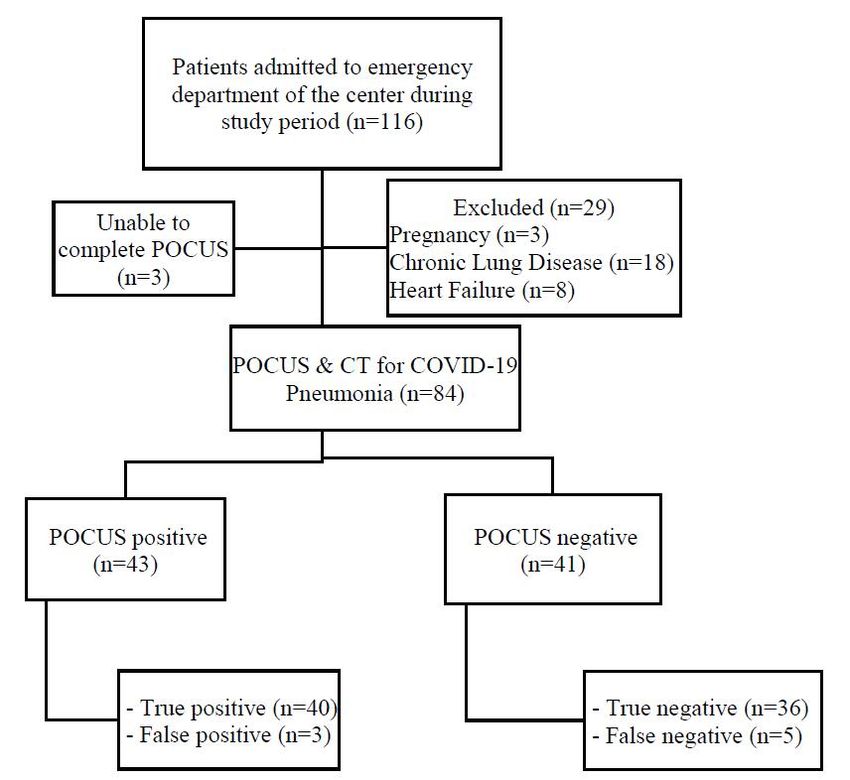
Flow diagram of the study is shown in Fig. 1. Probable case was asked to decide one of two conclusions of whether the
definition of COVID-19 was determined in accordance with patient has COVID-19 pneumonia or not. Soldati et al.’s [5]
the guidelines of scientific advisory board of Turkish Ministry article was taken as reference and 14-point lung ultrasound,
of Health [6]. Patients with heart failure, known chronic was determined (Fig. 3). Pleural movement and the presence
lung disease such as chronic obstructive pulmonary disease of B lines, white lung sign, were checked for the diagnosis
(COPD), chronic bronchitis, patients with lung malignancies of pneumonia in POCUS lung. Additionally, supporting the
or lung metastasis, pregnant patients and trauma patients were diagnosis of pneumonia, the presence of consolidation, air
excluded from the study. The sample size was conducted with bronchogram and pleural wall thickening (>3 mm) and its
previous studies. We assumed that the lung ultrasound has irregularity was checked (Fig. 4).
80% sensitivity and power of 80% with alpha = 0.05. Thus,
a minimum of 78 patients was planned to include in the study
2.5 Outcome
[7].
The ultrasounds performed within the scope of this study The primary outcome was the sensitivity of lung ultrasound at
were performed by emergency physicians who had a certi- identifying COVID-19 pneumonia against CCT as the refer-
fication of POCUS lung earned upon a bedside course of ence standard. The secondary results of this study; to deter-
POCUS lung. The study sonographers performed POCUS mine the presence of signs of pneumonia using 14-point lung
lung as blinded on the patient’s CCT and COVID-19 RT- ultrasound method.
PCR test results. The ultrasonographic protocol consisted of
scanning of the patient in the supine, right lateral decubitus, 2.6 Statistical analysis
left lateral decubitis and sitting erect positions. While applying All statistical analyses (sensitivity, specificity, negative pre-
ultrasound, transducer dressed with sterile gloves covered ca- dictive value, positive predictive value) were performed on
bles, other parts of the ultrasound were sterilized with sodium MedCalc Statistical Software version v19.4.1 (MedCalc Soft-
hypochlorite after each use against the risk of contamination ware, Ostend, Belgium). The data of the patients are expressed
(Fig. 2). The study sonographers took personal protective mea- as median (quartiles) for nonparametric data and percentage for
sures (N95 respirator mask, surgical gown, surgical gloves, eye categorical variables.
protection, surgical cap) while practicing.
3. Results
2.3 Interventions
Hitachi Aloka-ProSound F37 brand ultrasonography device 3.1 Characteristics of study subjects
and 6-2 MHz Convex (UTS9123) probe and linear (UST-568) During the study period, 29 of 116 patients were excluded
13-5 MHz probe was used in the study. CCT was performed from the study since they did not meet the inclusion criteria
using Toshiba Aquilion Prime 128 slice CT scanner. Chest CT and 3 did not complete the POCUS lung. The demographic
is the gold standard method used for COVID-19 pneumonia, characteristics of the participants according to the COVID-19
and COVID-19 pneumonia diagnosis was made in case of pneumonia status are shown in Table 1. Of the 84 participants
typical findings (crazy-paving pattern, air bronchogram, etc.) whose data were evaluated, 69.0% (n = 58) were male, the
were officially reported by radiology in favor of COVID-19. median (quartiles) age was 53.0 (30.7–67.5), 31.0% (n = 26)
The main hypothesis of this study is to evaluate the accu- was female, and the median (quartiles) age was 49.0 (31.50–
racy of lung ultrasound in diagnosing COVID-19 pneumonia. 63.0). When the vital signs of the patients included in the study
Therefore, it is more important whether the patients included were examined, the systolic blood pressure median (quartiles)
in the study have a COVID-19-related pathology in their lungs value was 120 (110–130) mmHg, the diastolic blood pressure
rather than having positive RT-PCR tests. It was shown in median (quartiles) value was 70 (66–80) mmHg, the median57
F I G U R E 1. Flowchart of the study population.
study had hypertension, 21.4% (n = 18) had diabetes mellitus,
and 10.7% (n = 9) had coronary artery disease.
3.2 Main results
Lung imaging assessments are shown in Table 2. According
to the CCT, 53.6% of the cases (n = 48) were found to have
findings in favor pneumonia, and 45 of them were reported
as COVID-19 pneumonia (18 mild, 14 moderate, 13 severe)
and 3 patients were reported as bacterial pneumonia. Bilateral
involvement was detected in 84.4% (n = 38) of patients with
pathological findings in favor of COVID-19 in CCT. Among
imaging results reported as COVID-19 pneumonia by CCT, in
order of frequency was; left lower lobe was involved in 91.1%
(n = 41), 88.8% (n = 40) had involvement in the right lower
lobe, the right middle lobe was involved in 77.7% (n = 35),
right upper lobe was involved in 51.1% (n = 23), left upper
F I G U R E 2. Covered cables and the probe were put in a lobe was involved in 37.7% (n = 17).
sterile gloves, helt by sonographer. According to 14-point lung ultrasound, the most common
findings of COVID-19 pneumonia (86.0% (n = 37)) were at
the 2nd point and then 69.7% (n = 30) at the 7th point. The
(quartiles) value of the pulse measured per minute was 84 (75– most common sonographic finding of pneumonia was B-Lines
92), oxygen saturation median (quartiles) value was 95.5% with a percentage of 47.6% (n = 40) (Table 2).
(92.0–97.0). 27.4% (n = 23) of the patients included in the According to POCUS lung, 51.2% (n = 43) of the patients58
F I G U R E 3. Fourteen areas of ultrasonographic examination.
TA B L E 1. Demographic characteristics of participants.
Parameter, n (%) COVID-19 pneumonia (+) COVID-19 pneumonia (−)
(n = 45) (n = 39)
Sex
Female 11 (24.4) 15 (38.5)
Male 34 (75.6) 24 (61.5)
Hypertension 14 (35.0) 9 (23.1)
Diabetes mellitus 16 (39.0) 2 (5.1)
Coronary artery disease 7 (17.5) 2 (5.1)
Hospitalization
Inpatient clinics 20 (47.6) 2 (5.1)
Intensive care unit 14 (33.3) 0 (0.0)
Shortness of breath 24 (53.3) 9 (23.0)
Sore throat 3 (6.6) 3 (7.6)
Cough 11 (24.4) 8 (20.5)
Fatigue 22 (48.8) 19 (48.7)
Fever 11 (24.4) 7 (17.9)
were evaluated as COVID-19 pneumonia. However, POCUS 4. Discussion
lung was found as false positive in 3 patients and false negative
in 5 patients. Accordingly, the sensitivity of POCUS lung CXR and CCT are used to detect the presence of lung pathol-
was calculated as 88.9%, specificity 92.3%, positive predictive ogy due to COVID-19 and to see its severity. However, due
value 93.0%, negative predictive value 87.8% (Table 3). to the known disadvantages of CXR (low sensitivity rate) and
CT (radiation risk, possibility of cross contamination), POCUS
lung, which is an alternative diagnosis method, started to be
used especially in the later stages of the pandemic [1, 2, 11].59 F I G U R E 4. Imaging features of COVID-19 patients. (a) Solid yellow arrow-signed consolidation and dashed yellow arrows showed nonconfluent B-lines in 2nd area of POCUS lung. Areas surrounded by yellow circles demonstrated ground glass opacity with consolidation in upper right lobe on CCT. (b) Solid yellow arrow showed consolidation with air bronchograms pointed by multiple yellow arrows in 1st area of POCUS lung. Yellow circles surrounded multiple ground glass opacities with consolidations in both lungs on CCT. This study revealed that POCUS lung had 88.9% sensitivity to the results of CCT. Detection of less pathology in POCUS and 92.3% specificity when CCT was taken as reference in de- 11th point, POCUS 12th point, POCUS 13th point and POCUS tecting lung pathology due to COVID-19 infection. Through- 14th point can be explained with involving the anterior region out the study, POCUS lung was more accessible and allowed of the lung is less in COVID-19 pneumonia. Additionally, due bedside application compared with CCT. BLUE and PLAPS to the presence of the heart image in the POCUS 13th point and points are used for sonography to detect lung pathologies. POCUS 14th point, pathology becomes less detectable in these However, COVID-19 pneumonia with its widespread lung areas. The POCUS Lung and CCT results of present study are involvement, would be better to be evaluated by covering consistent with the literature and it is seen that the posterior more areas. Therefore, the POCUS lung protocol in 14-point regions of the lower lobes of the lung are mostly affected [13]. recommended by Soldati et al. [5] for COVID-19 pneumonia, A limited number of articles exist on POCUS lung showing which provides a more detailed view to us, constituted the COVID-19 pathologies, most of them are technical reports and field of view of our study. COVID-19 pneumonia affects the narrative reviews. Of these, Moore S et al. [14] suggested middle and lower lobes in the right lung and the lower lobes that since POCUS lung had high sensitivity and specificity, in the left lung more frequently [12]. Similarly, CCT results and that the use of systemic ultrasound will be beneficial not in the current study, demonstrated that 41.7% of the patients only in emergency services but also in intensive care units had involvement in the right middle lobe, 47.6% in the right (ICU), in the follow-up of critical COVID-19 patients, thus lower lobe, and 48.8% in the left lower lobe. Looking at the reducing the reuse of radiographic imaging methods. A study, POCUS lung examination points, 44.0% of the patients had in which POCUS lung used in demonstrating and monitoring pathology in the POCUS 2nd point and 34.5% in the POCUS lung pathologies in COVID-19 ICU patients, 100% of the 5th point. The reason for detecting less pathology in POCUS patients had B-lines (confluent or separate) and 61.7% of them 3th point and POCUS 6th point in the POCUS lung is due to had consolidation at their first sonographic examinations on the upper lobe involvement of COVID-19 infection, similar admittance, and these findings were reported to be significantly
60

         TA B L E 2. Findings on POCUS lung for COVID-19 pneumonia, frequency rate of location of COVID-19
                                   pneumonia-positive areas of fourteen lung points.
         POCUS, n (%)                                    COVID-19 pneumonia (+) COVID-19 pneumonia (−)
                                                                         (n = 45)                        (n = 39)
          POCUS lung
             Pneumonia                                             43 (5 false positive)          41 (3 false negative)
             B-Lines                                                        40                               3
             Consolidation                                                  22                               3
             Pleural irregularity-thickening                                29                               2
          14-Point lung ultrasound
             1. Point (Right basal on the paravertebral line)               17                               0
             2. Point (Right middle on the paravertebral line)              37                               0
             3. Point (Right upper on the paravertebral line)               16                               0
             4. Point (Left basal on the paravertebral line)                21                               0
             5. Point (Left middle on the paravertebral line)               29                               0
             6. Point (Left upper on the paravertebral line)                14                               0
             7. Point (Right basal on the midaxillary line)                 30                               2
             8. Point (Right upper on the midaxillary line)                 29                               0
             9. Point (Left basal on the midaxillary line)                  19                               1
             10. Point (Left upper on the midaxillary line)                 18                               0
             11. Point (Right basal on the midclavicular line)              16                               0
             12. Point (Right upper on the midclavicular line)              19                               0
             13. Point (Left basal on the midclavicular line)               12                               0
             14. Point (Left upper on the midclavicular line)               14                               0

     TA B L E 3. Test characteristics of POCUS lung in             in CCT. All of those, who were evaluated as false negative,
            diagnosing COVID-19 pneumonia.                         were patients with mild pneumonia (unifocal opacity). Lu et al.
                               Value        95% CI                 [17] reported that the sensitivity of lung US for COVID-19 was
                                                                   68.8%, 77.8%, 100%, and the specificity was 85.7%, 76.2%,
     Sensitivity                  88.9%     75.95%–96.29%
                                                                   92.9%, respectively. Similarly, the present study revealed that
     Specificity                  92.3%     79.13%–98.38%          as the severity of the disease increases, the diagnostic power
     Positive likelihood ratio     11.5         3.88–34.44         of POCUS lung increases, so that all intensive care patients (n
     Negative likelihood ratio     0.12         0.05–0.28          = 14) were correctly diagnosed with POCUS lung.
     Positive predictive value    93.0%     81.73%–97.55%
     Negative predictive value    87.8%        75.8%–94.3%         5. Limitations

                                                                   This study has several limitations. Although it is the largest
regressed in the week of discharge [15]. In our study, there was   pandemic hospital in the region, the first limitation of this study
B-lines and consolidation, either, in 100% of 14 patients hospi-   is that it cannot be generalized with other hospitals because
talized in ICU while B-lines were present in 86.4% of patients     it is a single center. Despite ultrasound (USG) training is
hospitalized in pandemic ward (n = 22), and consolidation in       included in the curriculum of the emergency medicine clinic
70% of them. In this study, the scoring system or any classifi-    in our country, currently, the USG training of all emergency
cation was not used because of the CCT reports were reported       physicians, especially the POCUS lung training, is not at the
as “in favor of COVID-19” or “COVID-19 pneumonia was not           desired level. The fact that USG is a subjective tool, also
considered”. In a study on asymptomatic COVID-19 patients,         directly affects the sensitivity and specificity of POCUS lung.
POCUS lung detected the pneumonia findings of US with              Another limitation of the study was the fact that the study
66.7% sensitivity, 100% specificity, 100% positive predictive      directors were working in 24-hour shifts, which led to the
value, 85.71% negative predictive value [16]. Among all            exclusion of patients outside of their switch.
patients in the current study, the sensitivity of POCUS lung was      There were also some limitations due to the current disease
found to be 88.9%, specificity 92.3%, positive predictive value    state. First, since this study was conducted on COVID-19,
was 93.0%, and negative predictive value was 87.8%. All of         which has a high risk of contagion, sonographers had difficulty
the false positive cases were reported as bacterial pneumonia      to have the appropriate positions. Second, the use of only one
61
USG device for the study and the time required to sterilize this reasonable request.
USG device after each examination caused some patients who
came in the mean time to be excluded from the study. Third, in
case of lung pathology in patients with suspected COVID-19 R EF ERENCES
infection, they clinically give sonographers some clues (e.g., [1] Li M. Chest CT features and their role in COVID-19. Radiology of
tachypnea, tachycardia, high fever). This seems to cause an Infectious Diseases. 2020; 7: 51–54.
[2] Fiala MJ. A Brief Review of Lung Ultrasonography in COVID-19: is it
increase in accuracy in favor of USG.
Useful? Annals of Emergency Medicine. 2020; 75: 784–785.
[3] Lichtenstein DA, Mezière GA. Relevance of lung ultrasound in the
6. Conclusions diagnosis of acute respiratory failure: the BLUE protocol. Chest. 2008;
134: 117–125.
POCUS lung, with fourteen visual fields, provides sufficient [4] Remuzzi A, Remuzzi G. COVID-19 and Italy: what next? Lancet. 2020;
diagnostic opportunity. Although POCUS lung is not at the de- 395: 1225–1228.
[5] Soldati G, Smargiassi A, Inchingolo R, Buonsenso D, Perrone T, Briganti
sired level in distinguishing false-negativity risk and bacterial
DF, et al. Proposal for International Standardization of the Use of
pneumonia from COVID-19 pneumonia in patients with mild
Lung Ultrasound for Patients With COVID-19: A Simple, Quantitative,
pneumonia, it has a high level of sensitivity and specificity in Reproducible Method. Journal of Ultrasound in Medicine. 2020; 39:
severe COVID-19 pneumonia. Therefore, POCUS lung can be 1413–1419.
used as the method of choice due to its practical use, bedside [6] Scientific Advisory Board Republic of Turkey Ministry of Health.
availability and avoidance of radiation exposure for COVID- SARS-CoV-2 Infection: general information, epidemiology
19 associated lung lesions. and diagnosis April 21, 2020. 2020. Available at: https:
//covid19bilgi.saglik.gov.tr/depo/Sunumlar/COVID-19-
Epidemiyoloji-Tani-Tedavi.pdf (Accessed: 25 May 2020).
[7] Haak SL, Renken IJE, Jager LC, Lameijer H and Kolk BYM. Diagnostic
A U TH OR CO NT RI BU TI ONS
accuracy of point-of-care lung ultrasound in COVID-19. Emergency
RG, GE, RU and BC designed this study. RG, SF, BGY, EA, Medicine Journal. 2021; 38: 94–99.
[8] Fang Y, Zhang H, Xie J, Lin M, Pang P, Ji W. Sensitivity of Chest for
RU and GE, supervised the overall data collection process, had
COVID-19: Comparison to RT-PCR. Radiology. 2020; 296: E117.
full access to all the data in the study, and takes responsibility [9] Korkmaz I, Dikmen N, Keles FÖ, Bal T. Chest CT in COVID-19
for the integrity of the data. RG and GE conducted the pnumonia: correlations of imaging findings in clinically suspected but
data analysis. RG, EA, SF, BGY and BC wrote the initial repeatedly RT-PCR test-negative patients. Egyptian Journal of Radiology
draft of the article. All authors provided substantial review and Nuclear Medicine. 2021; 52: 96.
[10] Ai T, Yang Z, Hou H, Chenao Z, Chen C, Lv W, et al. Correlation of
and feedback on the final version of the article. RG takes
Chest CT and RT-PCR Testing in Coronavirus Disease 2019 (COVID-
responsibility for the paper as a whole. 19) in China: A Report of 1014 Cases. Radiology. 2020; 296: E32–E40.
[11] Cleverley J, Piper J, Jones MM. The role of chest radiography in
confirming covid-19 pneumonia. British Medical Journal. 2020; 370:
E T H I CS A PPR OVA L AN D CONS E N T TO m2426.
PA RT ICI PAT E [12] Li Y, Xia L. Coronavirus Disease 2019 (COVID-19): Role of Chest CT in
Diagnosis and Management. American Journal of Roentgenology. 2020;
All participants were included in the study following their 214: 1280–1286.
approval of written informed consent. The study was approved [13] Zhao W, Zhong Z, Xie X, Yu Q, Liu J. Relation between Chest CT
by the local ethics committee (Ethics Committee approval Findings and Clinical Conditions of Coronavirus Disease (COVID-19)
number: KAEK/2020.03.50) and Ministry of Health. Clini- Pneumonia: a Multicenter Study. American Journal of Roentgenology.
2020; 214: 1072–1077.
cal trial registration was made with a clinicaltrials.gov ID of [14] Moore S, Gardiner E. Point of care and intensive care lung ultrasound: a
NCT04399681. reference guide for practitioners during COVID-19. Radiography. 2020;
26: e297–e302.
[15] Alharthy A, Faqihi F, Abuhamdah M, Noor A, Naseem N, Balhamar
AC K NOW LED G ME N T A, et al. Prospective Longitudinal Evaluation of Point-of-Care Lung
Ultrasound in Critically Ill Patients With Severe COVID-19 Pneumonia.
The authors thanks to all emergency department staff for their Journal of Ultrasound in Medicine. 2020; 40: 443–456.
contribution during data collection. [16] Haidan LM, Bingqi ZM, Haiyan KM, Yuanyuan ZM, Keyan LM, Dudu
WM, et al. Application Value of Lung Ultrasound in Asymptomatic
Patients with Confirmed COVID-19. Advanced Ultrasound in Diagnosis
F U ND ING and Therapy. 2020; 4: 67.
[17] Lu W, Zhang S, Chen B, Chen J, Xian J, Lin Y, et al. A Clinical
This study received no external funding. Study of Noninvasive Assessment of Lung Lesions in Patients with
Coronavirus Disease-19 (COVID-19) by Bedside Ultrasound. Ultraschall
in Der Medizin—European Journal of Ultrasound. 2020; 41: 300–307.
CO NFL ICT OF IN T ERE ST
The authors declare no conflict of interest. How to cite this article: Ramazan Guven, Ramazan Unal, Burcu
Genc Yavuz, Ertugrul Ak, Gokhan Eyupoglu, Salih Fettahoglu, et
al. Diagnostic accuracy of bedside ultrasonography in COVID-19
AVA IL AB ILI T Y OF DATA AN D M AT E R I A L suspected patients admitted to the emergency department. Signa
Vitae. 2022;18(1):55-61. doi:10.22514/sv.2021.116.
The datasets generated during and/or analysed during the cur-
rent study are available from the corresponding author onYou can also read