Diagnostic Utility of Radiomics in Thyroid and Head and Neck Cancers
←
→
Page content transcription
If your browser does not render page correctly, please read the page content below
REVIEW
                                                                                                                                                  published: 07 July 2021
                                                                                                                                          doi: 10.3389/fonc.2021.639326

                                              Diagnostic Utility of Radiomics in
                                              Thyroid and Head and Neck Cancers
                                              Maryam Gul 1, Kimberley-Jane C. Bonjoc 2, David Gorlin 2, Chi Wah Wong 2,
                                              Amirah Salem 2, Vincent La 2, Aleksandr Filippov 2, Abbas Chaudhry 1,
                                              Muhammad H. Imam 3 and Ammar A. Chaudhry 2*
                                              1 Amaze Research Foundation, Department of Biomarker Discovery, Anaheim, CA, United States, 2 Department of Diagnostic

                                              and Interventional Radiology, City of Hope National Medical Center, Duarte, CA, United States, 3 Florida Cancer Specialists,
                                              Department of Oncology, Orlando, FL, United States

                                              Radiomics is an emerging field in radiology that utilizes advanced statistical data
                                              characterizing algorithms to evaluate medical imaging and objectively quantify
                                              characteristics of a given disease. Due to morphologic heterogeneity and genetic
                                              variation intrinsic to neoplasms, radiomics have the potential to provide a unique insight
                                              into the underlying tumor and tumor microenvironment. Radiomics has been gaining
                                              popularity due to potential applications in disease quantification, predictive modeling,
                                              treatment planning, and response assessment – paving way for the advancement of
                                              personalized medicine. However, producing a reliable radiomic model requires careful
                           Edited by:
                                              evaluation and construction to be translated into clinical practices that have varying
                        Davide Melisi,        software and/or medical equipment. We aim to review the diagnostic utility of radiomics in
            University of Verona, Italy
                                              otorhinolaryngology, including both cancers of the head and neck as well as the thyroid.
                       Reviewed by:
         Vito Carlo Alberto Caponio,          Keywords: radiomics, head and neck cancer, thyroid cancer, imaging biomakers, immunotherapy resistance
            University of Foggia, Italy
            Chandra Shekhar Dravid,
        Tata Memorial Hospital, India

                 *Correspondence:
                                              INTRODUCTION
                 Ammar A. Chaudhry
                 achaudhry@coh.org
                                              Head and neck cancer (HNC) malignancies include cancers within the upper aerodigestive tract –
                                              anatomically including cancers of the mucosal linings of the sinuses and air pathways from the
                   Specialty section:
                                              thoracic inlet up to the skull base (1). This group of malignancies is the seventh most common
         This article was submitted to        cancer worldwide and the ninth most common cancer within the United States (1). Considering the
              Head and Neck Cancer,           various anatomical regions pertaining to HNC, cutaneous neoplasms of the head and neck (e.g.
               a section of the journal       melanoma, cutaneous squamous cell carcinomas, basal cell carcinomas, etc.) are not discussed in
                 Frontiers in Oncology        this article. Instead, malignant neoplasms of the thyroid often present with similar clinical
      Received: 08 December 2020              symptoms as head and neck cancers, and both are often managed initially by
          Accepted: 08 June 2021              otorhinolaryngologists. The goal of this review is to illustrate the diagnostic utility the field of
          Published: 07 July 2021             radiomics can offer in differentiating pathology at the nascent setting of presentation.
                              Citation:          Radiomics - “radi” deriving from the science of radiology and “-omics” to indicate mapping of
        Gul M, Bonjoc K-JC, Gorlin D,         the human genome (2–4) - is a rapidly evolving field that aims to provide non-invasive ability to
            Wong CW, Salem A, La V,           comprehensively characterize tissues and organs from features extracted from standard-of-care
   Filippov A, Chaudhry A, Imam MH
                                              medical imaging (5), including techniques such as computed tomography (CT), positron emission
 and Chaudhry AA (2021) Diagnostic
   Utility of Radiomics in Thyroid and
                                              tomography (PET), magnetic resonance imaging (MRI), and so on. It is important to further
              Head and Neck Cancers.          explore the implications and significance of the clinical knowledge deduced from radiological
              Front. Oncol. 11:639326.        imaging to potentiate developing a radiomic pipeline that allows for improving diagnosis
      doi: 10.3389/fonc.2021.639326           development and clinical decision making when treating cancer.

Frontiers in Oncology | www.frontiersin.org                                          1                                           July 2021 | Volume 11 | Article 639326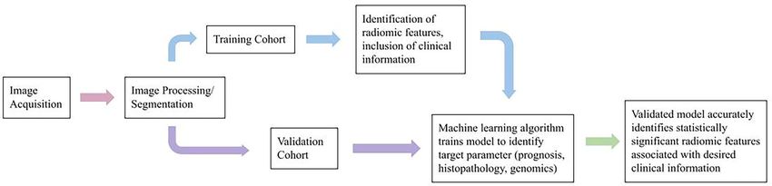
Gul et al. Radiomics and Head and Neck Cancers
Technological advancements in computer hardware and of developing oral and pharyngeal cancer, with an estimated 80%
artificial intelligence enable an integrative analysis of clinical, of that population being male and 61% being female (54).
radiomic, and bio-genomic data for cancer discovery (6–9). In Research has also indicated an etiological association of head
the case of radiomics, vast numbers of quantitative features can and neck cancer to viruses (56). The human papillomavirus
be derived from multi-modal medical images using (HPV), a virus known to cause common conditions such as
computational methods (3, 10). Phenotypes represented using warts, has developed a reputation for its association with cervical
radiomic features may have prognostic and diagnostic value, and and oropharyngeal cancers (53). Therefore, when diagnosing
potentially improve clinical decision support in cancer treatment HNC, patients will often be screened for HPV infection as a
(6, 11, 12). potential cause of disease. There are over 170 different types of
Radiomics can be performed using multimodal (CT, PET, HPV’s, categorized by the virus’s characteristics such as location
MRI, and ultrasound) and/or multiparametric (multiple MRI (mucosal or cutaneous anatomical sites), response to an external
sequences, e.g., diffusion MRI, perfusion MRI techniques (7–9, stimulus, and its risk for malignancy. The mucosal subgroup of
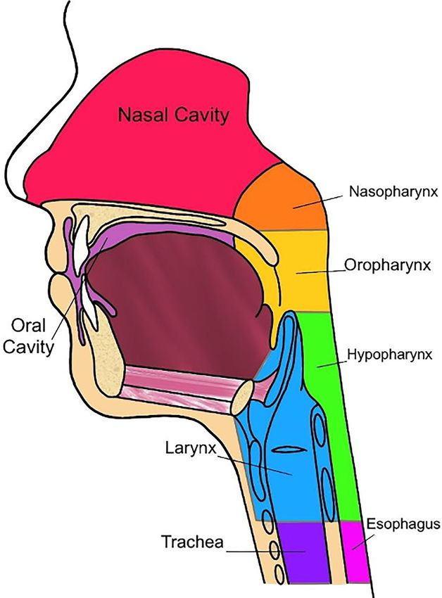
13–15). In a typical radiomic workflow (Figure 1), we first HPV is primarily associated with HNC as this subgroup contains
perform image registration and pre-processing, then image over 40 subtypes that are considered to be sexually transmitted
segmentation and annotation. Next, radiomic features are diseases (STD) and predominantly infect the reproductive and
calculated using computational methods. A variety of tools are respiratory tracts (53).
available to streamline the process (16–24). Radiomic features Additional etiological associations to HNC include the
are mostly sub-visual and can be coarsely grouped into intensity, Epstein-Barr virus (EBV), which is often associated with many
shape, and texture. In addition, before calculating the radiomic different types of human cancers, including those of lymphoid
values, we can apply spatial filters such as wavelets and Laplacian and epithelial cells (57). Considered one of the most common
of Gaussian filters to extract a variety of derivative and spatial- human viruses, EBV infection typically spreads undetected and
frequency information. can reside within the host over a span of ages in which infection
The radiomic features are then integrated with other data is dependent on several factors such as genetic predisposition,
sources for prognostic (7–9, 25–39), treatment response (40–42), diet, living conditions, hygiene, and sexual behavior (53, 58). To
histopathological (43–48), or radiogenomic (11, 49–51) analyses further validate the commonality of EBV infection, statistics
using statistical or machine learning modeling techniques. show by adulthood approximately 90-95% of the population will
sustain a permanent, asymptomatic infection of EBV (53, 57). As
a member of the Herpesviridae family, alternatively known as
HEAD AND NECK CANCER human herpesvirus type 4 (HHV4) (58), post-primary infection
of EBV is permanent and can subsequently result in the virus
Oncologic disease developing in the mucosal surfaces of shedding into genital and salivary secretions that increase the
anatomic subsites, such as the nasopharynx, oropharynx, risk of carcinogenesis into HNSSC.
hypopharynx, oral cavity, larynx, paranasal sinuses, and Currently, radiomics can predict some tumoral characteristics
salivary glands are considered HNC (Figure 2) (52, 53). The linked to patient survival in HNC (Table 1). In a study performed
International Classification of Diseases, Tenth Revision (ICD-10) by Mukherjee et.al., radiomic features were analyzed via CT
reports that oral and pharyngeal cancer accounts for imaging to non-invasively predict the histopathological features
approximately 2.3% of cancers within the United States. Oral of HNSCC. This study was performed retrospectively, utilizing CT
and pharyngeal cancer has a five-year survival of 27.8% and is images and data from clinically diagnosed patients with HNSCC.
internationally considered to be the sixth most common cancer An institutional test cohort (n = 71) and an HNSCC training
(54, 55). Risks of developing this disease are commonly cohort derived from The Cancer Genome Atlus (TCGA) (n = 113)
associated with the consumption of tobacco and alcoholic were analyzed within this study (43). A machine learning model,
products. Therefore, 74% of the general population that trained with 2,131 extracted radiomic features that were utilized to
practice tobacco and alcohol consumption have a greater risk predict tumor histopathological characteristics, was applied to the
FIGURE 1 | Typical radiomic workflow.
Frontiers in Oncology | www.frontiersin.org 2 July 2021 | Volume 11 | Article 639326Gul et al. Radiomics and Head and Neck Cancers FIGURE 2 | Anatomy of ear, nose, and throat, sagittal view. training and test cohort. These features included intensity, size pathologic features are specific to the individual regions of the and shape, texture, and filters (43). The cancer characteristics head and neck and will therefore be reviewed by region (Figure 2). investigated related to these features were tumor grade, perineural invasion, lymphovascular invasion, extracapsular spread, and Nasopharynx HPV status (p16 expression) (43). For dimensionality reduction Typically viewed as an endemic within the southern Chinese and classification of these features, principal component analysis, population, undifferentiated nasopharyngeal carcinoma (NPC) and regularized regression was applied, respectively (43). Results has the strongest association with EBV infection (57, 58). The from this study indicated that the radiomic model produced by World Health Organization (WHO) has characterized NPC into Mukherjee et al. showed strong-to-moderate power in predictive two primary histological types: keratinizing squamous cell prognosis for patients diagnosed with HNSCC, which was further carcinoma (Type I) and non-keratinizing squamous cell validated in an external institutional testing cohort. In other carcinoma (Type II and III). The undifferentiated histological words, this study concluded that radiomic CT models have subtype of NPC, such as Type II and III, has the closest significant value in predicting features typically indicating association with EBV infection, which particularly affects pathological assessment of HNSCC (43). Many of these regions such as Hong Kong, southern regions of China, and Frontiers in Oncology | www.frontiersin.org 3 July 2021 | Volume 11 | Article 639326
Gul et al.                                                                                                                 Radiomics and Head and Neck Cancers

TABLE 1 | Summary of radiomic applications in head and neck.

Classification                    Prediction Target                                         Radiomic and Clinical Features                                 Source

Nasopharynx     Progression free survival                        Multiparametric MRI features                                                              (37)
                Progression free survival                        EBV DNA, Gross tumor volume (GTVnx), lymph node (GTVnd), Dose Volume                      (59)
                                                                 Histogram
Oropharynx      HPV status                                       CT imaging: gross tumor volume (GTV)                                                      (63)
                HPV status                                       CE-CT imaging: gross tumor volume (GTV): high intensity, small lesions, greater           (64)
                                                                 sphericity, heterogeneity
                Local tumor control status post chemoradiation   CT imaging: shape, intensity, texture, wavelet transformation, heterogeneity, HPV         (32)
                                                                 status
Hypopharynx     Treatment response                               PET imaging: surface to volume ratio, spherical disproportion, TGV, local homogeneity,    (70)
                                                                 variance
                Disease progression                              CE-CT and NC-CT image features, clinical identification of peripheral Invasion             (71)
Larynx          T category prediction radiomics model            CT imaging: gradient skewness and mean, least axis, sphericity, wavelet kurtosis          (72)
                Overall survival                                 CT texture features                                                                       (73)
                Treatment response                               FLT PET tumor heterogeneity                                                               (28)
                Local control                                    CT imaging: entropy, kurtosis skewness, standard deviation                                (74)
Parotid gland   Differentiation of MALToma from benign           CT based hybrid radiomic and clinical demographic model                                   (82)
                lymphoepithelial lesion
Metastatic      PDL-1 expression                                 FDG PET textural features, HPV status, Ki-67 expression                                   (87)

Southeast Asia (58). Additional risks include are genetic                        K. et. al., the study aimed to develop and validate a nomogram
predisposition and dietary factors. It is important to note that                 that incorporated clinical data, gross tumor volume of the
although EBV infection is discovered in nearly all                               nasopharynx (GTVnx) and lymph nodes (GTVnd) radiomic
undifferentiated NPC cases, EBV is not detected in other head                    signatures, and multiparametric based therapeutic dose-volume
and neck cancers, excluding salivary gland tumors (58).                          histogram (DVH) signatures by Least Absolute Shrinkage and
                                                                                 Selection Operator (LASSO) to predict progression-free survival
Exploring the application of Radiomics to                                        (PFS) in patients diagnosed with locoregionally advanced NPC.
Nasopharyngeal Cancer                                                            The study concluded that the developed multidimensional
In a study performed by Zhang et. al., multiparametric magnetic                  nomogram incorporating radiomic signatures of lymph nodes,
resonance imaging (MRI)-based radiomics was utilized as a                        planning scores, and tumor-node-metastasis stage showed
prognostic factor in patients with advanced NPC. For this                        efficient predictive accuracy in determining PFS. However,
study, 118 advanced NPC patients were enrolled to determine                      incorporating pre-treatment plasma EBV-DNA status
the training cohort (n = 88) and the validation cohort (n = 30). A               improved the predictive accuracy of the nomogram model.
total of 970 radiomic features were extracted from two                           This implication was investigated via a sub-group analysis of
parameters: T2-weighted (T2-w) and contrast-enhanced T1-                         EBV-DNA (59). This data was confirmed by the study’s
weighted (CET1-w) MRI images. Application of LASSO                               validation cohort, and as a result, indicated that consideration
regression was utilized to select features for progression-free                  of pre-treatment EBV-DNA was a useful prognostic biomarker
survival (PFS) nomograms and the association between radiomic                    in NPC (59). Therefore, there is potential improvement in NPC
features and clinical data was evaluated via heatmaps (37). The                  screening when considering radiomics and EBV-status.
results indicated that there are significant associations between
the radiomic features and PFS. For example, radiomic signatures                  Oropharynx
derived from joint CET1-w and T2-w images displayed                              Oropharyngeal cancer (OPC) is one of the most frequent HNC,
improved prognostic performance when compared to                                 with squamous cell carcinoma (SCC) accounting for
signatures derived from the CET1-w and T2-w parameters                           approximately 90% of diagnosed cases (60). The oropharynx is
separately. These findings were confirmed in the validation                        a region in the pharynx located behind the oral cavity, including
cohort, suggesting the application of radiomics utilizing                        structures such as the soft palate and tonsils. This cancer has a 5-
multiparametric MRI-based radiomics provided improved                            year-survival rate of approximately 50% (60). The high mortality
prognosis in advanced NPC. Nonetheless, there is a need to                       rate is not always due to the malignancy or intensity of the
research features that can be utilized in radiomic application to                tumor, but simply due to late detection (60). OPC tumors rarely
profile these types of advanced NPC tumors. Producing these                       present symptoms that seem concerning upon initial screening.
findings will allow for treatment advancement and precise                         For example, symptoms typically include a sore throat or
clinical risk stratification (20).                                                difficulty swallowing (60). Therefore, the tumor is usually
                                                                                 detected late with little to no time to treat the disease, resulting
Exploring the application of Radiomics to the                                    in low survival rates and death shortly after diagnosis. OPC can
Epstein-Barr Virus in Head and Neck Cancer                                       also be characterized by its aggressive tumors, with a 70%
EBV in relation to HNSSC has the strongest association with                      prevalence of cervical metastases and the ability to disseminate
nasopharyngeal carcinoma (NPC). In a study performed by Yang                     quickly (60). Risk factors for oropharyngeal cancer include a

Frontiers in Oncology | www.frontiersin.org                                  4                                             July 2021 | Volume 11 | Article 639326
Gul et al. Radiomics and Head and Neck Cancers
history of smoking cigarettes and the presence of an HPV transformation (32). Results from this study indicated that 3
infection (61). features were significantly associated with LC, indicating that
The association between HPV status and HNSCC involves tumors with a heterogeneous CT density were at risk for decreased
distinct tumor morphology, younger patient’s age when LC (32). As a result, this study concluded that quantified CT
presented, and positive response to radiotherapy treatment. radiomics examining the heterogeneity of HNSCC tumor density
HPV-positive status is a significant prognostic feature is associated with LC after chemoradiation therapy and HPV
regarding favorable outcomes and overall survival in patients status (32). Utilizing this radiomic information from studies such
diagnosed with oropharyngeal squamous cell carcinoma as Bagher-Ebadian et al. and Bogowicz et al. will allow for
(OPSCC) (5). This is because HPV-positivity is considered a clinicians to further optimize oral screening for OPC and
strong, independent prognostic feature when diagnosing HNSCC, therefore optimizing patient diagnosis and clinical
OPSCC. HPV status of the tumor is determined by analyzing decision making in treatment planning.
p16 positivity using immunohistochemistry. The cyclin-
dependent kinase inhibitor p16 is a tumor suppressor gene
Hypopharynx
that is often overexpressed in HPV mediated cancers and leads
Hypopharyngeal cancer has the worst prognosis of all HNC with
to an overall better course of disease (62).
a 5-year-survival of only 25% to 41% (65–67). It is uncommon,
In a study performed by Leijenaar et. al., the study examined
with 2,500 new cases arising annually within the United States
that HPV-positive OPSCC is biologically and clinically different
(68). The hypopharynx can be divided into three distinct regions
than HPV-negative cases. The study then approached
to better distinguish the localized cancer cells: pyriform sinus,
understanding these significant differences through radiomics
postcricoid region, and the posterior wall (68). The pyriform
to evaluate the HPV status of OPSCC (63). The study included
sinus is where most squamous cell carcinomas occur, with 70%
four independent cohorts that encompassed a total of 778
of cases arising within this region. The postcricoid region
patients diagnosed with OPSCC. Of the 778 cases, the data was
accounts for approximately 20% of cases and the posterior wall
randomly assigned for the radiomic model training (n = 628) and
accounts for approximately 10% of cases (69). Because typical
validation (n = 150) cohorts. From pre-treatment CT imaging,
presentation is usually recognized by the growth of a neck mass
902 radiomic features were extracted from gross tumor volume.
or dysphonia, newly diagnosed patients are often presented at
Currently, there are no MRI-based radiomic reports available
Stage III or IV of disease, contributing to this disease history of
regarding radiomic signature prediction of HPV status.
poor prognosis (68). Hypopharyngeal cancer typically affects
individuals ranging between the ages of 50 to 60 years, occurring
Exploring the Application of Radiomics to
more often in men than women. Superior localization of the
Oropharyngeal Cancer
cancer cells is mostly associated with heavy drinking and
Application of radiomics has been practiced within this field of
smoking. Nutritional deficiencies account for the postcricoid,
disease and poses as a promising tool to noninvasively
the inferior part of the hypopharynx, being affected (68).
characterize tumor phenotypes (32, 64). In a study conducted
Hypopharyngeal tumors are classified as highly aggressive due
by Bagher-Ebadian et.al., a radiomic analysis of primary tumors
to their ability to metastasize early and infiltrate an abundant
extracted from pre-treatment contrast-enhanced computed
submucosal lymphatic network, sometimes even skipping
tomography (CE-CT) images was performed on patients
metastasis and reappearing in various locations distinct from
diagnosed with OPC (64). Within this study, Bagher-Ebadian
the primary site. Therefore, it is very common for multiple
et al. utilized radiomics to identify distinct features that construct
primary tumors to resurface (68). Treatment of hypopharyngeal
optimal characterization and prediction of HPV affecting OPC.
cancer is often controversial due to the desire for organ
Amongst the 172 radiomic features that were examined, only 12
preservation (65, 67). Early detection of this carcinoma may
radiomic features were significantly different between HPV-
only require radiotherapy, but treatment for later stages of the
positive and HPV-negative patients. Results from this study
disease is more complicated. Due to the complications of late-
indicate that gross tumor volumes (GTV) for HPV-positive
stage disease, the standard treatment is surgical resection and is
patients display higher intensity, smaller lesion size, greater
sometimes paired with postoperative chemoradiation therapy
sphericity, and higher patient intensity-variation/heterogeneity
with or without immunotherapy (69).
on CE-CT imaging (64). These results suggest that radiomic
features of HPV status in OPC patients are associated with
spatial arrangement and morphological appearance via CE- Exploring the Application of Radiomics to
CT imaging. Hypopharyngeal Cancer
Furthermore, in a retrospective study performed by Bogowicz Since early detection of this disease may only require treatment
et al. CT radiomics was utilized to predict local tumor control via radiotherapy, identifying significant markers that indicate the
(LC) after chemoradiation therapy of HNSCC, as well as carcinogenesis of hypopharyngeal cancers into a non-invasive
examining the effects of HPV infection on tumor radiomics. A radiomic pipeline could potentially improve prognosis. Utilizing
training cohort (n = 93) and a validation cohort (n = 56) were radiomics may allow clinicians to assess the progression of the
approved to be included in this study. 317 CT-radiomic features disease earlier, and, therefore, to construct a patient-specific
were calculated within the primary tumor region, including treatment plan that optimizes treatment response and reduces
features based on shape, intensity, texture, and wavelet unnecessary high-risk intervention. Fortunately, studies have
Frontiers in Oncology | www.frontiersin.org 5 July 2021 | Volume 11 | Article 639326Gul et al. Radiomics and Head and Neck Cancers
shown that early detection of the tumor can be found using relies heavily on tumor T categories defined by the National
radiomics. Liao et al. conducted a study including a total of 80 Comprehensive Cancer Network (NCCN) Guidelines (72).
OPC and hypopharyngeal cancer PET images were analyzed However, relapse occurrence resulting from these organ-
using radiomics to distinctively select imaging features indicative preserving treatment approaches remains high, with recurrence
of the diseases. These imaging features were then correlated with at 5-years approximately 30-40%, despite overall improvement
prognostic diagnosis, cancer stage detection, and prediction of in radiotherapy and systemic techniques (15). Exploring the
effective treatment. All cases included in the study had been radiomic study of one of the most anatomically complex
treated with chemoradiation therapy (70). This study found that structures within the head and neck region can provide
16 image features were significantly different between early and additional comprehensive information and characterization of
late stages within the several metabolic tumor volumes (MVT). intra-tumor heterogeneity.
The image features include surface area, surface to volume ratio,
compactness, spherical disproportion, TGV, energy, contrast, Exploring the Application of Radiomics to Laryngeal
local homogeneity, dissimilarity, variance, inverse variance, Squamous Cell Carcinoma
inverse difference moment, inverse difference, RLNU, and Surgical options for patients diagnosed with LSCC heavily
RPC. These features successfully differentiated early from late depend on preoperative T category classification, specifically
stages of OPC and hypopharyngeal cancer. As a result, these between T3 and T4 categories. This is because the distinction
findings assisted in evaluating prognosis and specific treatment between T3 and T4 categories for LSCC relies on the destruction
response for the patient (70). 5 and 2 features had an area under degree of the extralaryngeal spread and/or outer cortex of thyroid
curve (AUC) in receiver operating characteristic (ROC) greater cartilage (72). However, determining the T category pre-
than 0.7, indicating a promising predictor. The studied imaging operatively has its clinical challenges due to variable clinical
features resulted to prove to be essential indicators in tumor deductions between imaging modalities. Commonly used
differentiation, staging, overall survival (OS), relapse, and imaging techniques include CT and MRI, both techniques
treatment efficacy (70). harboring individual benefits and limitations (72). Therefore, a
Additionally, a study conducted by Mo et al. established a T category prediction radiomics (TCPR) model that combines
radiomics-based model to classify early versus late detection and radiomic signature and T category distinction could be beneficial
metastatic disease in patients with hypopharyngeal cancer. 113 in establishing optimal surgical outcomes. A study conducted by
patients diagnosed with this carcinoma were treated with Wang et al. was done to further validate the precise prediction of
chemoradiotherapy and divided into two cohorts, a training T categories using a radiomic nomogram and the TCPR model to
cohort (n = 80) and a validation cohort (n = 33) (71). The assess appropriate treatment management for each individual
radiomics model utilized the concordance index (C-index) to case. This study included a total of 211 patients with LSCC who
predict prognostic factors, resulting in C-indices of 0.804 with a had total laryngectomies separated into two cohorts. The
95% confidence interval (CI) of 0.688-0.920 and 0.756 with a training cohort (n=150) and the validation cohort (n=61)
95% CI between 0.605-0.907. Furthermore, the log-rank test and yielded results that demonstrate great capabilities of the TCPR
a nomogram were used in risk prediction of the model to assess model in predicting the preoperative T categories per patient.
disease progression. The significant results were p=0.00016 and The T category resulting from the study has an AUC of 0.775
p=0.00063, demonstrating an effective classification of patients (95% CI: 0.667–0.883). The radiomic signature resulted in a
into high and low-risk categories (71). Overall, the radiomics higher AUC, with AUC 0.862 (95% CI: 0.772–0.952). Finally, the
model in this study suggests being effective in predicting the nomogram incorporating the radiomic signature as well as the T
risk of progression for hypopharyngeal cancer along with category, the TCPR model, resulted in an AUC of 0.892 (95% CI:
chemoradiotherapy (71). 0.811–0.974). These results show that the predictive performance
of the T category improves with the application of the TCPR
Larynx model (72).
Laryngeal squamous cell carcinoma (LSCC) consists of 30-50% Moreover, in a study conducted by Chen et al., radiomic
of all neoplasms in the head and neck (15). Treatment analysis of laryngectomy CT imaging of 136 patients with LSCC
surrounding this disease is difficult due to considerable was performed to assess the prognostic value of radiomics
amounts of variability concerning the region’s anatomy, its derived from CT. All patients were divided into the training
surrounding structures, variable appearance of primary and cohort (n = 96) and the validation cohort (n = 40). A method was
recurrent tumors, significant anatomic changes resulting from designed to establish a radiomics signature from the CT texture
tumor response, and high intratumoral heterogeneity (15). features and a radiomics nomogram to predict overall survival
Standard-of-care treatment towards LSCC prioritizes organ- (OS) (73). The validation of the nomogram was done by a
preserving strategies, although treatment options may be calibration curve, C-index, and decision curve. The results
limited for more aggressive diseases. Although these strategies revealed the radiomics signature to have C-indices of 0.782
focus primarily on limiting the functional complications that are (95%CI: 0.656–0.909) and 0.752 (95%CI, 0.614–0.891). The
associated with complete surgical removal of the larynx, the most radiomics nomogram had outdone the cancer staging
appropriate therapy for patients with advanced LSCC is a total capability with a C-index of 0.817 vs. 0.682; P = 0.009 in the
laryngectomy (72). Conducting a surgical plan for treatment training cohort and a C-index of 0.913 vs. 0.699; P = 0.019 in the
Frontiers in Oncology | www.frontiersin.org 6 July 2021 | Volume 11 | Article 639326Gul et al. Radiomics and Head and Neck Cancers
validation cohort (73). The radiomics nomogram has had a the parotid gland, submandibular gland, and sublingual gland,
significant difference in its discrimination capability when respectively. Regarding the frequency of malignancy, 20%, 45%,
compared to other cancer staging techniques. The calibration and up to 81% of parotid tumors, submandibular gland tumors,
and decision curves have been shown to have an accurate 81% of sublingual gland tumors are malignant, respectively (77).
prediction for OS as well. This study has successfully utilized Although there are effective treatments for SGC, successful
radiomics in a way that predicts OS for LSCC, is critical in treatment for sublingual gland cancer is unknown due to lack
constructing a personalized treatment plan for each individual of clinical trials and the rarity of diagnosis (78). Standard of care
patient (73). treatment typically involves regional surgical resection of the
In another study conducted by Ulrich et al., radiomic feature parotid gland, otherwise known as a superficial parotidectomy
analysis from various 18F-fluorothymidine positron emission (77). Although more difficult to treat, cases of malignancy
tomography (FLT-PET) was done to evaluate the prediction of typically require a total parotidectomy. However, this
treatment response in patients with HNC. Thirty patients in the procedure is considered high risk as it involves contact with
late stages of OPC and LSCC who underwent chemoradiation critical facial nerves that may result in facial paralysis, in more
therapy and FLT-PET imaging before surgery were included in severe cases (77).
the study. 377 radiomic features of FLT uptake were extracted, 9
of which were indicated as significant (28). Within the 30 HNC Parotid Gland
cases, the study concluded that cases presenting smaller, Parotid tumors are the most common type of SGC, with the
homogeneous lesions at baseline resulted in a better prognosis. parotid gland accounting for approximately 25% of human saliva.
Furthermore, features extracted from the entire lesions had a It is the largest salivary gland and resides within the parotid space
higher C-index than primary tumor features for the majority of amongst the external carotid artery, retromandibular vein, and the
the 9 significant features. Overall, this study has shown that for intraparotid lymph nodes. In some cases, an accessory parotid gland
future studies integrating FLT-PET in predicting prognostic is present on the surface of the masseter muscle (77). The majority
outcome, radiomic features incorporating lesion shape, size, of parotid tumors are discovered as benign, though some lesions can
and texture features should be considered to ensure an be malignant (79). The different cancer subtypes of SGC that can
improved understanding of the disease (28). occur in the parotid gland include pleomorphic adenoma,
Additionally, the increasing application of radiomics to LSCC Warthin’s Tumor (War-T), parotid carcinoma (PCa), and
has demonstrated efficacy in predicting inferior local control and Kimura’s Disease (KD) (80). The most common of the subtypes
laryngectomy free survival (LFS). A study done by Agarwal et al. is pleomorphic adenoma. Pleomorphic adenoma composes of
explores if pre-treatment CT imaging features of the LSCC can epithelial cells along with myoepithelial cells, which are
predict long-term local control and LFS. This study analyzed 60 commonly referred to as benign mixed tumors (BMT) (81).
imaging texture features of patients undergoing chemoradiation Factors that may cause carcinogenesis of pleomorphic adenoma
(CTRT), which were further evaluated with a texture analysis include irradiation, dehydration, and tobacco use (81).
software (74). The data consisted of entropy, kurtosis, skewness,
standard deviation, mean intensity, and so on. After a median
follow-up of about 24 months, it was found that 39 patients were Exploring the Application of Radiomics
locally controlled and 10 had been treated with laryngectomy to Parotid Tumors
(74). Medium filtered-texture feature that had poor LFS were Regarding parotid tumors, one study implored radiomics to
entropy ≥4.54, (p = 0.006), kurtosis ≥4.18; p = 0.019, skewness improve diagnostic efficacy and, therefore, treatment options.
≤−0.59, p = 0.001, and standard deviation ≥43.18; p = 0.009). The To improve differentiation of a benign lymphoepithelial lesion
inferior local control was associated with medium filtered texture (BLEL) and a malignant mucosa-associated lymphoid tissue
features with entropy ≥4.54; p 0.01 and skewness ≤ – 0.12; p = lymphoma (MALToma) in the parotid gland, Y.-M. Zheng
0.02. The analysis of the study has shown medium texture et al. developed a CT-based radiomics nomogram that
entropy to be a predictor for local control and LFS (p = 0.001 integrated the radiomics signature alongside clinical data such
& p < 0.001). This advancement is undoubtedly efficient in as demographics (82). This integrated model was trained (n=70)
developing prognostic factors for LSCC and predicting and validated (n=31) on a total of 101 patients with BLEL or
treatment response (74). MALToma (82). In developing this model, 851 radiomics
features extracted from CT images were narrowed down to 7
features by removing features with poor inter- and intra-observer
Salivary Glands agreement between radiologists, including features that showed
Salivary gland cancer (SGC) is rare, compromising less than 1% significant differences between BLEL and MALToma (p < 0.000
of all cancers in the United States. This type of cancer is prevalent to 0.050) and applying LASSO regression (82). After performing a
in the older population, mostly affecting individuals between the multiple logistic regression analysis, statistically significant clinical
ages of 50 and 60 (75). The 5-year survival rate of SGC is factors of age (p = 0.0036) and maximum diameter (p = 0.019) were
approximately 7% (76). Residing within the facial region, three integrated with the radiomics signature resulting from the 7
major glands are used to classify different types of areas of SGC – radiomic features to produce a CT-based radiomics nomogram
the parotid, sublingual, and submandibular glands. Generally, that showed a statistically significant difference between BLEL and
about 80%, 11%, and less than 1% of SGC cases are found within MALToma (82).
Frontiers in Oncology | www.frontiersin.org 7 July 2021 | Volume 11 | Article 639326Gul et al. Radiomics and Head and Neck Cancers
Submandibular Gland studies. However, proper diagnosing of malignant sublingual
The submandibular gland is the second largest salivary gland. This glands from other types of malignancies has been a challenge
gland accounts for 70% of human saliva and is located underneath (85). Although advances in diagnostic imaging technology have
the jawbone (79). Despite the rarity of tumors in the submandibular helped with more effective identification, malignant sublingual
gland compared to the parotid gland, the probability of malignancy glands vary in degrees of malignancy and lead to difficulties in
in the submandibular gland is approximately 43% and results in a not only diagnosis but also management and treatment (85).
poorer prognosis (83). Due to rarity and high rates of malignancy, Radiomics has the potential to improve the initial evaluation of
there is a lack of knowledge pertaining to treating submandibular malignant gland tumors since there is a recurrence rate of 50%
gland tumors (83). There are no definitive treatments for for these tumors (85).
submandibular tumors, but there are numerous ways that have
been proven to be successful – all involving high-risk surgery. Radiomic Application to Advanced Head
A common procedure that is performed is submandibular and Neck Cancer
sialoadenectomy, which is to surgically remove the submandibular The management of metastatic and locally advanced head and
gland in its entirety (84). The efficacy of radiotherapy in targeting neck cancer has changed dramatically in the last several years.
these mass neoplasms is not well known with this type of cancer and Keynote 048 was a landmark trial that resulted in FDA approval
is still being evaluated. Chemotherapy in general is not shown to be for the use of immunotherapy either alone or in combination
successful in treating submandibular gland tumors but is sometimes with platinum-based chemotherapy as a first line treatment (78).
used for treatment if the tumor progressively spreads within the Specifically, this trial evaluated the efficacy of pembrolizumab, an
gland (83). immune checkpoint inhibitor that allows cytotoxic T cells to
recognize programmed death ligand 1 (PDL-1) overexpressed by
Exploring the Application of Radiomics to tumor cells, resulting in their destruction (78). In general, PDL-1
Submandibular Tumors expression by the tumor is evaluated by immunohistochemistry
In general, there remains uncertainty due to a lack of knowledge and serves as both a prognostic indicator and as a variable in the
for treatment of these diseases, demonstrating the necessity of decision-making process when selecting an appropriate
exploratory measures. Radiomic application to diseases such as immunotherapy regiment. The application of radiomics has
submandibular gland cancer illuminates characteristics that can further potential of evaluating the predictive power of PDL-1
be extracted into operational data. This data can then be utilized expression, and overall patient outcomes.
to improve detection and lead the course of treatment when While the radiomics of PDL-1 expression has been studied in
managing this disease. other tumors such as non-small cell lung cancer, data on
radiomic PDL-1 expression in head and neck cancer is lacking
Sublingual Gland (86). One pilot study by Chen et al. was able to predict PDL-1
Sublingual salivary gland tumors are the rarest tumors found in expression through FDG PET (87). This was accomplished by
SGC. The sublingual gland is the smallest of the three major glands, dichotomizing other biomarkers such as HPV status (p16
residing just below the floor of the mouth and is positioned under positivity) and Ki-67 expression. Textural features were also
the tongue, producing 5% of human saliva (79). Sublingual salivary used to predict PDL-1 expression. For example, gray-level
gland tumors typically affect individuals between 50 to 60 years old nonuniformity for run (GLNUr), run percentage (RP), and
and are not specific to gender (85). Sublingual gland tumors are short-zone low gray-level emphasis (SZLGE) were inversely
typically malignant, boasting an 81% probability of malignancy proportional with PDL-1 expression. While it is promising to
associated with this disease type. Adenoid cystic carcinoma and see evidence of the predictive power of PDL-1 expression
mucoepidermoid carcinoma are the most common neoplasms afforded by radiomics, this study is limited by its small cohort
found in the sublingual gland. Prognosis for adenocarcinoma of size. Further studies are needed to reproduce results and
the sublingual gland relies on the histology of the specific tumor. optimize the parameters relevant to head and neck cancer.
This tumor is commonly misinterpreted as minor salivary gland
tumors or other malignant lesions within the mouth due to its
compact mass (85). Patients normally present no symptoms, THYROID CANCERS
making the tumor difficult to identify and accurately diagnose.
When evaluating the tumor, it is important to distinguish if it lies in Defined as a malignancy of the thyroid gland by the International
the sublingual gland or any of the minor salivary glands. This cannot Classification of Diseases, Tenth Revision (ICD-10), thyroid
be done solely based on location on anatomy, but from a collection cancer accounts for 3.8% of all cancers in the United States
of imaging, surgical, and clinical data to ensure accurate and has a five-year survival of 98.3 (88). Thyroid cancers include
diagnosis (85). 3 main types: differentiated thyroid cancer (DTC), anaplastic
thyroid cancer (ATC), and medullary thyroid cancers (MTC)
Exploring the Application of Radiomics to Sublingual (89). Included in DTC, which accounts for over 90% of all
Gland Tumors thyroid cancers, are papillary thyroid cancer (PTC), follicular
Due to the rare nature of sublingual glands, specific suggestions thyroid cancer, Hurthle cell, and poorly differentiated thyroid
for treatment have not been developed, the lack of radiomic cancer (PDTC) (89). ATC accounts for less than 2% of call
Frontiers in Oncology | www.frontiersin.org 8 July 2021 | Volume 11 | Article 639326Gul et al. Radiomics and Head and Neck Cancers
thyroid cancers, and MTC accounts for about 1%-2% of all and a mortality rate of 1.2% at 20 years, patients with recurrent
thyroid cancers in the United States. Both DTC and MTC disease have poorer outcomes. Approximately 10% to 15% of
generally have good prognoses, with a 10-year survival rate of PTC cases recur, resulting in 35% of these patients ultimately
80–95% for PTC, 70–95% for follicular thyroid cancer, and 96% dying from this cancer. This is because recurrent PTC patients
for MTC (90, 91). However, ATC does not share such numbers, present aggressive features such as extrathyroidal extension
as it has a 5-year survival rate of 0-10%. Due to its rare and highly (ETE), aggressive pathological cell subtypes, the extent lymph
aggressive nature, ATC requires a multidisciplinary team node involvement, resistance to therapeutic treatments, and
approach with different treatment options of surgery, distant metastasis (93). To assess these aggressive features,
chemotherapy, or tracheotomy (89). Surgical resection is the clinicians use a variety of techniques such as ultrasound and
standard of care treatment option for DTC and MTC (89). ultrasound-guided fine-needle aspiration to develop a diagnosis.
An additional imaging modality that is often utilized is MRI.
Radiomic Application to Thyroid Cancers This allows for superior contrast of the soft tissues when
There is a need for establishing a non-invasive assessment examining the thyroid region, affording assessment of
technique that allows for the mapping of thyroid tumors in aggressive features such as ETE and neck nodal metastasis (93,
their entirety. It is important to expand the knowledge of 94). Although these imaging modalities are standard-of-care
radiomics and explore its implication to various disease types practices, both harbor limitations in accuracy and therefore
to improve clinical diagnosis and patient’s quality of life. inhibit optimal clinical assessment of the disease.
According to a study performed by Liang et. al., application of
radiomics showed good performance and potentially Exploring the Application of Radiomics to Papillary
outperformed ACR TI-RADS (American College of Radiology, Thyroid Cancer
Thyroid Imaging, Reporting, and Data System) scoring when In a retrospective study conducted by Park et. al., the association
predicting the malignancy of thyroid nodules (92). The objective between a radiomic signature of conventional ultrasound (US)
of this study was to produce a radiomic score utilizing US images and disease-free survival in PTC was investigated. The
imaging to predict the probability of malignancy in thyroid history of this disease type shows that PTC is considered a “good
nodules when compared to the ACR TI-RADS criteria. To do cancer” with regards to its treatability and relatively favorable
so, pathologically proven thyroid nodules were enrolled to survival rate (25). However, there is a small amount of PTC cases
produce a training cohort (one hospital, n=137) and a that show clinically aggressive behavior that results in 9% to 13%
validation cohort (separate hospital, n=95). The radiomic score of patients experiencing recurrence and 1% to 5% of patients
was developed utilizing the training cohort. US images were ultimately dying from thyroid cancer. Considering this
reviewed by two junior and one senior radiologist and scored the information, patients diagnosed with aggressive PTC would
nodules based on the 2017 ACR TI-RADS scoring criteria (92). greatly benefit from radiomic application with a preoperative
Results from this study indicated that the radiomic score had risk stratification tool that assists in assessing treatment plans
good discrimination, with an AUC of 0.921 in the training cohort and follow-up procedures (25).
and 0.931 in the validation cohort. This result suggests that the
radiomic score was significantly more accurate than the ACR Follicular Thyroid Cancer
scores when scoring suspicious thyroid nodules (Table 2). As a Follicular thyroid cancer (FTC) is known as the second most
result, a decision curve analysis showed that the radiomics score common differentiated thyroid cancer, accounting for 10% to
model potentially added more benefits than using the ACR TI- 15% of all cases. When considering age and gender, this disease
RADS scoring criteria (92). subtype typically affects women 50 to 60 years old. FTC presents
more aggressively in comparison to PTC, as this disease typically
Papillary Thyroid Cancer invades blood vessels and is capable of metastasizing via
Papillary thyroid cancer (PTC) is the most diagnosed thyroid hematogenous dissemination. Knowing this information, FTC
cancer, accounting for approximately 80% of well-differentiated is associated with a poorer prognosis in comparison to PTC, as
thyroid cancers. Although PTC typically has favorable outcomes FTC patients often present with more advanced staging of
TABLE 2 | Summary of radiomic applications in thyroid cancer.
Category Prediction Target Radiomic Features and Clinical Information Source
Thyroid nodules Malignancy US Thyroid radiomic score (92)
Papillary Thyroid Progression free survival US Thyroid: tumor size, cervical lymphadenopathy, extrathyroidal extension, gray (25)
Cancer level scores
Follicular Thyroid Metastatic disease US Thyroid: tumor shape, gray level scores (97)
Cancer
Medullary Thyroid Treatment response to PRRT SSTR- PET: textural features (gray level non uniformity) (101)
Cancer
Anaplastic Thyroid Treatment response/dose adjustment of Radiolabeled Trametinib (105)
Cancer Trametinib
Frontiers in Oncology | www.frontiersin.org 9 July 2021 | Volume 11 | Article 639326Gul et al. Radiomics and Head and Neck Cancers
disease due to vascular invasion (95). Long-term survival rates in to radiation therapy and/or chemotherapy. As a result, early
patients diagnosed with metastatic FTC range between 31% to detection and preventative surgery is often the standard-of-care
43%, taking into consideration the patient’s age at the time of treatment plan regarding MTC (98).
diagnosis, tumor size, capsular invasion, gender, and evidence of
metastases (96). FTC is typically classified into two categories: Exploring the Application of Radiomics to Medullary
minimally invasive or widely invasive. Thyroid Cancer
Regarding medullary thyroid cancers, there is great potential for
Exploring the Application of Radiomics to Follicular radiomics to be utilized here. One study shows promise in
Thyroid Cancer improving prognosis by exploring radiomic features involved
In a study conducted by Kwon et. al, radiomics was utilized to with PET images of advanced medullary thyroid cancer (101).
evaluate distant metastasis of FTC on gray-scale US images. This Lapa et al. assessed tumor heterogeneity by investigating the
retrospective study included 35 cases of FTC with distant association between textural parameters on somatostatin
metastases and 134 cases of FTC without distant metastasis receptor PET (SSTR-PET) and treatment response to peptide
(97). A total of 60 radiomic features were extracted, deriving receptor radionuclide therapy (PRRT) on 4 medullary thyroid
from the first order, shape, gray-level co-occurrence matrix, and cancer patients and 8 radioiodine-refractory differentiated
gray-level size zone matrix features utilizing US imaging thyroid cancer patients (101). They found that several textural
techniques. Results from this study indicated that the support parameters showed a significant capability to assess PFS, with
vector machine (SVM) classifier had an AUC of 0.90 on average “grey level non uniformity” ranking with the highest AUC (0.93)
on the test folds (97). Radiomic signature (pGul et al. Radiomics and Head and Neck Cancers
difficult to diagnose PC preoperatively because this disease type associated imaging data are typically acquired from just one or
has a lack of specific biochemical and clinical features (106). As a a few scanners from a single site. To deploy radiomic predictive
result, this disease is typically diagnosed postoperatively when models at scale and possibly across institutions, we need to
the disease is being examined histologically and/or when the address issues of potential data variability caused by scanners
disease recurs (106). from different vendors (114), and whether the models are still
predictive when they are applied to a different cohort from an
Exploring the Application of Radiomics to external site with similar disease types In summary, being able to
Parathyroid Cancer standardize image data acquisition and quality control using
Although there are no studies on the application of radiomics to phantoms, various calibration techniques, having large cohorts
parathyroid cancer, there is a need for clinicians to be able to from multiple locations for model training, and validation will
differentiate between parathyroid adenoma (benign) and provide more confidence for deployment in clinical settings.
parathyroid carcinoma because of the lack of specific The application of radiomics to HNC and thyroid cancers is
biochemical and clinical features (106). CT and MRI can both an advancement that allows for a deeper interpretation of a
help accurately localize the primary tumor, so the use of patient’s digital medical imaging data beyond visual assessment.
radiomics shows great promise in the parathyroid glands in Utilizing this practice, especially in cancer domains that lack
PC (106). radiomic studies such as anaplastic thyroid cancer and
parathyroid cancers, will allow for more personalized and
patient-specific cancer treatment. By gathering additional
DISCUSSION/CONCLUSION statistical data and conducting subsequent analysis, clinical
decision making is improved and therefore affects patient
Machine learning and deep learning models have been widely outcomes Court, Fave (115).
used for medical imaging research (6, 107). Although having
impressive predictive performance, these models are often AUTHOR CONTRIBUTIONS
difficult to interpret. Additionally, there may be hidden bias in
the model leading to potential ethical issues (108, 109). MG, K-JB, DG, CWH, AS, VL, AF, AC, MI, and AAC
Interpretability of predictive models has become one of the key contributed in literature search and manuscript preparation.
factors driving their adoption in clinical decision support MG and AAC performed final edits and revisions. All authors
environment. To ease the tension between the model contributed to the article and approved the submitted version.
prediction accuracy and interpretability, various approaches
have been proposed to generate intuitive interpretations of
predictive models (110–113). FUNDING
Radiomic studies are often exploratory in nature. They are
normally single institutional with limited cohort size. The The study was support by NIH grant # 2K12CA001727-26.
8. Bogowicz M, Riesterer O, Stark LS, Studer G, Unkelbach J, Guckenberger M,
REFERENCES et al. Comparison of PET and CT Radiomics for Prediction of Local Tumor
1. Rettig EM, D’Souza G. Epidemiology of Head and Neck Cancer. Surg Oncol Control in Head and Neck Squamous Cell Carcinoma. Acta Oncol (2017) 56
Clin N Am (2015) 24(3):379–96. doi: 10.1016/j.soc.2015.03.001 (11):1531–6. doi: 10.1080/0284186X.2017.1346382
2. Avanzo M, Stancanello J, El Naqa I. Beyond Imaging: The Promise of 9. Vallières M, Kay-Rivest E, Perrin LJ, Liem X, Furstoss C, Aerts H, et al.
Radiomics. Phys Med (2017) 38:122–39. doi: 10.1016/j.ejmp.2017.05.071 Radiomics Strategies for Risk Assessment of Tumour Failure in Head-and-
3. Rizzo S, Botta F, Raimondi S, Origgi D, Fanciullo C, Morganti AG, et al. Neck Cancer. Sci Rep (2017) 7(1):10117. doi: 10.1038/s41598-017-10371-5
Radiomics: The Facts and the Challenges of Image Analysis. Eur Radiol Exp 10. Gillies RJ, Kinahan PE, Hricak H. Radiomics: Images Are More Than
(2018) 2(1):36. doi: 10.1186/s41747-018-0068-z Pictures, They Are Data. Radiology (2016) 278(2):563–77. doi: 10.1148/
4. Yip SS, Aerts HJ. Applications and Limitations of Radiomics. Phys Med Biol radiol.2015151169
(2016) 61:R150. doi: 10.1088/0031-9155/61/13/R150 11. Aerts HJWL, Velazquez ER, Leijenaar RTH, Parmar C, Grossmann P,
5. Haider SP, Burtness B, Yarbrough WG, Payabvash S. Applications of Carvalho S, et al. Decoding Tumour Phenotype by Noninvasive Imaging
Radiomics in Precision Diagnosis, Prognostication and Treatment Using a Quantitative Radiomics Approach. Nat Commun (2014) 5(1):4006.
Planning of Head and Neck Squamous Cell Carcinomas. Cancers Head doi: 10.1038/ncomms5644
Neck (2020) 5:6. doi: 10.1186/s41199-020-00053-7 12. Aerts HJ. The Potential of Radiomic-Based Phenotyping in Precision
6. Langlotz CP, Allen B, Erickson BJ, Kalpathy-Cramer J, Bigelow K, Cook TS, Medicine: A Review. JAMA Oncol (2016) 2(12):1636–42. doi: 10.1001/
et al. A Roadmap for Foundational Research on Artificial Intelligence in jamaoncol.2016.2631
Medical Imaging: From the 2018 NIH/RSNA/ACR/The Academy 13. Wong AJ, Kanwar A, Mohamed AS, Fuller CD. Radiomics in Head and Neck
Workshop. Radiology (2019) 291(3):781–91. doi: 10.1148/radiol. Cancer: From Exploration to Application. Trans Cancer Res (2016) 5
2019190613 (4):371–82. doi: 10.21037/tcr.2016.07.18
7. Leger S, Zwanenburg A, Pilz K, Zschaeck S, Zöphel K, Kotzerke J, et al. CT 14. Jethanandani A, Lin TA, Volpe S, Elhalawani H, Mohamed ASR, Yang P,
Imaging During Treatment Improves Radiomic Models for Patients With et al. Exploring Applications of Radiomics in Magnetic Resonance Imaging
Locally Advanced Head and Neck Cancer. Radiother Oncol (2019) 130:10–7. of Head and Neck Cancer: A Systematic Review. Front Oncol (2018) 8:131.
doi: 10.1016/j.radonc.2018.07.020 doi: 10.3389/fonc.2018.00131
Frontiers in Oncology | www.frontiersin.org 11 July 2021 | Volume 11 | Article 639326Gul et al. Radiomics and Head and Neck Cancers
15. Chiesa-Estomba CM, Echaniz O, Larruscain E, Gonzalez-Garcia JA, 32. Bogowicz M, Riesterer O, Ikenberg K, Stieb S, Moch H, Studer G, et al.
Sistiaga-Suarez JA, Grana M. Radiomics and Texture Analysis in Computed Tomography Radiomics Predicts Hpv Status and Local Tumor
Laryngeal Cancer. Looking for New Frontiers in Precision Medicine Control After Definitive Radiochemotherapy in Head and Neck Squamous
Through Imaging Analysis. Cancers (Basel) (2019) 11(10):1409. doi: Cell Carcinoma. Int J Radiat Oncol Biol Phys (2017) 99(4):921–8. doi:
10.3390/cancers11101409 10.1016/j.ijrobp.2017.06.002
16. van Griethuysen JJM, Fedorov A, Parmar C, Hosny A, Aucoin N, Narayan 33. Ou D, Blanchard P, Rosellini S, Levy A, Nguyen F, Leijenaar RTH, et al.
V, et al. Computational Radiomics System to Decode the Radiographic Predictive and Prognostic Value of CT Based Radiomics Signature in Locally
Phenotype. Cancer Res (2017) 77(21):e104–e7. doi: 10.1158/0008- Advanced Head and Neck Cancers Patients Treated With Concurrent
5472.CAN-17-0339 Chemoradiotherapy or Bioradiotherapy and Its Added Value to Human
17. Pfaehler E, Zwanenburg A, de Jong JR, Boellaard R. Racat: An Open Source Papillomavirus Status. Oral Oncol (2017) 71:150–5. doi: 10.1016/
and Easy to Use Radiomics Calculator Tool. PloS One (2019) 14(2): j.oraloncology.2017.06.015
e0212223. doi: 10.1371/journal.pone.0212223 34. Kuno H, Qureshi MM, Chapman MN, Li B, Andreu-Arasa VC, Onoue K,
18. Nioche C, Orlhac F, Boughdad S, Reuzé S, Goya-Outi J, Robert C, et al. Lifex: et al. Ct Texture Analysis Potentially Predicts Local Failure in Head and
A Freeware for Radiomic Feature Calculation in Multimodality Imaging to Neck Squamous Cell Carcinoma Treated With Chemoradiotherapy. AJNR
Accelerate Advances in the Characterization of Tumor Heterogeneity. Am J Neuroradiol (2017) 38(12):2334–40. doi: 10.3174/ajnr.A5407
Cancer Res (2018) 78(16):4786–9. doi: 10.1158/0008-5472.CAN-18-0125 35. Ouyang FS, Guo BL, Zhang B, Dong YH, Zhang L, Mo XK, et al. Exploration
19. Dinapoli N, Alitto AR, Vallati M, Gatta R, Autorino R, Boldrini L, et al. and Validation of Radiomics Signature as an Independent Prognostic
Moddicom: A Complete and Easily Accessible Library for Prognostic Biomarker in Stage III-IVb Nasopharyngeal Carcinoma. Oncotarget (2017)
Evaluations Relying on Image Features. Conf Proc: Annu Int Conf IEEE 8(43):74869–79. doi: 10.18632/oncotarget.20423
Eng Med Biol Soc IEEE Eng Med Biol Soc Annu Conf (2015) 2015:771–4. doi: 36. Zhang B, Ouyang F, Gu D, Dong Y, Zhang L, Mo X, et al. Advanced
10.1109/EMBC.2015.7318476 Nasopharyngeal Carcinoma: Pre-Treatment Prediction of Progression Based
20. Liang Z-G, Tan HQ, Zhang F, Rui Tan LK, Lin L, Lenkowicz J, et al. on Multi-Parametric MRI Radiomics. Oncotarget (2017) 8(42):72457–65.
Comparison of Radiomics Tools for Image Analyses and Clinical Prediction doi: 10.18632/oncotarget.19799
in Nasopharyngeal Carcinoma. Br J Radiol (2019) 92(1102):20190271. doi: 37. Zhang B, Tian J, Dong D, Gu D, Dong Y, Zhang L, et al. Radiomics Features
10.1259/bjr.20190271 of Multiparametric MRI as Novel Prognostic Factors in Advanced
21. Götz M, Nolden M, Maier-Hein K. Mitk Phenotyping: An Open-Source Nasopharyngeal Carcinoma. Clin Cancer Res (2017) 23(15):4259–69. doi:
Toolchain for Image-Based Personalized Medicine With Radiomics. 10.1158/1078-0432.CCR-16-2910
Radiother Oncol (2019) 131:108–11. doi: 10.1016/j.radonc.2018.11.021 38. Bogowicz M, Jochems A, Deist TM, Tanadini-Lang S, Huang SH, Chan B,
22. Fang YH, Lin CY, Shih MJ, Wang HM, Ho TY, Liao CT, et al. Development et al. Privacy-Preserving Distributed Learning of Radiomics to Predict
and Evaluation of an Open-Source Software Package “Cgita” for Quantifying Overall Survival and HPV Status in Head and Neck Cancer. Sci Rep
Tumor Heterogeneity With Molecular Images. BioMed Res Int (2014) (2020) 10(1):4542. doi: 10.1038/s41598-020-61297-4
2014:248505. doi: 10.1155/2014/248505 39. Zhang B, He X, Ouyang F, Gu D, Dong Y, Zhang L, et al. Radiomic Machine-
23. Brown AM, Nagala S, McLean MA, Lu Y, Scoffings D, Apte A, et al. Multi- Learning Classifiers for Prognostic Biomarkers of Advanced
Institutional Validation of a Novel Textural Analysis Tool for Preoperative Nasopharyngeal Carcinoma. Cancer Lett (2017) 403:21–7. doi: 10.1016/
Stratification of Suspected Thyroid Tumors on Diffusion-Weighted MRI. j.canlet.2017.06.004
Magnet Resonance Med (2016) 75(4):1708–16. doi: 10.1002/mrm.25743 40. Jansen JF, Lu Y, Gupta G, Lee NY, Stambuk HE, Mazaheri Y, et al. Texture
24. Zhang L, Fried DV, Fave XJ, Hunter LA, Yang J, Court LE. IBEX: An Open Analysis on Parametric Maps Derived From Dynamic Contrast-Enhanced
Infrastructure Software Platform to Facilitate Collaborative Work in Magnetic Resonance Imaging in Head and Neck Cancer. World J Radiol
Radiomics. Med Phys (2015) 42(3):1341–53. doi: 10.1118/1.4908210 (2016) 8(1):90–7. doi: 10.4329/wjr.v8.i1.90
25. Park VY, Han K, Lee E, Kim E-K, Moon HJ, Yoon JH, et al. Association 41. Liu J, Mao Y, Li Z, Zhang D, Zhang Z, Hao S, et al. Use of Texture Analysis
Between Radiomics Signature and Disease-Free Survival in Conventional Based on Contrast-Enhanced MRI to Predict Treatment Response to
Papillary Thyroid Carcinoma. Sci Rep (2019) 9(1):4501. doi: 10.1038/ Chemoradiotherapy in Nasopharyngeal Carcinoma. J Magnet Resonance
s41598-018-37748-4 Imaging: JMRI (2016) 44(2):445–55. doi: 10.1002/jmri.25156
26. Head MDACC and Neck Quantitative Imaging Working G. Investigation of 42. Scalco E, Marzi S, Sanguineti G, Vidiri A, Rizzo G. Characterization of Cervical
Radiomic Signatures for Local Recurrence Using Primary Tumor Texture Lymph-Nodes Using a Multi-Parametric and Multi-Modal Approach for an
Analysis in Oropharyngeal Head and Neck Cancer Patients. Sci Rep (2018) 8 Early Prediction of Tumor Response to Chemo-Radiotherapy. Phys Med: PM
(1):1524. doi: 10.1038/s41598-017-14687-0 (2016) 32(12):1672–80. doi: 10.1016/j.ejmp.2016.09.003
27. Parmar C, Grossmann P, Rietveld D, Rietbergen MM, Lambin P, Aerts HJ. 43. Mukherjee P, Cintra M, Huang C, Zhou M, Zhu S, Colevas AD, et al. CT-
Radiomic Machine-Learning Classifiers for Prognostic Biomarkers of Head Based Radiomic Signatures for Predicting Histopathologic Features in Head
and Neck Cancer. Front Oncol (2015) 5:272. doi: 10.3389/fonc.2015.00272 and Neck Squamous Cell Carcinoma. Radiol: Imaging Cancer (2020) 2(3):
28. Ulrich EJ, Menda Y, Boles Ponto LL, Anderson CM, Smith BJ, Sunderland JJ, e190039. doi: 10.1148/rycan.2020190039
et al. Flt PET Radiomics for Response Prediction to Chemoradiation 44. Fruehwald-Pallamar J, Hesselink JR, Mafee MF, Holzer-Fruehwald L,
Therapy in Head and Neck Squamous Cell Cancer. Tomography (2019) 5 Czerny C, Mayerhoefer ME. Texture-Based Analysis of 100 MR
(1):161–9. doi: 10.18383/j.tom.2018.00038 Examinations of Head and Neck Tumors - Is It Possible to Discriminate
29. Guezennec C, Robin P, Orlhac F, Bourhis D, Delcroix O, Gobel Y, et al. Between Benign and Malignant Masses in a Multicenter Trial? RoFo:
Prognostic Value of Textural Indices Extracted From Pretherapeutic 18-F Fortschr Auf Dem Gebiete Der Rontgenstrahlen Und Der Nuklearmed
FDG-PET/CT in Head and Neck Squamous Cell Carcinoma. Head Neck (2016) 188(2):195–202. doi: 10.1055/s-0041-106066
(2019) 41(2):495–502. doi: 10.1002/hed.25433 45. Yang X, Wu N, Cheng G, Zhou Z, Yu DS, Beitler JJ, et al. Automated
30. Zhang H, Graham CM, Elci O, Griswold ME, Zhang X, Khan MA, et al. Segmentation of the Parotid Gland Based on Atlas Registration and Machine
Locally Advanced Squamous Cell Carcinoma of the Head and Neck: CT Learning: A Longitudinal MRI Study in Head-and-Neck Radiation Therapy. Int
Texture and Histogram Analysis Allow Independent Prediction of Overall J Radiat Oncol Biol Phys (2014) 90(5):1225–33. doi: 10.1016/j.ijrobp.2014.08.350
Survival in Patients Treated With Induction Chemotherapy. Radiology 46. Fruehwald-Pallamar J, Czerny C, Holzer-Fruehwald L, Nemec SF, Mueller-
(2013) 269(3):801–9. doi: 10.1148/radiol.13130110 Mang C, Weber M, et al. Texture-Based and Diffusion-Weighted
31. Bogowicz M, Leijenaar RTH, Tanadini-Lang S, Riesterer O, Pruschy M, Discrimination of Parotid Gland Lesions on MR Images at 3.0 Tesla.
Studer G, et al. Post-Radiochemotherapy PET Radiomics in Head and Neck NMR Biomed (2013) 26(11):1372–9. doi: 10.1002/nbm.2962
Cancer - The Influence of Radiomics Implementation on the Reproducibility 47. Wu W, Ye J, Wang Q, Luo J, Xu S. Ct-Based Radiomics Signature for the
of Local Control Tumor Models. Radiother Oncol (2017) 125(3):385–91. doi: Preoperative Discrimination Between Head and Neck Squamous Cell
10.1016/j.radonc.2017.10.023 Carcinoma Grades. Front Oncol (2019) 9:821. doi: 10.3389/fonc.2019.00821
Frontiers in Oncology | www.frontiersin.org 12 July 2021 | Volume 11 | Article 639326You can also read