Diffuse Large B-Cell Lymphoma of the Colon in an Asymptomatic Patient - Cureus
←
→
Page content transcription
If your browser does not render page correctly, please read the page content below
Open Access Case
Report DOI: 10.7759/cureus.26003
Diffuse Large B-Cell Lymphoma of the Colon in
an Asymptomatic Patient
Review began 06/13/2022
Sai Samyuktha Bandaru 1 , Vishal Busa 1 , Sanjay Juneja 2
Review ended 06/13/2022
Published 06/16/2022 1. Internal Medicine, Baton Rouge General Medical Center, Baton Rouge, USA 2. Hematology and Medical Oncology,
© Copyright 2022 Mary Bird Perkins Cancer Center, Baton Rouge, USA
Bandaru et al. This is an open access
article distributed under the terms of the Corresponding author: Sai Samyuktha Bandaru, samyukthareddy269@gmail.com
Creative Commons Attribution License CC-
BY 4.0., which permits unrestricted use,
distribution, and reproduction in any
medium, provided the original author and
source are credited. Abstract
Extranodal lymphomas of the gastrointestinal (GI) tract are known entities, but primary lymphoma of the
colon is extremely rare. Symptoms are non-specific, such as abdominal pain, bloody diarrhea, unintentional
weight loss, night sweats, and changes in bowel habits. Some patients do not have any specific symptoms,
which makes diagnosis extremely difficult. We present a 69-year-old asymptomatic male who was
incidentally found to have an inflammatory mass in the descending colon on screening colonoscopy; the
initial biopsy was inconclusive. However, due to high suspicion of any underlying malignancy, a repeat
colonoscopy with biopsy was done, which revealed diffuse large B-cell lymphoma (DLBCL). Prompt and early
diagnosis is extremely crucial for timely management. Management includes chemotherapy, radiotherapy,
and surgery.
Categories: Internal Medicine, Oncology, Hematology
Keywords: histopathology (hp), chemotherapy, colon, diffuse large b cell lymphoma, lymphoma
Introduction
Non-Hodgkin's lymphoma is a lymphoproliferative disorder originating from B lymphocytes or T
lymphocytes or natural killer cells. Diffuse large B-cell lymphoma (DLBCL) is the most common subtype of
non-Hodgkin's lymphoma. Primary diffuse large B-cell lymphoma of the colon is extremely rare. It affects
both men and women, with male predominance and the peak incidence seen in the sixth to seventh decades
[1,2]. There are only a few cases of DLBCL of the colon reported so far. Histopathological diagnosis can
confirm DLBCL. Treatment modalities include chemoradiotherapy; however, the role of surgery in the
treatment of DLBCL is still controversial. Some authors believe surgery should be limited to obstruction,
perforation, and hemorrhage, while others believe it should be an initial treatment modality [2,3].
Case Presentation
A 69-year-old Caucasian male presented to a gastroenterology clinic for a routine screening colonoscopy. He
had a two-year history of night sweats; otherwise, he did not have any symptoms. He denied unintentional
weight loss, fatigue, and change in bowel habits; abdominal pain; nausea; and vomiting. The medical history
was significant for hypertension, dyslipidemia, and coronary artery disease. Surgical history was significant
for percutaneous coronary intervention for coronary artery disease and hernia repair. He had a 20-year
history of smoking and no alcohol or drug use. No significant family history. On physical examination, vitals
were stable. The lungs were clear on examination, the heart sounds were regular in rate and rhythm, there
was no abdominal tenderness, bowel sounds were normal, and no frank blood was seen on rectal
examination. The rest of the physical examination was unremarkable.
Initial screening colonoscopy showed a 4 cm ulcerated, non-bleeding, non-obstructing mass in the
descending colon (Figure 1). A biopsy of the descending colon mass showed ulcerated mucosa with fibrino-
purulent exudate and granulation tissue, negative for any dysplasia or malignancy. Immunostains for
cytokeratin, synaptophysin, and CDX2 were negative for evidence of invasive carcinoma or neuroendocrine
tumor. However, due to high suspicion of any underlying malignancy, a repeat colonoscopy was done three
months later, which revealed localized inflammation characterized by congestion, erosions, and erythema in
the descending colon, and biopsies were taken for further histological examination. A biopsy from a
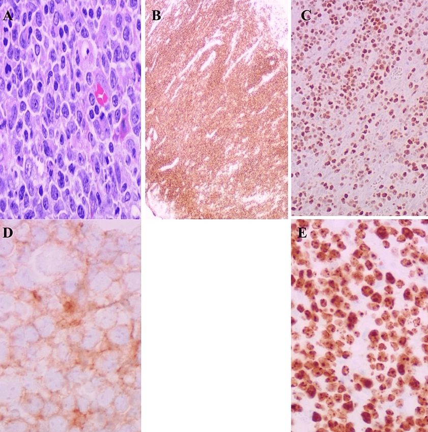
descending colon lesion showed high-grade CD10-positive B-cell lymphoma with intermediate to large-
sized lymphoid cells. Immunohistochemistry staining was positive for CD20, CD10, and BCL6, but negative
for CD 30 (Figure 2). Fluorescence in situ hybridization (FISH) analysis did not reveal any abnormalities in
BCL2, BCL6, or MYC. The Ki-67 proliferative index is high (around 80 to 90%). The initial staging positron
emission tomography (PET) scan showed no evidence of 18F-fluorodeoxyglucose (FDG) avid disease. Based
on pathology and PET scan findings, he was diagnosed with diffuse large B-cell lymphoma stage I of the
descending colon. He established care with a hematologist for further management.
How to cite this article
Bandaru S, Busa V, Juneja S (June 16, 2022) Diffuse Large B-Cell Lymphoma of the Colon in an Asymptomatic Patient. Cureus 14(6): e26003.
DOI 10.7759/cureus.26003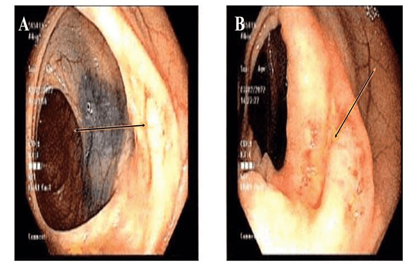
FIGURE 1: Image showing localized inflammation in descending colon
                                                   (arrows in panel A, B).

2022 Bandaru et al. Cureus 14(6): e26003. DOI 10.7759/cureus.26003                                                      2 of 5
FIGURE 2: Image shows pathological confirmation of DLBCL with
immunofixation staining (panels A, B, C, D, E).
Panel B positive for CD20, panel C positive for BCL-6, panel D positive for CD10, and panel E positive for Ki-67
high proliferation rate.
He underwent a laparoscopic colon resection. 20.1 cm of splenic flexure and proximal descending colon
segment were resected; the serosa of the resected lesion was pink- tan and smooth; multiple possible lymph
nodes were resected and sent for immunohistological examination. The histology of the resected colon
revealed a 1.4 cm atypical lymphoid lesion involving mucosal, submucosal, and superficial midmuscularis
propria; it is composed of intermediate to large lymphocytes with diffuse pattern involvement.
Immunohistochemistry was positive for CD10, CD20, BCL6, and negative for CD30 and MUM1. The
proliferative index Ki-67 is high, around 95%. Morphological and immunophenotypic findings are consistent
with diffuse large B-cell lymphoma. Negative FISH for MYC and large cell size make Burkitt lymphoma
unlikely. All the resected lymph nodes were negative for any metastatic disease. He was seen by a
hematologist and was started on chemotherapy with six cycles of rituximab, cyclophosphamide,
hydroxydaunorubicin, oncovin, and prednisone (R-CHOP). He was seen after two cycles of R-CHOP. He is
tolerating chemotherapy well without any dose-limiting toxicities.
Discussion
DLBCL is one of the aggressive lymphomas that can be cured, even in advanced cases, with effective
treatment regimens. It is the most common lymphoma in adults, accounting for 37% of B-cell lymphomas
and 30-40% of non-Hodgkin's lymphomas [4]. These commonly originate from lymph nodes, spleen, and
thymus, which are grouped as nodal DLBCL. However, 30-40% of patients could be extra-nodal in origin,
which includes sites like the gastro-intestinal tract, skin and soft tissue, head and neck, nervous system,
genitourinary, breast, musculoskeletal, pancreaticobiliary, respiratory tract, etc. [4,5]. Among these extra-
nodal sites, the GI tract is the most commonly involved site, especially the stomach in 65%, the small
intestine in 20-35%, and the colon and rectum in 5-15% [6]. Typically, these patients present with
gastrointestinal (GI) symptoms like abdominal pain, nausea, vomiting, weight loss, diarrhea, and rarely, can
present as acute complete or partial bowel obstructions. Presentations of these patients vary widely based
on the site involved. Although B symptoms are common in nodal DLBCL, the majority of patients with
2022 Bandaru et al. Cureus 14(6): e26003. DOI 10.7759/cureus.26003 3 of 5colonic DLBCL might be asymptomatic or with mild GI symptoms like in our patient, which was an
incidental finding on screening colonoscopy. Given the extra-nodal origin, the patient might not have
swollen or enlarged lymph nodes, which is a common presentation in nodal lymphomas.
Risk factors are mostly similar to nodal DLBCL with some differences, like associations with inflammatory
bowel disease, celiac disease, and infections like Campylobacter jejuni and Helicobacter pylori. Other risk
factors include viral infections like HIV, Epstein-Barr virus (EBV), hepatitis C virus (HCV), human T-cell
leukemia virus-1 (HTLV-1), and human herpesvirus-8 (HHV-8); immunosuppressants and
immunodeficiencies [7]. Pathogenesis includes activation of protooncogenes, chromosomal mutations by
various mechanisms like translocation t (14;18), c-rel amplification, histone methyl-transferase mutations,
and PTEN deletion [8,9]. Among these translocations, t (14;18) is the most common one seen in 30-40% of
cases that leads to BCL2 overexpression, resulting in unchecked growth of B-cells in germinal centers [9].
Diagnosing colonic lymphomas needs imaging modalities like contrast-enhanced CT of the abdomen along
with double-contrast barium enema followed by colonoscopy and biopsy for confirmation, as imaging only
provides information about size, depth of invasion, and nodal involvement. They also needed a wide range
of hematological testing and further staging of lymphoma, along with prognostication of the type of DLBCL.
Basic labs include complete blood count (CBC) with a differential that helps to find the bone marrow
involvement; a peripheral smear, comprehensive metabolic panel (CMP), serum lactate dehydrogenase
(LDH), and uric acid levels usually correspond to tumor burden and are also used as an early marker for
relapse [10]. Testing for hepatitis B virus (HBV), HCV, HIV, and EBV is necessary as these are highly
lymphoproliferative viruses and these can be possible etiologies. These serologies are not only needed in
diagnosis but also needed before starting treatment as they tell you the need for anti-viral prophylaxis to
prevent re-activation [11,12]. Although biopsy confirms the diagnosis, further comprehensive testing like
immunohistochemistry and flow cytometry needs to be done in order to identify the molecular and
phenotypic variants [12]. Immunophenotyping is almost mandatory in all DLBCL cases as this confirms the
B-cell lineage and type of DLBCL. The World Health Organization (WHO) has suggested an
immunohistochemical panel for phenotyping that includes CD20, CD79a, BCL6, CD10, MYC, BCL2, Ki67,
IRF4, CyclinD1, CD5, CD23, and EBER-1 staining for EBV [10]. Flow cytometry and cytogenetic tests like
fluorescent in situ hybridization (FISH) analysis are also part of investigations that are used especially to
differentiate the subtypes of DLBCL [12]. FISH studies help in identifying translocations, amplifications, and
deletions, which can further guide the therapies. Imaging studies include computed tomography (CT) of the
neck, chest, abdomen, and pelvis as a routine standard of care along with a whole-body PET as it helps in
prognostication and staging [10]. Staging is important in any malignancy as it helps in determining the
treatment plans, predicting the outcomes and survival rates, and helps in the aggressiveness of the
management. Even in colonic lymphomas, the Ann-Arbor staging system is the standard classification
system widely used worldwide [13].
Treatment modalities include multi-agent chemotherapy, surgical resection, and radiotherapy, and these
are used alone or in combination based on the staging of lymphoma, subtype, and other co-morbidities that
vary between individuals. Even in colonic lymphomas, R-CHOP induction therapy is the gold standard
regimen that includes rituximab in combination with cyclophosphamide, doxorubicin
(hydroxydaunorubicin), vincristine (Oncovin), and prednisone [14]. The role of surgery is still debated, and
studies recommend that it should be reserved for patients with complications like obstruction, perforation,
and bleeding [14]. Evidence has shown improved survival with chemotherapy +/- radiotherapy alone. In early
stages and localized colonic lymphomas, a few studies showed a better outcome with surgical resection
followed by chemotherapy [7]. In advanced stages, anthracycline-based multiagent chemotherapy with or
without radiotherapy is indicated [7]. Typically, each cycle lasts for 14-21 days, and the number of cycles of
chemotherapy is based on staging, prognosis, and need for radiation. Remission is achieved in two-thirds of
patients with this regimen. Management of relapse and refractory diseases depends on whether the patient
is a candidate for a hematopoietic stem cell transplant. High-dose chemotherapy with autologous stem cell
rescue is shown to improve survival in these patients [1]. The overall prognosis of colonic lymphomas is
poor, and these patients need to be monitored at regular intervals with frequencies based on response to
initial chemotherapy and staging of the disease. Follow-ups are recommended every three to six months for
three to five years, then annually or as clinically indicated with basic blood workup and LDH during each
visit [4].
Our patient was completely asymptomatic, and his lymphoma was identified on routine screening
colonoscopy. This case helps us realize that DLBCL of the colon can be asymptomatic. Any concerning
lesions on colonoscopy need proper histopathological examination. Without an appropriate and timely
diagnosis, the prognosis is poor.
Conclusions
DLBCL of the colon is an extremely rare entity. Patients can be asymptomatic or have non-specific
symptoms that can delay diagnosis. Histopathological examination findings are key for diagnosis. Early
diagnosis is really important for the timely management of these patients. Treatment includes
chemotherapy and radiotherapy, and the role of surgery is still controversial.
2022 Bandaru et al. Cureus 14(6): e26003. DOI 10.7759/cureus.26003 4 of 5Additional Information
Disclosures
Human subjects: Consent was obtained or waived by all participants in this study. Conflicts of interest: In
compliance with the ICMJE uniform disclosure form, all authors declare the following: Payment/services
info: All authors have declared that no financial support was received from any organization for the
submitted work. Financial relationships: All authors have declared that they have no financial
relationships at present or within the previous three years with any organizations that might have an
interest in the submitted work. Other relationships: All authors have declared that there are no other
relationships or activities that could appear to have influenced the submitted work.
References
1. Erginoz E, Askar A, Cavus GH, Velidedeoglu M: Primary diffuse large B-cell lymphoma of the sigmoid colon .
Int J Surg Case Rep. 2021, 87:106454. 10.1016/j.ijscr.2021.106454
2. Pandey M, Swain J, Iyer HM, Shukla M: Primary lymphoma of the colon: report of two cases and review of
literature. World J Surg Oncol. 2019, 17:18. 10.1186/s12957-018-1548-6
3. Haddad I, El Kurdi B, El Iskandarani M, Babar S, Young M: Primary diffuse large B-cell lymphoma of the
sigmoid colon. Cureus. 2019, 11:e5048. 10.7759/cureus.5048
4. Zelenetz AD, Gordon LI, Chang JE, et al.: NCCN Guidelines® insights: B-cell lymphomas, version 5.2021 . J
Natl Compr Canc Netw. 2021, 19:1218-30. 10.6004/jnccn.2021.0054
5. Vitolo U, Seymour JF, Martelli M, et al.: Extranodal diffuse large B-cell lymphoma (DLBCL) and primary
mediastinal B-cell lymphoma: ESMO Clinical Practice Guidelines for diagnosis, treatment and follow-up.
Ann Oncol. 2016, 27:v91-v102. 10.1093/annonc/mdw175
6. Gupta V, Singh V, Bajwa R, et al.: Site-specific survival of extra nodal diffuse large B-cell lymphoma and
comparison with gastrointestinal diffuse large B-cell lymphoma. J Hematol. 2022, 11:45-54. 10.14740/jh984
7. Boussios S, Zerdes I, Vassou A, et al.: Extranodal diffuse large B-cell lymphomas: a retrospective case series
and review of the literature. Hematol Rep. 2018, 10:7070. 10.4081/hr.2018.7070
8. Martelli M, Ferreri AJ, Agostinelli C, Di Rocco A, Pfreundschuh M, Pileri SA: Diffuse large B-cell lymphoma.
Crit Rev Oncol Hematol. 2013, 87:146-71. 10.1016/j.critrevonc.2012.12.009
9. Sharma B, Pavelock N, Antoine M, Shah M, Galbraith K, Rawlins S: Primary diffuse large B-cell lymphoma of
the descending colon. Am J Med Sci. 2019, 358:164-7. 10.1016/j.amjms.2019.05.004
10. Sweetenham JW: Diffuse large B-cell lymphoma: risk stratification and management of relapsed disease .
Hematology Am Soc Hematol Educ Program. 2005, 252-9. 10.1182/asheducation-2005.1.252
11. Mert D, Merdin A, Ceken S, Dal MS, Ertek M, Altuntas F: Evaluation of hepatitis B virus, hepatitis C virus,
and human immunodeficiency virus seroprevalence in patients with diffuse large B cell lymphoma and
Hodgkin's lymphoma. J Cancer Res Ther. 2021, 17:951-5. 10.4103/jcrt.JCRT_465_19
12. Tilly H, Gomes da Silva M, Vitolo U, et al.: Diffuse large B-cell lymphoma (DLBCL): ESMO Clinical Practice
Guidelines for diagnosis, treatment and follow-up. Ann Oncol. 2015, 26 Suppl 5:v116-25.
10.1093/annonc/mdv304
13. Carbone PP, Kaplan HS, Musshoff K, Smithers DW, Tubiana M: Report of the committee on Hodgkin's
disease staging classification. Cancer Res. 1971, 31:1860-1.
14. Candelaria M, Dueñas-Gonzalez A: Rituximab in combination with cyclophosphamide, doxorubicin,
vincristine, and prednisone (R-CHOP) in diffuse large B-cell lymphoma. Ther Adv Hematol. 2021,
12:2040620721989579. 10.1177/2040620721989579
2022 Bandaru et al. Cureus 14(6): e26003. DOI 10.7759/cureus.26003 5 of 5You can also read