Extracorporeal shock wave as adjuvant therapy for wrist and hand spasticity in post-stroke patients: a randomized controlled trial
←
→
Page content transcription
If your browser does not render page correctly, please read the page content below
Tabra et al. Egyptian Rheumatology and Rehabilitation              (2021) 48:21
https://doi.org/10.1186/s43166-021-00068-z
                                                                                                                            Egyptian Rheumatology
                                                                                                                            and Rehabilitation

 RESEARCH                                                                                                                                          Open Access

Extracorporeal shock wave as adjuvant
therapy for wrist and hand spasticity in
post-stroke patients: a randomized
controlled trial
Samar Abd Alhamed Tabra* , Mohammad Ibrahim Zaghloul and Doaa Shawky Alashkar

  Abstract
  Background: Stroke patients often present with upper limb spasticity which impairs the functional status of patients.
  Recently, extracorporeal shock wave therapy (ESWT) is reported to be a safe, non-invasive, alternative treatment for
  spasticity. Many articles have been published on the effect of ESWT on lower limb spasticity, but only few of them had
  focused on upper limb spasticity, so the aim of this study is to evaluate the clinical and electrophysiological effect of
  ESWT on wrist and hand spasticity of chronic stroke patients and its impact on functional performance. In this
  monocentric study, forty chronic stroke patients with upper limb spasticity were recruited and randomly allocated into
  two groups. Both groups continued to receive conventional stroke rehabilitative program, while group I received three
  sessions of radial extracorporeal shock wave therapy (rESWT) 1 week apart.
  Results: There was a significant decrease in wrist and hand spasticity after treatment and at follow-up in group I
  compared to group II (Modified Ashworth Scale after rESWT 1.45 ± 0.16, 2.90 ± 0.18 and follow-up 1.55 ± 0.13, 3.00 ±
  .0.15 in groups I and II, respectively). Also, there was a significant improvement of wrist control and hand function after
  treatment and at follow-up in group I compared to group II (p < 0.001). The improvement of pinch grip was noticed at
  follow-up with a significant difference relative to baseline in group I (p < 0.05). Hmax/Mmax ratio was significantly
  decreased at follow-up in group I compared to group II (p < 0.001).
  Conclusion: ESWT is a valuable adjuvant treatment for spasticity of the hand and wrist in stroke patients which is
  reflected as improvement of functional activity.
  Trial registration: ClinicalTrials.gov, NCT04312581. Registered on 18 March 2020.
  Keywords: Extracorporeal shock wave therapy, Rehabilitation, Spasticity, Stroke, Upper limb

Background                                                                            post-stroke spasticity is variable ranging from 20 to 40%
Spasticity is defined as “a velocity-dependent enhance-                               and associated with a significant impact on patient’s
ment in muscle tone in response to passive stretching                                 functional status and quality of life [1]. Persistent pain,
because of supraspinal disinhibition of stretch reflexes.”                            reduced mobility, contractures, and skeletal deformities
Post-stroke upper limb spasticity can be disabling and                                are the drawbacks of spasticity that may limit the poten-
can result in some functional limitations, representing                               tial effect of rehabilitation. Therapeutic regimens for
an example of maladaptive plasticity. The incidence of                                spasticity management depend on passive stretching,
                                                                                      splints, antispastic drug, phenol, botulinum toxin (BTX)
                                                                                      injection, physical modalities as electric therapy and
* Correspondence: dr_stabra_113@yahoo.com
Rheumatology and Rehabilitation Department, Faculty of Medicine, Tanta                ultrasound therapy, and surgery [2].
University, El-Geish Street, Tanta, Gharbia 31527, Egypt

                                      © The Author(s). 2021 Open Access This article is licensed under a Creative Commons Attribution 4.0 International License,
                                      which permits use, sharing, adaptation, distribution and reproduction in any medium or format, as long as you give
                                      appropriate credit to the original author(s) and the source, provide a link to the Creative Commons licence, and indicate if
                                      changes were made. The images or other third party material in this article are included in the article's Creative Commons
                                      licence, unless indicated otherwise in a credit line to the material. If material is not included in the article's Creative Commons
                                      licence and your intended use is not permitted by statutory regulation or exceeds the permitted use, you will need to obtain
                                      permission directly from the copyright holder. To view a copy of this licence, visit http://creativecommons.org/licenses/by/4.0/.
Tabra et al. Egyptian Rheumatology and Rehabilitation (2021) 48:21 Page 2 of 10
Extracorporeal shock wave therapy (ESWT) is defined Study setting
as “a sequence of single, highly energetic, biphasic acous- Patients were selected from the outpatient clinic of
tic impulses characterized by rapid propagation of sud- Physical Medicine, Rheumatology and Rehabilitation
denly increased pressure in three-dimensional space.” Department of Tanta University Hospitals, Egypt.
Two types of ESWT are defined, focused (fESWT) and
radial (rESWT); focused ESWT (fESWT) is generated by
electromagnetic, electrohydraulic, and piezoelectric Participant
sources. In fESWT, the pressure increases rapidly from This study was carried out on forty chronic stroke pa-
under 10 ns up to 100–1000 bars (energy absorption up tients who presented at the time of study with upper
to 12-cm depth). In the radial ESWT (rESWT), the pres- limb spasticity at different degrees. The sample size esti-
sure increases slightly up to 5 μs and reaching 1–10 bars mation was performed using the G-Power 3.1.9.2 soft-
(energy absorption up to 3 cm). So fESWT is more in- ware. It was found that 11 individuals for each group
tensive than rESWT within the focal area of the highest must have been recruited to have 85% power with 5%
energy exposure when rESWT has the superficial region type 1 error level. The average expected value in the first
of interest. The rESWT is less invasive than fESWT and group was − 0.5 (with a standard deviation of 0.09), and
more appropriate for physiotherapy purposes [3]. the average expected value in the second group was 0.05
ESWT has been successfully used in orthopedic dis- (with a standard deviation of 0.05) based on the previous
eases such as tendinitis, epicondylitis, plantar fasciitis, research conducted by Li et al. evaluating the effect of
and several inflammatory tendon diseases [4]. radial shock wave therapy on spasticity of the upper limb
In the last 5 years, a few review studies have pro- in patients with chronic stroke.
vided evidence to support the use of ESWT for the Informed written consent from all patients was ob-
spasticity: one meta-analysis of clinical trials on all tained in accordance with the local ethical committee.
types of spasticity in patients after brain injury [5], Privacy of all patients’ data was granted as there was a
two meta-analyses of randomized controlled trials code number for every patient file.
(RCTs) on spasticity in post-stroke patients [2, 6],
and one authorized narrative review on upper and
lower limb spasticity in post-stroke patients [7]. Sev- Inclusion criteria
eral studies have confirmed the use of ESWT in Patients with chronic stroke with a disease duration of
decreasing spasticity in patients with cerebral palsy more than 1 year were included in the study with a
[8–13] and multiple sclerosis [14]. stable Modified Ashworth Scale for upper limb spasticity
The underlying mechanisms that explain the beneficial ranged from 1+ to 4.
effects of ESWT on spasticity are still undefined. Previ-
ous studies have suggested the effect of ESWT on nitric
oxides (NO) production [15], muscle fibrosis [16], spinal Exclusion criteria
cord excitability modification [17], or affect the Golgi Patients more than 65 years (to limit the effect of age on
tendon directly which suppresses motor nerve excitabil- muscle bulk and power); patients with double stroke; pa-
ity by inhibiting muscle spindle activity [18]. tients with fixed contractures of the wrist and hand; pa-
As many articles that have been published on the ef- tients who had received antispastic measures (botulinum
fect of ESWT were performed on the lower limb spasti- toxins, nerve block) within 6 months; patients with
city [8–10, 12–14, 19–21], only a few of them had contraindication to extracorporeal shock wave therapy,
focused on upper limb spasticity [22]. We aimed to i.e., malignancy at the treatment area, coagulopathies,
evaluate the clinical and electrophysiological effect of and active infection (viral or TB); patients with oral anti-
extracorporeal shock wave therapy (ESWT) on the wrist coagulants and bleeding wounds; and patients with
and hand spasticity of chronic stroke patients and its pacemakers.
impact on functional performance.
Randomization
Methods The patients were randomly divided into two groups (20
Study design patients for each) by using a computer-generated ran-
This is a prospective, single-center, double-blind ran- dom number of sequences. The group assignment was
domized controlled parallel-group trial. Ethical approval recorded on a card. This card was folded in half such
for this study was obtained from the ethics committee of that the label with the patient’s group assignment was
Tanta. University Faculty of Medicine (approval number: on the inside of the fold. The folded card was then
33693/2/20). placed inside the envelope, and the envelope was sealed.Tabra et al. Egyptian Rheumatology and Rehabilitation (2021) 48:21 Page 3 of 10
Interventions from 0 (unable to perform), 1 (partial ability to perform),
First group to 2 (near-normal ability to perform). The items that
The first group received three sessions of radial extra- measure wrist control and hand function have been re-
corporeal shock wave therapy (rESWT) 1 week apart, vealed to have excellent intrarater reliability and high
2000–3000 impulses at 0.25–0.84 mJ/mm2 with a pres- interrater reliability [25].
sure 2.8 bar and 15 Hz frequency. rESWT was applied
on flexor carpi ulnaris, flexor carpi radialis, flexor digi- Motricity Index [26]
torum muscles nearly at the midpoint of their fleshy The Motricity Index can be used to assess the motor im-
part, intrinsic hand muscles, and tendons of flexor digi- pairment in a patient with stroke; only one item (pinch
torum on the palm. The patients continued their med- grip) was tested using a 2.5-cm cube between the thumb
ical treatment and conventional rehabilitation program. and forefinger and the score was graded as:
Second group 0 no movement
The second group received conventional rehabilitation
11 beginnings of prehension (any movement of
in form of range of motion exercise of upper limb joints;
fingers or thumb)
passive stretching exercise for fingers, wrist, and elbow
19 able to grip cube but not hold it against gravity
flexors; and occupational therapy, in addition to medical
22 able to hold cube against gravity but not against
treatment for spasticity management. The patients in
a weak pull
both groups received the same conventional rehabilita-
26 able to hold the cube against a weak pull but
tion program: three sessions/week (45 min each session)
for the whole period of the study till the time of follow- strength is weaker than normal
up. 33 normal pinch grip
Outcome measurements The validity of the Motricity Index for the upper ex-
Patients were examined by the same physiatrist, who tremity is supported by the high degree of association
was blinded to the randomization and treatment proced- between its components and its correlation with both
ure. The evaluation was performed at the baseline, 2 grip strength and a measure of upper extremity function
weeks, and 3 months after the last session of rESWT as [27].
a follow-up.
Electrophysiological assessment of spasticity by Hmax/
Modified Ashworth Scale (MAS) [23] Mmax amplitude ratio
Scoring (taken from Bohannon and Smith [23]) is a valid Nihon Kohden neuropack 2 electromyography (EMG)
scoring system for spasticity; it consists of six grades {0 machine, 2 channel, surface electrodes were used. H re-
no increase in muscle tone; 1 slight increase in muscle flexes occur consistently in a variety of upper extremity
tone, manifested by a catch and release or by minimal muscles during an isometric contraction. Hmax/Mmax
resistance at the end of the range of motion when the af- amplitude ratio was obtained from the patient’s spastic
fected part(s) is moved in flexion or extension; 1+ slight upper limb. Flexor carpi radialis muscle (FCR) which
increase in muscle tone, manifested by a catch, followed was the recording site with active electrode placement
by minimal resistance throughout the remainder (less was placed at a point between the proximal one-fourth
than half) of the range of motion (ROM); 2 more and the distal three-fourths (recording point) on a line
marked increase in muscle tone through most of the between the medial epicondyle of the humerus and the
ROM, but affected part(s) easily moved; 3 considerable styloid process of the radius, reference over its tendon.
increase in muscle tone, passive movement difficult; 4 The ground electrode was put between the recording
affected part(s) rigid in flexion or extension}. Grade 1+ electrode and the stimulator. Submaximal stimulation
was pointed as 2 in the study; therefore, 1 point was was done for the median nerve between the biceps bra-
added to each of the following grades, so the scale was chii and brachialis muscles at the elbow using a bipolar
pointed from 0 to 5. The patient was supine with the stimulator with the cathode proximal to the anode [28].
elbow as straight as possible and the wrist was moved After that, supramaximal stimulation was done (at the
passively from flexion to extension. same previous site of stimulation with the cathode distal
to the anode) to produce the maximal direct muscle (M)
Fugl-Meyer Assessment (FMA) [24] response from FCR. The H/M ratio was calculated as
The FMA assesses motor function recovery after stroke each value is a mean of three consecutive values of both
and consists of 33 and 17 performance items in the H reflex and M response. The Hmax/Mmax ratio is a
upper and lower limbs, respectively. The scores range reliable measure of the α motor neuron excitability [29].Tabra et al. Egyptian Rheumatology and Rehabilitation (2021) 48:21 Page 4 of 10
Statistical analysis Fugl-Myer Assessment (FMA) for wrist control and
Data were fed to the computer and analyzed using IBM hand function, and Motricity Index at the baseline.
SSPS software package version 20.0 (Armonk, NY: IBM
Corp.). Qualitative data were described using number Outcome measurements
and percent, and quantitative data were described using There was a significant improvement of MAS and FMA at
range, mean, standard deviation. The significance of the 2 weeks after rESWT and at 3-month follow-up in the
obtained results was adjusted at the 5% level. first group compared to the second. Also, there was a sig-
A paired t test was used for normally distributed quan- nificant improvement of MAS and FMA in patients of the
titative variables, to compare between two periods, and a first group after treatment and at follow-up relative to
Student t test to compare between two studied groups. baseline, but there was no significant difference at follow-
Statistical significance was set at p ≤ 0.05 [30]. up compared to after treatment except for hand function
of FMA. Patients of the second group showed no signifi-
Results cant change at the two periods (Tables 2 and 3).
Participants Regarding Motricity Index for pinch grip, there was no
The flow diagram of the patients who participated in this significant difference between patients of both groups
study is shown in Fig. 1. The demographic and clinical after treatment and at follow-up; however, the only sig-
data of patients are listed in Table 1. Both groups nificant improvement of this index was recorded at the
showed no significant difference regarding Modified follow-up period when compared to baseline in the first
Ashworth Scale (MAS) for wrist and hand muscles, group (Table 3).
Fig. 1 The flow diagram of the patientsTabra et al. Egyptian Rheumatology and Rehabilitation (2021) 48:21 Page 5 of 10
Table 1 Demographic and clinical data of the studied groups showing that the two groups are matched regarding patient’s age
and disease duration
Group I (n = 20) Group II (n = 20) Unpaired t test (p)
Age (years)
Mean ± SD 55.70 ± 9.30 53.85 ± 10.20 0.557
Sex No. % No. % –
Male 18 90 17 85
Female 2 10 3 15
Type of stroke No. % No. % –
Ischemic 13 65 11 55
Hemorrhagic 7 35 9 45
Side affected No. % No. % –
Right 15 75 12 60
Left 5 25 8 40
Disease duration (months)
Mean ± SD 33.50 ± 5.60 31.70 ± 9.15 0.462
Group I, radial extracorporeal shock wave therapy and conventional rehabilitation; group II, conventional rehabilitation
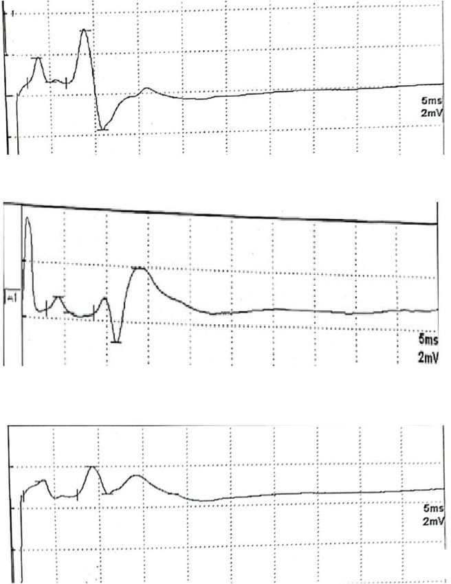
Hmax/Mmax ratio was significantly decreased at 0.84, 0.67, and 0.40 before treatment, 2 weeks after treat-
follow-up in patients of the first group (Fig. 2) compared ment, and at follow-up, respectively.
to the second but not after treatment. Also, this ratio No adverse events were observed in either group.
was significantly decreased after the treatment and at
follow-up in the first group (Table 4). Discussion
Figure 2 shows H reflex in group I before treatment, 2 The present study suggests that rESWT has a beneficial
weeks after treatment, and at follow-up with H/M ratio effect on spasticity and functional status of the upper
Table 2 Modified Ashworth Scale (MAS) for the wrist and hand in the studied groups at baseline, after rESWT, and at follow-up
showing significant improvement in GI after receiving rESWT therapy relative to baseline and also a significant improvement in
comparison to GII
Group I Group II Unpaired t test (p)
Mean ± SD Mean ± SD
MAS (wrist)
Baseline 3.05 ± 0.18 2.95 ± 0.19 0.094
After rESWT (2 weeks after the last session) 1.45 ± 0.16 2.90 ± 0.18 < 0.001*
Follow-up (3 months after the last session) 1.55 ± 0.13 3.00 ± .0.15 < 0.001*
Paired t test
P1 < 0.001* 0.396
P2 < 0.001* 0.064
P3 0.372 0.361
MAS (hand)
Baseline 3.35 ± 0.22 3.25 ± 0.21 0.145
After rESWT (2 weeks after last session) 2.15 ± 0.19 3.15 ± 0.18 < 0.001*
Follow-up (3 months after the last session) 2.20 ± 0.17 3.20 ± 0.16 < 0.001*
Paired t test
P1 < 0.001* 0.107
P2 < 0.001* 0.359
P3 0.388 0.391
Values are mean ± SD
MAS Modified Ashworth Scale, P1 2 weeks after rESWT versus baseline, P2 3-month follow-up versus baseline, P3 3-month follow-up versus 2 weeks after rESWT
*Significant valuesTabra et al. Egyptian Rheumatology and Rehabilitation (2021) 48:21 Page 6 of 10
Table 3 Fugl-Myer Assessment (FMA) of hand function and wrist control and Motricity Index (pinch grip) in the studied groups at
baseline, after rESWT, and at follow-up showing significant improvement of FMA in GI after receiving rESWT therapy relative to
baseline and in comparison to GII and significant improvement of Motricity Index at follow-up period in GI
Group I Group II Unpaired t test (p)
Mean ± SD Mean ± SD
FMA (wrist control)
Baseline 1.20 ± 0.17 1.25 ± 0.23 0.436
After rESWT (2 weeks after the last session) 2.10 ± 0.35 1.30 ± 0.21 < 0.001*
Follow-up (3 months after the last session) 2.00 ± 0.16 1.35 ± 0.19 < 0.001*
Paired t test
P1 < 0.001* 0.476
P2 < 0.001* 0.140
P3 0.266 0.439
FMA (hand function)
Baseline 3.65 ± 0.24 3.75 ± 0.32 0.272
After rESWT (2 weeks after the last session) 5.20 ± 0.36 3.70 ± 0.34 < 0.001*
Follow-up (3 months after the last session) 4.70 ± 0.18 3.80 ± 0.28 < 0.001*
Paired t test
P1 < 0.001* 0.632
P2 < 0.001* 0.600
P3 < 0.001* 0.313
Motricity Index (pinch grip)
Baseline 20.45 ± 3.37 21.05 ± 2.48 0.521
After rESWT (2 weeks after the last session) 22.70 ± 4.10 21.40 ± 3.23 0.272
Follow-up (3 months after the last session) 23.15 ± 4.62 21.85 ± .3.70 0.336
Paired t test
P1 0.064 0.707
P2 0.043* 0.432
P3 0.747 0.682
Values are mean ± SD
FMA Fugl-Meyer Assessment, P1 2 weeks after rESWT versus baseline, P2 3-month follow-up versus baseline, P3 3-month follow-up versus 2 weeks after rESWT
*Significant values
limb (using Fugl-Meyer Assessment for hand function neuromuscular junctions, and synaptic plasticity in the
and wrist control and Motricity Index for pinch grip). central nervous system [4]. Similar to the inhibitory
Neuronal plasticity can lead to an extreme degree of action of botox (BTX) on neuromuscular transmission,
spontaneous recovery of early stroke patients in few Kenmoku et al. observed rapid degeneration of acetyl-
months. However, neural plasticity concerning compen- choline receptors after ESWT application but without
satory movement, activated ipsilateral motor projections, muscle weakness. Also, the amplitude of the compound
and competitive interaction after stroke contributes to muscle action potential was significantly decreased
maladaptive plasticity, which negatively affects motor re- immediately after ESWT and lasted for 8 weeks in an
covery. Rehabilitation programs should be selected ac- animal study [32].
cording to the motor impairment of stroke patients to Modification of spinal excitability is another mechan-
minimize maladaptive plasticity [31]. We conducted this ism; as abnormal stretch reflexes may not completely ex-
study on patients with a disease duration of more than 1 plain the development of spasticity, the effects of ESWT
year aiming to investigate the individual effect of treat- on spinal excitability may support the idea that ESWT
ment regimens on spastic muscles. acts on nonreflex hypertonia [33]. Although having a
Despite no established standard guidelines, several temporary effect, mechanical vibration could be attrib-
mechanisms could explain the therapeutic role of ESWT uted as a contributing factor decreasing spinal excitabil-
in spasticity; nitric oxides (NO) generated by ESWT are ity [16]. Structural and mechanical changes in the
involved in neurotransmission, memory formation, spastic muscle lead to fibrosis of inactive connectiveTabra et al. Egyptian Rheumatology and Rehabilitation (2021) 48:21 Page 7 of 10
Fig. 2 H reflex in group I. a Before treatment. b Two weeks after treatment. c Follow-up (3 months after treatment)
tissue. ESWT in the long run can diminish spasticity by significant reduction in spasticity of the hand and wrist
reduction of fibrosis of chronic hypertonic muscles [2, 16]. for at least 16 and 8 weeks in the group receiving 3 ses-
Dymarek et al. [34] revealed a significant reduction in sions of rESWT and in the group receiving a single ses-
MAS in 20 patients with upper limb spasticity treated sion of rESWT, respectively. Three sessions of rESWT
with a single session of rESWT. Li et al. [35] reported a had a longer-lasting effect than one session.
Table 4 Hmax/Mmax ratio in the studied groups at baseline, after rESWT, and at follow-up showing a significant decrease of Hmax/
Mmax ratio in GI after rESWT therapy relative to baseline and significant improvement in comparison to GII at follow-up
Group I Group II Unpaired t test (p)
Mean ± SD Mean ± SD
Hmax/Mmax ratio
Baseline 0.79 ± 0.14 0.77 ± 0.22 0.735
After rESWT (2 weeks after the last session) 0.71 ± 0.10 0.75 ± 0.23 0.283
Follow-up (3 months after the last session) 0.58 ± 0.12 0.76 ± .0.19 0.0014*
Paired t test
P1 0.048* 0.727
P2 < 0.001* 0.879
P3 0.006* 0.847
Values are mean ± SD
P1 2 weeks after rESWT versus baseline, P2 3-month follow-up versus baseline, P3 3-month follow-up versus 2 weeks after rESWT
*Significant valuesTabra et al. Egyptian Rheumatology and Rehabilitation (2021) 48:21 Page 8 of 10
Coinciding with the previous data [34, 35], MAS for be a reasonable mechanism. One recent study revealed a
clinical assessment of the wrist and hand muscle spasti- reduction of the Hmax/Mmax ratio after ESWT indicat-
city in stroke patients pointed out significant improve- ing a change in alpha motor neuron excitability [39].
ment after treatment and at follow-up compared to Other studies revealed that no significant changes of F
baseline in the first group with a significant difference wave or H wave latency or amplitude were demonstrated
between the two groups (Table 2). in human studies after ESWT application [40, 41].
FMA for wrist control and hand function was our tool To our knowledge, this study is the first one using
to assess the sensorimotor function of the wrist and combined different assessment measures (clinical and
hand in this study; improvement of functional ability electrophysiological) to evaluate the effect of radial
was noticed in the first group relative to the second one extracorporeal shock wave therapy (rESWT) on wrist
after treatment and at follow-up with significant differ- and hand functions of spastic chronic stroke patients.
ences. Also, there was a significant improvement in pa- Some limitations of this study should be considered as
tients of the first group after treatment and at follow-up relatively small sample size of patients and short dur-
relative to baseline. ation of follow-up to reveal the sustained effects of this
Spastic hand muscles may serve as a reasonable factor modality of treatment. More studies are needed with
for an average hand grip more than weak muscles; this more flexible inclusion criteria for the disease duration
can explain the results of the Motricity Index for pinch to include spastic patients during the first year to pre-
grip after treatment, where there was no significant vent the development of maladaptive plasticity. Also,
change after treatment and at follow-up between both more research should be done to see if the rESWT could
groups, with significant improvement at follow-up substitute other modalities of spasticity treatment such
period when compared to baseline in the first group. as the muscle relaxant and botox injection.
Few studies measured the motor functional outcome
of the upper limb in spastic stroke patients. Troncati Conclusions
et al. [36] revealed significant improvement of FMA of ESWT is a valuable adjuvant treatment for spasticity of
twelve patients with stroke after treatment with two ses- the hand and wrist in stroke patients which is reflected
sions of ESWT, and the effects were maintained 3 and 6 as improvement of functional activity.
months after treatment.
In a prospective randomized, single-blind controlled Recommendation
trial by Li et al. [35], a significant improvement in FMA rESWT is recommended as adjuvant physical treatment
scores for hand function and wrist control after three if the muscle relaxants failed to achieve the desired effect
sessions of rESWT was maintained for 16 and 12 weeks, or had side effects or contraindications.
respectively, compared with those of sham or one ses-
sion of rESWT group and there was no significant im- Abbreviations
rESWT: Radial extracorporeal shock wave therapy; BTX: Botulinum toxin;
provement in FMA scores after one session of rESWT fESWT: Focused extracorporeal shock wave therapy; RCTs: Randomized
compared with sham control, indicating that repeated controlled trials; NO: Nitric oxides; MAS: Modified Ashworth Scale;
sessions of ESWT are necessary to ameliorate functional ROM: Range of motion; FMA: Fugl-Meyer Assessment
motricity. Thus, clinical experience indicates repeated Acknowledgements
sessions of ESWT could be superior to a single applica- Not applicable.
tion. Li et al. [35] confirmed that repeated sessions of
Authors’ contributions
rESWT result in a more noticeable and longer-lasting TSA and ADS designed the study protocol. ZMI, TSA, and ADS monitored the
effect. progress and collected the data. ADS did the statistical analysis. All authors
In 1974, Deschuytere and colleagues [37] showed resting contributed to drafting the manuscript and have read and approved the
final version of the manuscript.
H reflexes in the flexor carpi radialis (FCR) and palmaris
longus in most of their volunteers. H reflex amplitudes are Funding
increased with respect to maximal M responses recorded The authors received no financial support for the research, authorship, and/
or publication of this article.
in the spastic muscle as a result of enhanced excitability in
the monosynaptic stretch reflex arc [38]. Availability of data and materials
Our results revealed a significant reduction in Hmax/ The data will be available upon reasonable request.
Mmax amplitude ratio at follow-up in the first group
Declarations
with a significant difference between the two groups at
follow-up (Table 4); these results pointed out the de- Ethics approval and consent to participate
layed effect of ESWT on reflex arc excitability. Ethical approval for this study was obtained from the Ethics Committee of
University faculty of Medicine (approval number: 33693/2/20). Informed
The effect of ESWT on spinal excitability and Golgi written consents from all patients were obtained in accordance with the
tendon organs to suppress motor nerve excitability can local ethical committee.Tabra et al. Egyptian Rheumatology and Rehabilitation (2021) 48:21 Page 9 of 10
Consent for publication 16. Manganotti P, Amelio E (2005) Long-term effect of shock wave therapy on
Not applicable. upper limb hypertonia in patients affected by stroke. Stroke 36(9):1967–
1971. https://doi.org/10.1161/01.STR.0000177880.06663.5c
17. Leone JA, Kukulka CG (1988) Effects of tendon pressure on alpha motor
Competing interests neuron excitability in patients with stroke. Phys Ther 68(4):475–480. https://
The authors declare that they have no competing interests. doi.org/10.1093/ptj/68.4.475
18. Bae H, Lee JM, Lee KH (2010) The effects of extracorporeal shock wave
Received: 30 December 2020 Accepted: 26 February 2021 therapy on spasticity in chronic stroke patients. J Korean Acad Rehabil Med
34:663–669
19. Wu Y-T, Chang C-N, Chen Y-M, Hu G-C (2018) Comparison of the effect of
focused and radial extracorporeal shock waves on spastic equinus in
References
patients with stroke: a randomized controlled trial. Eur J Phys Rehabil Med
1. Watkins CL, Leathley MJ, Gregson JM, Moore AP, Smith TL, Sharma AK
54(4):518–525. https://doi.org/10.23736/S1973-9087.17.04801-8
(2002) Prevalence of spasticity post stroke. Clin Rehabil 16(5):515–522.
20. Taheri P, Vahdatpour B, Mellat M, Ashtari F, Akbari M (2017) Effect of
https://doi.org/10.1191/0269215502cr512oa
extracorporeal shock wave therapy on lower limb spasticity in stroke
2. Xiang J, Wang W, Jiang W, Qian Q (2018) Effects of extracorporeal
patients. Arch Iran Med 20(6):338–343
shockwave therapy on spasticity in post-stroke patients: a systematic review
21. Santamato A, Micello MF, Panza F, Fortunato F, Logroscino G, Picelli A,
and meta-analysis of randomized controlled trials. J RehabilMed 50(10):852–
Manganotti P, Smania N, Fiore P, Ranieri M (2014) Extracorporeal shock
859
wave therapy for the treatment of post stroke plantar-flexor muscles
3. Dymarek R, Ptaszkowski K, Ptaszkowska L, Kowal M, Sopel M, Taradaj J,
spasticity: a prospective open-label study. Top Stroke Rehabil 21(1):S17–S24.
Rosińczuk J (2020) Shock waves as a treatment modality for spasticity
https://doi.org/10.1310/tsr21S1-S17
reduction and recovery improvement in post-stroke adults–current
22. Guoxing, X, , Zhang, Y, Yinmeng Z, Haoyang D, Ping L, Na L, Fuqian L,
evidence and qualitative systematic review. Clin Interv Aging 15:9–28.
Zhenlan L (2018) Therapeutic effect of slow stretching training combined
https://doi.org/10.2147/CIA.S221032
with extracorporeal shock wave on biceps brachii spasticity in stroke
4. Mariotto S, de Prati AC, Cavalieri E, Amelio E, Marlinghaus E, Suzuki H (2009)
patients. J Jilin Univ (Med Ed) 44(2): 374–378.
Extracorporeal shock wave therapy in inflammatory diseases: molecular
23. Bohannon R, Smith M (1987) Interrater reliability of a modified Ashworth
mechanism that triggers anti-inflammatory action. Curr Med Chem 16(19):
scale of muscle spasticity. Phys Ther 67(2):206
2366–2372. https://doi.org/10.2174/092986709788682119
5. Lee J-Y, Kim S-N, Lee I-S, Jung H, Lee K-S, Koh S-E (2014) Effects of 24. Fugl-Meyer AR, Jaasko L, Leyman I, Olsson S, Steglind S (1975) The post-
extracorporeal shock wave therapy on spasticity in patients after brain stroke hemiplegic patient. A method for evaluation of physical
injury: a meta-analysis. J Phys Ther Sci 26(10):1641–1647 performance. Scand J Rehabil Med 7(1):13–31
6. Guo P, Gao F, ZhaoT SW, Wang B, Li Z (2017) Positive effects of 25. Duncan PW, Propst M, Nelson SG (1983) Reliability of the Fugl-Meyer
extracorporeal shock wave therapy on spasticity in poststroke patients: a assessment of sensorimotor recovery following cerebrovascular accident.
meta-analysis. J Stroke Cerebrovasc Dis Off J Natl Stroke Assoc 26(11):2470– Phys Ther 63(10):1606–1610. https://doi.org/10.1093/ptj/63.10.1606
2476. https://doi.org/10.1016/j.jstrokecerebrovasdis.2017.08.019 26. Collin C, Wade D (1990) Assessing motor impairment after stroke: a pilot
7. Dymarek R, Ptaszkowski K, Słupska L, Halski T, Taradaj J, Rosińczuk J (2016) reliability study. J Neurol Neurosurg Psychiatry 53(7):576–579. https://doi.
Effects of extracorporeal shock wave on upper and lower limb spasticity in org/10.1136/jnnp.53.7.576
post-stroke patients: A narrative review. Top Stroke Rehabil 23(4):293–303. 27. Bohannon RW (1999) Motricity index scores are valid indicators of paretic
https://doi.org/10.1080/10749357.2016.1141492. upper extremity strength following stroke. J Phys Ther Sci 11(2):59–61
8. Park D-S, Kwon DR, Park G-Y, Lee MY (2015) Therapeutic effect of 28. Zheng C, Zhu Y, Lv F, Ma X, Xia X, Wang L, Jin X, Weber R, Jiang J, Anuvat K
extracorporeal shock wave therapy according to treatment session on (2014) Abnormal flexor carpi radialis H-reflex. J Clin Neurophysiol 31(6):529–
gastrocnemius muscle spasticity in children with spastic cerebral palsy: a 534
pilot study. Ann Rehabil Med 39(6):914–921. https://doi.org/10.5535/arm.201 29. Angel RW, Hoffmann WW (1963) The H reflex in normal, spastic and rigid
5.39.6.914 subjects. Arch Neurol 8(6):591–596. https://doi.org/10.1001/archneur.1963.
9. Picelli A, Marchina EL, Gajofatto F, Pontillo A, Vangelista A, Filippini R, 00460060021002
Baricich A, Cisari C, Smania N (2017) Sonographic and clinical effects of 30. Kirkpatrick LA, Feeney BC (2013) A simple guide to IBM SPSS statistics for
botulinum toxin type A combined with extracorporeal shockwave therapy version 20.0 student ed. Wadsworth, Cengage Learning, Belmont
on spastic muscles of children with cerebral palsy. Dev Neurorehabil 20(3): 31. Takeuchi N, Izumi S (2012) Maladaptive plasticity for motor recovery after
160–164 stroke: mechanisms and approaches. Neural Plast 2012:359728
10. Amelio E, Manganotti P (2010) Effect of shock wave stimulation on 32. Kenmoku T, Ochiai N, Ohtori S, Saisu T, Sasho T, Nakagawa K, Iwakura N,
hypertonic plantar flexor muscles in patients with cerebral palsy: a placebo- Miyagi M, Ishikawa T, Tatsuoka H, Inoue G, Nakamura J, Kishida S, Saito A,
controlled study. J Rehabil Med 42(4):339–343. https://doi.org/10.2340/1 Takahashi K (2012) Degeneration and recovery of the neuromuscular
6501977-0522 junction after application of extracorporeal shock wave therapy. J Orthop
11. Vidal X, Morral A, Costa L, Tur M (2011) Radial extracorporeal shock wave Res 30(10):1660–1665. https://doi.org/10.1002/jor.22111
therapy (rESWT) in the treatment of spasticity in cerebral palsy: a 33. Galiana L, Fung J, Kearney R (2005) Identification of intrinsic and reflex ankle
randomized, placebo-controlled clinical trial. NeuroRehabilitation 29(4):413– stiffness components in stroke patients. Exp Brain Res 165(4):422–434.
419. https://doi.org/10.3233/NRE-2011-0720 https://doi.org/10.1007/s00221-005-2320-z
12. Gonkova MI, Ilieva EM, Ferriero G, Chavdarov I (2013) Effect of radial shock 34. Dymarek R, Taradaj J, Rosińczuk J (2016) Extracorporeal shock wave stimulation
wave therapy on muscle spasticity in children with cerebral palsy. Int J as alternative treatment modality for wrist and fingers spasticity in poststroke
Rehabil Res Int Z Für Rehabil Rev Int Rech Réadapt 36(3):284–290 patients: a prospective, open-label, preliminary clinical trial. Evid Based
13. Wang T, Du L, Shan L, Dong H, Feng J, Kiessling MC, Angstman NB, Schmitz Complement Alternat Med 2016:1–10. https://doi.org/10.1155/2016/4648101
C, Jia F (2016) A prospective case-control study of radial extracorporeal 35. Li TY, Chang CY, Chou YC, Chen LC, Chu HY, Chiang SL, Chang ST, Wu YT
shock wave therapy for spastic plantar flexor muscles in very young (2016) Effect of radial shock wave therapy on spasticity of the upper limb in
children with cerebral palsy. Medicine (Baltimore) 95(19):e3649. https://doi. patients with chronic stroke. A prospective, randomized, single blind,
org/10.1097/MD.0000000000003649 controlled trial. Medicine 95(18):e3544
14. Marinelli LM (2014) Effect of radial shock wave therapy on pain and muscle 36. Troncati F, Paci M, Myftari T, Lombardi B (2013) Extracorporeal shock wave
hypertonia: a double-blind study in patients with multiple sclerosis. Mult therapy reduces upper limb spasticity and improves motricity in patients
Scler Houndmills Basingstoke Engl 21:5 with chronic hemiplegia: a case series. NeuroRehabilitation 33(3):399–405.
15. Mariotto S, Cavalieri E, Amelio E, Ciampa AR, Carcereri de Prati A, https://doi.org/10.3233/NRE-130970
Marlinghaus E, Russo S, Suzuki H (2005) Extracorporeal shock waves: from 37. Deschuytere J, DeKeyser C, Deschuttere M, Rosselle N (1983) H reflexes in
lithotripsy to anti-inflammatory action by NO production. Nitric Oxide 12(2): muscles of the lower and upper limbs in man: identification and clinical
89–96. https://doi.org/10.1016/j.niox.2004.12.005 significance. Adv Neurol 39(9):51–60Tabra et al. Egyptian Rheumatology and Rehabilitation (2021) 48:21 Page 10 of 10
38. Powers RK, Campbell DL, Rymer WZ (1989) Stretch reflex dynamics in
spastic elbow flexor muscles. Ann Neurol 25(1):32–42. https://doi.org/10.1
002/ana.410250106
39. Daliri SS, Forogh B, Emami Razavi SZ, Ahadi T, Madjlesi F, Ansari NN (2015) A
single blind, clinical trial to investigate the effects of a single session
extracorporeal shock wave therapy on wrist flexor spasticity after stroke.
NeuroRehabilitation 36(1):67–72. https://doi.org/10.3233/NRE-141193
40. Sohn MK, Cho KH, Kim YJ, Hwang SL (2011) Spasticity and
electrophysiologic changes after extracorporeal shock wave therapy on
gastrocnemius. Ann Rehabil Med 35(5):599–604. https://doi.org/10.5535/a
rm.2011.35.5.599
41. Marinelli L, Mori L, Solaro C, Uccelli A, Pelosin E, Currà A, Molfetta L,
Abbruzzese G, Trompetto C (2014) Effect of radial shock wave therapy on
pain and muscle hypertonia: a double-blind study in patients with multiple
sclerosis. Mult Scler 21:622–629
Publisher’s Note
Springer Nature remains neutral with regard to jurisdictional claims in
published maps and institutional affiliations.You can also read