Inheritance of Monogenic Hereditary Skin Disease and Related Canine Breeds - MDPI
←
→
Page content transcription
If your browser does not render page correctly, please read the page content below
veterinary
             sciences

Review
Inheritance of Monogenic Hereditary Skin Disease and Related
Canine Breeds
Pablo Jesús Marín-García                   and Lola Llobat *

                                           Department of Animal Production and Health, Veterinary Public Health and Food Sciences and
                                           Technology (PASAPTA), Facultad de Veterinaria, Universidad Cardenal Herrera-CEU, CEU Universities,
                                           46113 Valencia, Spain
                                           * Correspondence: maria.llobatbordes@uchceu.es

                                           Simple Summary: The high prevalence of genetic diseases in dog breeds and the structure of their
                                           populations has led to detailed studies of the canine genome, which are important for understanding
                                           the origin of these pathologies. The location of certain genes involved in a few autosomal recessive
                                           monogenic diseases, including genodermatosis. The most prevalent canine genodermatosis are
                                           non-epidermolytic ichthyosis, epidermolytic ichthyosis, and junctional epidermolysis bullosa. Other
                                           genodermatoses are nasal paraqueratosis, cutaneous mucinosis, dermoid sinus, lethal acrodermatitis,
                                           palmoplantar hyperkeratosis, or exfoliative cutaneous lupus erythematosus. Most of this genoder-
                                           matosis is associated with a specific and known number of mutations, which have a higher prevalence
                                           in certain canine breeds. The main objective of this review is to analyze each of these genodermatoses,
                                           the genes and mutations associated with them, and the breeds with the greatest predisposition to
                                           suffer from them.

                                           Abstract: The plasticity of the genome is an evolutionary factor in all animal species, including
                                           canines, but it can also be the origin of diseases caused by hereditary genetic mutation. Genetic
                                           changes, or mutations, that give rise to a pathology in most cases result from recessive alleles that
                                           are normally found with minority allelic frequency. The use of genetic improvement increases the
Citation: Marín-García, P.J.; Llobat, L.
                                           consanguinity within canine breeds and, on many occasions, also increases the frequency of these
Inheritance of Monogenic Hereditary
                                           recessive alleles, increasing the prevalence of these pathologies. This prevalence has been known for
Skin Disease and Related Canine
                                           a long time, but mutations differ according to the canine breed. These genetic diseases, including
Breeds. Vet. Sci. 2022, 9, 433.
https://doi.org/10.3390/
                                           skin diseases, or genodermatosis, which is narrowly defined as monogenic hereditary dermatosis. In
vetsci9080433                              this review, we focus on genodermatosis sensu estricto, i.e., monogenic, and hereditary dermatosis,
                                           in addition to the clinical features, diagnosis, pathogeny, and treatment. Specifically, this review
Academic Editor: Patrick Butaye
                                           analyzes epidermolytic and non-epidermolytic ichthyosis, junctional epidermolysis bullosa, nasal
Received: 9 June 2022                      parakeratosis, mucinosis, dermoid sinus, among others, in canine breeds, such as Golden Retriever,
Accepted: 12 August 2022                   German Pointer, Australian Shepherd, American Bulldog, Great Dane, Jack Russell Terrier, Labrador
Published: 15 August 2022                  Retriever, Shar-Pei, and Rhodesian Ridgeback.
Publisher’s Note: MDPI stays neutral
with regard to jurisdictional claims in
                                           Keywords: skin disorders; genodermatosis; monogenic hereditary dermatosis; ichthyosis; epidermol-
published maps and institutional affil-    ysis; parakeratosis; mucinosis
iations.

                                           1. Introduction
Copyright: © 2022 by the authors.
                                                During recent centuries, genetic pathologies in canine breeds have increased consid-
Licensee MDPI, Basel, Switzerland.
                                           erably, possibly because of a reduction in the effective number of individuals in canine
This article is an open access article
                                           populations due to genetic selection. Such a focus on morphological characteristics has
distributed under the terms and
                                           limited the number of alleles, thereby increased consanguinity, and reduced genetic di-
conditions of the Creative Commons
                                           versity. This has mainly occurred due to inadequate crossing practices, together with
Attribution (CC BY) license (https://
creativecommons.org/licenses/by/
                                           insufficient selective pressure on canine well-being and health characteristics [1]. In fact,
4.0/).
                                           the effective number of some canine breeds has been estimated at 30–70%, and inbred dogs

Vet. Sci. 2022, 9, 433. https://doi.org/10.3390/vetsci9080433                                                https://www.mdpi.com/journal/vetsci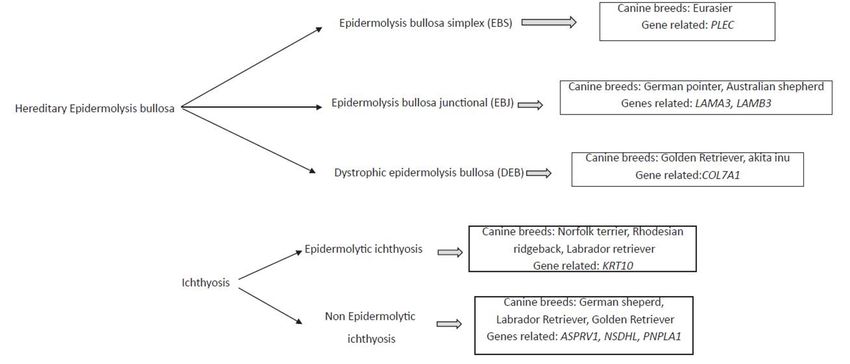
Vet. Sci. 2022, 9, 433 2 of 15
after two generations have ranged from 1 to 8%, depending on mating practices [2]. Genetic
selection has focused on aesthetics rather than function or health, so a small number of
breeders have been crossed with closed relatives, producing significantly reduced genetic
diversity and increasing the prevalence of specific deleterious alleles [3–5]. Dermatological
pathologies are no exception, and some have increased considerably in certain breeds. For
these, it is important to distinguish genodermatosis (dermatoses of a monogenic origin)
from polygenic dermatoses with racial predisposition, the latter being more frequent than
others [6]. In this review, we focused strictly on genodermatosis, i.e., monogenic, and
hereditary dermatosis.
The high prevalence of genetic diseases in dog breeds and the structure of their
populations has led to detailed studies of the canine genome, which are important for
understanding the origin of these pathologies. For this reason, it was possible to determine
that the canine genome has about 2.42 gigabases (Gb) composed of 20,000 genes distributed
on 78 chromosomes, 38 pairs of acrocentric autosomes, and a pair of sex chromosomes: the
X chromosome, the largest karyotype with 128 megabases (Mb), and the Y chromosome
with the smallest karyotype of 27 Mb [7]. Whole canine genome mapping, sequencing,
and linkage analyses has made possible the highlighting of several disease-related sources.
First, the high incidence of SINE (short interspersed nuclear element), which are repeated
sequences associated with many diseases [8]; the frequent occurrence of SNPs (single
nucleotide polymorphisms), which are variations of a single nucleotide in the genome [9],
and the location of certain genes involved in several autosomal recessive monogenic
diseases, including genodermatosis, have made it possible to reveal multiple causes of
disease occurrence.
The canine genodermatosis are mainly non-epidermolytic ichthyosis in the Golden
Retriever and the Jack Russell Terrier, epidermolytic ichthyosis in the Norfolk Terrier, junc-
tional epidermolysis bullosa in the German Shorthaired Pointer, and Shar-Pei mucinosis,
although other types of canine genodermatosis exist (Table 1).
Table 1. Genodermatosis with known causative genetic variants in dog breeds.
Phenotype Breed Inheritance 1 Reference
Junctional German Pointer, Australian
AR [10,11]
epidermolysis bullosa Shepherd
Epidermolysis
Golden Retriever, Akita Inu AR [12,13]
bullosa dystrophic
American Bulldog, Great Dane,
Ichthyosis Jack Russell Terrier, Golden AR [14–18]
Retriever
Nasal parakeratosis Labrador Retriever AR [19]
Mucinosis Shar-Pei ASD [20]
Dermoid sinus Rhodesian Ridgeback AR [21]
1 AR: autosomal recessive inheritance; ASD: autosomal semi-dominant inheritance.
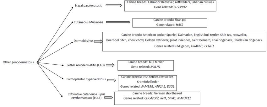
Different genodermatosis has been correlated to canine breed, and the several genes
seems to be the responsible for these diseases (Figure 1).Vet.
Vet. Sci. 2022, 9,
Sci. 2022, 9, 433
x FOR PEER REVIEW 33 of
of 15
15
Figure 1. Diagram
Figure 1. Diagramof
ofmainly
mainlymonogenic
monogenichereditary
hereditaryskin disease,
skin thethe
disease, associated canine
associated breed,
canine andand
breed, the
the related
related genes.
genes.
2.
2. Hereditary
Hereditary Epidermolysis
Epidermolysis Bullosa
Bullosa
Hereditary
Hereditary epidermolysis bullosa
epidermolysis bullosa constitutes
constitutes aa heterogeneous
heterogeneous group
group of
of hereditary
hereditary
blistering diseases of the skin and mucous membranes [22,23]. These pathologies
blistering diseases of the skin and mucous membranes [22,23]. These pathologies are char-are
characterized by the spontaneous development of vesicles, erosions, and ulcers because
acterized by the spontaneous development of vesicles, erosions, and ulcers because of
of minimal trauma to the excessively fragile dermal–epidermal junction (DEJ) [24]. This
minimal trauma to the excessively fragile dermal–epidermal junction (DEJ) [24]. This
group of dermal diseases is classified according to the level of cleavage as epidermolysis
group of dermal diseases is classified according to the level of cleavage as epidermolysis
bullosa simplex (EBS), epidermolysis bullosa junctional (EBJ), and dystrophic epidermolysis
bullosa simplex (EBS), epidermolysis bullosa junctional (EBJ), and dystrophic epidermol-
bullosa (DEB). Different dog breeds have been associated with each of these diseases and
ysis bullosa (DEB). Different dog breeds have been associated with each of these diseases
have presented different types of ulcers. Some genes with recessive autosomal inheritance
and have presented different types of ulcers. Some genes with recessive autosomal inher-
have been associated with them (Table 2).
itance have been associated with them (Table 2).
Table 2. Classification of canine epidermolysis, related ulcerations, and the associated genes and
Table 2. Classification of canine epidermolysis, related ulcerations, and the associated genes and
canine breeds.
canine breeds.
Classification
Classification Epider- Type
Type of of
Ulcer- Gene
Dog Breeds Reference
Epidermolysis Dog Breeds Ulceration Gene Associated Reference
Associated
molysis ation
Epidermolysis bullosa
Epidermolysis bullosa Eurasier dog Multifocal
Multifocal ul-
ulcers PLEC [25]
simplex (EBS) Eurasier dog PLEC [25]
simplex (EBS) cers
Skin and
German Pointer,
Epidermolysis bullosa German Pointer, Skin and mu-
mucous LAMA3,
Epidermolysis bullosa Australian [10,11,26,27]
junctional (EBJ) Australian Shep- membrane
cous mem- LAMB3
LAMA3, LAMB3 [10,11,26,27]
junctional (EBJ) Shepherd
herd ulcers
brane ulcers
Dystrophic Golden
Dystrophic epidermol- Golden Retriever, Oralcavity
Oral cavity ul-
epidermolysis bullosa Retriever, Akita COL7A1
COL7A1 [12,13]
[12,13]
ysis bullosa ulcers
(DEB) (DEB) Akita
Inu Inu cers
Epidermolysis bullosa simplex (EBS) is a skin disease, in which the keratinocytes or
Epidermolysis bullosa simplex (EBS) is a skin disease, in which the keratinocytes
basal and suprabasal are related [25]. This disease is not unique to dogs, so different sub-
or basal and suprabasal are related [25]. This disease is not unique to dogs, so different
types of EBS have also been observed in humans, and different genes play a role [28–36].
subtypes of EBS have also been observed in humans, and different genes play a role [28–36].
However, in dogs, only two genes have been associated with it. The mutation of the PLEC
However, in dogs, only two genes have been associated with it. The mutation of the PLEC
gene has
gene has been
been associated
associated so
so far
far with
with EBS
EBS in in Eurasier
Eurasier dogs
dogs [25].
[25]. The
The product
product ofof the
the PLEC
PLEC
gene is plectin, a 500 kDa protein found in skin and other tissues, such as
gene is plectin, a 500 kDa protein found in skin and other tissues, such as bone, muscle,bone, muscle,
and the
and thenervous
nervoussystem
system[37].
[37].There
There areare likely
likely different
different isoforms
isoforms of plectin
of plectin thatthat are cell-
are cell-type
type dependent and/or developmentally regulated [38]. Mauldin et
dependent and/or developmentally regulated [38]. Mauldin et al. (2017) demonstratedal. (2017) demon-
that
strated that in dogs with a homozygous G-to-A variant in the PLEC gene,
in dogs with a homozygous G-to-A variant in the PLEC gene, a tryptophan is converted toa tryptophan is
converted to a premature stop codon in exon 27, resulting in this disease with autosomalVet. Sci. 2022, 9, 433 4 of 15
a premature stop codon in exon 27, resulting in this disease with autosomal recessive inher-
itance [39]. On the other hand, Olivry et al. (2012) showed the association between a single
mutation in the first intron of PKP1 gene. This single mutation results in a premature stop
codon, and the absence of the protein plakophilin-1, a protein that stabilizes desmosomes
in the skin [40,41]. The inheritance of this mutation was also autosomal recessive, and
this detection occurred in dog breed Chesapeake Bay Retriever, resulting in an ectodermal
dysplasia-skin fragility syndrome [40].
In epidermolysis bullosa junctional (EBJ), cleft formation occurs through the lamina
lucida of the basement membrane zone. Affected individuals exhibit blisters, deep erosions,
and ulcers [22]. In humans, mutations in several genes have been associated with this
pathology, including genes encoding subunits of integrins (ITGA6, ITGB4, and ITGA3),
collagen (COL17A1), and laminin 332 (LAMA3, LAMB3, and LAMC2) [42,43]. Recently,
mutations in the LAMA3 and LAMB3 genes have been associated with EBJ in Australian
shepherd dogs [11,26]. In the study by Kiener et al. (2020), a LAMB3:c.1174T > C mutation
was reported as the cause of EBJ, suggesting an autosomal recessive inheritance of this
mutation [11]. The LAMB3 gene encodes the β3-polypeptide chain of laminin-1 [44] and
has been associated with the progression of several human tumors [45]. The recent study by
Herrmann et al. (2021) reported a LAMA3 mutation associated with EBJ and severe upper
respiratory disease in Australian Shepherd dogs [26]. This mutation (Asp2867Val) results
in a missense variant in the laminin-α3 chain with autosomal recessive inheritance. Other
mutations in the same gene have been found in the German Pointer dog breed associated
with EBJ. Specifically, an insertion of repetitive satellite DNA in intron 35 of this gene has
been associated with EBJ [10,27]. This insertion results in an α3-pre-messenger RNA that is
not well matured and a decrease in laminin 5 expression, thereby impairing adhesion and
the clonogenic potential of the EBJ keratinocytes.
Finally, in dystrophic epidermolysis bullosa (DEB), blistering occurs in the sublamina
densa, and the skin and mucosa are extremely sensitive. The blisters heal with scarring,
and end with progressive disability and the deformation of the fingers [46]. This disease,
which affects dogs, sheep, cattle, cats, and humans, is caused by mutations in the COL7A1
gene, which encodes collagen type VII [47]. A total of 500 mutations of this gene have been
associated with DEB, and the severity of the phenotype depends on the type of mutations
and their location [48]. Most of these mutations were observed in the golden retriever,
although Nagata et al. (1995) reported a case of DEB in Akita Inu dog breed [12]. The
authors did not perform a genetic study on the animal, and the results they observed
when analyzing the bladders by electronic microscopy and immunohistochemistry were
comparable to those in humans and other dogs suffering with this disease [12]. Several
studies reported new therapies to control and eradicate this disease. In one study, canine
keratinocytes were used to generate autologous epidermal layers in dogs with homozygous
missense mutation in the COL7A1 gene, which expressed an aberrant protein, with good
results [49]. Other authors attempted gene therapy with retroviral vectors [50]. Recently,
Gretzmeier et al. (2021) published good results when recombinant protein collagen VII (C7)
was administered to mice and dogs [51].
3. Ichthyosis
The term ichthyosis describes rare congenital or hereditary pathologies caused by
primary defects in the formation of the stratum corneum [52]. This ichthyosis could be
epidermolytic or non-epidermolytic, depending on whether they are vacuoles and lysis of
keratinocytes within the spinous and granular cell layers [53]. Epidermolytic ichthyosis
has been described in the Norfolk Terrier concerning a mutation to the epidermal keratin
gene (KRT10) with autosomal recessive inheritance [54], although the same pathology has
been described in the Rhodesian Ridgeback and Labrador Retriever.
However, the most common ichthyosis is non-epidermolytic and presents autosomal
recessive inheritance. In humans, there are X-linked dominant forms [55], but in dogs these
forms are yet to be documented [52], with two exceptions. The first is the autosomal domi-Vet. Sci. 2022, 9, 433 5 of 15
nant inheritance of mutation c.1052C > T in the ASPRV1 gene in the German Shepherd [56];
the second is the deletion identified in the NSDHL gene of two female Labrador Retrievers,
which encoded an NAD(P)-dependent steroid dehydrogenase-like protein related to choles-
terol biosynthesis and with monogenic X-chromosomal semidominant inheritance [57].
Different mutations in several genes have been related (Table 3).
Table 3. Ichthyosis with known gene associated in canine breed.
Dog Breeds Gene Associated Inheritance 1 Reference
ABHD5 AR [58]
Golden Retriever
PNPLA1 AR [17]
German Shepherd ASPRV1 AD [56]
American Bulldog NIPAL4 AR [59]
Great Dane SLC27A4 AR [15]
Jack Russell Terrier TGM1 AR [16]
1 AR: autosomal recessive inheritance; AD: autosomal dominant inheritance.
One of the canine breeds most affected by non-epidermolytic ichthyosis is the golden
retriever. In this breed, the clinical signs include a generalized scaling and hyperpigmented
and rough ventral glabrous skin. The histopathology shows a laminated orthokeratosis
and an epidermal hyperkeratosis without significant involvement of the stratum gran-
ulosum [39,60]. The PNPLA1 variant that produces this pathology reached more than
50% frequency in the breeding population now of identification [17]. The frequencies
of genotypes are estimated around 32% in affected dogs (homozygous recessives), 49%
heterozygous, and 20% homozygous dominant, thus clean of defective variants [61]. More
recently, these frequencies have been estimated at 21% in wild-type, 48% in heterozygous,
and 31% in recessive homozygous [18]. The PNPLA protein family has nine patatin-like
phospholipases (PNPLA1-PNPLA9) with lipolytic and acyltransferase activities and are
related to lipid metabolism [62,63]. In humans, five mutations of PNPLA1 caused auto-
somal recessive congenital ichthyosis, which affects the composition and organization
of epidermal lipids. All five mutations provoke a PNPLA1 amino acid change [64]. In
dogs, specifically Golden Retrievers with this mutation, an indel in exon 8 is reported to
cause non-epidermolytic ichthyosis by GWAS analysis [17]. To evaluate the efficacy of
treatment with shampoo and lotion containing gluconolactone and other hydroxylated
acids, a prospective study was carried out, and the results were encouraging: the extension
and size of the scales was reduced between 60 and 75% after 14 and 30 days of treatment,
respectively [65]. Recently, Kiener et al. (2021) reported a ABHD5 gene frameshift deletion
in Golden Retrievers with non-epidermolytic ichthyosis [59]. The mutation is a 14 bp
deletion that provokes a frameshift that alters the last 14 codons. The ABHD5 gene en-
codes an acyltransferase related to lipid metabolism, and defects in this gene are related to
Chanarin–Dorfman syndrome, a neutral lipid storage disease with ichthyosis [66,67]. To
date, these mutations have not been reported in other breeds; however, they have presented
mutations related to non-epidermolytic ichthyosis. For example, a variant of ASPRV1 gene
has been found in German Shepherds [56]. This gene encodes a retroviral-like protease
involved in profilaggrin-to-filaggrin processing and plays a relevant role in skin barrier
formation [68]. The missense variant of c.1052T < C has found in this breed, which affects a
conserved residue and produces the amino acid change Leu351Pro. This change provokes a
deficient ASPRV1 protein, which produces a lower level of stratum corneum hydration [69].
In the American Bulldog, mutations in the NIPAL4 gene are related to non-epidermolytic
ichthyosis [14,70] and in humans to autosomal recessive congenital ichthyosis [71]. In dogs,
the frameshift deletion of the NIPAL4 gene produces a premature stop codon that results in
a truncated and defective NIPAL4 protein [59]. This protein seems to have a relevant role in
lipid metabolism, and it is associated with keratins and desmosomes in the epidermis [72].
Therefore, animals with deficient NIPAL4 protein fail to form normal lamellar bilayers,
leading to the appearance of the typical clinical signs of non-epidermolytic ichthyosis [73].Vet. Sci. 2022, 9, x FOR PEER REVIEW 6 of 15
Vet. Sci. 2022, 9, 433 6 of 15
fail to form normal lamellar bilayers, leading to the appearance of the typical clinical signs
of non-epidermolytic ichthyosis [73].
In Great Danes, a mutant transcript of the SLC27A4 gene has been correlated to the
In Great Danes, a mutant transcript of the SLC27A4 gene has been correlated to the
ichthyosis phenotype by sequence analysis [15]. The mutation provokes an in-frame loss
ichthyosis phenotype by sequence analysis [15]. The mutation provokes an in-frame loss
of 54 bp in exon 8, that probably affects protein expression. The mutant dogs presented a
of 54 bp in exon 8, that probably affects protein expression. The mutant dogs presented
truncated
a truncated protein
protein levels elevated.
levels elevated.TheTheSLC27A4
SLC27A4 protein hashas
protein acyl-CoA
acyl-CoA synthetase
synthetase activity,
activ-
which is related to fatty-acid and phospholipid synthesis and, consequently,
ity, which is related to fatty-acid and phospholipid synthesis and, consequently, to lipid to lipid me-
tabolism
metabolism [74] and fatty-acid transport in the cell membrane [75]. Some mutations inthe
[74] and fatty-acid transport in the cell membrane [75]. Some mutations in the
SLCC27A4
SLCC27A4 gene have been associated with ichthyosis in human patients [76–78], so it isis
gene have been associated with ichthyosis in human patients [76–78], so it
probable
probable thatthat the
the mutation
mutationin inGreat
GreatDanes
Danesisisnot notthe
theonly
onlyoneonein in this
this gene
gene related
related to to the
the
disease in dogs. In Jack Russell Terriers, [16] related the lamellar
disease in dogs. In Jack Russell Terriers, [16] related the lamellar ichthyosis to a LINE-1 ichthyosis to a LINE-1
insertion
insertionin inthe
thetransglutaminase
transglutaminase11(TGM1) (TGM1)gene, gene,which
whichencodes
encodesan anenzyme
enzymewith withaaroleroleinin
cornified envelope formation, and 30–40% of humans with non-epidermolytic
cornified envelope formation, and 30–40% of humans with non-epidermolytic (lamellar) (lamellar)
ichthyosis
ichthyosis present
present mutations
mutations in in this
this gene
gene[79].
[79].TheTheauthors
authorsidentified
identifieda aLINE-1
LINE-1 insertion
insertion in
in this
this gene
gene related
related to to non-epidermolytic
non-epidermolytic ichthyosis
ichthyosis phenotype
phenotype as as found
found in in humans.
humans.
Finally,
Finally, mutations
mutationsininCERS3 CERS3 have
havebeenbeenrelated to autosomal
related to autosomal recessive congenital
recessive congenitalich-
thyosis in humans [80]. Even though these mutations have
ichthyosis in humans [80]. Even though these mutations have not yet been found in not yet been found in dogs, it
would be interesting to analyze the prevalence of these mutations
dogs, it would be interesting to analyze the prevalence of these mutations to see if the to see if the phenotype
they produce
phenotype theyis produce
like that is oflike
humans.
that ofThis gene This
humans. encodes
geneaencodes
proteinawithproteina relevant role in
with a relevant
sphingolipid metabolism
role in sphingolipid and is essential
metabolism for the maintenance
and is essential of epidermal
for the maintenance lipid homeo-
of epidermal lipid
stasis. In fact, mutations
homeostasis. found infound
In fact, mutations other inhuman
othergenes
human related
genestorelated
ichthyosis have beenhave
to ichthyosis re-
lated
been to different
related types of types
to different canineofgenodermatosis. For example,
canine genodermatosis. For Caroppo
example,etCaroppo al. (2020)etre- al.
cently
(2020) reported a novel keratin
recently reported a novel1keratin
(KRT1)1c.1433A
(KRT1) >c.1433A
G mutation related torelated
> G mutation human toepider-
human
molytic ichthyosis
epidermolytic [81]. Other
ichthyosis [81].mutations in the same
Other mutations in thegene
same [82]
geneor others
[82] orof the same
others of the family
same
[83] have
family [83]been
have related to human
been related ichthyosis
to human and toand
ichthyosis different caninecanine
to different skin pathologies:
skin pathologies: epi-
epidermolytic
dermolytic ichthyosis,
ichthyosis, epidermolytic
epidermolytic hyperkeratosis,
hyperkeratosis, andand nasal
nasal parakeratosis
parakeratosis [54,84,85].
[54,84,85].
4.Other
4. OtherGenodermatosis
Genodermatosis
Othergenodermatosis
Other genodermatosishave
havebeen
beendescribed
describedin
indifferent
differentcanine
caninebreeds
breedsand
andthe
the genes
genes
candidateshave
candidates have been
been studied
studied (Figure
(Figure 2).
2).
Figure
Figure2.2.Diagram
Diagramofofother
othergenodermatosis
genodermatosisfound
foundthe
theassociated
associatedcanine
caninebreed
breedand
andthe
thegenes
genescandi-
candi-
dates to be responsible.
dates to be responsible.
Nasal
Nasalparakeratosis
parakeratosisisisaavariety
varietyof of genodermatosis
genodermatosischaracterized
characterizedby byaathick,
thick,slightly
slightly
verrucous, brown scale on the nasal planum with variable depigmentation [86].
verrucous, brown scale on the nasal planum with variable depigmentation [86]. Detected Detected
in
inLabrador
LabradorRetrievers,
Retrievers,Rottweilers
RottweilersandandSiberian
SiberianHuskies,
Huskies,this
thispathology
pathologyisischaracterized
characterized
by
bythe
theaccumulation
accumulationofof serum
serum in the nasal
in the epidermis
nasal and and
epidermis numerous intracorneal
numerous vacuoles
intracorneal vac-
[87,88]. Afterwards,
uoles [87,88]. severalseveral
Afterwards, studies connected
studies a mutation
connected in theinSUV39H2
a mutation gene gene
the SUV39H2 with with
this
this pathology in Labrador Retrievers [19] and Greyhounds [39]. The gene encoded histone
3 methyltransferase, which helps regulate protein stability and activity, protein–proteinVet. Sci. 2022, 9, 433 7 of 15
interactions, and epigenetic silencing [89,90]. Jagannathan et al. (2013) detected a mis-
sense variant c.972T > G, with the amino acid change Asn324Lys in Labrador Retrievers
affected by nasal parakeratosis [19]. Later, the same group related nasal parakeratosis in
Greyhounds with a 4 bp deletion at the 50 -splice site of intron 4 [39]. These data suggest
that mutations in the SUV39H2 gene could be related to nasal parakeratosis in different
breeds. More recently, Bannoehr et al. (2020) analyzed Labrador Retrievers affected by nasal
parakeratosis and the c.972T > G mutation in the SUV39H2 gene [85]. The results showed
an up-regulation of genes that encode keratins 1, 10, and 14, although their expression
did not cause changes in the nasal planum, suggesting that the SUV39H2 enzyme affected
several genes or pathways related to epidermal differentiation.
Cutaneous mucinosis was described for the first time in seven Shar-Peis that presented
asymptomatic nodules, papules, or plaques on the skin or oral mucosa and an excess
accumulation of mucin within the dermis or submucosa [91]. Immunohistochemical
techniques revealed the sulphated acid glycosaminoglycans in mast cell granules and other
mast cell subtypes [92,93]. An analysis of those with mucinosis revealed a high serum
concentration of hyaluronic acid, the main component of mucin [94]. In fact, there was a
higher transcription of hyaluronan synthase 2 and protein expression in fibroblasts [95,96],
indicating a relationship between cutaneous mucinosis and the genetic cause related to
this enzyme. In humans, the HAS2 gene expresses a protein that correlates with malignant
transformation [97]. Its activity is regulated by the phosphorylation of protein kinase C [98]
and adenosine monophosphate-activated protein kinase [99], which can induce HAS2
transcript accumulation in dermal fibroblasts [100]. HAS proteins facilitated the extrusion
of hyaluronan to the extracellular space [101], and this could explain the relationship
between mucinosis in the Shar-Peis and high levels of HAS2 protein expression and
hyaluronan accumulation. However, more study is necessary to determine the causal
mutation related to this genodermatosis.
Dermoid sinus is caused by incomplete separation of the skin and neural tube during
embryonic development [102]. This congenital malformation has been found in different
species, including humans [103,104] and dogs. Up to now, the canine breeds where it
has been reported are the American Cocker Spaniel [105], Dalmatian [106], English Bull
Terrier [107], Shih Tzu [108], Rottweiler [109], Boerboel Bitch [110], Chow Chow [111],
Golden Retriever [112], Great Pyrenees [113], Saint Bernard [114], Thai Ridgeback [21] and
Rhodesian Ridgeback [115–118]. In the last one, several authors concluded that the Ridge-
back has an autosomal dominant mutation related to dermoid sinus emergence [21,119].
This mutation is a 133 Kb duplication of three fibroblast growth factor (FGF) genes (FGF3,
FGF4, FGF19), the oral cancer overexpressed gene (ORAOV1), and the CCND1 gene, which
encodes cyclin D1 [21]. The FGF family comprises 17 members with mitogenic or metabolic
activity (FGF19, FGF21 and FGF23). The FGFs with mitogenic activity play a critical role
in metabolic development, while those with metabolic activity play a role in its regula-
tion [120]. On the other hand, the ORAOV1 gene is associated with different types of cancer
in human patients [121–124] because it is a regulator of the cell cycle and apoptosis [125].
Furthermore, the expression of cyclin D1 (encoded by CCND1 gene) is reduced in ORAOV1-
silenced cells [126], which could indicate dysregulation of the cell cycle mediated by this
gene and cyclin D1 in animals with this mutation. However, few studies have been carried
out in this regard.
Lethal acrodermatitis (LAD) is a genetically determined metabolic disease of Bull
Terriers that was found in the U.S. in the 1980s [125]. This disease is not exclusively a
pathology of the skin, so different characteristics are also reported: stunting, splayed
digits, eating difficulties, and increased susceptibility to microbial infections [125,127].
After analyzing the liver-soluble proteome, 13 differentially expressed proteins, including
chaperones, for calcium binding, energy metabolism, and inflammatory response were
identified [128]. In a genome-wide association study and haplotype analysis, Bauer et al.
(2018) showed a splice-region variant in the MKLN1 gene associated with the presence
of disease [129].Vet. Sci. 2022, 9, 433 8 of 15
Palmoplantar hyperkeratosis in Irish Terriers was associated with autosomal recessive
inheritance in a retrospective analysis by Binder et al. (2000) and it was associated with
a complex mutation in the KRT16 gene, corresponding to an insertion/deletion of four
nucleotides downstream in exon 6 [130]. The last one is a good model for human focal non-
epidermolytic palmoplantar keratoderma (FNEPPK) [131]. This disease is characterized
by the abnormal development of the footpad epidermis, and the affected dogs developed
smooth parchment-like footpads at the age of six months. The pad epidermis hardened
and grew lateral cone-like protrusions of up to 5 mm in diameter and developed fissures
and cracks, which predisposed the affected dogs to secondary infections [132]. Several
mutations in different genes have been associated with this disease, including mutations in
the genes encoding keratin 2 and 9, and desmoglein 1 [132], and a variant is the missense
c.155G > C in the FAM83G gene, which encodes a protein that has a largely unknown
function [133]. In this same breed, the heterozygous SINE insertion into the ATP2A2 gene
is associated with Darier canine disease, a rare form of genodermatosis that affects different
breeds [134,135]. Concretely, Linek et al. (2020) showed a demarcated ulcerative and
crusting lesion in the ear canal in one Irish Terrier, related to canine Darier disease [135].
The dog presented a splicing defect and marker allelic imbalance in ATP2A2 mRNA from
skin. In the Kromfhrländer canine breed, a variant FAM83G:c155G > C has been related to
palmoplantar hyperkeratosis [136], and Backel et al. (2020) found recently a DSG1 gene
variant in a single male rottweiler [137]. This gene encoding desmoglein 1 and variants
of this gene have been related to palmoplantar keratoderma in humans [138]. Therefore,
future studies about the relationship between this gene and this disease in different dog
breeds would be interesting.
Exfoliative cutaneous lupus erythematosus (ECLE) has been described in German Short-
haired Pointer dogs with monogenic autosomal recessive inheritance [139]. The treatment
with ciclosporin, hydroxychloroquine, and adalimumab does not seem to have good long-
term results [140], whereas the treatment with mycophenolate mofetil seems to achieve a
complete remission of the disease [141]. This disease seemed to be related to a SNP allele
on canine chromosome 18 [139]. These authors concluded that different candidate genes
could be related to ECLE, including genes CDC42EP2 (a Rho GPase regulates downstream
effector proteins for the assembly of the actin cytoskeleton [142]), RelA (part of the KFkB
complex, involved in immune processes [143]), SIPA1 (involved cell cycle progression [144]),
and MAP3K11, which is required for the activation of JNK, p38, and ERK [145]. Leeb et al.
(2020) realized a genome-wide association study and they concluded that the p. Pro480Thr
mutation in the UNC93B1 gene is causing ECLE in dogs [146].
Finally, hereditary sensory and autonomic neuropathies (HSAN) should be noted
in this review, as lesions (gross or microscopical) are only detected in the skin. These
diseases are characterized by progressive sensory loss, chronic skin ulcerations, and nail
dystrophic changes [147]. Several mutations have been correlated with these HSAN in
canine breeds. In Border Collies, the inversion disrupting FAM134B and the missense
variant in the RETREG1 (reticulophagy regulator 1) gene and are associated with HSAN
has been detected in Border Collies, Spaniels, and Pointers [148–150]. The last one variant
has also been associated with these diseases in other canine breeds, such as Spaniels and
Pointers [150]. In Siberian Huskies, the polyneuropathy has been related to five different
mutations in NDRG1, ARHGEF10, and RAB3GAP1 genes [151], and a point mutation in a
lincRNA of GDNF gene has been associated with HSAN in French Spaniels by genome-wide
association study (GWAS) [152].
5. Conclusions
Hereditary diseases affect a great number of canine breeds. These diseases include
genodermatosis, narrowly defined as monogenic hereditary dermatoses, and could be epi-
dermolysis, ichthyosis, nasal parakeratosis, mucinosis, or dermoid sinus. All these present
with a genetic inheritance in certain canine breeds and the specific canine genodermatosis
of a dog is life-threatening and the animal welfare is markedly reduced, which could beVet. Sci. 2022, 9, 433 9 of 15
treated with reducing the skin problems. In some, the causal mutation and its type of
inheritance is well known for certain breeds, while for others, only the breed with the
highest prevalence of the pathology is known. Several studies are necessary to elucidate
the causal mutations and their prevalence in different breeds to incorporate the studies
of genetic selection programs of the different breeds to minimize or eradicate this type of
dermatologic disease for which there is still no definitive cure.
Author Contributions: Conceptualization: L.L.; Resources: L.L.; Writing-original draft preparation:
P.J.M.-G. and L.L.; Writing-review and editing: L.L.; Supervision: L.L.; Project administration: L.L.;
All authors have read and agreed to the published version of the manuscript.
Funding: This research received no external funding.
Institutional Review Board Statement: Not applicable.
Informed Consent Statement: Not applicable.
Data Availability Statement: Not applicable.
Acknowledgments: We are grateful to the Veterinary Medicine Faculty of Universidad Cardenal
Herrera-CEU.
Conflicts of Interest: The authors declare no conflict of interest.
References
1. Wade, C.M. Inbreeding and Genetic Diversity in Dogs: Results from DNA Analysis. Vet. J. 2011, 189, 183–188. [CrossRef]
[PubMed]
2. Leroy, G.; Baumung, R. Mating Practices and the Dissemination of Genetic Disorders in Domestic Animals, Based on the Example
of Dog Breeding. Anim. Genet. 2011, 42, 66–74. [CrossRef] [PubMed]
3. Cruz, F.; Vilà, C.; Webster, M.T. The Legacy of Domestication: Accumulation of Deleterious Mutations in the Dog Genome. Mol.
Biol. Evol. 2008, 25, 2331–2336. [CrossRef]
4. Mooney, J.A.; Yohannes, A.; Lohmueller, K.E. The Impact of Identity by Descent on Fitness and Disease in Dogs. Proc. Natl. Acad.
Sci. USA 2021, 118, e2019116118. [CrossRef] [PubMed]
5. Makino, T.; Rubin, C.-J.; Carneiro, M.; Axelsson, E.; Andersson, L.; Webster, M.T. Elevated Proportions of Deleterious Genetic
Variation in Domestic Animals and Plants. Genome Biol. Evol. 2018, 10, 276–290. [CrossRef] [PubMed]
6. Leeb, T.; Roosje, P.; Welle, M. Genetics of Inherited Skin Disorders in Dogs. Vet. J. 2021, 279, 105782. [CrossRef] [PubMed]
7. Switonski, M.; Szczerbal, I.; Nowacka, J. The Dog Genome Map and Its Use in Mammalian Comparative Genomics. J. Appl. Genet.
2004, 45, 195–214.
8. Wang, W.; Kirkness, E.F. Short Interspersed Elements (SINEs) Are a Major Source of Canine Genomic Diversity. Genome Res. 2005,
15, 1798–1808. [CrossRef]
9. Lindblad-Toh, K.; Wade, C.M.; Mikkelsen, T.S.; Karlsson, E.K.; Jaffe, D.B.; Kamal, M.; Clamp, M.; Chang, J.L.; Kulbokas, E.J.;
Zody, M.C.; et al. Genome Sequence, Comparative Analysis and Haplotype Structure of the Domestic Dog. Nature 2005, 438,
803–819. [CrossRef]
10. Capt, A.; Spirito, F.; Guaguere, E.; Spadafora, A.; Ortonne, J.-P.; Meneguzzi, G. Inherited Junctional Epidermolysis Bullosa in the
German Pointer: Establishment of a Large Animal Model. J. Investig. Dermatol. 2005, 124, 530–535. [CrossRef]
11. Kiener, S.; Laprais, A.; Mauldin, E.A.; Jagannathan, V.; Olivry, T.; Leeb, T. LAMB3 Missense Variant in Australian Shepherd Dogs
with Junctional Epidermolysis Bullosa. Genes 2020, 11, 1055. [CrossRef] [PubMed]
12. Nagata, M.; Shimizu, H.; Masunaga, T.; Nishikawa, T.; Nanko, H.; Kariya, K.; Washizu, T.; Ishida, T. Dystrophic Form of Inherited
Epidermolysis Bullosa in a Dog (Akita Inu). Br. J. Dermatol. 1995, 133, 1000–1003. [CrossRef]
13. Magnol, J.-P.; Pin, D.; Palazzi, X.; Lacour, J.-P.; Gache, Y.; Meneguzzi, G. Characterization of a canine model of dystrophic bullous
epidermolysis (DBE). Development of a gene therapy protocol. Bull. Acad. Natl. Med. 2005, 189, 107–119; discussion 119–121.
[PubMed]
14. Mauldin, E.A.; Wang, P.; Evans, E.; Cantner, C.A.; Ferracone, J.D.; Credille, K.M.; Casal, M.L. Autosomal Recessive Congenital
Ichthyosis in American Bulldogs Is Associated with NIPAL4 (ICHTHYIN) Deficiency. Vet. Pathol. 2015, 52, 654–662. [CrossRef]
[PubMed]
15. Metzger, J.; Wöhlke, A.; Mischke, R.; Hoffmann, A.; Hewicker-Trautwein, M.; Küch, E.-M.; Naim, H.Y.; Distl, O. A Novel SLC27A4
Splice Acceptor Site Mutation in Great Danes with Ichthyosis. PLoS ONE 2015, 10, e0141514. [CrossRef]
16. Credille, K.M.; Minor, J.S.; Barnhart, K.F.; Lee, E.; Cox, M.L.; Tucker, K.A.; Diegel, K.L.; Venta, P.J.; Hohl, D.; Huber, M.; et al.
Transglutaminase 1-Deficient Recessive Lamellar Ichthyosis Associated with a LINE-1 Insertion in Jack Russell Terrier Dogs. Br. J.
Dermatol. 2009, 161, 265–272. [CrossRef] [PubMed]Vet. Sci. 2022, 9, 433 10 of 15
17. Grall, A.; Guaguère, E.; Planchais, S.; Grond, S.; Bourrat, E.; Hausser, I.; Hitte, C.; Le Gallo, M.; Derbois, C.; Kim, G.-J.; et al.
PNPLA1 Mutations Cause Autosomal Recessive Congenital Ichthyosis in Golden Retriever Dogs and Humans. Nat. Genet. 2012,
44, 140–147. [CrossRef] [PubMed]
18. Graziano, L.; Vasconi, M.; Cornegliani, L. Prevalence of PNPLA1 Gene Mutation in 48 Breeding Golden Retriever Dogs. Vet. Sci.
2018, 5, 48. [CrossRef]
19. Jagannathan, V.; Bannoehr, J.; Plattet, P.; Hauswirth, R.; Drögemüller, C.; Drögemüller, M.; Wiener, D.J.; Doherr, M.; Owczarek-
Lipska, M.; Galichet, A.; et al. A Mutation in the SUV39H2 Gene in Labrador Retrievers with Hereditary Nasal Parakeratosis
(HNPK) Provides Insights into the Epigenetics of Keratinocyte Differentiation. PLoS Genet. 2013, 9, e1003848. [CrossRef]
[PubMed]
20. von Bomhard, D.; Kraft, W. Idiopathic mucinosis cutis in Chinese Shar pei dogs: Epidemiology, clinical features, histopathologic
findings and treatment. Tierarztl. Prax. Ausg. K Kleintiere Heimtiere 1998, 26, 189–196.
21. Salmon Hillbertz, N.H.C.; Isaksson, M.; Karlsson, E.K.; Hellmén, E.; Pielberg, G.R.; Savolainen, P.; Wade, C.M.; von Euler, H.;
Gustafson, U.; Hedhammar, A.; et al. Duplication of FGF3, FGF4, FGF19 and ORAOV1 Causes Hair Ridge and Predisposition to
Dermoid Sinus in Ridgeback Dogs. Nat. Genet. 2007, 39, 1318–1320. [CrossRef]
22. Fine, J.-D.; Bruckner-Tuderman, L.; Eady, R.A.J.; Bauer, E.A.; Bauer, J.W.; Has, C.; Heagerty, A.; Hintner, H.; Hovnanian, A.;
Jonkman, M.F.; et al. Inherited Epidermolysis Bullosa: Updated Recommendations on Diagnosis and Classification. J. Am. Acad.
Dermatol. 2014, 70, 1103–1126. [CrossRef] [PubMed]
23. Medeiros, G.X.; Riet-Correa, F. Epidermolysis Bullosa in Animals: A Review. Vet. Dermatol. 2015, 26, 3–13, e1–e2. [CrossRef]
24. Uitto, J.; Pulkkinen, L. Molecular Genetics of Heritable Blistering Disorders. Arch. Dermatol. 2001, 137, 1458–1461. [CrossRef]
[PubMed]
25. Mauldin, E.A.; Wang, P.; Olivry, T.; Henthorn, P.S.; Casal, M.L. Epidermolysis Bullosa Simplex in Sibling Eurasier Dogs Is Caused
by a PLEC Non-Sense Variant. Vet. Dermatol. 2017, 28, 10-e3. [CrossRef]
26. Herrmann, I.; Linder, K.E.; Meurs, K.M.; Friedenberg, S.G.; Cullen, J.; Olby, N.; Bizikova, P. Canine Junctional Epidermolysis
Bullosa Due to a Novel Mutation in LAMA3 with Severe Upper Respiratory Involvement. Vet. Dermatol. 2021, 32, 379-e108.
[CrossRef]
27. Frattini, S.; Polli, M.; Cortellari, M.; Negro, A.; Bionda, A.; Riva, J.; Rizzi, R.; Marelli, S.; Crepaldi, P. Genetic Trend of the Junctional
Epidermolysis Bullosa in the German Shorthaired Pointer in Italy. Vet. Rec. Open 2021, 8, e15. [CrossRef]
28. Pigors, M.; Schwieger-Briel, A.; Leppert, J.; Kiritsi, D.; Kohlhase, J.; Bruckner-Tuderman, L.; Has, C. Molecular Heterogeneity of
Epidermolysis Bullosa Simplex: Contribution of EXPH5 Mutations. J. Investig. Dermatol. 2014, 134, 842–845. [CrossRef]
29. McGrath, J.A.; Stone, K.L.; Begum, R.; Simpson, M.A.; Dopping-Hepenstal, P.J.; Liu, L.; McMillan, J.R.; South, A.P.; Pourreyron,
C.; McLean, W.H.I.; et al. Germline Mutation in EXPH5 Implicates the Rab27B Effector Protein Slac2-b in Inherited Skin Fragility.
Am. J. Hum. Genet. 2012, 91, 1115–1121. [CrossRef]
30. McGrath, J.A.; Mellerio, J.E. Ectodermal Dysplasia-Skin Fragility Syndrome. Dermatol. Clin. 2010, 28, 125–129. [CrossRef]
31. Pigors, M.; Kiritsi, D.; Cobzaru, C.; Schwieger-Briel, A.; Suárez, J.; Faletra, F.; Aho, H.; Mäkelä, L.; Kern, J.S.; Bruckner-Tuderman,
L.; et al. TGM5 Mutations Impact Epidermal Differentiation in Acral Peeling Skin Syndrome. J. Investig. Dermatol. 2012, 132,
2422–2429. [CrossRef] [PubMed]
32. Pigors, M.; Kiritsi, D.; Krümpelmann, S.; Wagner, N.; He, Y.; Podda, M.; Kohlhase, J.; Hausser, I.; Bruckner-Tuderman, L.; Has, C.
Lack of Plakoglobin Leads to Lethal Congenital Epidermolysis Bullosa: A Novel Clinico-Genetic Entity. Hum. Mol. Genet. 2011,
20, 1811–1819. [CrossRef] [PubMed]
33. Bolling, M.C.; Veenstra, M.J.; Jonkman, M.F.; Diercks, G.F.H.; Curry, C.J.; Fisher, J.; Pas, H.H.; Bruckner, A.L. Lethal Acantholytic
Epidermolysis Bullosa Due to a Novel Homozygous Deletion in DSP: Expanding the Phenotype and Implications for Desmoplakin
Function in Skin and Heart. Br. J. Dermatol. 2010, 162, 1388–1394. [CrossRef]
34. Hobbs, R.P.; Han, S.Y.; van der Zwaag, P.A.; Bolling, M.C.; Jongbloed, J.D.H.; Jonkman, M.F.; Getsios, S.; Paller, A.S.; Green, K.J.
Insights from a Desmoplakin Mutation Identified in Lethal Acantholytic Epidermolysis Bullosa. J. Investig. Derm. 2010, 130,
2680–2683. [CrossRef] [PubMed]
35. Jonkman, M.F.; Pasmooij, A.M.G.; Pasmans, S.G.M.A.; van den Berg, M.P.; Ter Horst, H.J.; Timmer, A.; Pas, H.H. Loss of
Desmoplakin Tail Causes Lethal Acantholytic Epidermolysis Bullosa. Am. J. Hum. Genet. 2005, 77, 653–660. [CrossRef]
36. Kiritsi, D.; Cosgarea, I.; Franzke, C.-W.; Schumann, H.; Oji, V.; Kohlhase, J.; Bruckner-Tuderman, L.; Has, C. Acral Peeling Skin
Syndrome with TGM5 Gene Mutations May Resemble Epidermolysis Bullosa Simplex in Young Individuals. J. Investig. Dermatol.
2010, 130, 1741–1746. [CrossRef]
37. Castañón, M.J.; Walko, G.; Winter, L.; Wiche, G. Plectin-Intermediate Filament Partnership in Skin, Skeletal Muscle, and Peripheral
Nerve. Histochem. Cell Biol. 2013, 140, 33–53. [CrossRef]
38. Wiche, G. Role of Plectin in Cytoskeleton Organization and Dynamics. J. Cell Sci. 1998, 111, 2477–2486. [CrossRef]
39. Mauldin, E.A.; Credille, K.M.; Dunstan, R.W.; Casal, M.L. The Clinical and Morphologic Features of Nonepidermolytic Ichthyosis
in the Golden Retriever. Vet. Pathol. 2008, 45, 174–180. [CrossRef]
40. Olivry, T.; Linder, K.E.; Wang, P.; Bizikova, P.; Bernstein, J.A.; Dunston, S.M.; Paps, J.S.; Casal, M.L. Deficient Plakophilin-1
Expression Due to a Mutation in PKP1 Causes Ectodermal Dysplasia-Skin Fragility Syndrome in Chesapeake Bay Retriever Dogs.
PLoS ONE 2012, 7, e32072. [CrossRef]
41. South, A.P. Plakophilin 1: An Important Stabilizer of Desmosomes. Clin. Exp. Dermatol. 2004, 29, 161–167. [CrossRef] [PubMed]Vet. Sci. 2022, 9, 433 11 of 15
42. Has, C.; Bauer, J.W.; Bodemer, C.; Bolling, M.C.; Bruckner-Tuderman, L.; Diem, A.; Fine, J.-D.; Heagerty, A.; Hovnanian, A.;
Marinkovich, M.P.; et al. Consensus Reclassification of Inherited Epidermolysis Bullosa and Other Disorders with Skin Fragility.
Br. J. Dermatol. 2020, 183, 614–627. [CrossRef] [PubMed]
43. Bardhan, A.; Bruckner-Tuderman, L.; Chapple, I.L.C.; Fine, J.-D.; Harper, N.; Has, C.; Magin, T.M.; Marinkovich, M.P.; Marshall,
J.F.; McGrath, J.A.; et al. Epidermolysis Bullosa. Nat. Rev. Dis. Primers 2020, 6, 78. [CrossRef] [PubMed]
44. Buchroithner, B.; Klausegger, A.; Ebschner, U.; Anton-Lamprecht, I.; Pohla-Gubo, G.; Lanschuetzer, C.M.; Laimer, M.; Hintner, H.;
Bauer, J.W. Analysis of the LAMB3 Gene in a Junctional Epidermolysis Bullosa Patient Reveals Exonic Splicing and Allele-Specific
Nonsense-Mediated MRNA Decay. Lab. Investig. 2004, 84, 1279–1288. [CrossRef]
45. Liu, L.; Jung, S.-N.; Oh, C.; Lee, K.; Won, H.-R.; Chang, J.W.; Kim, J.M.; Koo, B.S. LAMB3 Is Associated with Disease Progression
and Cisplatin Cytotoxic Sensitivity in Head and Neck Squamous Cell Carcinoma. Eur. J. Surg. Oncol. 2019, 45, 359–365. [CrossRef]
46. Fine, J.-D.; Eady, R.A.J.; Bauer, E.A.; Bauer, J.W.; Bruckner-Tuderman, L.; Heagerty, A.; Hintner, H.; Hovnanian, A.; Jonkman, M.F.;
Leigh, I.; et al. The Classification of Inherited Epidermolysis Bullosa (EB): Report of the Third International Consensus Meeting
on Diagnosis and Classification of EB. J. Am. Acad. Dermatol. 2008, 58, 931–950. [CrossRef]
47. Bruckner-Tuderman, L.; Nilssen, O.; Zimmermann, D.R.; Dours-Zimmermann, M.T.; Kalinke, D.U.; Gedde-Dahl, T.; Winberg, J.O.
Immunohistochemical and Mutation Analyses Demonstrate That Procollagen VII Is Processed to Collagen VII through Removal
of the NC-2 Domain. J. Cell Biol. 1995, 131, 551–559. [CrossRef]
48. Dang, N.; Murrell, D.F. Mutation Analysis and Characterization of COL7A1 Mutations in Dystrophic Epidermolysis Bullosa. Exp.
Dermatol. 2008, 17, 553–568. [CrossRef]
49. Gache, Y.; Pin, D.; Gagnoux-Palacios, L.; Carozzo, C.; Meneguzzi, G. Correction of Dog Dystrophic Epidermolysis Bullosa by
Transplantation of Genetically Modified Epidermal Autografts. J. Investig. Dermatol. 2011, 131, 2069–2078. [CrossRef]
50. Baldeschi, C.; Gache, Y.; Rattenholl, A.; Bouillé, P.; Danos, O.; Ortonne, J.-P.; Bruckner-Tuderman, L.; Meneguzzi, G. Genetic
Correction of Canine Dystrophic Epidermolysis Bullosa Mediated by Retroviral Vectors. Hum. Mol. Genet. 2003, 12, 1897–1905.
[CrossRef]
51. Gretzmeier, C.; Pin, D.; Kern, J.S.; Chen, M.; Woodley, D.T.; Bruckner-Tuderman, L.; de Souza, M.P.; Nyström, A. Systemic
Collagen VII Replacement Therapy for Advanced Recessive Dystrophic Epidermolysis Bullosa. J. Investig. Dermatol. 2022, 142,
1094–1102.e3. [CrossRef] [PubMed]
52. Mauldin, E.A. Canine Ichthyosis and Related Disorders of Cornification. Vet. Clin. N. Am. Small Anim. Pract. 2013, 43, 89–97.
[CrossRef] [PubMed]
53. Guaguère, É. A Practical. Guide to Canine Dermatology; Merial: Ingelheim am Rhein, German, 2008; ISBN 978-2-915758-11-5.
54. Credille, K.M.; Barnhart, K.F.; Minor, J.S.; Dunstan, R.W. Mild Recessive Epidermolytic Hyperkeratosis Associated with a Novel
Keratin 10 Donor Splice-Site Mutation in a Family of Norfolk Terrier Dogs. Br. J. Dermatol. 2005, 153, 51–58. [CrossRef] [PubMed]
55. Alperin, E.S.; Shapiro, L.J. Characterization of Point Mutations in Patients with X-Linked Ichthyosis. Effects on the Structure and
Function of the Steroid Sulfatase Protein. J. Biol. Chem. 1997, 272, 20756–20763. [CrossRef]
56. Bauer, A.; Waluk, D.P.; Galichet, A.; Timm, K.; Jagannathan, V.; Sayar, B.S.; Wiener, D.J.; Dietschi, E.; Müller, E.J.; Roosje, P.; et al. A
de Novo Variant in the ASPRV1 Gene in a Dog with Ichthyosis. PLoS Genet. 2017, 13, e1006651. [CrossRef]
57. Bauer, A.; De Lucia, M.; Jagannathan, V.; Mezzalira, G.; Casal, M.L.; Welle, M.M.; Leeb, T. A Large Deletion in the NSDHL Gene
in Labrador Retrievers with a Congenital Cornification Disorder. G3 (Bethesda) 2017, 7, 3115–3121. [CrossRef]
58. Kiener, S.; Wiener, D.J.; Hopke, K.; Diesel, A.B.; Jagannathan, V.; Mauldin, E.A.; Casal, M.L.; Leeb, T. ABHD5 Frameshift Deletion
in Golden Retrievers with Ichthyosis. G3 (Bethesda) 2021, 12, jkab397. [CrossRef]
59. Casal, M.L.; Wang, P.; Mauldin, E.A.; Lin, G.; Henthorn, P.S. A Defect in NIPAL4 Is Associated with Autosomal Recessive
Congenital Ichthyosis in American Bulldogs. PLoS ONE 2017, 12, e0170708. [CrossRef]
60. Guaguere, E.; Bensignor, E.; Küry, S.; Degorce-Rubiales, F.; Muller, A.; Herbin, L.; Fontaine, J.; André, C. Clinical, Histopathological
and Genetic Data of Ichthyosis in the Golden Retriever: A Prospective Study. J. Small Anim. Pract. 2009, 50, 227–235. [CrossRef]
61. Owczarek-Lipska, M.; Thomas, A.; André, C.; Hölzer, S.; Leeb, T. Frequency of gene defects in selected European retriever
populations. Schweiz. Arch. Tierheilkd. 2011, 153, 418–420. [CrossRef]
62. Kienesberger, P.C.; Oberer, M.; Lass, A.; Zechner, R. Mammalian Patatin Domain Containing Proteins: A Family with Diverse
Lipolytic Activities Involved in Multiple Biological Functions. J. Lipid Res. 2009, 50, S63–S68. [CrossRef] [PubMed]
63. Grond, S.; Eichmann, T.O.; Dubrac, S.; Kolb, D.; Schmuth, M.; Fischer, J.; Crumrine, D.; Elias, P.M.; Haemmerle, G.;
Zechner, R.; et al. PNPLA1 Deficiency in Mice and Humans Leads to a Defect in the Synthesis of Omega-O-Acylceramides. J.
Investig. Dermatol. 2017, 137, 394–402. [CrossRef]
64. Pichery, M.; Huchenq, A.; Sandhoff, R.; Severino-Freire, M.; Zaafouri, S.; Opálka, L.; Levade, T.; Soldan, V.; Bertrand-Michel, J.;
Lhuillier, E.; et al. PNPLA1 Defects in Patients with Autosomal Recessive Congenital Ichthyosis and KO Mice Sustain PNPLA1
Irreplaceable Function in Epidermal Omega-O-Acylceramide Synthesis and Skin Permeability Barrier. Hum. Mol. Genet. 2017, 26,
1787–1800. [CrossRef]
65. Puigdemont, A.; Furiani, N.; De Lucia, M.; Carrasco, I.; Ordeix, L.; Fondevila, D.; Ramió-Lluch, L.; Brazis, P. Topical Polyhydroxy
Acid Treatment for Autosomal Recessive Congenital Ichthyosis in the Golden Retriever: A Prospective Pilot Study. Vet. Dermatol.
2018, 29, 323-e113. [CrossRef] [PubMed]
66. Nakhaei, S.; Heidary, H.; Rahimian, A.; Vafadar, M.; Rohani, F.; Bahoosh, G.R.; Amirkashani, D. A New Case of Chanarin-Dorfman
Syndrome with a Novel Deletion in ABHD5 Gene. Iran. Biomed. J. 2018, 22, 415–419. [CrossRef] [PubMed]Vet. Sci. 2022, 9, 433 12 of 15
67. Eskiocak, A.H.; Missaglia, S.; Moro, L.; Durdu, M.; Tavian, D. A Novel Mutation of ABHD5 Gene in a Chanarin Dorfman Patient
with Unusual Dermatological Findings. Lipids Health Dis. 2019, 18, 232. [CrossRef]
68. Golda, M.; Mótyán, J.A.; Nagy, K.; Matúz, K.; Nagy, T.; Tőzsér, J. Biochemical Characterization of Human Retroviral-Like Aspartic
Protease 1 (ASPRV1). Biomolecules 2020, 10, 1004. [CrossRef]
69. Matsui, T.; Miyamoto, K.; Kubo, A.; Kawasaki, H.; Ebihara, T.; Hata, K.; Tanahashi, S.; Ichinose, S.; Imoto, I.; Inazawa, J.; et al.
SASPase Regulates Stratum Corneum Hydration through Profilaggrin-to-Filaggrin Processing. EMBO Mol. Med. 2011, 3, 320–333.
[CrossRef]
70. Briand, A.; Cochet-Faivre, N.; Reyes-Gomez, E.; Jaraud-Darnault, A.; Tiret, L.; Chevallier, L. NIPAL4 Deletion Identified in an
American Bully with Autosomal Recessive Congenital Ichthyosis and Response to Topical Therapy. Vet. Med. Sci. 2019, 5, 112–117.
[CrossRef]
71. Pigg, M.H.; Bygum, A.; Gånemo, A.; Virtanen, M.; Brandrup, F.; Zimmer, A.D.; Hotz, A.; Vahlquist, A.; Fischer, J. Spectrum of
Autosomal Recessive Congenital Ichthyosis in Scandinavia: Clinical Characteristics and Novel and Recurrent Mutations in 132
Patients. Acta Derm Venereol. 2016, 96, 932–937. [CrossRef]
72. Dahlqvist, J.; Westermark, G.T.; Vahlquist, A.; Dahl, N. Ichthyin/NIPAL4 Localizes to Keratins and Desmosomes in Epidermis
and Ichthyin Mutations Affect Epidermal Lipid Metabolism. Arch. Dermatol. Res. 2012, 304, 377–386. [CrossRef]
73. Mauldin, E.A.; Crumrine, D.; Casal, M.L.; Jeong, S.; Opálka, L.; Vavrova, K.; Uchida, Y.; Park, K.; Craiglow, B.; Choate, K.A.; et al.
Cellular and Metabolic Basis for the Ichthyotic Phenotype in NIPAL4 (Ichthyin)-Deficient Canines. Am. J. Pathol. 2018, 188,
1419–1429. [CrossRef] [PubMed]
74. Yen, M.-C.; Chou, S.-K.; Kan, J.-Y.; Kuo, P.-L.; Hou, M.-F.; Hsu, Y.-L. Solute Carrier Family 27 Member 4 (SLC27A4) Enhances Cell
Growth, Migration, and Invasion in Breast Cancer Cells. Int. J. Mol. Sci. 2018, 19, 3434. [CrossRef] [PubMed]
75. Schwenk, R.W.; Holloway, G.P.; Luiken, J.J.F.P.; Bonen, A.; Glatz, J.F.C. Fatty Acid Transport across the Cell Membrane: Regulation
by Fatty Acid Transporters. Prostaglandins Leukot Essent Fat. Acids 2010, 82, 149–154. [CrossRef]
76. Simpson, J.K.; Martinez-Queipo, M.; Onoufriadis, A.; Tso, S.; Glass, E.; Liu, L.; Higashino, T.; Scott, W.; Tierney, C.;
Simpson, M.A.; et al. Genotype-Phenotype Correlation in a Large English Cohort of Patients with Autosomal Recessive
Ichthyosis. Br. J. Dermatol. 2020, 182, 729–737. [CrossRef] [PubMed]
77. Saldaña-García, N.; Espinosa-Fernández, M.G.; Serrano-Martín, M.D.M.; Vera Casaño, Á. A new SLC27A4 mutation associated
with ichthyosis prematurity syndrome and compartment syndrome. An. Pediatr. 2020, 92, 308–310. [CrossRef] [PubMed]
78. Li, S.; Green, J.F.; Jin, M. Impacts of Deletion and Ichthyosis Prematurity Syndrome-Associated Mutations in Fatty Acid Transport
Protein 4 on the Function of RPE65. FEBS Lett. 2020, 594, 540–552. [CrossRef]
79. Oji, V.; Traupe, H. Ichthyoses: Differential Diagnosis and Molecular Genetics. Eur. J. Dermatol. 2006, 16, 349–359.
80. Radner, F.P.W.; Marrakchi, S.; Kirchmeier, P.; Kim, G.-J.; Ribierre, F.; Kamoun, B.; Abid, L.; Leipoldt, M.; Turki, H.;
Schempp, W.; et al. Mutations in CERS3 Cause Autosomal Recessive Congenital Ichthyosis in Humans. PLoS Genet. 2013, 9,
e1003536. [CrossRef]
81. Caroppo, F.; Cama, E.; Salmaso, R.; Bertolin, C.; Salviati, L.; Belloni Fortina, A. A Novel KRT1 c.1433A>G p.(Glu478Gly) Mutation
in a Newborn with Epidermolytic Ichthyosis. Clin. Case Rep. 2020, 8, 3079–3081. [CrossRef]
82. Nellen, R.G.L.; Nagtzaam, I.F.; Hoogeboom, A.J.M.; Bladergroen, R.S.; Jonkman, M.F.; Steijlen, P.M.; van Steensel, M.A.M.; van
Geel, M. Phenotypic Variation in Epidermolytic Ichthyosis: Clinical and Functional Evaluation of the Novel p.(Met339Lys)
Mutation in the L12 Domain of KRT1. Exp. Dermatol. 2015, 24, 883–885. [CrossRef] [PubMed]
83. Al Raddadi, A.A.; Habibullah, T.H.; Abdelaal, A.M.; Felimban, A.M.; Al Raddadi, H.A.; Satti, M.B. Epidermolytic Ichthyosis
without Keratin 1 or 10 Mutations: A. Case Report. Saudi. J. Med. Med. Sci. 2018, 6, 36–39. [CrossRef] [PubMed]
84. Bannoehr, J.; Balmer, P.; Stoffel, M.H.; Jagannathan, V.; Gaschen, V.; Kühni, K.; Sayar, B.; Drögemüller, M.; Howald, D.;
Wiener, D.J.; et al. Abnormal Keratinocyte Differentiation in the Nasal Planum of Labrador Retrievers with Hereditary Nasal
Parakeratosis (HNPK). PLoS ONE 2020, 15, e0225901. [CrossRef]
85. Mecklenburg, L.; Hetzel, U.; Ueberschär, S. Epidermolytic Ichthyosis in a Dog: Clinical, Histopathological, Immunohistochemical
and Ultrastructural Findings. J. Comp. Pathol. 2000, 122, 307–311. [CrossRef] [PubMed]
86. Mauldin, E.A.; Elias, P.M. Ichthyosis and Hereditary Cornification Disorders in Dogs. Vet. Dermatol. 2021, 32, 567-e154. [CrossRef]
[PubMed]
87. Peters, J.; Scott, D.W.; Erb, H.N.; Miller, W.H. Hereditary Nasal Parakeratosis in Labrador Retrievers: 11 New Cases and a
Retrospective Study on the Presence of Accumulations of Serum (‘serum Lakes’) in the Epidermis of Parakeratotic Dermatoses
and Inflamed Nasal Plana of Dogs. Vet. Dermatol. 2003, 14, 197–203. [CrossRef]
88. Senter, D.A.; Scott, D.W.; Miller, W.H.; Erb, H.N. Intracorneal Vacuoles in Skin Diseases with Parakeratotic Hyperkeratosis in the
Dog: A Retrospective Light-Microscopy Study of 111 Cases (1973-2000). Vet. Dermatol. 2002, 13, 43–47. [CrossRef] [PubMed]
89. Weirich, S.; Khella, M.S.; Jeltsch, A. Structure, Activity and Function of the Suv39h1 and Suv39h2 Protein Lysine Methyltrans-
ferases. Life 2021, 11, 703. [CrossRef] [PubMed]
90. Wang, L.; Chakraborty, D.; Iqbal, K.; Soares, M.J. SUV39H2 Controls Trophoblast Stem Cell Fate. Biochim. Biophys. Acta Gen. Subj.
2021, 1865, 129867. [CrossRef] [PubMed]
91. Dillberger, J.E.; Altman, N.H. Focal Mucinosis in Dogs: Seven Cases and Review of Cutaneous Mucinoses of Man and Animals.
Vet. Pathol. 1986, 23, 132–139. [CrossRef] [PubMed]You can also read