Lactobacillus plantarum 299v probiotic supplementation in men with stable coronary artery disease suppresses systemic inflammation
←
→
Page content transcription
If your browser does not render page correctly, please read the page content below
www.nature.com/scientificreports
OPEN Lactobacillus plantarum 299v
probiotic supplementation in men
with stable coronary artery disease
suppresses systemic inflammation
Benjamin C. Hofeld1, Venkata K. Puppala1, Sudhi Tyagi1, Kwang Woo Ahn2, Amberly Anger1,
Shuang Jia3, Nita H. Salzman3,4, Martin J. Hessner3 & Michael E. Widlansky1,5*
Recent trials demonstrate that systemic anti-inflammatory therapy reduces cardiovascular events
in coronary artery disease (CAD) patients. We recently demonstrated Lactobacillus plantarum
299v (Lp299v) supplementation improved vascular endothelial function in men with stable CAD.
Whether this favorable effect is in part due to anti-inflammatory action remains unknown. Testing
this hypothesis, we exposed plasma obtained before and after Lp299v supplementation from these
subjects to a healthy donor’s PBMCs and measured differences in the PBMC transciptome, performed
gene ontological analyses, and compared Lp299v-induced transcriptome changes with changes in
vascular function. Daily alcohol users (DAUs) (n = 4) had a significantly different response to Lp299v
and were separated from the main analyses. Non-DAUs- (n = 15) showed improved brachial flow-
mediated dilation (FMD) and reduced circulating IL-8, IL-12, and leptin. 997 genes were significantly
changed. I.I.com decreased (1.01 ± 0.74 vs. 0.22 ± 0.51; P < 0.0001), indicating strong anti-inflammatory
effects. Pathway analyses revealed downregulation of IL-1β, interferon-stimulated pathways, and
toll-like receptor signaling, and an increase in regulator T-cell (Treg) activity. Reductions in GBP1,
JAK2, and TRAIL expression correlated with improved FMD. In non-DAU men with stable CAD, post-
Lp299v supplementation plasma induced anti-inflammatory transcriptome changes in human PBMCs
that could benefit CAD patients. Future studies should delineate changes in circulating metabolites
responsible for these effects.
Abbreviations
I.I.com Composite inflammatory index
CAD Coronary artery disease
SOCS1 Cytokine signaling-1
DAVID Database for annotation, visualization, and integrated discovery
DAU Daily alcohol user
TREX1 DNA exonuclease 3′-repair exonuclease 1
FMD Flow-mediated dilation
GBP1 Guanyl binding protein 1
IFNα Interferon gamma
JAK2 Janus kinase 2
Lp299v Lactobacillus plantarum 299v
PBMCs Peripheral blood mononuclear cells
Blimp1 PRDM1
Treg Regulator T-cell
1
Department of Medicine, Division of Cardiovascular Medicine, Medical College of Wisconsin, Milwaukee,
WI, USA. 2Department of Biostatistics, Medical College of Wisconsin, Milwaukee, WI, USA. 3Department of
Pediatrics, Medical College of Wisconsin, Milwaukee, WI, USA. 4Department of Microbiology and Immunology,
Medical College of Wisconsin, Milwaukee, WI, USA. 5Division of Cardiovascular Medicine, Professor of Medicine
and Pharmacology, Medical College of Wisconsin, Hub for Collaborative Medicine, 5th Floor A5743, 8701
W. Watertown Plank Road, Milwaukee, WI 53226, USA. *email: mwidlans@mcw.edu
Scientific Reports | (2021) 11:3972 | https://doi.org/10.1038/s41598-021-83252-7 1
Vol.:(0123456789)www.nature.com/scientificreports/
TNFSF10 TNF Superfamily Member 10
TRAIL TNFSF10
Over the last several decades, appreciation has grown for the critical role of systemic inflammation regulated by
peripheral blood mononuclear cells (including lymphocytes, monocytes, and granulocytes) in the development
of atherosclerotic disease and plaque vulnerability1,2. The mechanistic role of PBMC-driven inflammation in
the development and activity of coronary artery disease has led to the completion of large randomized trials
targeting pro-inflammatory cytokine and using anti-inflammatory medications that have shown some benefit
in the secondary prevention of atherosclerotic events3,4.
Emerging data also implicate the gut microbiota in the regulation of systemic inflammation. Proinflam-
matory gut microbiota in murine models leads to lower levels of short chain fatty acid production, increased
production of pro-inflammatory cytokines including interferon gamma (IFNα), IL-1β, and IL-2, increased
PBMC production and accelerated atherosclerosis with increased neutrophil i nfiltration5. In humans, the rela-
tive abundance of certain genus of bacteria have been shown to be associated with coronary artery disease and
carotid atherosclerosis6,7. Metagenomic analysis suggests the gut microbiomes of patients with atherosclerosis
are enriched with genes that may stimulate innate immunity, with suppressed levels of genes expressing anti-
inflammatory molecules including S CFAs8. The gut microbiota also influences gut barrier function which may
influence the inflammatory impact of the gut microbiota and its m etabolites9.
Murine studies have demonstrated worsened endothelial dysfunction accompanied by increased systemic
inflammatory markers and immune cell activity in two models: conventionally raised versus germ-free A poE-/-
mice, and germ-free WT mice colonized with normal gut flora versus non-colonized GF WT mice10,11. In an
LDLR-/- model, GF mice demonstrated lower carotid artery thrombus area accompanied by decreased leukocyte
adhesion and decreased adhesion-dependent platelet activation ex vivo12. A study in angiotensin II-infused
LysM transgenic mice showed reduced eNOS glutathionylation (uncoupling) and aortic endothelial superoxide
formation with LysM( +) monocyte ablation13. Together, these murine data suggest the attractive hypothesis that
targeting the gut microbiota with an intervention that leads to anti-inflammatory effect on PBMCs may also
favorably impact the health of the vasculature in humans.
We recently reported that six weeks of supplementation with a probiotic, Lactobacillus plantarum 299v
(Lp299v), results in improved vascular endothelial function, reduced circulating levels of interleukins 8 and 12,
and increased propionic acid production in men with stable coronary artery disease14. We hypothesized that
a portion of the favorable impact of Lp299v is due to a systemic, genomic anti-inflammatory effect, and that a
portion of these transcriptome changes would be associated with improvements in in vivo endothelial function.
To test these hypotheses, we used plasma from subjects in our interventional study of Lp299v supplementation
to determine whether post-probiotic supplementation plasma induces a systemic anti-inflammatory impact that
could be favorable in men with CAD.
Results
Study subjects. The characteristics of the 15 subjects included in these analyses prior to and following
Lp299v supplementation are included in Table 1. Study subjects (n = 15) were all men with an average age of
63 ± 7 years. Medical therapies included the following: aspirin (86%), beta blocker (73%), ezetimibe (6.7%),
fibrate (6.7%), HMG-CoA Reductase Inhibitor (86.7%), P2Y12 inhibitor (40%), niacin (6.7%), and ranolazine
(6.7%). Self-reported alcohol intake frequency were as follows: rarely/never (n = 8), several times per month
(n = 1), once per week (n = 2), several times per week (n = 4). Supplemental Table S1 provides subject characteris-
tic data on the four subjects who were excluded due to daily ETOH intake. Of note, post-supplementation, both
the 15 non-daily drinking subjects and the 4 daily drinking subjects showed significantly increased circulating
propionic acid levels. (Table 1 and Supplemental Table S1). There were no significant changes in butyric acid
for either group. Acetic acid concentrations trended downward in both groups without statistically significant
changes.
Measures of brachial artery flow‑mediated dilation, ILs‑8,12, and leptin. As seen in Fig. 1A-D,
brachial artery flow-mediated dilation significantly increased (1A, 3.70 ± 0.51 vs. 4.58 ± 0.61%, P = 0.016) while
IL-8 (1B, 13.8 ± 2.0 vs. 10.1 ± 1.1 pg/mL, P = 0.032), IL-12 (1C, 54.6 ± 8.1 vs. 33.0 ± 7.9 pg/mL, P = 0.042), and
leptin (1D, 136 ± 25 vs. 111 ± 23, ng/mL P = 0.002) all significantly decreased. These findings confirm that this
subset of subjects showed similar degrees of improvements in all these parameters as previously reported for the
full study cohort.
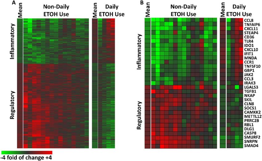
Plasma‑induced Transcriptome changes with Lp299v supplementation. Overall, 1,443 probe
sets (505 downregulated and 938 upregulated) representing 997 unique genes met our criteria for differen-
tial induction after Lp299v supplementation; these data are depicted as heat maps (Fig. 2) where the data are
expressed as a ratio of post-supplement versus pre-supplement. As seen in Fig. 2, the pattern of changes in
gene transcription of the 15 non-DAU subjects differs from DAUs (Fig. 2A). In general, the 15 non-DAU sub-
jects exhibited reduced induction of inflammatory transcripts and increased induction of regulatory transcripts
after Lp299v supplementation. A subset of well-annotated transcripts representing these changes is depicted in
Fig. 2B. A full list of the gene transcripts that were significantly changed are included in Supplemental Table S2.
Ingenuity pathway analysis: regulated pathways and predicted upstream regulators. Canon-
ical pathways considered to be significantly changed (|z-score|≥ 2 and P < 0.01) by exposure to post-Lp299v sup-
plementation plasma are listed in Table 2. Additionally, IPA identified 101 upstream regulators whose suppression
Scientific Reports | (2021) 11:3972 | https://doi.org/10.1038/s41598-021-83252-7 2
Vol:.(1234567890)www.nature.com/scientificreports/
Pre-Lp299v Post-Lp299v p Value
Physical measurements
Systolic blood pressure (mmHg) 136 ± 15 137 ± 14 0.80
Diastolic blood pressure (mmHg) 75 ± 6 75 ± 8 0.84
Body mass index (kg/m2) 31.3 ± 4.2 31.2 ± 4.2
Plasma biomarkers
Fasting glucose (mg/dL) 112 ± 45 112 ± 50 0.91
Total cholesterol (mg/dL) 170 ± 34 164 ± 24 0.39
HDL (mg/dL) 49 ± 12 47 ± 10 0.40
LDL (md/dL) 95 ± 31 89 ± 25 0.32
Triglycerides (mg/dL) 130 ± 61 140 ± 81 0.44
Leptin (ng/mL) 136 ± 98 111 ± 90 0.002
Short chain fatty acids
Propionic acid (µM) 31.9 ± 3.5 36.2 ± 3.6 0.005
Butryic acid (µM) 0.8 ± 0.4 0.8 ± 0.3 0.99
Acetic acid (µM) 42.1 ± 9.6 37.2 ± 7.4 0.16
Vascular function measurements
Resting brachial diameter (mm) 4.14 ± 0.43 4.01 ± 0.39 0.14
Peak hyperermic shear (dynes/cm2) 74.8 ± 14.3 82.9 ± 15.1 0.58
Baseline peak shear (dynes/cm2) 41.4 ± 10.4 40.8 ± 12.2 0.77
Nitroglycerin-mediated dilation (%) 19.9 ± 5.7 19.2 ± 5.1 0.75
Table 1. Subject characteristics. *All data presented as mean ± SD. N = 6 for Nitroglycerin-Mediated Dilation.
N = 14 short chain fatty acid measurements. N = 15 for all other comparisons. p Values derived from Paired
t-Test. † Lp299v = Lactobacillus plantarum 299v.
Figure 1. Effects of Lp299v supplementation on Brachial Artery FMD, ILs-8, 12, and Leptin in Non-Daily
Alcohol Drinkers: Lp299v supplementation resulted in (A) improved brachial flow mediated dilation (FMD)%
(3.70 ± 0.51 vs. 4.58 ± 0.61%, n = 15; P = 0.016) while IL-8 (B, 13.8 ± 2.0 vs. 10.1 ± 1.1 pg/mL, n = 15; P = 0.032),
IL-12 (C, 54.6 ± 8.1 vs. 33.0 ± 7.9 pg/mL, n = 15; P = 0.042), and leptin (D, 136 ± 25 vs. 111 ± 23 ng/mL, n = 15;
P = 0.002) all significantly decreased. All statistical tests paired t-tests. All values given as mean ± SE. *p < 0.05.
Scientific Reports | (2021) 11:3972 | https://doi.org/10.1038/s41598-021-83252-7 3
Vol.:(0123456789)www.nature.com/scientificreports/
Figure 2. Post-Lp299v Plasma-Induced PBMC Signatures‒Stratified by Daily and Non-Daily Alcohol
Drinkers: (A) 1,443 probe sets representing 997 unique genes were altered by at least 20% with a qval FDR < 0.2)
in 15 individuals with stable CAD who did not report daily alcohol use (left) and four individuals with stable
CAD who did report daily alcohol use (right). The mean for each group is indicated and each subsequent
column is a subject. Data are expressed as a fold of change post- vs pre-supplementation with Lp299v. Red
represents upregulated probe sets (n = 505); green represents downregulated probe sets (n = 938). (B) Well
annotated transcripts associated with inflammatory and regulatory activity that showed significant change with
Lp299v supplementation in these subjects.
Canonical Pathway p Value z-score Genes
Tryptophan degradation to 2-amino-3-carboxymuconate semialdehyde 0.00024 − 2.000 IDO1,IDO2,KMO,KYNU
ARHGEF7,CALM1,CHP1,FYN,GNB4, GNB5,HDAC2,HDAC9,L
Phospholipase C signaling 0.00041 2.132 CP2,LYN,MARCK, MEF2A,MRAS,MYL12A,NAPEPLD,NFATC2,
NFATC3,PLA2G4A,PPP1CB,PPP1R12A, PRKD3,RAP1A,RAP2B,RHOF,SOS2
Interferon signaling 0.00048 − 2.828 IFIT1,IFIT3,IFITM3,IFNB1,ISG15,JAK2,OAS1,SOCS1
ATM,CACNA1A,CD80,CD86,CHP1,FGFR1, FYN,IKBKB,LCP2,MAP3K2,MAP3K
PKCθ signaling in T lymphocytes 0.0017 2.183
3,MRAS, NFATC2,NFATC210 ± ± ± 0,PIK3R1,RAP1A, RAP2B,SOS2
AKT3,ATM,CALM1,CD80,CD86,CHP1,FGFR1,IKBKB,IL2RB,LCP2,NFATC2,NFA
iCOS-iCOSL signaling in T helper cells 0.0030 2.309
TC3,PIK3R,PLEKHA2
ACTR2,AKT3,ATM,CALM1,CD80,CD86,CHP1,FGFR1,FYN,IKBKB,LCP2,NFATC
CD28 signaling in T helper cells 0.0060 2.138
2,NFATC3,PIK3R1
AKT3,ATM,CALM1,CHP1,FGFR1,IKBKB, MAP3K2,MAP3K3,MAPK14,MITF,NF
RANK signaling in osteoclasts 0.0076 2.111
ATC2, PIK3R1
ACTR2,ATM,CALM1,CHP1,FGFR1,GNB4,
fMLP signaling in neutrophils 0.0079 2.309
GNB5,MRAS,NFATC2,NFATC3,PIK3R1, PRKD3,RAP1A,RAP2B
Table 2. Canonical pathways significantly impacted by Lp299v supplementation. *Lp299v = Lactobacillus
plantarum 299v genes that were differentially expressed by at least 20% between pre- and post-Lp299v plasma
sample exposure with an FDR < 0.2 were considered significantly changed in this exploratory pilot study.
Hierarchical clustering was performed using Genesis53. In addition, using this set of genes, we calculated the
composite inflammatory index (I.I.com)54. I I.com was determined by calculating the ratio between the mean log
intensity of the inflammatory genes (n = 505) versus the mean log intensity of the regulatory genes (n = 938)
as described in Chen et al.54 A high score reflects greater inflammatory bias and a low score reflects greater
regulatory bias. The changes in differentially expressed genes were correlated with changes in brachial FMD%
using Pearson’s r with significant correlations considered for P < 0.00035 following Bonferroni correction
for multiple testing. The set of genes considered to be differentially expressed were further analyzed using
Ingenuity Pathway Analysis (Qiagen) and the Database for Annotation, Visualization, and Integrated
Discovery (DAVID) to determine up- and down-regulated pathways and functional gene clusters as well as
potential upstream regulators for the transcriptome results[55].
Scientific Reports | (2021) 11:3972 | https://doi.org/10.1038/s41598-021-83252-7 4
Vol:.(1234567890)www.nature.com/scientificreports/
Canonical pathway p Value Number of genes/fold enrichment Genes
CCL3L1,CCL3L3,CCL3,CXCL10,CXCL11, CXCL9,CD80,CD86,IFNB1,
Toll-like receptor signaling pathway 0.000012 13/4.8
LY96,TLR4,TLR7,TLR8
CCL24,CCL3L1,CCL3L3,CCL3,CCL8,CXCL10,CXCL11, CXCL9,CXCL2,TNFRSF10D,
Cytokine-cytokine receptor interaction 0.000035 19/3.1
ACVR2A,INHBA,IFNB1,IL1R2,IL15,PF4V1,TNFSF10, TNFSF13B
Complement and coagulation cascades 0.000051 10/5.7 A2M,C1QB,C1QC,C3AR1,C5AR1,CFB,CFD,SERPINA1,SERPING1,TFP1
OAS1,OAS2,OAS3,CXCL10,DDX58, TNFRSF10, CASP1,IFNB1,IFIH1,NXT2,RSAD2,
Influenza A 0.00011 15/3.4
TLR4,TLR7,TNFSF10
CCL24,CCL3L1,CCL3L3,CCL3,CCL8,CCR1,CXCL10,CXCL11,CXCL9,
Chemokine signaling pathway 0.00023 15/3.5
CXCL2,GNB4,JAK2,LYN,ARRB1, PF4V1
Rheumatoid arthritis 0.00034 10/4.5 ATP6V1C1,CCL3L1,CCL3L3,CCL3, CD80,CD86,ANGPT1,IL15, TLR4,TNFSF13B
Measles 0.00049 12/3.6 OAS1,OAS2,OAS3, DDX58,JAK2,TNFSF10,CCNE2,IFNB1,IFIH1,TLR4,TLR7
Staphylococcus aureus infection 0.0023 7/5.1 FGCR1A,C1QB,C1QC,C3AR1,C5AR1,CFB,CFD
Table 3. KEGG pathways downregulated by Lp299v. *Analysis performed using DAVID 6.8. Clustering
Parameters: Threshold:2, EASE: 0.05. p Value Threshold: P < 0.005. † Lp299v = Lactobacillus plantarum 299v.
Canonical pathway p Value Number of genes/fold enrichment Genes
Chronic myeloid leukemia 0.000032 11/5.3 ABL1,AKT3,CRKL,MDM2,SMAD4,SOS2,IKBKB,PIK3R1,STAT5B, TGFB1,TGFBR2
AKT3,AMT,BCL2L11,FBXO32,MDM2,SKP2,SMAD4,SOS2,FOXO3,IKBKB,MAPK14,PI
FoxO signaling pathway 0.00010 14/3.6
K3R1, TGFB1,TGFBR2
p53 signaling pathway 0.00011 10/5.2 ATM,MDM2,MDM4,CASP8,CNND3,CYCS,SERPINE1,SESN3,TSC2,ZMAT3
ABL1,AKT3,CRKL,RAP1A,SOS2, CALM1,FOXO3,IKBKB,IRAK4, MAPK14,
Neurotrophin signaling pathway 0.00015 13/3.8
AP3K3,PIK3R1,ZNF274
Cell cycle 0.00077 12/3.4 ABL1,ATM,MDM2,RBL1,SKP2, SMAD4,ANAPC5,CDC14A,CCND3,TGFB1,YWHAZ
AKT3,SMAD4,ATF6B,CASP8,CYCS, IKBKB,MAVS,NFATC2,NFATC3,
Hepatitis B 0.00084 13/3.1
PIK3R1,TGFB1,YWHAZ
T cell receptor signaling pathway 0.0021 10/3.5 AKT3,FYN,SOS2,DLF1,IKBKB,LCP2,MAPK14,NFATC2,NFATC3,PIK3R1
Prolactin signaling pathway 0.0039 8/3.9 HERC1,MDM2,SKP2,SMURF2, ANAPC5,CUL4A,SOCS1,UBE2G2, UBE2I,UBE2W,VHL
Table 4. KEGG pathways upregulated by Lp299v. *Analysis performed using DAVID 6.8. Clustering
Parameters: Threshold:2, EASE: 0.05. p Value Threshold: P < 0.005. † Lp299v = Lactobacillus plantarum 299v.
was suggested by the patterns in gene expression changes (Supplemental Table S3), and 25 upstream regulators
that appear to be activated (Supplemental Table S4). These data suggest a significant anti-inflammatory effect of
Lp299v supplementation, including downregulation of interferon-signaling (including IFN-γ and IL-1β), TNF-α
signaling, toll-like receptor activation (TLRs 2,3,4,7, and 9), tryptophan degradation, and upregulation of IL-10
receptor subunit alpha which suppresses pro-inflammatory cytokine activation and stimulates IL-1 receptor
antagonist activity. Further, upregulation of both the CD28 and iCOS signaling pathways for T helper cells along
with genes regulated by suppressor of cytokine signaling-1 (SOCS1), PRDM1 (Blimp1), and DNA exonucle-
ase 3′-repair exonuclease 1 (TREX1) suggest Lp299v supplementation shifts T cell activity towards suppressive
immunity through regulatory T-cells (Tregs) and suppression of innate immune activation15–17.
DAVID functional annotation clustering and pathway analyses. DAVID 6.8 recognized 423 of the
down-regulated Homo sapien genes transcripts in our dataset. Pathway analysis identified 7 KEGG Pathways
significantly downregulated by Lp299v supplementation (Table 3). Consistent with our findings from the IPA
upstream regulator analysis, DAVID identified downregulation of cytokine and chemokine signaling and innate
immunity activation through toll-like receptors. These analyses additionally identified downregulation of path-
ways activated by acute infections.
DAVID 6.8 recognized 608 of the up-regulated Homo sapien genes transcripts in our dataset. Pathway analysis
identified 8 KEGG Pathways significantly upregulated by Lp299v supplementation (Table 4). Consistent with
our findings from the IPA upstream regulator analysis, DAVID identified upregulation of CD4 + T cell signaling
pathways that regulate adaptive immunity. These analyses additionally identified two pathways involved with
cell cycle checkpoints and DNA repair (FoxO and p53 signaling pathways).
Ontology‑based quantitative scoring of plasma‑induced signatures. As defined by the gene
ontological analyses, the induced probe sets regulated by Lp299v supplementation can be broadly considered as
“inflammatory” or “regulatory.” Therefore, signatures were quantitatively scored with a composite inflammatory
index (I.I.com) determined by calculating an average ratio between the mean log intensity of the inflammatory
genes versus the mean log intensity of the regulatory genes22. High scores reflect greater inflammatory bias
and low scores reflect greater regulatory bias. The mean I.I.com of the 15 CAD patients was significantly lower
after Lp299v supplementation (1.01 ± 0.74 vs 0.22 ± 0.51 P < 0.0001, Fig. 3A) suggesting a strong anti-inflamma-
Scientific Reports | (2021) 11:3972 | https://doi.org/10.1038/s41598-021-83252-7 5
Vol.:(0123456789)www.nature.com/scientificreports/
Figure 3. Impact of Lp299v supplementation on the Composite Inflammatory Index (I.I.com) for Non-Daily and
Daily Alcohol Drinkers. (A) Exposure to post-Lp299v supplementation plasma from non-daily alcohol using
subjects drove an anti-inflammatory gene expression profile in PBMCs compared to plasma obtained prior
to Lp299v supplementations (0.22 ± 0.51 vs 1.01 ± 0.74, n = 15; P < 0.0001 by paired t-test). (B) Lp299v had no
significant impact on the inflammatory gene transcription expression in subjects who reported drinking alcohol
daily (0.49 ± 0.78 vs 1.01 ± 1.02, n = 4; P = 0.23 by paired t-test). All values given as mean ± SD.
Gene name N36 gene symbol Fold change Pearson’s r p Value
Guanylate binding protein 1 GBP1 − 1.48011 − 0.85 0.000044
Janus kinase 2 JAK2 − 1.27588 − 0.79 0.000069
TNF superfamily member 10 TNFSF10 − 1.51119 − 0.81 0.00020
Table 5. Changes in PBMC expression associated with changes in FMD% following Lp299v supplementation.
*Changes in differentially expressed genes were correlated with changes in brachial FMD% using Pearson’s r
with significant correlations considered for P < 0.00035 following Bonferroni correction for multiple testing.
tory influence of Lp299v supplementation. No such impact was seen in the four DAU subjects (0.49 ± 0.78 vs
1.01 ± 1.02, P = 0.23, Fig.e 3B). Baseline I.I.com did not differ when comparing DAUs versus non-DAUs (P = 0.24).
Correlations between changes in PBMC gene expression and brachial artery endothelial func-
tion. Of the 1373 genes differentially expressed following Lp299v supplementation, only three genes, guanyl
binding protein 1 (GBP1), Janus kinase 2 (JAK2), and TNF Superfamily Member 10 (TNFSF10), were signifi-
cantly correlated with changes in FMD% (Table 5). Transcription of all 3 genes was suppressed overall and the
correlations all reflected an inverse association with changes in FMD%. Correlations between changes in gene
expression and changes in FMD% for the additional genes in this set is included as Supplemental Table S5.
Discussion
In the absence of daily ETOH use, six weeks of supplementation with 20 billion colony-forming units of Lp299v
in men with stable CAD resulted not only improved vascular endothelial function, but also induced changes in
plasma composition to yield a strong anti-inflammatory effect. Favorable effects observed include downregu-
lation of inflammation driven by IL-1β and TNF-α which have been successfully targeted with canakinumab
(IL-1β monoclonal antibody) and colchicine to reduce secondary cardiovascular risk in large clinical t rials3,4. In
addition, Lp299v supplementation broadly suppressed activation of innate immunity through TLR signaling as
well as favoring suppression of inflammation through upregulation of regulatory T-cells ( Tregs). Additionally, we
identified suppression of transcription of three genes (GBP1, JAK2, and TNFSF10) that correlated inversely with
flow-mediated dilation of the brachial artery. Whether these genes mechanistically mediate the development
of inflammation-associated vascular endothelial dysfunction deserves further investigation. Lp299v’s favorable
effects do not appear to extend to DAUs, suggesting that alcohol ingestion diminishes the anti-inflammatory effect
of Lp299v supplementation on circulating plasma. Levels of SCFAs (acetic, propionic, and butyric acid) were
similarly changed in individuals who drank daily and those who did not suggesting that these SCFAs were not
mediating Lp299v’s anti-inflammatory effects. Taken together, our data support a paradigm of Lp299v contrib-
uting to improved vascular health in individuals with CAD at least in part through reductions in inflammation
driven by yet-to-be identified gut-derived metabolites that should fuel future investigations.
Prior in vitro work exposing human PBMCs and L. plantarum species demonstrate the potential for L.
plantarum to modulate the PBMC inflammatory response18. Our initial studies with Lp299v were stimulated by
the favorable effects we observed with Lp299v supplementation on infarct size and reducing pro-inflammatory
leptin levels in male salt-sensitive rats19. Different species of L. plantarum appear to have differing effects on the
activation state and differentiation of human PBMCs in vitro20. Importantly, these data also demonstrate the
Scientific Reports | (2021) 11:3972 | https://doi.org/10.1038/s41598-021-83252-7 6
Vol:.(1234567890)www.nature.com/scientificreports/
in vitro effect of the direct exposure of L. plantartum species on PBMCs poorly predicts the in vivo response to
this class of probiotics20–22. Our data significantly extend these prior findings by focusing on highly clinically rel-
evant human in vivo supplementation actions with Lp299v and clearly delineating this supplementation changes
the composition of plasma so as to exert a systemic anti-inflammatory effect that can be observed in PBMCs.
Lp299v supplementation in the present study had favorable anti-inflammatory effects on plasma composition
including lowering of leptin, IL-12, and IL-8, and the reductions in the latter two may be, in part, leptin-mediated.
Prior studies have mechanistically shown increases in serum leptin resulting in increases in ILs 12 and 8 in
macrophages. Leptin enhanced secretion of LPS-stimulated IL-12 by peritoneal macrophages and stimulated
IL-8 expression via p38 and ERK signaling pathways in M2 m acrophages23,24. Given this prior work, it is quite
possible that the decrease in leptin is causal in the reductions we see in IL-12 and IL-18.
Daily alcohol ingestion appears to blunt the anti-inflammatory effects of Lp299v in our study as demonstrated
by individual inflammatory indices calculated from the transcriptome data (Fig. 3). Prior work demonstrates
that alcohol ingestion promotes the growth of gram negative bacteria in the intestine and increases overall gut
permeability and circulating endotoxin, triggering systemic inflammation25,26. Any of these established physi-
ologic changes could account for the blunted anti-inflammatory effects of Lp299v in DAUs in our study. Our
transcriptome data is consistent with these earlier findings, showing significant reductions in signaling pathways
triggered by circulating lipopolysaccharide and peptidoglycan, including TLR4 activation, in our 15 non-DAU
subjects (Supplemental Table S3). However, we had only four subjects who self-reported daily alcohol drinking
with some variability in response, making our findings only hypothesis-generating. The dose and frequency
relationships between alcohol intake and the anti-inflammatory impact of Lp299v supplementation, as well as the
underlying mechanisms driving this mitigating effect, remain to be delineated and merit further investigation.
Over the past two decades, there has been an increasing recognition of the critical role played by activation
of the innate immune system through toll-like receptors in the development of atherosclerotic disease. IPA
analyses of our transcriptome data suggest Lp299v supplementation downregulates TLR signaling, including
TLRs 2,3,4,7, and 9. Each of these has been shown to be expressed by human coronary artery and microvascular
endothelial cells with activation leading to increased expression of cell adhesion molecules and pro-inflammatory
cytokines27. In the peripheral human circulation, TLRs 2 and 4 are ubiquitously expressed and are enhanced
in atherosclerotic p laques22, 23. TLR3 is most expressed in the carotid arteries and aorta, TLR7 in the iliac and
carotid arteries, and TLR9 with overall low expression but most prominent expression in the iliac arteries28
TLR 2,3,4,7, and 9 stimulation have been implicated in the development of atherosclerosis through combina-
tion of promoting of lipid uptake into the plaques, monocyte activation, endothelial dysfunction, and foam cell
formation29–34. Recently TLR2 has been shown to facilitate microbiota-induced VWF expression and systemic
interferon responses mice35,36. Our data suggest Lp299v may reduce inflammation in part through reducing TLR
activation in humans. Additional work will be necessary to better determine how Lp299v interacts with the innate
immune system to suppress inflammation and atherogenesis.
Functional analyses of our transcriptome data support a paradigm of Lp299v supplementation increasing
the number and activity of regulatory T-cells (Tregs). Tregs generally exert an anti-inflammatory influence and
suppress self-antigen recognition. In mouse models of hypertension and atherosclerosis, T regs reduce monocyte
activation and infiltration into the vasculature and atherosclerosis formation, preserve endothelium-dependent
vasodilation of coronary arterioles in mice through increased IL-10 expression, reduced IL-1β and TNF-α expres-
sion leading to suppression of endothelial expression of adhesion molecules like VCAM-1 and E-selectins37,38.
Our data reporting that Lp299v-induces reductions in IL-1β and TNF signaling as well as upregulation of IL-10
receptor signaling in mononuclear cells are consistent with these mouse data and suggest an association between
anti-inflammatory effect of Treg upregulation and improvements in endothelial function extend to humans with
CAD. Additional work to delineate the mechanistic connections in human are warranted, particularly in light
of the efficacy of IL-1β inhibition in reducing cardiovascular r isk3.
We identified three genes (JAK2, GBP1, and TNFSF10) whose transcription was significantly reduced in
PBMCs by post-Lp299v supplementation plasma. These changes inversely correlated with increases in brachial
FMD%. JAK2, a tyrosine kinase involved in cytokine signaling, increases plaque and plaque necrotic core size
in hemopoietic cells and increases IL-1β from mononuclear cells challenged with lipopolysaccharide, when
activated; it’s suppression increases expression and activity of anti-inflammatory Tregs39,40. Here, the reduction
in JAK2 transcription appears consistent with the increases in expression of canonical pathways involved with
increased Treg pathways (Table 2) and as well as the decrease in expression in genes regulated by IL-1β signaling
detected by our Ingenuity Pathway Analysis for upstream regulators (Supplemental Table S3). GBP1 is an IFNγ-
induced GTPase found in T-cells, B-cells, and endothelial cells whose activation in the innate immune response
to pathogens results in an inflammatory activation of endothelial cells and inhibited angiogenesis35–38. TNFSF10
(TRAIL) is an apoptosis-inducing ligand expressed in T cells and induced by IFNα and β to enhance T-cell
cytotoxicity41. It has been detected in CD3 + T-cells of human atherosclerotic plaques with higher concentrations
in vulnerable p laques42. Further studies determining the impact of Lp299v on endothelial cell expression of all
three genes and their concomitant impact on interactions between PBMCs and endothelium, and endothelial
function, are warranted.
Our study has some limitations. This study included a small number of only male subjects; whether these
findings generalize to women and hold for a large group of male subjects await further elucidation which we
will be pursing though a randomized control trial (NCT03267758). We did not perform transcriptome analyses
on PBMCs directly from study subjects prior to and following Lp299v supplementation given concerns for con-
founding by differences in the types and amounts of pharmacological therapies. However, our approach more
clearly delineates that Lp299v’s anti-inflammatory effects are mediated by factors in the circulating plasma. We
did not examine the direct impact of Lp299v on cultured endothelial cells which may reveal a direct impact of
Lp299v supplementation on the activation state of the endothelium itself. Nevertheless, PBMCs play a critical
Scientific Reports | (2021) 11:3972 | https://doi.org/10.1038/s41598-021-83252-7 7
Vol.:(0123456789)www.nature.com/scientificreports/
role in the development of coronary atherosclerosis and the stability of atherosclerotic plaques making our
findings highly relevant to vascular disease. Balanced against these limitations is the novelty of the findings
suggesting the anti-inflammatory effect of supplementation with probiotic Lp299v may account for part of its
favorable vascular effects.
Conclusions
In this pilot study of 15 men with stable CAD, we demonstrated that six weeks of Lp299v supplementation not
only improved vascular endothelial function, but also had a significant anti-inflammatory effect. This anti-
inflammatory effect appears to be at least in part due to suppression of innate immune signaling and increased
activity of Tregs. We additionally identified three genes (JAK2, GBP1, and TNFSF10) whose mRNA transcription
suppression by Lp299v in PBMCs was inversely correlated with brachial FMD% suggesting a potential more direct
role for these proteins in inflammation-induced endothelial dysfunction in humans. The changes in circulating
metabolites induced by Lp299v responsible for these favorable effects remain to elucidate and hold promise for
further delineating the mechanisms behind improvements in vascular health with this probiotic intervention.
Methods
Subjects and study protocol. The study protocol was reviewed and approved by the Institutional
Research Board at the Medical College of Wisconsin, and all experiments were performed in accordance with
their regulations and guidelines. Informed consent was given by all participants within this study. 23 men with
stable CAD (ages 40–75) were recruited as previously d escribed14. This study includes analyses on 15 of the 23
subjects that were enrolled on the initial report on the impact of Lp299v supplementation on endothelial func-
tion in men with stable CAD14. As noted in the initial report, two subjects enrolled were disqualified because
they failed to pass the initial screening visit. One subject had a stroke while on protocol and did not complete
the study. We excluded an additional four subjects from these study analyses who were self-reported daily alco-
hol users (DAUs), but did perform transcriptome analyses using their plasma to validate their exclusion. One
subject did not have plasma samples from all study visits and we were therefore unable to perform transcrip-
tome analyses for this subject. After exclusions, a total of 15 subjects were available for this set of analyses. The
inclusion and exclusion criteria are identical to the previously described study p rotocol8. Individuals with left-
ventricular ejection fraction less than 45% within one year of enrollment, uncontrolled hypertension (blood
pressure > 170/110 mmHg), a creatinine clearance less than 60 mL/min, recent antibiotic or probiotic adminis-
tration (within 12 weeks of enrollment), chronic liver disease, cancer requiring chemotherapy within five years
of enrollment, or changes in vasoactive medications (within six weeks of enrollment) were excluded.
All subjects underwent focused medical history reviews and physical exams to screen out occult disease or
instability of cardiac status that would exclude a subject. In addition, each subject was asked to self-report their
daily alcohol intake in one of five categories: rarely/never, once per month, several times per month, once per
week, several times per week, and daily. Subjects who passed screening and enrolled in the study underwent an
initial set of measurements (heart rate and blood pressure in triplicate; height and waist circumference; IL-8,
IL-12, leptin, and glycosylated hemoglobin from peripheral upper extremity venous blood, which was saved for
the plasma induced transcriptome studies; and endothelial function by vascular artery ultrasound). Subjects were
subsequently placed on once daily supplementation with 80 ml (2.7 oz.) of GoodBelly Straight (Probi, Sweden).
Each daily supplement contained 20 billion colony forming units of L.plantarum 299v. Subjects returned to the
study center approximately six weeks following beginning supplementation and repeated the studies from their
initial study visit.
Measurement of endothelial function by vascular ultrasound. Measurements of vascular endothe-
lial function were performed using high-resolution vascular ultrasound as previously described14,43–48. All image
acquisition and analyses were performed by trained technicians. Our measurements are highly reproducible
between different operators (average intra-class correlation between four technicians for ten studies of 0.97
[0.92–0.99, P < 0.001] and a single rater intraclass correlation coefficient of 0.88 [0.74–0.97, P < 0.001].
Measurement of circulating IL‑8, IL‑12, and leptin plasma levels. Serum cytokines and leptin lev-
reviously14. Sample collections were taken from all subjects between the hours
els were measured as described p
of 08:00 AM and 12:00 PM on the day of study visit. IL-8 and IL-12 measurements were performed by Eve Tech-
nologies (Calgary, Canada). Human plasma leptin was measured using a quantitative ELISA kit (R&D Systems,
Minneapolis, MN, #DLP00) on EDTA-preserved plasma both pre- and post-probiotic treatment; assays were
read with a SpectraMax M5e Microplate Reader (Molecular Devices, Sunnyvale, CA).
Plasma‑induced regulation of mononuclear cell transcription. Using a validated assay49, we incu-
bated 200 µL of pre- and post-Lp299v supplementation plasma from each subject in separate wells of Costar
24-well plates seeded with 5 × 10⁵ UPN119 PBMCs (Cellular Technology LTD, Shaker Heights, OH). Wells also
contained RPMI 1,640 medium supplemented with 100 U/ml penicillin and 100 µg/mL streptomycin to a total
volume of 500 µL. RNA from PBMCs was subsequently isolated, labeled, and hybridized to the Affymetrix
escribed50,51. Affymetrix microarray data were uploaded to NCMI Gene Expres-
U133 + 2.0 array as previously d
sion Omnibus (GEO, http://www.ncbi.nlm.nih.gov/geo/).
Statistical analyses. All data analyses were performed using SPSS 24.0, SigmaPlot 12.5, and GraphPad
Prism. Subject characteristics prior to and following Lp299v supplementation were compared by paired t-test,
Scientific Reports | (2021) 11:3972 | https://doi.org/10.1038/s41598-021-83252-7 8
Vol:.(1234567890)www.nature.com/scientificreports/
Wilcoxon signed-rank test, or chi-square tests as appropriate. Changes in brachial artery parameters and inflam-
matory cytokines were compared by paired t-test or the signed-rank test as appropriate. For these data, P < 0.05
was considered significant. For the plasma-induced transcriptome data, genes that were differentially expressed
by at least 20% between pre- and post-Lp299v plasma sample exposure with an FDR < 0.2 were considered signif-
icantly changed in this exploratory pilot study. Hierarchical clustering was performed using G enesis52. In addi-
tion, using this set of genes, we calculated the composite inflammatory index (I.I.com)53. I I.com was determined
by calculating the ratio between the mean log intensity of the inflammatory genes (n = 505) versus the mean log
intensity of the regulatory genes (n = 938) as described in Chen et al.53 A high score reflects greater inflammatory
bias and a low score reflects greater regulatory bias. The changes in differentially expressed genes were correlated
with changes in brachial FMD% using Pearson’s r with significant correlations considered for P < 0.00035 fol-
lowing Bonferroni correction for multiple testing. The set of genes considered to be differentially expressed were
further analyzed using Ingenuity Pathway Analysis (Qiagen) and the Database for Annotation, Visualization,
and Integrated Discovery (DAVID) to determine up- and down-regulated pathways and functional gene clusters
as well as potential upstream regulators for the transcriptome r esults54.
Data availability
Transcriptome microarray data is publicly available in the Gene Expression Omnibus (GEO) repository (acces-
sion number GSE156357). All other data analyzed for this study can be found within this published manuscript
and the supplementary materials.
Received: 14 October 2020; Accepted: 25 January 2021
References
1. Ross, R. Atherosclerosis–an inflammatory disease. N Engl J Med 340, 115–126 (1999).
2. Ridker, P. M. et al. Rosuvastatin to prevent vascular events in men and women with elevated C-reactive protein. N Engl J Med 359,
2195–2207 (2008).
3. Ridker, P. M. et al. Antiinflammatory therapy with canakinumab for atherosclerotic disease. N Engl J Med 377, 1119–1131 (2017).
4. Tardif, J. C. et al. Efficacy and safety of low-dose colchicine after myocardial infarction. N Engl J Med 381, 2497–2505 (2019).
5. Brandsma, E. et al. A proinflammatory gut microbiota increases systemic inflammation and accelerates atherosclerosis. Circ. Res.
124, 94–100 (2019).
6. Yin J, Liao SX, He Y et al. Dysbiosis of gut microbiota with reduced trimethylamine-N-oxide level in patients with large-artery
atherosclerotic stroke or transient ischemic attack. J. Am. Heart Assoc. 2015;4.
7. Toya, T. et al. Coronary artery disease is associated with an altered gut microbiome composition. PLoS ONE 15, e0227147 (2020).
8. Karlsson, F. H. et al. Symptomatic atherosclerosis is associated with an altered gut metagenome. Nat. Commun. 3, 1245 (2012).
9. Li, J., Lin, S., Vanhoutte, P. M., Woo, C. W. & Xu, A. Akkermansia muciniphila protects against atherosclerosis by preventing
metabolic endotoxemia-induced inflammation in apoe-/- mice. Circulation 133, 2434–2446 (2016).
10. Karbach SH, Schönfelder T, Brandão I et al. Gut microbiota promote angiotensin II-induced arterial hypertension and vascular
dysfunction. J. Am. Heart Assoc. 2016;5.
11. Lindskog Jonsson, A. et al. Impact of gut microbiota and diet on the development of atherosclerosis in Apoe(-/-) Mice. Arterioscler.
Thromb. Vasc. Biol. 38, 2318–2326 (2018).
12. Kiouptsi K, Jäckel S, Pontarollo G et al. The Microbiota promotes arterial thrombosis in low-density lipoprotein receptor-deficient
mice. mBio 2019;10.
13. Kossmann, S. et al. Inflammatory monocytes determine endothelial nitric-oxide synthase uncoupling and nitro-oxidative stress
induced by angiotensin II. J. Biol. Chem. 289, 27540–27550 (2014).
14. Malik, M. et al. Lactobacillus plantarum 299v supplementation improves vascular endothelial function and reduces inflammatory
biomarkers in men with stable coronary artery disease. Circ. Res. 123, 1091–1102 (2018).
15. Lu, L. F. et al. Foxp3-dependent microRNA155 confers competitive fitness to regulatory T cells by targeting SOCS1 protein.
Immunity 30, 80–91 (2009).
16. Chen, X. et al. CD4+CD25+ regulatory T cells in tumor immunity. Int Immunopharmacol 34, 244–249 (2016).
17. Garg, G. et al. Blimp1 prevents methylation of Foxp3 and loss of regulatory T cell identity at sites of inflammation. Cell Rep.
26(1854–1868), e5 (2019).
18. Borchers, A. T., Selmi, C., Meyers, F. J., Keen, C. L. & Gershwin, M. E. Probiotics and immunity. J. Gastroenterol. 44, 26–46 (2009).
19. Lam, V. et al. Intestinal microbiota determine severity of myocardial infarction in rats. FASEB J. 26, 1727–1735 (2012).
20. Rask, C., Adlerberth, I., Berggren, A., Ahren, I. L. & Wold, A. E. Differential effect on cell-mediated immunity in human volunteers
after intake of different lactobacilli. Clin. Exp. Immunol. 172, 321–332 (2013).
21. Garcia-Gonzalez, N., Prete, R., Battista, N. & Corsetti, A. Adhesion properties of food-associated lactobacillus plantarum strains
on human intestinal epithelial cells and modulation of IL-8 release. Front. Microbiol. 9, 2392 (2018).
22. van Hemert, S. et al. Identification of Lactobacillus plantarum genes modulating the cytokine response of human peripheral blood
mononuclear cells. BMC Microbiol. 10, 293 (2010).
23. Loffreda, S. et al. Leptin regulates proinflammatory immune responses. Faseb J 12, 57–65 (1998).
24. Cao, H. et al. Leptin promotes migration and invasion of breast cancer cells by stimulating IL-8 production in M2 macrophages.
Oncotarget 7, 65441–65453 (2016).
25. Parlesak, A., Schafer, C., Schutz, T., Bode, J. C. & Bode, C. Increased intestinal permeability to macromolecules and endotoxemia
in patients with chronic alcohol abuse in different stages of alcohol-induced liver disease. J. Hepatol. 32, 742–747 (2000).
26. Lambert, J. C. et al. Prevention of alterations in intestinal permeability is involved in zinc inhibition of acute ethanol-induced liver
damage in mice. J. Pharmacol. Exp. Ther. 305, 880–886 (2003).
27. Khakpour, S., Wilhelmsen, K. & Hellman, J. Vascular endothelial cell Toll-like receptor pathways in sepsis. Innate. Immun. 21,
827–846 (2015).
28. Pryshchep, O., Ma-Krupa, W., Younge, B. R., Goronzy, J. J. & Weyand, C. M. Vessel-specific Toll-like receptor profiles in human
medium and large arteries. Circulation 118, 1276–1284 (2008).
29. Lee, J. G. et al. A combination of Lox-1 and Nox1 regulates TLR9-mediated foam cell formation. Cell Signal 20, 2266–2275 (2008).
30. Funk, J. L., Feingold, K. R., Moser, A. H. & Grunfeld, C. Lipopolysaccharide stimulation of RAW 264.7 macrophages induces lipid
accumulation and foam cell formation. Atherosclerosis 98, 67–82 (1993).
31. Mullick, A. E., Tobias, P. S. & Curtiss, L. K. Modulation of atherosclerosis in mice by Toll-like receptor 2. J. Clin. Invest. 115,
3149–3156 (2005).
Scientific Reports | (2021) 11:3972 | https://doi.org/10.1038/s41598-021-83252-7 9
Vol.:(0123456789)www.nature.com/scientificreports/
32. Fukuda, D. et al. Toll-like receptor 9 plays a pivotal role in angiotensin II-induced atherosclerosis. J. Am. Heart Assoc. 8, e010860
(2019).
33. Zimmer, S. et al. Activation of endothelial toll-like receptor 3 impairs endothelial function. Circ. Res. 108, 1358–1366 (2011).
34. Liu, C. L. et al. Toll-like receptor 7 deficiency protects apolipoprotein E-deficient mice from diet-induced atherosclerosis. Sci. Rep.
7, 847 (2017).
35. Jäckel, S. et al. Gut microbiota regulate hepatic von Willebrand factor synthesis and arterial thrombus formation via Toll-like
receptor-2. Blood 130, 542–553 (2017).
36. Schaupp, L. et al. Microbiota-induced type i interferons instruct a poised basal state of dendritic cells. Cell 181(1080–1096), e19
(2020).
37. Matrougui, K. et al. Natural regulatory T cells control coronary arteriolar endothelial dysfunction in hypertensive mice. Am. J.
Pathol. 178, 434–441 (2011).
38. Maganto-Garcia, E. et al. Foxp3+-inducible regulatory T cells suppress endothelial activation and leukocyte recruitment. J. Immu-
nol. 187, 3521–3529 (2011).
39. Iamsawat, S. et al. Stabilization of Foxp3 by targeting JAK2 enhances efficacy of CD8 induced regulatory T cells in the prevention
of graft-versus-host disease. J. Immunol. 201, 2812–2823 (2018).
40. Desai, H. R. et al. Macrophage JAK2 deficiency protects against high-fat diet-induced inflammation. Sci. Rep. 7, 7653 (2017).
41. Kayagaki, N. et al. Type I interferons (IFNs) regulate tumor necrosis factor-related apoptosis-inducing ligand (TRAIL) expression
on human T cells: a novel mechanism for the antitumor effects of type I IFNs. J. Exp. Med. 189, 1451–1460 (1999).
42. Michowitz, Y. et al. The involvement of tumor necrosis factor-related apoptosis-inducing ligand (TRAIL) in atherosclerosis. J. Am.
Coll. Cardiol. 45, 1018–1024 (2005).
43. Kizhakekuttu TJ, Gutterman DD, Phillips SA et al. Measuring FMD in the brachial artery: how important is QRS-gating? J. Appl.
Physiol. 2010.
44. Babar, G. S. et al. Impaired endothelial function in preadolescent children with type 1 diabetes. Diabet. Care 34, 681–685 (2011).
45. Kizhakekuttu, T. J. et al. Adverse alterations in mitochondrial function contribute to type 2 diabetes mellitus-related endothelial
dysfunction in humans. Arterioscler. Thromb. Vasc. Biol. 32, 2531–2539 (2012).
46. Suboc TB, Knabel D, Strath SJ et al. Associations of reducing sedentary time with vascular function and insulin sensitivity in older
sedentary adults. Am J Hypertens 2015.
47. Widlansky, M. E. et al. Impact of DPP-4 inhibition on acute and chronic endothelial function in humans with type 2 diabetes on
background metformin therapy. Vasc. Med. 22, 189–196 (2017).
48. Kakarla, M. et al. Circulating levels of mitochondrial uncoupling protein 2, but not prohibitin, are lower in humans with type 2
diabetes and correlate with brachial artery flow-mediated dilation. Cardiovasc. Diabetol. 18, 148 (2019).
49. Speake, C. & Odegard, J. M. Evaluation of candidate biomarkers of Type 1 diabetes via the core for assay validation. Biomark.
Insights 10, 19–24 (2015).
50. Cabrera, S. M., Chen, Y. G., Hagopian, W. A. & Hessner, M. J. Blood-based signatures in type 1 diabetes. Diabetologia 59, 414–425
(2016).
51. Wang, X. et al. Identification of a molecular signature in human type 1 diabetes mellitus using serum and functional genomics. J.
Immunol. 180, 1929–1937 (2008).
52. Sturn, A., Quackenbush, J. & Trajanoski, Z. Genesis: cluster analysis of microarray data. Bioinformatics 18, 207–208 (2002).
53. Chen, Y. G. et al. Molecular signatures differentiate immune states in type 1 diabetic families. Diabetes 63, 3960–3973 (2014).
54. da Huang, W., Sherman, B. T. & Lempicki, R. A. Systematic and integrative analysis of large gene lists using DAVID bioinformatics
resources. Nat Protoc 4, 44–57 (2009).
Author contributions
B.C.H. drafted the work and was involved in data analysis and interpretation. V.K.P. and S.T. participated in
data acquisition, data analyses, and revised the draft. A.A. contributed to data acquisition and draft revision. S.J.
contributed to data analyses, interpretation, and draft revision. N.H.S. and M.J.H. contributed to study design,
data interpretation, and draft revision. M.E.W. contributed to study conception, design, data analyses and inter-
pretation, and substantially revised the draft.
Funding
The work was funded in part by a grant from the Clinical Translational Research Initiative of Southeast Wiscon-
sin (UL1TR001436, Medical College of Wisconsin 8701 Watertown Plank Road, Suite M1350 Milwaukee, WI
53226). Dr. Widlansky is supported by HL125409, HL128240, HL144098, and R38HL143561. Dr. Hessner is
supported by DK121528. Dr. Salzman is supported by DK088831 and GM122503. Dr. Puppala received support
from T32HL007792. Dr. Hofeld is supported by R38HL143561.
Competing interests
Dr. Widlansky is supported in part by HL144098 that focuses on the impact of Lp299v supplementation. Probi
(Lund, Sweden), the manufacturer of Lp299v, supports HL144098 through the donation of product. Probi did not
donate any product for studies reported in this manuscript. Dr. Hessner is also supported in part by DK125014
which studies the effect of Lp299v on innate inflammation in type 1 diabetes.
Additional information
Supplementary Information The online version contains supplementary material available at https://doi.
org/10.1038/s41598-021-83252-7.
Correspondence and requests for materials should be addressed to M.E.W.
Reprints and permissions information is available at www.nature.com/reprints.
Publisher’s note Springer Nature remains neutral with regard to jurisdictional claims in published maps and
institutional affiliations.
Scientific Reports | (2021) 11:3972 | https://doi.org/10.1038/s41598-021-83252-7 10
Vol:.(1234567890)www.nature.com/scientificreports/
Open Access This article is licensed under a Creative Commons Attribution 4.0 International
License, which permits use, sharing, adaptation, distribution and reproduction in any medium or
format, as long as you give appropriate credit to the original author(s) and the source, provide a link to the
Creative Commons licence, and indicate if changes were made. The images or other third party material in this
article are included in the article’s Creative Commons licence, unless indicated otherwise in a credit line to the
material. If material is not included in the article’s Creative Commons licence and your intended use is not
permitted by statutory regulation or exceeds the permitted use, you will need to obtain permission directly from
the copyright holder. To view a copy of this licence, visit http://creativecommons.org/licenses/by/4.0/.
© The Author(s) 2021
Scientific Reports | (2021) 11:3972 | https://doi.org/10.1038/s41598-021-83252-7 11
Vol.:(0123456789)You can also read