Left ventricular free wall rupture as a result of delayed presentation of an inferior ST- elevation myocardial infarction due to fear of COVID-19: ...
←
→
Page content transcription
If your browser does not render page correctly, please read the page content below
Nasr et al. Journal of Cardiothoracic Surgery            (2021) 16:106
https://doi.org/10.1186/s13019-021-01495-x

 CASE REPORT                                                                                                                                           Open Access

Left ventricular free wall rupture as a result
of delayed presentation of an inferior ST-
elevation myocardial infarction due to fear
of COVID-19: case report
George H. Nasr1* , Diana Glovaci2, Andrew Mikhail3, Steven Sinfield1, Kevin Chen2, Hardikkumar Patel2,
Michael Johl2, Bharath Chakravarthy3, Siddharth Singh4, Fabio Sagebin5 and Ailin Barseghian El-Farra2

  Abstract
  Background: Left ventricular free wall rupture (LVFWR) is a rare complication after myocardial infarction and usually
  occurs 1 to 4 days after the infarct. Over the past decade, the overall incidence of LVFWR has decreased given the
  advancements in reperfusion therapies. However, during the COVID-19 pandemic, there has been a significant delay
  in hospital presentation of patients suffering myocardial infarctions, leading to a higher incidence of mechanical
  complications from myocardial infarctions such as LVFWR.
  Case presentation: We present a case in which a patient suffered a LVFWR as a mechanical complication from
  myocardial infarction due to delay in seeking care over fear of contracting COVID-19 from the medical setting. The
  patient had been having chest pain for a few days but refused to seek medical care due to fear of contracting
  COVID-19 from within the medical setting. He eventually suffered a cardiac arrest at home from a massive inferior
  myocardial infarction and found to be in cardiac tamponade from a left ventricular perforation. He was emergently
  taken to the operating room to attempt to repair the rupture but he ultimately expired on the operating table.
  Conclusions: The occurrence of LVFWR has been on a more significant rise over the course of the COVID-19
  pandemic as patients delay seeking care over fear of contracting COVID-19 from within the medical setting.
  Clinicians should consider mechanical complications of MI when patients present as an out-of-hospital cardiac
  arrest, particularly during the COVID-19 pandemic, as delay in seeking care is often the exacerbating factor.
  Keywords: Left ventricular free wall rupture, Inferior STEMI, Cardiac tamponade, Pericardial effusion, MI
  complications, CTA, COVID-19, Case report

* Correspondence: gnasr@hs.uci.edu
1
 Department of Medicine, University of California, Irvine, USA
Full list of author information is available at the end of the article

                                           © The Author(s). 2021 Open Access This article is licensed under a Creative Commons Attribution 4.0 International License,
                                           which permits use, sharing, adaptation, distribution and reproduction in any medium or format, as long as you give
                                           appropriate credit to the original author(s) and the source, provide a link to the Creative Commons licence, and indicate if
                                           changes were made. The images or other third party material in this article are included in the article's Creative Commons
                                           licence, unless indicated otherwise in a credit line to the material. If material is not included in the article's Creative Commons
                                           licence and your intended use is not permitted by statutory regulation or exceeds the permitted use, you will need to obtain
                                           permission directly from the copyright holder. To view a copy of this licence, visit http://creativecommons.org/licenses/by/4.0/.
                                           The Creative Commons Public Domain Dedication waiver (http://creativecommons.org/publicdomain/zero/1.0/) applies to the
                                           data made available in this article, unless otherwise stated in a credit line to the data.
Nasr et al. Journal of Cardiothoracic Surgery   (2021) 16:106                                                                         Page 2 of 5

Background                                                                of contracting COVID-19 from within the medical set-
Left ventricular free wall rupture (LVFWR) is a rare                      ting. When emergency medical services (EMS) arrived to
complication that can occur after suffering a myocardial                  his house, the patient was complaining of severe crush-
infarction (MI). The incidence of LVFWR has decreased                     ing chest pain and a 12 lead electrocardiogram (ECG)
dramatically over the years with the increased use of re-                 performed revealed ST-segment elevations and patho-
perfusion strategies such as percutaneous coronary                        logical q-waves in leads II, III, and aVF with reciprocal
intervention (PCI) and fibrinolytic therapy, with an over-                ST-segment depressions in leads I and aVL, consistent
all incidence ranging from 0.8% to 6.2% [1]. LVFWR is                     with inferior ST-segment elevation myocardial infarction
most likely to occur 1–4 days after the initial myocardial                (STEMI) (Fig. 1). En route to the hospital the patient
insult, and is one of the more deadly complications of                    subsequently lost pulses and underwent cardiopulmo-
MI [2]. The early diagnosis of LVFWR is critical and                      nary resuscitation (CPR) for 3 min prior to arrival to the
point of care ultrasound (POCUS) can help establish the                   emergency department (ED). On arrival to the ED, the
diagnosis quickly by revealing evidence of pericardial ef-                patient underwent 10 more minutes of CPR until return
fusion and tamponade.                                                     of spontaneous circulation (ROSC) was achieved. After
  The COVID-19 pandemic has had a dramatic effect                         appropriate donning of personal protective equipment
on life in the United States (U.S.) and has significantly                 (PPE) by ED personnel, the patient was intubated and
impacted and burdened the medical community. An in-                       point of care (POC) ultrasound revealed a depressed
creasing number of patients have avoided seeking med-                     ejection fraction (EF) and a large pericardial effusion
ical care due to fear of contracting COVID-19 from the                    with tamponade physiology. A pericardiocentesis was
medical setting. This is especially concerning for patients               attempted twice, however aspiration of the pericardial
who may be suffering from myocardial infarctions, as a                    effusion was unsuccessful which raised concern for a
delay in treatment can lead to devastating consequences.                  hematoma formation from a possible contained old rup-
Since the onset of the COVID-19 pandemic in March                         ture. A chest x-ray revealed a widened mediastinum
2020 in the U.S., there has been a much higher incidence                  (Fig. 2). The patient was taken emergently for computed
of mechanical complications, such as valvular or ven-                     tomography angiography (CTA) scan of his aorta. The
tricular wall ruptures, from myocardial infarctions due                   CT scan revealed active extravasation through a small
to delay in treatment [3–6]. Here we present a case of a                  channel in the inferior wall of the left ventricle into the
patient who suffered a left ventricular free wall rupture                 pericardial cavity consistent with left ventricular myocar-
as a mechanical complication of myocardial infarction                     dial perforation. There was also evidence of reflux of
due to delay in seeking treatment over fear of contract-                  contrast into the inferior vena cava, distal hepatic veins,
ing COVID-19.                                                             and right renal vein suggestive of low cardiac output in
                                                                          the setting of acute cardiac tamponade, left ventricular
Case presentation                                                         perforation and hemopericardium (Fig. 3). The right cor-
A 67 year old male with a history of heavy smoking was                    onary artery was noted to be opacified in the proximal
witnessed by his wife to have suddenly collapsed at                       segment with significant acute thrombosis (Fig. 4). The
home. The patient had been complaining of chest pain                      patient lost pulses multiple times in the ER and was too
for a few days but did not seek medical care due to fear                  hemodynamically unstable to be taken to the

 Fig. 1 ECG by EMS performed on arrival at patient’s home showing ST-segment elevations and pathological q-waves leads II, III, and aVF with
 reciprocal ST-segment depressions in leads I and aVL consistent with delayed inferior myocardial infarction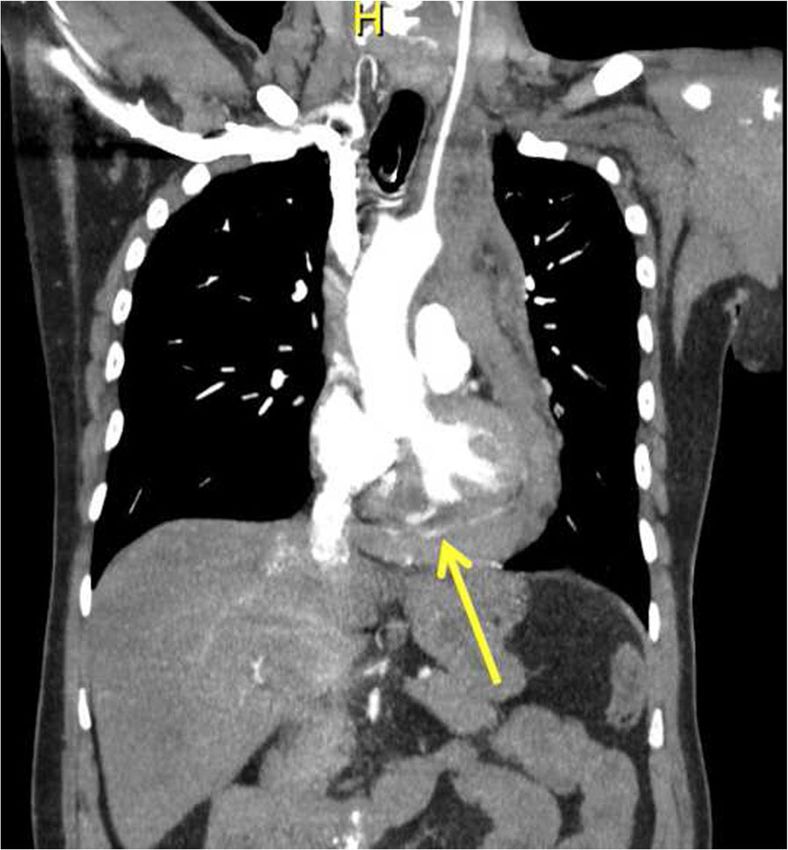
Nasr et al. Journal of Cardiothoracic Surgery    (2021) 16:106                                                                   Page 3 of 5

 Fig. 2 CXR on presentation revealing a widened mediastinum

catheterization lab for PCI at the time. In an effort to               alongside the pericardium was opened. One liter of
prevent further cardiac arrests, he was immediately                    blood was evacuated. Given ongoing blood loss, a large
taken to the operation room (OR) for further explor-                   catheter was directly placed into the right atrium for
ation and possible evacuation of the large pericardial                 transfusion and another catheter was directly placed into
effusion.                                                              the aorta to transduce blood pressure. The patient
  In the OR, the hemopericardium was evacuated                         underwent aggressive resuscitation with a max systolic
through an emergent subxiphoid incision and the chest                  blood pressure of 50 mmHg, and his left ventricle was
                                                                       noted to be empty throughout this time. On closer
                                                                       examination of the left ventricle, there was a large free
                                                                       wall rupture and surrounding fat pad hematoma on the
                                                                       diaphragmatic surface of the left ventricle, likely from
                                                                       the inferior STEMI he suffered. They also noted similar

 Fig. 3 Coronal view of Aortic CTA revealing contrast leakage in the    Fig. 4 Axial view of Aortic CTA demonstrating occlusion of the
 pericardium (yellow arrow) consistent with LVFWR                       proximal segment of the right coronary artery (yellow arrow)
Nasr et al. Journal of Cardiothoracic Surgery (2021) 16:106 Page 4 of 5
discoloration and a hematoma lateral to the left anterior CPR via intraoperative open thoracotomy cardiac mas-
descending (LAD) artery. The open free wall of the left sage (Video 1 A-B). After about another hour of
ventricle was attempted to be sutured to control the attempted resuscitation to control the bleeding, his sys-
bleeding, however, the myocardium was noted to be ex- tolic pressure was no longer able to be generated and
tremely fragile and could not hold the suture (Fig. 5). In- the patient eventually expired on the table.
traoperative transesophageal echocardiogram (TEE)
following evacuation of blood revealed inferior and sep- Discussion
tal wall hypokinesis, no evidence of a flap suggestive of The overall incidence of free wall rupture has dramatic-
aortic dissection, and multiple cardiac arrests requiring ally decreased with advancements and widespread use of
PCI [1]. Over half the deaths of LVFWR occur as an
out-of-the-hospital sudden death [7]. The mortality rate
of LVFWR is extremely high, estimated to be around
88.2% [8].
It is important to make note of mechanical complica-
tions of acute MI, such as LVFWR, when a patient pre-
sents as an out-of-hospital cardiac arrest. Although rare,
LVFWR is more common than other complications such
as papillary muscle or interventricular septal rupture [1].
Free wall rupture may occur sooner than anticipated
and a quick bedside POC ultrasound can help identify
the pericardial effusion to further guide management.
Although mortality is extremely high, attempted early
reperfusion and surgical management is the mainstay of
treatment.
The most common presentation of a free wall rupture
is an acute MI most often in the anterior and lateral re-
gion of the left ventricle with an associated pericardial
effusion, usually complicated by cardiac arrest. One of
the leading risk factors for LVFWR is absence of imme-
diate perfusion [1]. During the COVID-19 pandemic, it
has been demonstrated that the median time for STEMI
presentation from symptom onset to hospital arrival has
significantly increased from 2 h to 15 h [9]. This delay in
presentation is associated with an increased incidence of
post-MI mechanical complications during the COVID-
19 pandemic, as seen in two large observational studies
conducted in Italy and Germany [4, 6] and multiple case
reports in the U.S. [3, 5]. Given that the patient had been
complaining of chest pain for a few days and had patho-
logical q-waves in the inferior leads on his ECG, it is
likely that this dramatic presentation of free wall rupture
was due to delay in seeking medical care.
The COVID-19 pandemic has had a major impact on
the U.S. healthcare system and there have been multiple
reports of its’ influence on myocardial infarction presen-
tation. It has been shown that since the start of COVID-
19 lockdowns in March 2020, there has been a signifi-
cant 38% decrease in STEMI presentations in the U.S.
and a 48% decrease in hospitalization rate for myocardial
infarction [10, 11]. It is hypothesized that this is due in
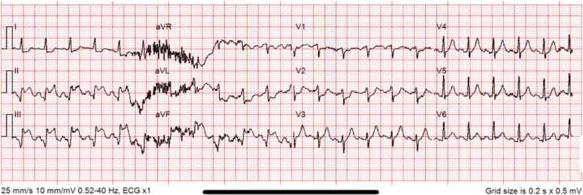
Fig. 5 OR view of attempted suture in the open free wall of the part to social distancing and patients avoiding hospital
diaphragmatic surface of the left ventricle (yellow arrow). Also, note settings in fear of contracting COVID-19. It is important
the bruising surrounding the friable myocardial around the inferior
for clinicians to consider mechanical complications of
free wall of the left ventricle (green arrow)
MI, such as LVFWR, when patients present as an out-Nasr et al. Journal of Cardiothoracic Surgery (2021) 16:106 Page 5 of 5
of-hospital cardiac arrest, particularly during the Competing interests
COVID-19 pandemic where there is often a delay of The authors declare that they have no competing interests to report.
presentation from initial onset of symptoms. Author details
1
Department of Medicine, University of California, Irvine, USA. 2Department
of Medicine, Division of Cardiology, University of California, Irvine, USA.
Conclusion 3
Department of Emergency Medicine, University of California, Irvine, USA.
LVFWR, albeit rare, occurs 1–4 days after suffering an 4
Department of Anesthesia & Perioperative Care, University of California,
MI. The incidence of LVFWR has decreased over the Irvine, USA. 5Department of Surgery, Division of Cardiothoracic Surgery,
University of California, Irvine, USA.
past decade with advancements in reperfusion therapy,
however, there has been an increased incidence of Received: 9 December 2020 Accepted: 9 April 2021
LVFWR during the COVID-19 pandemic as patients
delay seeking care over fear of contracting COVID-19
References
from within the medical setting. Clinicians should con- 1. Figueras J, Alcalde O, Barrabés J, et al. Changes in hospital mortality rates in
sider mechanical complications of MI when patients 425 patients with acute ST-elevation myocardial infarction and cardiac
rupture over a 30-year period. Circulation. 2008;118(25):2783–9. https://doi.
present as an out-of-hospital cardiac arrest, particularly
org/10.1161/CIRCULATIONAHA.108.776690.
during the COVID-19 pandemic. 2. Purcaro A, Costantini C, Ciampani N, Mazzanti M, Silenzi C, Gili A, et al.
Diagnostic criteria and management of subacute ventricular free wall
Abbreviations rupture complicating acute myocardial infarction. Am J Cardiol. 1997;80(4):
LVFWR: Left ventricular free wall rupture; MI: Myocardial infarction; STEMI: ST- 397–405. https://doi.org/10.1016/s0002-9149(97)00385-8.
segment elevation myocardial infarction; PCI: Percutaneous coronary 3. Otero D, Singam N, Barry N, Raheja P, Solankhi A, Solankhi N. Complications
intervention; POCUS: Point of care ultrasound; US: United States; of late presenting STEMI due to avoidance of medical care during the
ED: Emergency department; EMS: Emergency medical services; COVID-19 pandemic. JACC Case Rep. 2020;2(10):1610–3. https://doi.org/10.1
ECG: Electrocardiogram; CPR: Cardiopulmonary resuscitation; ROSC: Return of 016/j.jaccas.2020.05.045.
spontaneous circulation; EF: Ejection fraction; CTA: Computed tomography 4. De Rosa S, Spaccarotella C, Basso C, et al. Reduction in hospitalizations for
angiography; OR: Operation room; LAD: Left anterior descending; myocardial infarction in Italy in the COVID-19 era. Eur Heart J. 2020;41(22):
TEE: Transesophageal echocardiogram 2083–8. https://doi.org/10.1093/eurheartj/ehaa409.
5. Ahmed T, Nautiyal A, Kapadia S, Nissen S. Delayed presentation of STEMI
complicated by ventricular septal rupture in the era of the COVID-19
Supplementary Information pandemic. JACC Case Rep. 2020;2(10):1599–602. https://doi.org/10.1016/j.ja
The online version contains supplementary material available at https://doi. ccas.2020.05.089.
org/10.1186/s13019-021-01495-x. 6. Primessnig U, Pieske B, Sherif M. Increased mortality and worse cardiac
outcome of acute myocardial infarction during the early COVID-19
Video 1A. TEE transgastric short axis view (left) and transgastric long axis pandemic. ESC Heart Fail. 2021;8(1):333–43. https://doi.org/10.1002/ehf2.13
view (right) showing hypokinetic inferior wall (yellow arrow) 075.
7. Becker R, Gore J, Lambrew C, et al. A composite view of cardiac rupture in
Video 1B. TEE midesophageal four chamber view showing attempted
the United States National Registry of myocardial infarction. J Am Coll
CPR via intraoperative cardiac massage
Cardiol. 1996;27(6):1321–6. https://doi.org/10.1016/0735-1097(96)00008-3.
8. Nozoe M, Sakamoto T, Taguchi E, Miyamoto S, Fukunaga T, Nakao K. Clinical
Acknowledgements manifestation of early phase left ventricular rupture complicating acute
Not Applicable. myocardial infarction in the primary PCI era. J Cardiol. 2014;63(1):14–8.
https://doi.org/10.1016/j.jjcc.2013.06.012.
Authors’ contributions 9. Gramegna M, Baldetti L, Beneduce A, Pannone L, Falasconi G, Calvo F, et al.
GN, DG, AM, KC, HP, MJ, BC, and AE were all involved in the management of ST-segment-elevation myocardial infarction during covid-19 pandemic.
the patient and participated in the writing of the paper. SS (Singh) and FS Insights from a regional public health service healthcare hub. Circ
were involved in the operative management of the patient and in the Cardiovasc Interv. 2020;13(8):9413. https://doi.org/10.1161/
writing of the paper. SS (Sinfield) participated in the performance of the CIRCINTERVENTIONS.120.009413.
substantive revise of the work. All authors read and approved the final 10. Garcia S, Albaghdadi M, Meraj P, et al. Reduction in ST- segment elevation
manuscript and have agreed both to be personally accountable for the cardiac catheter laboratory activations in the United States during COVID-19
author’s own contributions and to ensure that questions related to the pandemic. J Am Coll Cardiol. 2020;75(22):2871–2. https://doi.org/10.1016/j.ja
accuracy or integrity of any part of the work, even ones in which the author cc.2020.04.011.
was not personally involved, are appropriately investigated, resolved, and the 11. Solomon M, McNulty E, Rana J, et al. The Covid-19 pandemic and the
resolution documented in the literature. incidence of acute myocardial infarction. N Engl J Med. 2020;383(7):691–3.
https://doi.org/10.1056/NEJMc2015630.
Funding
The authors declare that there is no funding to report. Publisher’s Note
Springer Nature remains neutral with regard to jurisdictional claims in
Availability of data and materials published maps and institutional affiliations.
Data sharing is not applicable to this case report as no datasets were
generated or analyzed in our article.
Declarations
Ethics approval and consent to participate
Not Applicable.
Consent for publication
Informed consent for publication was obtained.You can also read