Looking into live cells with in-cell NMR spectroscopy
←
→
Page content transcription
If your browser does not render page correctly, please read the page content below
Journal of
                                                                                                                             Structural
                                                                                                                              Biology
                                              Journal of Structural Biology 158 (2007) 244–253
                                                                                                                      www.elsevier.com/locate/yjsbi

                                                                      Review

                  Looking into live cells with in-cell NMR spectroscopy
                                               Philipp Selenko *, Gerhard Wagner
Harvard Medical School, Department of Biological Chemistry and Molecular Pharmacology (BCMP), 240 Longwood Avenue, Boston, MA 02115, USA

                                 Received 24 May 2006; received in revised form 2 April 2007; accepted 3 April 2007
                                                          Available online 10 April 2007

Abstract

    In-cell NMR spectroscopy has gained recent popularity since it provides means to analyze the conformational and functional properties
of proteins inside living cells and at atomic resolution. High-resolution in-cell NMR spectroscopy was originally established in bacterial cells
and based on a rationale that relies on protein over-expression and sample analysis within the same cellular environment. Here, we review in-
cell NMR approaches in Xenopus laevis oocytes and evaluate potential future applications in other eukaryotic cell types.
Ó 2007 Elsevier Inc. All rights reserved.

Keywords: High-resolution NMR spectroscopy; In-cell NMR spectroscopy; Protein transduction; Xenopus laevis oocytes

1. High-resolution in-cell NMR spectroscopy                                  high-resolution snapshots of intracellular protein confor-
                                                                             mations. These structural ‘fingerprints’ may change upon
   Biophysical methods for the structural characterization                   biological interactions, post-translational protein modifica-
of biomolecules are often confined to artificial, in vitro                     tions or due to structural rearrangements. In-cell NMR
experimental setups. X-ray crystallography and high-reso-                    measurements then detect these biological events and read
lution electron microscopy are intrinsically restricted from                 out residue specific changes in a time dependent- and quan-
in vivo approaches due to their requirement for pure sam-                    titative manner. Why would these measurements be of any
ples and crystalline or vitrified specimens. Nuclear mag-                     additional value compared to conventional analyses by
netic resonance (NMR) spectroscopy, the only other                           in vitro methods? First, most proteins function inside cells
method for structural investigations at the atomic level,                    and in a highly crowded, viscous solution that harbors an
allows for the direct observation of NMR-active nuclei                       intricate network of biological activities simultaneously
within any NMR-inactive environment and can thus be                          exerted by a large number of macromolecules. Whereas
employed to analyze biomolecules in vivo and inside cells                    in vitro structural analyses on pure samples have shaped
(Serber et al., 2005). Historically, small molecule in vivo                  our 3-dimensional understanding of many biological pro-
NMR spectroscopy denotes the observation of metabolites                      cesses, they do not necessarily reflect the true nature of
in suspensions of bacteria and other cells by means of                       the cellular environment. What are the biological questions
investigating a few characteristic proton or phosphor reso-                  that can be addressed by in-cell NMR techniques? Does an
nance signals in 1-dimensional NMR spectra (Cohen et al.,                    in vitro determined protein structure represent the cellular
1989; Szwergold, 1992). In-cell NMR spectroscopy, in con-                    in vivo conformation? How do proteins that do not exhibit
trast, employs modern methods of isotope labeling and                        folded properties in their pure states behave in a cellular
multi-dimensional, isotope-edited correlation experiments                    environment? How do in vitro investigated conformational
to obtain structural information on proteins within living                   changes, upon ligand binding for example, relate to
cells (Reckel et al., 2005). In brief, this method enables                   equivalent structural alterations in vivo? How do post-
                                                                             translational protein modifications affect protein structure
 *
     Corresponding author.                                                   and in what ways does a protein respond structurally to
     E-mail address: philipp_selenko@hms.harvard.edu (P. Selenko).           cellular processes like apoptosis, cell cycle progression or

1047-8477/$ - see front matter Ó 2007 Elsevier Inc. All rights reserved.
doi:10.1016/j.jsb.2007.04.001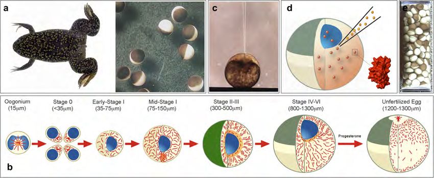
P. Selenko, G. Wagner / Journal of Structural Biology 158 (2007) 244–253                        245

differentiation? Clearly, these are important issues that can         lution NMR spectra of the intracellular recombinant
only be adequately addressed in a cellular setting and by a          protein (Fig. 1a). Alternatively, labeled protein samples
high-resolution method. Second, biological reactions often           can be conventionally purified from bacteria and then
involve the dynamic modulation of a protein’s activity. In           transferred into other cells for in-cell NMR analyses
eukaryotes, this is often achieved by reversible post-trans-         (Fig. 1b). For eukaryotic in-cell NMR applications in
lational modifications of one, or many amino acid residues            Xenopus laevis oocytes, for example, labeled proteins are
in a protein. Such modifications involve the exchange of              deposited by microinjection. It is evident that the same
various functional groups in highly regulated processes              underlying principle constitutes the experimental feasibility
that are typically controlled by intrinsic cellular mecha-           of both of these methods.
nisms. Moreover, these alterations often result in substan-
tial conformational rearrangements, which lead to specific            3. NMR parameters for in-cell NMR experiments
modulations in a protein’s function or activity. Eukaryotic
in-cell NMR techniques in particular enable the direct                   In the following paragraph we will briefly outline impor-
observation of these cellular reactions, and of their struc-         tant NMR parameters that are routinely employed for the
tural implications, and can thus provide important new               qualitative and quantitative interpretation of in-cell, and
insights into the biological behavior of a protein.                  other, NMR spectra.
   In the following chapters we will introduce the basic                 Two-dimensional correlation experiments of 1H and
                                                                     15
concepts of NMR experiments inside living cells, provide                N, or 13C, serve as the primary in-cell NMR techniques.
a brief introduction to NMR properties of labeled proteins           The correlation of these NMR-active atomic nuclei, by
in a cellular environment, review in-cell NMR approaches             means of specifically tailored NMR pulse-sequences, yields
in bacterial and eukaryotic cells and outline future in-cell         individual NMR signals (or resonance cross-peaks) at the
NMR applications.                                                    respective resonance frequencies (or chemical shift values,
                                                                     d[1H], d[15N] and/or d[13C]) (Fig. 2a). For a folded protein,
2. Basic concepts                                                    the resonance frequencies of individual residues are deter-
                                                                     mined by the amino acid specific chemical environments,
   Today, many in-cell NMR applications employ bacte-                which in turn are defined by the protein’s 3-dimensional
rial cells and follow the experimental rationale proposed            structure. This leads to characteristic patterns of resonance
by Serber et al. (2001). Their approach exploits the fact            cross-peaks in 2-dimensional correlation spectra, which
that most atomic nuclei in natural substances are NMR-               reflect the overall conformational state of the labeled pro-
inactive and hence not directly observable by NMR                    tein. Local changes in the chemical environment of labeled
spectroscopy. Modern NMR methods utilize recombinant                 residues, either by ligand binding or conformational rear-
protein over-expression and isotope labeling to substitute           rangements, result in differences in resonance frequencies
some of these inactive nuclei (14N, 12C) with NMR-active             and changes in chemical shift values (Fig. 2b). The overall
isotopes (15N, 13C). Multi-dimensional correlation experi-           number of resonance peaks remains the same because both
ments then allow the detection of pairs of coupled nuclei            reactions do not introduce additional observable spin-
in labeled proteins, such as 1H and 15N or 1H and 13C, in            pairs. Differences in resonance frequencies of participating
any aqueous environments that do not contain significant              residues are measured as chemical shift changes (Ddtotal =
amounts of such isotope pairs. In essence, isotope labeling          Dd[1H] + Dd[15N or 13C]) and mapped onto the 3-dimen-
functions as a selective filter that renders the NMR-inactive         sional protein structure. Chemical shift changes serve as
cellular environment invisible to spectroscopic eye. In that         unique indicators of conformational alterations or localized
sense, the underlying principle of in-cell NMR spectros-             binding events both in vitro and in vivo (Zuiderweg, 2002).
copy is very similar to applications in cellular microscopy              Second, the characteristic appearance of each NMR
that employ fluorescent labeled proteins.                             signal contains additional information about the spin
   Isotope labeling involves recombinant protein produc-             system under investigation (Fig. 3). A prerequisite for
tion in growth media that provide isotope-substituted met-           liquid state NMR spectroscopy is that the molecule of
abolic precursors (15N-ammonium chloride, 13C-glucose).              interest tumbles freely in solution. The resulting overall
Due to the high protein expression levels that are typically         tumbling rate depends on the size of the molecule and
achieved with viral promoters and polymerases in Esche-              the temperature and viscosity of the NMR sample solu-
richia coli, recombinant polypeptides accumulate rapidly             tion. These parameters determine the overall line widths
post-induction and generally outperform protein synthesis            of the respective NMR resonance signals. Yet, NMR res-
rates of endogenous E. coli gene products. When, in addi-            onances from one protein do not all exhibit identical
tion, bacterial cells are grown in unlabeled medium first             peak intensities or uniform NMR line widths. Additional
and only switched to labeled growth conditions before                parameters like internal residue mobility, or solvent and
the induction of recombinant protein expression, selective           conformational exchange, differentially affect the relaxa-
isotope labeling is restricted to the recombinant protein            tion properties of individual spins and hence the appear-
only. Hence, and without further purification, isotope-edi-           ance of the respective NMR signals (Fischer et al., 1998;
ted correlation experiments on intact cells yield high-reso-         Palmer, 2001; Peng and Wagner, 1994). Amino acids of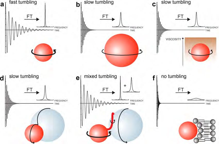
246                                    P. Selenko, G. Wagner / Journal of Structural Biology 158 (2007) 244–253

Fig. 1. (a) Prokaryotic in-cell NMR approaches typically employ recombinant protein over-expression, isotope labeling and in-cell NMR analyses within
the same cell type. Suspensions of bacterial cells are directly analyzed without purification of the recombinant protein. (b) Eukaryotic in-cell NMR
applications can involve isotope labeling in bacterial cells and recombinant protein purification prior to in-cell NMR sample preparation. Labeled proteins
are then transferred into eukaryotic cells by microinjection, or other vector-based transduction techniques.

unstructured loop regions typically display narrower and                       rates that correspond to the sum of their individual
more intense resonance signals than residues in second-                        masses (Fig. 3d), or individual contributions to a mixed
ary structure elements. These dynamic relaxation proper-                       set of rates, when the interaction is restricted to a subset
ties may undergo differential alterations in a cellular                         of residues (Fig. 3e). The latter results in residue specific
environment and the comparative analysis of changes                            line broadening, which yields information on the dynam-
in NMR line widths can therefore provide information                           ics and localization of the cellular interaction. Binding to
about in vivo dynamics and exchange behaviors. In gen-                         quasi-static cellular structures like organelles or mem-
eral, small proteins display large tumbling rates, which                       branes results in severe line broadening (Fig. 3f), which
lead to slow overall relaxation and narrow NMR line                            can serve as a qualitative indicator for the kind of inter-
widths (Fig. 3a). Molecules of larger size tumble more                         action. Many biological binding events are dynamic and
slowly, relax faster and exhibit broader resonance signals                     modulated by cellular signaling, which often leads to
(Fig. 3b). Because the overall rotational tumbling rate is                     transient and interpretable changes of NMR line widths.
a direct function of the viscosity of the medium in which                      It is apparent that a biologically active protein can expe-
the macromolecule is dissolved, intracellular viscosity                        rience any of the aforementioned conditions, and super-
becomes a crucial parameter for in-cell NMR experi-                            positions thereof, in a cellular environment. Complicated
ments. Any molecule in a cellular environment exhibits                         or poor quality in-cell NMR spectra are the likely result.
a reduced tumbling rate due to intracellular viscosity                         In such cases, the researcher needs to reduce the com-
and hence displays broader NMR line widths (Fig. 3c).                          plexity of the system under investigation, which can be
In the absence of protein binding to endogenous cellular                       achieved either by ‘chopping up’ full-length proteins into
factors, a direct comparison of individual protein line                        individual domains in order to selectively probe differen-
widths in buffer and in in-cell NMR experiments will                            tial biological activities or by introducing site-directed
readily yield a qualitative estimate about intracellular vis-                  mutations that abolish certain functional characteristics
cosity. Upon sample binding to cellular components, the                        and similarly enable one to discriminate between specific
resulting protein complexes can either display tumbling                        cellular contributions.
P. Selenko, G. Wagner / Journal of Structural Biology 158 (2007) 244–253                                      247

                                                                                                                                   ppm

                                                    X                                                        X
                                                1
                                                H        15
                                                              N

                                                                                   R                             Y                 δNy
                                                                           Y
                                                                               1
                                                                  15
                                                                       N       H

                                                    Z                                                  Z
                                                                                                                                    15
                                                1
                                                H        15
                                                              N                                                                          N
                                                                                       ppm                               δHy
                                                                                                                               1
                                                                                                                               H

                                                                                                                                   ppm

                     1
                         H
                                                    X’                                                       X
                                                                                                           ΔδN
                                                1
                                                H        15
                                                              N

                                                                                   R             ΔδH             Y                 δNy
                                       H
                                       1                                                X’
                                                                           Y
                                                                               1
                                                                  15
                                                                       N       H                       Z’
                                                                                             Z
                     1
                      H                                                                                              N
                                                    Z’                                                 ΔδH
                                                H
                                                1        15
                                                              N                                                                     15
                                                                                                                                         N
                                                                                       ppm                               δHy
                                                                                                                               1
                                                                                                                               H
Fig. 2. (a) Schematic representation of a 2-dimensional 1H, 15N isotope-edited NMR spectrum. Backbone cross-peaks of amide spin-pairs give rise to a
characteristic pattern of NMR resonance signals. The chemical shift (d) of these peaks depends on the environment of the labeled spin-pairs, which is
defined by their structural context. (b) Upon ligand binding, a limited set of amide spin-pairs experience a novel chemical environment, which results in
selective chemical shift changes (Dd). Since those changes are more pronounced for residues directly involved in binding, their mapping onto the 3-
dimensional structure of the protein identifies the interaction surface.

   In summary, a thorough and comparative analysis of                                  of these prokaryotic in-cell NMR applications have been
NMR chemical shift values and NMR line widths of labeled                               excellently reviewed in recent publications (Reckel et al.,
protein samples in different in vitro and in vivo environments                          2005; Serber et al., 2005). We will not further elaborate
can yield a wealth of structural and dynamic information                               on these bacterial studies but restrict ourselves, for the
about a protein’s cellular behavior and about its potential                            remainder of the manuscript, to outline the methodological
interactions with endogenous cellular components.                                      considerations for in-cell NMR applications in eukaryotic
                                                                                       cells.
4. In-cell NMR spectroscopy in prokaryotic cells
                                                                                       5. In-cell NMR spectroscopy in eukaryotic cells
   Several in-cell NMR applications for structural and
functional studies of proteins in bacteria have been                                      Why do we wish to extend the applicability of in-cell
reported (Dedmon et al., 2002; Hubbard et al., 2003; McN-                              NMR measurements to eukaryotic cells? Above all, pro-
ulty et al., 2006; Serber et al., 2004; Wieruszeski et al.,                            karyotic organisms exhibit a limited range of biological
2001). Bacterial in-cell NMR techniques have been success-                             activities and many of the cellular processes that define
fully employed to analyze protein dynamics (Bryant, 2006;                              the prevalent topics in modern biological research are
Bryant et al., 2005), protein–protein interactions (Burz                               absent in bacteria. Post-translational protein modifica-
et al., 2006), for de-novo resonance assignments (Reardon                              tions, for example, serve to ubiquitously regulate biological
and Spicer, 2005) and automated structure determinations                               activities in eukaryotes, but are much less common in pro-
in crude cell extracts (Etezady-Esfarjani et al., 2006). Some                          karyotic organisms. The presence of organelles and the
248 P. Selenko, G. Wagner / Journal of Structural Biology 158 (2007) 244–253
Fig. 3. (a) Small molecules display large rates of molecular tumbling, indicated by a thick arrow. This results in a slow decay of the experimentally
observed time domain NMR signal (the free induction decay, or FID). Upon Fourier transformation (FT) of the time-domain FID signal, the frequency-
domain NMR signal displays the characteristic Lorentzian line shape. (b) Large molecules tumble more slowly, indicated by a thin arrow, which results in
fast relaxation rates and broad NMR line widths with reduced overall signal intensities. (c) An increase in viscosity of the sample solution results in an
overall reduction in tumbling rates. Under these circumstances small molecules display relaxation properties, and hence NMR line widths, comparable to
those of larger molecules in low viscosity environments. (d) Intermolecular interactions with components of larger size yield relaxation rates that
correspond to the overall molecular weight of the complex (typically the sum of the individual rates). (e) Interactions with subsets of residues of the labeled
molecule result in a mixed set of relaxation rates. These may yield non-uniform degrees of line broadening. (f) Interactions with immobile cellular
structures, like membranes, effectively abolish molecular tumbling of the labeled molecule. Relaxation rates become so large that the resulting frequency
domain signals are virtually undetectable.
concomitant requirement of regulated cellular transport nale, the manipulation of X. laevis oocytes by microinjec-
constitutes another characteristic of eukaryotic cell iden- tion represents a powerful alternative approach. Before
tity. Compartmentalization per se results in the creation we outline the experimental conditions for both methods,
of sub-cellular environments with different physical and we will address some of the conceptual differences behind
biological properties and little is known about the effects these two techniques.
that these compartments exert on a protein’s structure or Protein over-expression and in-cell NMR measurements
function. Moreover, eukaryotic organisms display a high within the same cell type do not require purification of the
degree of cellular differentiation, which leads to functional labeled polypeptide, which can be beneficial when a protein
specification and differences in biological activities in is difficult to isolate or complicated to stabilize in its pure
neighboring cell types. Do these specialized cellular envi- form. On the other hand, in-cell protein production cannot
ronments differentially affect protein structure? Some pro- be easily quantified or reproduced under exactly identical
tein examples indeed suggest so (Dyson and Wright, cellular conditions. Therefore, in-cell NMR analyses in
2005). Together, we believe that eukaryotic model systems bacteria, for example, are of qualitative nature. At low lev-
for in-cell NMR measurements will prove instrumental in els of recombinant protein over-expression, other cellular
structurally addressing a multitude of fundamental biolog- components become increasingly labeled, which results in
ical activities that are present in higher organisms. the generation of signal artefacts that have to be actively
What constitutes a suitable eukaryotic in-cell NMR sys- suppressed (Rajagopalan et al., 2004). These adverse effects
tem? First, we have to decide on the rationale for introduc- are absent when a labeled compound is introduced into the
ing labeled protein samples into the native, intracellular cellular environment at defined concentrations, as can be
environment. We could either choose the original approach achieved by microinjection, for example. This approach,
of sample over-expression and analysis within the same cell however, is restricted to a few eukaryotic cell types, which
type, or turn towards intracellular sample delivery by alter- can be manipulated in such a way, and requires the labeled
native methods. Whereas recombinant protein over-expres- protein samples to be sufficiently soluble at high concentra-
sion in yeast-, insect- or mammalian cells would represent a tions, since the maximum injection volume per single cell is
‘eukaryotic solution’ along the originally proposed ratio- typically on the order of nano-liters.P. Selenko, G. Wagner / Journal of Structural Biology 158 (2007) 244–253 249
Alternatively, one could envisage intracellular sample which is likely to increase the amount of background label-
delivery by means of cell-permeable synthetic vectors. We ing artefacts.
are particularly intrigued by the potential application of In summary, we believe that in-cell NMR measurements
‘Trojan’ peptide tags, which confer efficient cell membrane in these eukaryotic cells will be technically feasible but are
transduction activities to a wide range of fused protein sub- probably not going to constitute practicable routine
strates (Derossi et al., 1998; Dietz and Bahr, 2004). These approaches. Some proteins may require expression within
internalization peptides are composed of short, positively a eukaryotic cellular environment in order to properly fold
charged amino acid residues, which can be genetically engi- or to express in a soluble form. For these cases, in-cell
neered to be part of virtually any recombinant polypeptide NMR experiments in yeast, insect or mammalian cells
(Li et al., 2002). Upon labeled expression and purification may provide valuable insights into the structural mecha-
of tagged fusion proteins, these substrates are simply added nisms of protein folding or could enable the production
to the growth medium of a variety of cultured laboratory of labeled, functional proteins for the delivery into other
cell lines and readily internalized. In theory, this method cells.
should be generally applicable to a wide range of eukary-
otic cells and quantitatively accomplishable for a large 5.2. In-cell NMR spectroscopy in X. laevis oocytes
number of cells. The method of choice for eukaryotic in-
cell NMR measurements will hence be dictated by both We, and others, have recently reported the usage of X.
the suitability of the protein of interest for either approach laevis oocytes for eukaryotic in-cell NMR measurements
and by the overall biological question to be addressed. (Selenko et al., 2006; Serber et al., 2006; Sakai et al.,
2006). These amphibian cells have long served as important
laboratory tools in the disciplines of developmental and
5.1. In-cell NMR spectroscopy in yeast-, insect- and cellular biology (Fig. 4a) (Liu, 2006; Murray, 1991b).
mammalian cells Mature oocyte cells (stage VI) arrest in prophase at the
G2/M boundary of the first meiotic division (Fig. 4b)
Recent advances in structural genomics have also led to and contain large cell volumes (1lL, compared to a few
a more thorough investigation of possible alternatives to pL as for most somatic cells), 20% of which comprises
bacterial recombinant protein expression and isotope label- the nuclear organelle (or germinal vesicle). During oocyte
ing (Goto and Kay, 2000; Yokoyama, 2003). Amongst to egg maturation, a hormonal trigger activates synchro-
these, a few exotic approaches in mammalian CHO and nized cell cycle progression into metaphase of meiosis II.
HEK cells have been reported for NMR sample prepara- For isolated oocytes in an in vitro setting, these events
tions (Hansen et al., 1992; Lustbader et al., 1996; Wyss can be executed by the external addition of hormones,
et al., 1995). More prominent systems include the yeast which renders this system an important laboratory tool
Pichia pastoris and Baculovirus-infected insect cells (Chen for studying signaling events during cell cycle progression.
et al., 2006; Pickford and O’Leary, 2004; Strauss et al., Cellular extracts from Xenopus oocytes or from Xenopus
2003, 2005). All of these eukaryotic cells have been eggs are easily obtained in a virtually undiluted form and
employed to prepare labeled NMR samples for in vitro similarily recapitulate most of the intact cells’ biological
analyses and we can therefore only speculate about their activities. They are frequently used as alternative, cell-free
experimental suitability for in-cell NMR measurements. systems for ex vivo analyses of cellular processes (Crane
The major obstacles for the selective labeling of recombi- and Ruderman, 2006; Murray, 1991a).
nant proteins with NMR-active isotopes in eukaryotic cells Stage VI oocytes are conveniently manipulated by
have been the difficulty to achieve adequate levels of pro- microinjection, which permits the direct deposition of
tein over-expression, sufficient isotope incorporation and defined quantities of exogenous compounds into the cellu-
the costs of isotope-enriched growth media. Growth media lar environment (Fig. 4c). Sophisticated setups and proto-
for NMR labeling in E. coli are simple in their composi- cols for manipulating Xenopus oocytes have been devised
tion, easily prepared and, depending on the type of label- over the years, which include a fully automated injection
ing, relatively cheap. Bacteria will also incorporate setup (Schnizler et al., 2003). We use this robotic device
isotopes with high efficiency (98%). Labeling media for to routinely introduce labeled protein samples into large
eukaryotic cells are sophisticated, they must often be numbers of Xenopus oocytes for in-cell NMR measure-
obtained commercially for satisfactory results, and they ments (Fig. 4d). About 200 manipulated oocytes are
are expensive. The yeast P. pastoris represents an exception required for an in-cell NMR sample, which corresponds
to this notion since cells can be grown in glycerol/glucose to >250 lL in sample volume and, inside a Shigemiä
and labeled in 15N-ammoniumchloride and 13C-methanol NMR tube, will suffice to span transmitter- and receiver-
(Wood and Komives, 1999). Due to the complexity of most coil extensions of most commercial NMR spectrometer
eukaryotic metabolisms, isotope incorporation in the probes (Fig. 4e). Considering the small injection volume
above cells is typically less than 90%. With regard to in-cell per cell (50 nL), an oocyte sample requires only about
NMR measurements, induction times for recombinant pro- 10 lL of labeled protein. The necessary concentration of
tein expression are on the order of days rather than hours, injected protein for the minimally sufficient experimental250 P. Selenko, G. Wagner / Journal of Structural Biology 158 (2007) 244–253
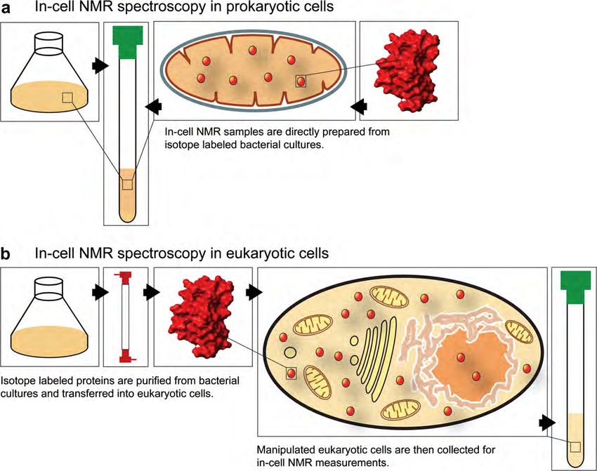
Fig. 4. (a) Oocyte cells from Xenopus laevis are surgically removed from the ovary lobes of female frogs. Stage VI oocytes are sorted and collected for
microinjections. (b) An overview of the oocyte maturation process and oocyte to egg transition is depicted. The developmental stages and average cell sizes
are indicated. Note that stage VI oocytes are interphase cells at the G2/M transition and contain an intact nuclear organelle (or germinal vesicle, shown in
blue). Upon progesterone treatment, oocytes mature into eggs, which are arrested in metaphase of meiosis II. The rearrangement of cellular microtubules
is shown in red. (Figure courtesy of David L. Gard, University of Utah). (c) Cell injections with pulled glass capillaries enable the quantitative deposition
of exogenous proteins into the intracellular environment of Xenopus oocytes. (d) For in-cell NMR applications in Xenopus oocytes, the labeled protein
sample is introduced into the unlabeled intracellular environment of intact oocyte cells by this approach. (e) The resulting in-cell NMR sample consists of
200 oocytes settled in a Shigemiä NMR tube.
NMR signal has to be evaluated empirically for each sam- Fig. 5a shows the characteristic appearance of 1-dimen-
ple. In addition, the protein’s size, expected cellular activ- sional proton-only NMR spectra (no isotope-edited corre-
ity, and type of labeling, will determine the minimally lation ‘filter’) of a 15N-labeled protein sample in its pure
sufficient amount of labeled compound. Injection concen- form (top panel), resuspended in crude Xenopus egg extract
trations are typically in the range of 0.5–3.0 mM. These (middle panel) or upon oocyte injection (bottom panel). It
quantities might not be achievable for all recombinant pro- is apparent that by conventionally recording all NMR sig-
tein samples nor generally suffice for a satisfying experi- nals from 1H nuclei only, these spectra do not allow to dis-
mental readout, especially for biomolecules with criminate between resonances from endogenous, cellular
promiscuous binding activities towards endogenous cellu- components and the ones from the labeled compound.
lar components, and within a reasonable amount of exper- Moreover, it is evident that the quality of in-cell NMR
imental time. It is evident that a compromise between the experiments, recorded in this mode, is too poor to provide
experimentally achievable signal-to-noise, the duration of any information on the injected protein sample. When,
individual NMR experiments, the cellular concentration however, these same samples are measured with the appli-
of labeled proteins and its physiological relevance, will cation of a 1H, 15N correlation pulse-sequence, similar 1-
have to be found if in-cell NMR measurements in X. laevis dimensional traces selectively display NMR resonances
oocytes are to yield biologically meaningful results. from the labeled protein only. The good quality of these
We also conduct NMR experiments in X. laevis oocyte background-suppressed spectra enables the selective detec-
and egg extracts, and find that in these ‘cellular’ settings tion of protein NMR signals under extract and in-cell con-
the overall spectral quality is generally better due to the ditions (Fig. 5b). Changes in NMR line widths of
homogenous nature of the sample solutions. Concentra- individual resonance signals in these different aqueous solu-
tions of labeled specimens in the lower micro-molar range tions are readily visible (Fig. 3). Clearly, these spectra dem-
(10–50 lM) typically yield interpretable 2-dimensional cor- onstrate the great potential of isotope-edited correlation
relation spectra of satisfying quality. Additionally, sample techniques and the experimental feasibility of high-resolu-
dilutions upon extract suspension are lower than for oocyte tion NMR experiments in Xenopus egg extracts and oocyte
injections. Most biological reactions are executed with cells.
comparable activities in Xenopus cell extracts and can eas- We have recently delineated the experimental reference
ily be modulated by the addition of small molecule inhibi- conditions for in-cell NMR measurements in X. laevis
tors or activators. These extracts can also be selectively oocytes and provided a detailed structural analysis of the
depleted of certain cellular components, and replenished conformational in vivo properties of a small, biologically
with labeled, NMR-active substitutes, which enables the inert protein domain (Selenko et al., 2006). Whereas our
isolated investigation of biological processes without com- sample did not engage in biological interactions in these
promising the qualitative nature of the experimental read- cells, a similar approach by Sakai et al., presented experi-
out by residual endogenous activities. A more thorough mental evidence for the cellular in vivo characteristics of
description of experimental procedures is given in (Selenko ubiquitin, another small, but highly abundant protein with
et al., 2006; Serber et al., 2006) a large array of cellular functions (Sakai et al., 2006).P. Selenko, G. Wagner / Journal of Structural Biology 158 (2007) 244–253 251
1D proton NMR spectrum 1D proton/nitrogen correlation spectrum
buffer buffer
cell cell
extract extract
Scaled down by
a factor of 2
intact intact
cells cells
Scaled down by
a factor of 4
11.0 10.0 9.0 8.0 7.0 6.0 ppm 11.0 10.0 9.0 8.0 7.0 6.0 ppm
Fig. 5. (a) The amide/aromatic region (5–11 ppm) of a 1D proton spectrum of the Streptococcal protein GB1 (GB1) domain recorded with a standard 1H
NMR experiment. Solvent suppression was achieved by a Watergate NMR pulse-sequence, the experiment was recorded at 295 K, with 1024 points and
128 scans. The sample concentration was 0.5 mM of 15N-labeled GB1 (No 15N decoupling was employed). Individual panels display the respective NMR
spectra in buffer (top), in crude Xenopus egg extracts (middle) and upon oocyte injection (bottom). Scaling was performed as indicated. (b) Identical
samples as in (a) but recorded as 1D 1H(15N) hetero-nuclear single quantum coherence (HSQC) correlation spectra, using a Watergate version of a
standard, sensitivity-enhanced HSQC pulse-sequence with the same number of scans as in (a). No scaling was applied to the experimental data. Some
corresponding resonance frequencies are indicated as lines at the respective chemical shift values.
Indeed, given the multitude of cellular binding partners for be regarded as discouraging, even poor quality in-cell
ubiquitin in Xenopus oocytes, in-cell NMR experiments for NMR experiments provide a wealth of information about
the wild-type protein yielded poor quality NMR spectra. the in vivo biological activity of a given protein and can still
Only upon mutating conserved binding sites of known serve as valuable functional assays. In-cell NMR measure-
interaction surfaces, did ubiquitin provide interpretable ments in combination with mutant screening approaches,
in-cell NMR data. This work thus serves a fine example for example, can identify protein residues critically
for what kind of biological studies will be amenable to required for in vivo binding even if the nature of the biolog-
in-cell NMR approaches. Once cellular proteins engage ical activity that causes NMR signal deterioration is
the labeled specimen in too many generic interactions, or unknown. If, on the other hand, an expected cellular activ-
scavenge the NMR-active protein into complexes of molec- ity is highly selective, or engages the labeled protein of
ular weights too large for detection by conventional solu- interest in a unique form of interaction, in-cell NMR
tion-state NMR methods, the envisaged in-cell NMR experiments will succeed in providing a wealth of structural
approach is likely to fail. In most instances, such unfavor- and functional insights. In the following chapter, we out-
able binding behaviors are easily pre-assessed by NMR line two biological areas of eukaryotic in-cell NMR
experiments in cellular extracts (Serber et al., 2006). None- research that we actively and successfully pursue in our
theless, and despite the fact that these considerations might laboratory.252 P. Selenko, G. Wagner / Journal of Structural Biology 158 (2007) 244–253
6. Future applications the in situ observation of the establishment of these modi-
fications in a time-dependent and residue-specific manner.
6.1. In-cell NMR analyses of intrinsically disordered proteins We have recently confirmed that these in vivo approaches
can indeed resolve cellular phosphorylation reactions in
Intrinsically disordered proteins (IDPs) represent a X. laevis oocytes (Selenko et al., submitted). They point
growing class of gene products (Dyson and Wright, to a plethora of possible in-cell NMR applications in
2005), which are characterized by lack of secondary and/ eukaryotic post-translational protein modification
or tertiary structure in their pure forms and at physiologi- research.
cal pH (Uversky, 2002). IDPs are estimated to account for
20% of all human proteins (Dunker et al., 2000) and exert Acknowledgments
important functions in key cellular processes (Dunker
et al., 2002). A significant number of IDPs are implicated We thank Matthew Dedmon for carefully reading the
in human protein deposition diseases, in which a normally manuscript. P.S. is a Human Science Frontier Program
soluble polypeptide forms insoluble aggregates in a subset Organization (HSFPO) long-term fellow (LT00686/2004-
of cells and precipitates in the form of amyolid fibrils C). This work was additionally supported by a National
(Fink, 2005). Little is known about the general 3-dimen- Institute of Health Grant GM47467 to G.W.
sional conformation of IDPs in cellular environments and
it is tempting to speculate whether unfolded protein con-
References
formations are preserved under native, intracellular condi-
tions. Could it be possible that some IDPs are not per se Bryant, J.E., 2006. In-cell protein dynamics. Mol. Biosyst. 2, 406–410.
structurally deficient protein entities but that their Bryant, J.E., Lecomte, J.T., Lee, A.L., Young, G.B., Pielak, G.J., 2005.
unfolded state results as a consequence of the isolated Protein dynamics in living cells. Biochemistry 44, 9275–9279.
in vitro experimental setup employed for their characteriza- Burz, D.S., Dutta, K., Cowburn, D., Shekhtman, A., 2006. Mapping
structural interactions using in-cell NMR spectroscopy (STINT-
tion? In-cell NMR experiments on the natively unfolded
NMR). Nat. Methods 3, 91–93.
FlgM protein indeed suggest a more folded conformation Chen, C.Y., Cheng, C.H., Chen, Y.C., Lee, J.C., Chou, S.H., Huang, W.,
in the cellular environment of live bacteria (Dedmon Chuang, W.J., 2006. Preparation of amino-acid-type selective isotope
et al., 2002). Are there cell type specific differences in the labeling of protein expressed in Pichia pastoris. Proteins 62, 279–287.
conformation of unfolded proteins and to what extent do Cohen, J.S., Lyon, R.C., Daly, P.F., 1989. Monitoring intracellular
metabolism by nuclear magnetic resonance. Methods Enzymol. 177,
different cellular environments modulate the pathological
435–452.
conversion of IDPs during amyloid formation? In-cell Crane, R., Ruderman, J., 2006. Using Xenopus oocyte extracts to study
NMR spectroscopy appears to be a most appropriate tool signal transduction. Methods Mol. Biol., 435–445.
to address these questions in live cells. Dedmon, M.M., Patel, C.N., Young, G.B., Pielak, G.J., 2002. FlgM gains
structure in living cells. Proc. Natl. Acad. Sci. USA 99, 12681–12684.
Derossi, D., Chassaing, G., Prochiantz, A., 1998. Trojan peptides: the
6.2. In situ observation of post-translational protein
penetratin system for intracellular delivery. Trends Cell Biol. 8, 84–87.
modifications Dietz, G.P., Bahr, M., 2004. Delivery of bioactive molecules into the cell:
the Trojan horse approach. Mol. Cell Neurosci. 27, 85–131.
A limitation of in-cell NMR spectroscopy in bacteria is Dunker, A.K., Brown, C.J., Lawson, J.D., Iakoucheva, L.M., Obradovic,
the inability to study post-translational protein modifica- Z., 2002. Intrinsic disorder and protein function. Biochemistry 41,
6573–6582.
tions. Whereas the function of most eukaryotic proteins
Dunker, A.K., Obradovic, Z., Romero, P., Garner, E.C., Brown, C.J.,
is regulated by a variety of sometimes transient, covalent 2000. Intrinsic protein disorder in complete genomes. Genome.
modifications, mammalian proteins expressed in bacterial Inform. Ser. Workshop Genome. Inform. 11, 161–171.
organisms are typically not modified. Eukaryotic protein Dyson, H.J., Wright, P.E., 2005. Intrinsically unstructured proteins and
modifications, with the exception of post-translational gly- their functions. Nat. Rev. Mol. Cell Biol. 6, 197–208.
Etezady-Esfarjani, T., Herrmann, T., Horst, R., Wuthrich, K., 2006.
cosylation, involve the covalent attachment of small chem-
Automated protein NMR structure determination in crude cell-
ical entities, acetyl-, methyl-, or phosphate-groups, which extract. J. Biomol. NMR 34, 3–11.
do not greatly alter the overall molecular weight of the Fink, A.L., 2005. Natively unfolded proteins. Curr. Opin. Struct. Biol. 15,
modified substrates. This is particularly favorable for bio- 35–41.
molecular NMR analyses as the spectral quality of a Fischer, M.W., Zeng, L., Majumdar, A., Zuiderweg, E.R., 1998. Char-
acterizing semilocal motions in proteins by NMR relaxation studies.
unmodified protein is likely to be preserved upon covalent
Proc. Natl. Acad Sci. USA 95, 8016–8019.
modification. In addition, post-translational protein modi- Goto, N.K., Kay, L.E., 2000. New developments in isotope labeling
fications greatly change the chemical environment of tar- strategies for protein solution NMR spectroscopy. Curr. Opin. Struct.
geted residues, which translates into large chemical shift Biol. 10, 585–592.
changes. In theory, transferring an unmodified protein sub- Hansen, A.P., Petros, A.M., Mazar, A.P., Pederson, T.M., Rueter, A.,
Fesik, S.W., 1992. A practical method for uniform isotopic labeling of
strate into a eukaryotic cellular environment should result
recombinant proteins in mammalian cells. Biochemistry 31, 12713–
in its post-translational modification by endogenous 12718.
enzymes and according to a specific, biologically relevant Hubbard, J.A., MacLachlan, L.K., King, G.W., Jones, J.J., Fosberry,
pattern. In-cell NMR measurements should then enable A.P., 2003. Nuclear magnetic resonance spectroscopy reveals theP. Selenko, G. Wagner / Journal of Structural Biology 158 (2007) 244–253 253
functional state of the signalling protein CheY in vivo in Escherichia laevis egg extracts and intact oocytes. Proc. Natl. Acad. Sci. USA 32,
coli. Mol. Microbiol. 49, 1191–1200. 11904–11909.
Li, Y., Rosal, R.V., Brandt-Rauf, P.W., Fine, R.L., 2002. Correlation Serber, Z., Corsini, L., Durst, F., Dotsch, V., 2005. In-cell NMR
between hydrophobic properties and efficiency of carrier-mediated spectroscopy. Methods Enzymol. 394, 17–41.
membrane transduction and apoptosis of a p53 C-terminal peptide. Serber, Z., Keatinge-Clay, A.T., Ledwidge, R., Kelly, A.E., Miller, S.M.,
Biochem. Biophys. Res. Commun. 298, 439–449. Dotsch, V., 2001. High-resolution macromolecular NMR spectroscopy
Liu, J.X., 2006. Xenopus Protocols, vol. 322. inside living cells. J. Am. Chem. Soc. 123, 2446–2447.
Lustbader, J.W., Birken, S., Pollak, S., Pound, A., Chait, B.T., Mirza, Serber, Z., Selenko, P., Hansel, R., Reckel, S., Lohr, F., Ferrell, J.E.,
U.A., Ramnarain, S., Canfield, R.E., Brown, J.M., 1996. Expression of Wagner, G., Dotsch, V., 2006. Investigating macromolecules inside
human chorionic gonadotropin uniformly labeled with NMR isotopes cultured and injected cells by in-cell NMR spectroscopy. Nat.
in Chinese hamster ovary cells: an advance toward rapid determination Protocols 1, 2701–2709.
of glycoprotein structures. J. Biomol. NMR 7, 295–304. Serber, Z., Straub, W., Corsini, L., Nomura, A.M., Shimba, N., Craik,
McNulty, B.C., Young, G.B., Pielak, G.J., 2006. Macromolecular C.S., Ortiz de Montellano, P., Dotsch, V., 2004. Methyl groups as
crowding in the Escherichia coli periplasm maintains alpha-synuclein probes for proteins and complexes in in-cell NMR experiments. J. Am.
disorder. J. Mol. Biol. 355, 893–897. Chem. Soc. 126, 7119–7125.
Murray, A.W., 1991a. Cell cycle extracts. Methods Cell Biol. 36, 581–605. Strauss, A., Bitsch, F., Cutting, B., Fendrich, G., Graff, P., Liebetanz, J.,
Murray, A.W., 1991b. Xenopus laevis: practical uses in cell and molecular Zurini, M., Jahnke, W., 2003. Amino-acid-type selective isotope
biology. Methods Cell Biol. 36, 1–718. labeling of proteins expressed in Baculovirus-infected insect cells useful
Palmer 3rd, A.G., 2001. NMR probes of molecular dynamics: overview for NMR studies. J. Biomol. NMR 26, 367–372.
and comparison with other techniques. Annu. Rev. Biophys. Biomol. Strauss, A., Bitsch, F., Fendrich, G., Graff, P., Knecht, R., Meyhack, B.,
Struct. 30, 129–155. Jahnke, W., 2005. Efficient uniform isotope labeling of Abl kinase
Peng, J.W., Wagner, G., 1994. Investigation of protein motions via expressed in Baculovirus-infected insect cells. J. Biomol. NMR 31,
relaxation measurements. Methods Enzymol. 239, 563–596. 343–349.
Pickford, A.R., O’Leary, J.M., 2004. Isotopic labeling of recombinant Szwergold, B.S., 1992. NMR spectroscopy of cells. Annu. Rev. Physiol.
proteins from the methylotrophic yeast Pichia pastoris. Methods Mol. 54, 775–798.
Biol. 278, 17–33. Uversky, V.N., 2002. What does it mean to be natively unfolded? Eur. J.
Rajagopalan, S., Chow, C., Raghunathan, V., Fry, C.G., Cavagnero, S., Biochem. 269, 2–12.
2004. NMR spectroscopic filtration of polypeptides and proteins in Wieruszeski, J.M., Bohin, A., Bohin, J.P., Lippens, G., 2001. In vivo
complex mixtures. J. Biomol. NMR 29, 505–516. detection of the cyclic osmoregulated periplasmic glucan of Ralstonia
Reardon, P.N., Spicer, L.D., 2005. Multidimensional NMR spectroscopy solanacearum by high-resolution magic angle spinning NMR. J. Magn.
for protein characterization and assignment inside cells. J. Am. Chem. Reson. 151, 118–123.
Soc. 127, 10848–10849. Wood, M.J., Komives, E.A., 1999. Production of large quantities of
Reckel, S., Lohr, F., Dotsch, V., 2005. In-cell NMR spectroscopy. isotopically labeled protein in Pichia pastoris by fermentation. J.
Chembiochem 6, 1601–1606. Biomol. NMR 13, 149–159.
Sakai, T., Tochio, H., Tenno, T., Ito, Y., Kokubo, T., Hiroaki, H., Wyss, D.F., Choi, J.S., Li, J., Knoppers, M.H., Willis, K.J., Arulanan-
Shirakawa, M., 2006. In-cell NMR spectroscopy of proteins inside dam, A.R., Smolyar, A., Reinherz, E.L., Wagner, G., 1995. Confor-
Xenopus laevis oocytes. J. Biomol. NMR 36, 179–188. mation and function of the N-linked glycan in the adhesion domain of
Schnizler, K., Kuster, M., Methfessel, C., Fejtl, M., 2003. The roboocyte: human CD2. Science 269, 1273–1278.
automated cDNA/mRNA injection and subsequent TEVC recording on Yokoyama, S., 2003. Protein expression systems for structural genomics
Xenopus oocytes in 96-well microtiter plates. Receptors Channels 9, 41–48. and proteomics. Curr. Opin. Chem. Biol. 7, 39–43.
Selenko, P., Serber, Z., Gadea, B., Ruderman, J., Wagner, G., 2006. Zuiderweg, E.R., 2002. Mapping protein–protein interactions in solution
Quantitative NMR analysis of the protein GB1 domain in Xenopus by NMR spectroscopy. Biochemistry 41, 1–7.You can also read