Niacinamide and undenatured type II collagen modulates the inflammatory response in rats with monoiodoacetate induced osteoarthritis
←
→
Page content transcription
If your browser does not render page correctly, please read the page content below
www.nature.com/scientificreports
OPEN Niacinamide and undenatured
type II collagen modulates
the inflammatory response in rats
with monoiodoacetate‑induced
osteoarthritis
Kazim Sahin1*, Osman Kucuk2, Cemal Orhan1, Mehmet Tuzcu3, Ali Said Durmus4,
Ibrahim Hanifi Ozercan5, Nurhan Sahin1 & Vijaya Juturu6
The current work aimed to examine the properties of oral supplementation of niacinamide and
undenatured type II collagen (UCII) on the inflammation and joint pain behavior of rats with
osteoarthritis (OA). Forty-nine Wistar rats were allocated into seven groups; control (no MIA), MIA
as a non-supplemental group with monosodium iodoacetate (MIA)-induced knee osteoarthritis,
MIA + undenatured type II collagen (UCII) at 4 mg/kg BW, MIA + Niacinamide at 40 mg/kg BW (NA40),
MIA + Niacinamide at 200 mg/kg BW (NA200), MIA + UCII + NA40 and MIA + UCII + NA200. Serum IL‐1β,
IL‐6, TNF-α, COMP, and CRP increased in rats with OA and decreased in UCII and NA groups (p < 0.05).
Rats with osteoarthritis had greater serum MDA and knee joint MMP-3, NF-κB, and TGβ protein levels
and decreased in treated groups with UCII and NA (p < 0.05). The rats with OA also bore elevated joint
diameters with joint pain behavior measured as decreased the stride lengths, the paw areas, and the
paw widths, and increased the Kellgren-Lawrence and the Mankin scores (p < 0.05) and decreased in
UCII treated groups. These results suggest the combinations with the UCII + NA supplementation as
being most effective and reduce the inflammation responses for most OA symptoms in rats.
Osteoarthritis (OA) is a degenerative condition of articular cartilage, in which the knee is the most affected
joint. Over 10% of the world population suffers from knee OA, including 14 million A mericans1. The patients
with knee OA go through surgical treatment (arthroscopy and total knee arthroplasty) due to the pain, stiffness,
and deformation typically seen in the d isease2. However, up to 20% of patients complain of persisting pain after
surgery3. Therefore, preventive thoughts, including relevant nutrient supplementations to support the joints,
should also be considered for OA of all kinds.
Niacinamide is the amide form of vitamin B3 (niacin) not only involves in the synthesis of NAD+, repair-
men of damaged DNA and pigmentary disorders, and being part of antioxidant defense mechanisms4–6, but also
takes part in the regulation of cellular inflammation, which leads to arthritis through the inhibition of collagen
II expression7. In addition, Jonas et al.8 found that niacinamide held a beneficial role in treating osteoarthritis
measured as better joint flexibility and decreased inflammation and arthritis impact (arthritis severity).
As the main part of collagen fibrils in hyaline cartilage of the articular surfaces, type II collagen is also present
in the nucleus pulposo of the intervertebral disc and vitreous of the eye. Natural type II collagen derived from
chicken sternum cartilage has been revealed to be beneficial in patients with rheumatoid a rthritis9,10 as well as
patients with knee osteoarthritis11. Undenatured type II collagen (UCII) is also a native type II collagen derived
from chicken sternum c artilage12 and has been proven to improve OA symptoms in dogs13.
Niacinamide prevents cytokine-mediated induction of nitric oxide synthase, thus, decreases inflammation in
various cell types14. Knockout of the GPR109a gene encoding the niacin receptor led to a reduction in Foxp3 + T
cells (regulatory T cells or Tregs), increases of CD4 + T cells producing IL-10 and IL-18, increases of CD4 + T
1
Department of Animal Nutrition, Faculty of Veterinary Medicine, Firat University, Elazig, Turkey. 2Department
of Animal Nutrition, Faculty of Veterinary Medicine, Erciyes University, Kayseri, Turkey. 3Department of Biology,
Faculty of Science, Firat University, Elazig, Turkey. 4Department of Surgery, Faculty of Veterinary Medicine, Firat
University, Elazig, Turkey. 5Department of Pathology, Faculty of Medicine, Firat University, Elazig, Turkey. 6Lonza
Inc., Consumer Health and Nutrition, Morristown, NJ, USA. *email: ozercanih@firat.edu.tr
Scientific Reports | (2021) 11:14724 | https://doi.org/10.1038/s41598-021-94142-3 1
Vol.:(0123456789)
www.nature.com/scientificreports/
cells producing proinflammatory cytokine IL-17, and the inability of CD 103 + to induce the Treg differentiation
in vitro15. These effects suggest that niacin may influence the Treg activation leading to restoration of the joint
deterioration. Therefore, the niacin receptor knockout led to the downregulation of the T regulator pathway
modulated by UCII, potential synergy/additivity. Although this receptor (GPR109a) may have a high affinity for
niacin16, this can possibly be a shared pathway between both UCII and niacinamide and may show some synergy
between both ingredients. In addition, nicotinates have been described to inhibit the SIRT1, a biomarker for joint
also linked to UCII17. Niacin could affect the oral tolerance pathway similarly to UCII. A combination of UCII
and niacin may be used as a joint health product to lead to better efficacy (additivity/synergy).
The number of works conducted on the effects of niacinamide and UCII as single supplementations is just a
few in the literature, and the combination of the two supplements has not been investigated in humans or animal
models for knee OA. The rationale for the present work was a need for an alternative as a combination of collagen
and niacinamide in the treatment of OA in terms of relieving the pain and/or other symptoms. Therefore, the
objective of this work was to examine the properties of niacinamide and UCII supplementation as single or as a
combination on some serum biochemical and inflammation parameters, MDA and antioxidant enzymes levels,
and stride lengths, paw areas, diameters, and inflammation parameters of the knee joint along with histopatho-
logic and radiographic images in monosodium iodoacetate (MIA)-induced knee osteoarthritis of rat models.
Materials and methods
Animals and experimental design. Male Wistar rats with eight weeks (mean weight of 180 ± 200 g) were
purchased from Firat University Experimental Research Centre. Animals were housed in cages of three to five
rats with a 12 light-12 h dark cycle at constant temperature and humidity. The research was approved by the
Animal Ethics Committee of Firat University (2019/88-135) and all experimental methods were conducted in
accordance with relavent ethical guidelines for laboratory animal use and c are18. The present study was also car-
ried out in compliance with the ARRIVE guidelines. All animals were given ad libitum to feed and water.
Forty-nine male Wistar albino rats were randomly allocated into seven groups (n = 7 each), namely; Control
as a non-supplemental group with no osteoarthritis-induced rats, MIA as a non-supplemental group with 1 mg
monosodium iodoacetate (MIA)-induced knee osteoarthritis, MIA + UCII as MIA group gavage-fed a supple-
mental undenatured type II collagen (UCII) at 4 mg/kg BW, MIA + NA40 as MIA group gavage-fed supplemen-
tal niacinamide (NA) at 40 mg/kg, MIA + NA200 as MIA group gavage-fed a supplemental NA at 200 mg/kg,
MIA + UCII + NA40 as MIA group gavage-fed both supplemental UCII at 4 mg/kg mg/kg and NA at 40 mg/kg and
MIA + UCII + NA200 as MIA group gavage-fed both supplemental UCII at 4 mg/kg mg/kg and NA at 200 mg/kg.
The OA rat model was performed as previously d escribed19,20. To induce OA rat model, the right knee of
the rats was shaved and disinfected with 70% alcohol following anaesthetization using xylazine (10 mg/kg) and
ketamine hydrochloride (50 mg/kg). 1.0 mg of MIA (Sigma, St. Louis, U.S.A.) was dissolved in 50 μL saline and
injected into right knee joints through the infrapatellar ligament using a 0.3 ml insulin syringe fitted with a 29-G
needle. The control group received an injection of 50 μL saline. A week before injection with MIA, the niaci-
namide at 40 or 200 mg/kg BW and UCII (Lonza, New Jersey, U.S.A.) at 4 mg/kg BW were delivered through
oral gavage until day 30 (i.e., from d7 to d30). The regular diet and water were offered ad libitum. The dose of
niacinamide use at the present work was determined based on the work published in the literature21,22, and the
dose of 4 mg UCII was calculated based on a previous s tudy23.
Measurement of joint swelling (edema). All rats were observed every other alternate day to assess
knee joint swelling. The clinical assessment consisted of pain evaluation and inflammation by measuring joint
diameter size. Three right knee joint thickness measures were taken under anesthesia using an electronic digital
caliper. The results were expressed as an average in mm.
Gait test. Gait test (paw area, paw width, stride length) of the knee joint was analyzed. The ink was smeared
on the hind paws, and rats were permitted to run on a 60 cm long and 7 cm wide path covered with white paper.
A dark chamber was located at the end of the road to persuade the animals. Upon the end of the test, the paper
was scanned at 300dpi. The size around the paw was described as paw area ( cm2), the distance between the first
and fifth toes as paw width (cm), the distance of the same hind paw between two steps as stride length (cm). The
footsteps were measured by Image J software (version 1.43u, National Institutes of Health, USA).
Determination of the Kellgren–Lawrence score and cartilage evaluation. Experienced senior
radiologists determined the severity of OA in all rats. The severity in each joint was evaluated according to the
Kellgren–Lawrence scoring system24 (Table 1). The extent of articular cartilage damage for each joint compart-
ment was assessed using the Mankin system25 by an experienced senior surgeon who was blind to the study
groups (Table 2).
Biochemical analysis. At the end of the study, the rats were sacrificed, and blood samples were collected.
The blood samples were centrifuged, and the collected sera were kept at − 80 °C. Serum biochemical param-
eters, namely glucose, blood urea nitrogen (BUN), and creatine levels, as well as ALT and AST activities, were
assessed biochemistry analyzer (Samsung Electronics Co., Suwon, Korea). Enzyme-linked immunosorbent assay
(ELISA) kits (Cayman Chemical, Ann Arbor, MI, USA) were used in analyzing serum inflammation parameters
of IL‐1β, IL‐6, TNF-α, cartilage oligomeric matrix protein (COMP), and C-reactive protein (CRP) according to
the manufacturer instructions. Serum malondialdehyde (MDA) was analyzed using an HPLC apparatus of Shi-
madzu (Shimadzu, Japan) equipped with UV–vis SPD-10 AVP detector, a CTO-10 AS VP column, and 30 mM
KH2PO4 and methanol (82.5: 17.5, v/v, pH 3.6) at a flow rate of 1.2 mL/min26. Column waste was monitored at
Scientific Reports | (2021) 11:14724 | https://doi.org/10.1038/s41598-021-94142-3 2
Vol:.(1234567890)
www.nature.com/scientificreports/
Stage Radiologic findings
0 None
1 Doubtful: Suspicious narrowing of the joint space and possible osteophyte formation
2 Minimal: Definite osteophyte and possible narrowing of the joint space
Moderate: Numerous moderate osteophytes, definite narrowing of the joint space, some sclerosis, and possible deformity of the
3
bone ends
4 Severe: Large osteophytes, marked narrowing of the joint space, sclerosis, and deformity of the bone ends
Table 1. Kellgren–Lawrence scoring system24.
Criteria Score Histological finding
0 Smooth intact surface
1 Slight surface irregularities
2 Pannus/surface fibrillation
Structure 3 Clefts into the transitional zone
4 Clefts into the radial zone
5 Clefts into the calcified zone
6 Total disorganization
0 Uniform cell distribution
1 Diffuse cell proliferation
Cells
2 Cell clustering
3 Cell loss
0 Intact
Tidemark integrity
1 Vascularity
Table 2. Cartilage evaluation according to the Mankin system25.
250 nm. Antioxidant levels of superoxide dismutase (SOD), catalase (CAT), and glutathione peroxidase (GPx)
were measured using the relevant commercial kits (Cayman Chemical, Ann Arbor, MI, USA) according to the
ELISA method.
Western blot analysis. Joint tissue protein levels (IL-1β, IL-6, IL-10, TNF-α, COMP, collagen type II,
MMP-3, NF-κB, and TGF)-β1 levels from the articular cartilage samples were analyzed using the Western blot
technique as defined by Yabas et al.27. Firstly, joint tissue samples were homogenized and 20 μg of protein was
electrophoresed and transferred to a nitrocellulose membrane. The membranes were incubated with primary
antibodies (IL-1β, IL-6, IL-10, TNF-α, COMP, MMP-3, and NFkB; Abcam, Cambridge, UK) that were diluted.
In the following stage, nitrocellulose membranes were incubated with a peroxidase-conjugated secondary anti-
body. Finally, the relative densities of the bands, visualized by diaminobenzidine solution, were examined using
the Image analysis system (Image J National Institute of Health Bethesda, USA). Data are expressed as a percent
of the control. Full blots are included in the supplementary file (Supplementary Fig. S2,S3).
Histological evaluates. Histological alterations were assessed to check the effects of the product on car-
tilage degeneration in the knee joints of MIA-induced OA rats. Following the rat sacrifice, each knee joint was
resected, fixed in 10% formalin for 24 h at 4 °C, and decalcified with 5% hydrochloric acid for four days at 4 °C.
Following decalcification, specimens were dehydrated in graded acetone and embedded in paraffin. Sections
(thickness, 2–3 µm) were stained with 0.2% hematoxylin and 1% eosin (H&E) for 5 min and 3 min, respectively.
The histological preparations were analyzed and photographed with a microscope using a digital image capture
camera by an experienced histopathologist blind to the study groups.
Statistical analyses. The sample size of the work was figured out by the G* Power program (Version 3.1.9.2)
with alpha error 0.05 and 85% power with effect size 0.65 calculated from earlier s tudies28,29. In this study, con-
formism to normality from the prerequisites of the parametric tests was implemented using the “Shapiro–Wilk”
test, and the homogeneity of the variances was checked with the “Levene” test. Analysis of variance (ANOVA)
test was performed to determine the differences between the groups, and post-hoc Tukey test was used for multi-
ple comparisons of the groups. For nonparametric data, the radiologic and histopathologic scores were analyzed
using Kruskal–Wallis followed by Mann–Whitney U. Statistical significance was accepted as p < 0.05.
Ethics approval and consent to participate. The research was approved by the Animal Ethics Com-
mittee of Firat University (2019/88-135) and conducted following the ethical guidelines for laboratory animal
use and care.
Scientific Reports | (2021) 11:14724 | https://doi.org/10.1038/s41598-021-94142-3 3
Vol.:(0123456789)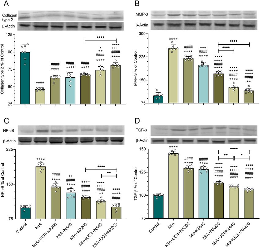
www.nature.com/scientificreports/

                                                      Groups
                                  Items               Control            MIA                  MIA + UCII                MIA + NA40            MIA + NA200               MIA + UCII + NA40      MIA + UCII + NA200
                                  Glucose (mg/dL)     114.71 ± 5.31      116.57 ± 7.28        116.00 ± 10.71            114.71 ± 7.48         116.14 ± 8.11             115.14 ± 9.67          116.43 ± 4.31
                                  BUN (mg/dL)          24.24 ± 3.02          24.24 ± 0.71          23.10 ± 4.20             24.37 ± 2.20          24.30 ± 2.28           24.63 ± 0.78           24.10 ± 2.71
                                  Creatine (mg/dL)      0.48 ± 0.10           0.47 ± 0.10           0.48 ± 0.10              0.48 ± 0.09           0.46 ± 0.08            0.46 ± 0.11            0.47 ± 0.11
                                  ALT (U/L)            70.57 ± 8.30          68.29 ± 4.72          70.29 ± 5.88             69.86 ± 3.89          71.71 ± 6.97           72.14 ± 11.78          69.00 ± 3.70
                                  AST (U/L)            88.43 ± 12.99         87.00 ± 11.85         87.43 ± 12.47            88.14 ± 11.17         86.86 ± 6.47           89.57 ± 6.50           85.14 ± 6.96

                                              Table 3.  Effects of niacinamide (NA) and undenatured type II collagen (UCII) on serum biochemical
                                              parameters in rats (n = 7). Data are presented as mean and standard deviation (p > 0.05; ANOVA and Tukey’s
                                              post-hoc test). NA: niacinamide; MIA: monosodium iodoacetate; UCII: undenatured type II collagen; BUN:
                                              Blood urea nitrogen; ALT: Alanine aminotransferase; AST: Aspartate aminotransferase. NA40 and NA200
                                              represent 40 and 200 mg/kg niacinamide dose applications, respectively.

                                                         Groups
                                      Items              Control                MIA                   MIA + UCII               MIA + NA40            MIA + NA200 MIA + UCII + NA40 MIA + UCII + NA200
                                      IL-1β (pg/mL)      20.13 ± 2.86d          47.62 ± 4.63a         36.59 ± 2.43b            38.78 ± 3.36b         35.82 ± 2.04b         24.97 ± 3.65cd       26.02 ± 3.86c
                                                                       d                       a                        b                     b                     b                      c
                                      IL-6 (pg/mL)        8.92 ± 1.47           39.46 ± 1.83          31.00 ± 1.85             32.72 ± 2.98          31.47 ± 2.72          21.18 ± 2.38         20.23 ± 1.99c
                                                                        d                     a                         b                     b                     b                      c
                                      TNF-α (pg/mL)      24.02 ± 3.60           68.03 ± 3.41          47.17 ± 3.15             51.10 ± 5.67          49.06 ± 3.76          34.27 ± 6.46         31.62 ± 2.21c
                                                                       d                      a                         b                     b                     b                      c
                                      COMP (pg/mL)        7.28 ± 1.06           32.10 ± 3.03          25.76 ± 2.98             27.69 ± 2.31          26.40 ± 3.07          16.72 ± 1.71         15.10 ± 2.46c
                                                                       d                      a                         b                    b                     b                      c
                                      CRP (pg/mL)         1.79 ± 0.19           10.76 ± 1.45            7.18 ± 0.67             7.54 ± 0.61           7.17 ± 0.64            4.39 ± 0.58         4.23 ± 0.64c

                                              Table 4.  Effects of niacinamide (NA) and undenatured type II collagen (UCII) supplementation on serum
                                              inflammation parameters in rats (n = 7). Data are presented as mean and standard deviation (p > 0.05;
                                              ANOVA and Tukey’s post-hoc test). NA: niacinamide; MIA: monosodium iodoacetate; UCII: undenatured
                                              type II collagen; IL-1β, Interleukin 1 beta; IL-6, Interleukin 6; TNF-α, tumor necrosis factor-alpha; COMP,
                                              cartilage oligomeric matrix protein; CRP, C-reactive protein. NA40 and NA200 represent 40 and 200 mg/kg
                                              niacinamide dose applications, respectively. (a–d): Means in the same line without a common superscript differ
                                              significantly.

                                                        Groups
                                      Items             Control               MIA                   MIA + UCII               MIA + NA40            MIA + NA200 MIA + UCII + NA40               MIA + UCII + NA200
                                      MDA (nmol/mL)        0.71 ± 0.08d          2.94 ± 0.11a          2.81 ± 0.12a             2.28 ± 0.11b          2.00 ± 0.08c          2.35 ± 0.13b          2.31 ± 0.31b
                                                                         a                   d                     cd                       bc                   b                       b
                                      SOD (U/mL)           81.2 ± 5.08         39.05 ± 4.53          42.19 ± 3.27             50.20 ± 7.90          53.24 ± 4.62           52.29 ± 6.85          56.69 ± 4.06b
                                                                         a                   d                     d                        c                    bc                      bc
                                      GPx (U/mL)         58.97 ± 2.80          20.01 ± 2.01          22.41 ± 2.41             27.84 ± 1.48          30.10 ± 2.94           30.36 ± 2.36          32.68 ± 2.54b
                                                                         a                    c                     c                        b                     b                      b
                                      CAT (U/mL)        159.29 ± 7.90         110.18 ± 7.95         113.18 ± 5.37            122.48 ± 3.93         128.88 ± 3.52          129.34 ± 3.96        130.93 ± 3.94b

                                              Table 5.  Effects of niacinamide (NA) and undenatured type II collagen (UCII) supplementation on serum
                                              MDA and antioxidant enzymes levels in rats (n = 7). Data are presented as mean and standard deviation
                                              (p > 0.05; ANOVA and Tukey’s post-hoc test). NA: niacinamide; MIA: monosodium iodoacetate; UCII:
                                              undenatured type II collagen; MDA, malondialdehyde; SOD, superoxide dismutase; CAT, catalase; GPx,
                                              glutathione peroxidase. NA40 and NA200 represent 40 and 200 mg/kg niacinamide dose applications,
                                              respectively. (a–d): Means in the same line without a common superscript differ significantly.

                                              Results
                                              Serum glucose, BUN, and creatine levels as well as ALT and AST activities, remained unchanged among treat-
                                              ments (p > 0.05; Table 3). Serum IL‐1β, IL‐6, TNF-α, COMP, and CRP concentrations increased in rats with OA
                                              compared with control rats (p < 0.05; Table 4). Supplementing UCII, NA40, and NA200 alone equally reduced
                                              the measured concentrations (p < 0.05). However, the combination of UCII and NA at both 40 and 200 mg/kg
                                              treatments equally provided further decreases in the concentrations of the inflammation parameters. Numeri-
                                              cally, the UCII + NA40 treatment provided the lowest IL‐1β concentrations compared with that of MIA.
                                                  Rats with osteoarthritis had greater MDA but lower SOD, CAT, and GPx activities compared with those of rats
                                              in the control group (p < 0.05; Table 5). However, the rats received each supplementation as single or as a combi-
                                              nation except for UCII treatments, which were similar to those of MIA, which reversed the responses (p < 0.05).
                                              Numerically but not statistically, MDA concentrations were lowest with the treatment of NA200, although
                                              concentrations of SOD, CAT, and GPx were greatest with UCII + NA200 treatment compared with those of MIA.
                                                  The stride lengths and their representative images at d7, d14, and d21 are shown in Fig. 1. The stride
                                              length decreased in rats with OA compared to rats of control (p < 0.05) at d7, d14, and d21. The rats receiving

          Scientific Reports |   (2021) 11:14724 |                       https://doi.org/10.1038/s41598-021-94142-3                                                                                                 4

Vol:.(1234567890)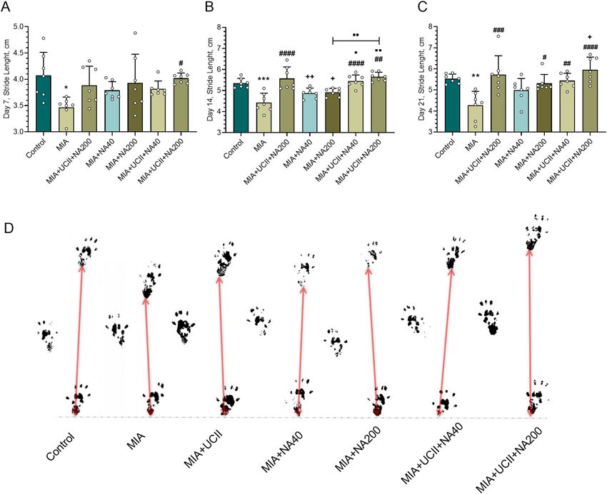
www.nature.com/scientificreports/

                                  Figure 1.  Effects of niacinamide (NA) and undenatured type II collagen (UCII) supplementation on stride
                                  length [on days 7 (A), 14 (B), and 21 (C)] in monosodium iodoacetate (MIA)-induced osteoarthritis in rats
                                  (n = 7). Representative images of the stride length measured on day 21 of the study are shown (D). Control
                                  as a non-supplemental group with no osteoarthritis-induced rats, MIA as a non-supplemental group with
                                  monosodium iodoacetate (MIA)-induced knee osteoarthritis, MIA + UCII as MIA group gavage-fed a
                                  supplemental UCII at 4 mg/kg, MIA + NA40 as MIA group gavage-fed supplemental niacinamide (NA) at
                                  40 mg/kg, MIA + NA200 as MIA group gavage-fed a supplemental NA at 200 mg/kg, MIA + UCII + NA40 as
                                  MIA group gavage-fed both supplemental UCII at 4 mg/kg and NA at 40 mg/kg and MIA + UCII + NA200
                                  as MIA group gavage-fed both supplemental UCII at 4 mg/kg and NA at 200 mg/kg. Oral gavage delivery of
                                  supplements was applied from d7 to d30. The error bars point out the standard deviation of the mean. ANOVA
                                  and Tukey’s post-hoc test were used to compare the results among different treatment groups. Statistical
                                  significance between groups is shown by: *p < 0.05; **p < 0.01; ***p < 0.001 compared as Control group and,
                                  #
                                   p < 0.05; ##p < 0.01; ###p < 0.001; ####p < 0.0001 compared as MIA group and, +p < 0.05; ++p < 0.01 compared as
                                  MIA + UCII group and ⬛p < 0.05; ⬛⬛p < 0.01 compared as MIA + NA40 group and, ••p < 0.01 compared as
                                  pairwise comparisons between the groups) (ANOVA and Tukey’s post-hoc test; p < 0.05).

                                  supplements of UCII or NA increased the stride length with various degrees (p < 0.05) in comparison to those of
                                  MIA rats (p < 0.05), the treatment of the combination of UCII + NA200 providing the greatest stride length even
                                  greater than that of control at d14 and d21. The representative images from the rats of control or the treatments
                                  showed similar trends to those of stride lengths at d21.
                                      Although the paw areas and the paw widths remained similar among treatments at d7 (p > 0.05; Fig. 2), the
                                  rats with OA had reduced paw areas and paw widths at d14 and d21 (p < 0.05). The rats supplemented only with
                                  a combination of UCII + NA200 increased (p < 0.05) the paw areas at d14. However, all supplements equally
                                  increased the paw areas in rats with OA bringing the values to those of control at d21. The paw widths in rats with
                                  OA increased equally (p < 0.05) with single supplements, and further increases (p < 0.05) were equally observed
                                  with the combination of UCII and NA treatments at d14. Similar responses were also observed at d21, with the

Scientific Reports |   (2021) 11:14724 |               https://doi.org/10.1038/s41598-021-94142-3                                                    5

                                                                                                                                              Vol.:(0123456789)
www.nature.com/scientificreports/
Figure 2. Effects of niacinamide (NA) and undenatured type II collagen (UCII) supplementation on paw
area [on days 7 (A), 14 (B), and 21 (C)] and pad width [on days 7 (D), 14 (E), and 21 (F)] in monosodium
iodoacetate (MIA)-induced osteoarthritis in rats (n = 7). Representative images of the paw area and pad width
measured on day 21 of the study are shown (G). Control as a non-supplemental group with no osteoarthritis-
induced rats, MIA as a non-supplemental group with monosodium iodoacetate (MIA)-induced knee
osteoarthritis, MIA + UCII as MIA group gavage-fed a supplemental UCII at 4 mg/kg, MIA + NA40 as MIA
group gavage-fed supplemental niacinamide (NA) at 40 mg/kg, MIA + NA200 as MIA group gavage-fed a
supplemental NA at 200 mg/kg, MIA + UCII + NA40 as MIA group gavage-fed both supplemental UCII at 4 mg/
kg and NA at 40 mg/kg, and MIA + UCII + NA200 as MIA group gavage-fed both supplemental UCII at 4 mg/
kg and NA at 200 mg/kg. Oral gavage delivery of supplements was applied from d7 to d30. ANOVA and Tukey’s
post-hoc test were used for comparing the results among different treatment groups. Statistical significance
between groups is shown by: *p < 0.05; **p < 0.01; ***p < 0.001; ****p < 0.0001 compared as Control group and,
#
p < 0.05; ##p < 0.01; ###p < 0.001; ####p < 0.0001 compared as MIA group and, +p < 0.05; ++++p < 0.0001 compared
as MIA + UCII group and, ⬛p < 0.05; ⬛⬛⬛p < 0.001; ⬛⬛⬛⬛p < 0.0001 compared as MIA + NA40 group and,
•p < 0.05; ••p < 0.01; ••••p < 0.0001 compared as pairwise comparisons between the groups). (ANOVA and
Tukey’s post-hoc test; p < 0.05).
combination of UCII+NA200 treatment having the greatest paw width values even similar to those of control. The
representative images of the paw area and pad width from the rats of control or the treatments showed similar
trends to those of stride lengths at d21.
Representative radiographic images indicated minimal Kellgren-Lawrence scores (grade 0; Fig. 3) with pres-
ervation of the joint space along with no signs of joint space narrowing and no formation of osteophytes in intact
rats. However, rats with OA had a high score reaching grade 3 with common radiographic features, including
joint space, narrowing, reduced articular space, sclerosis with articular surface irregularity, and the intense for-
mation of osteophytes and intense osteophyte formation. The rats treated with UCII and NA alone experienced
less evidence of the common radiographic features of OA, along with the scores not exceeded grade 2. However,
Scientific Reports | (2021) 11:14724 | https://doi.org/10.1038/s41598-021-94142-3 6
Vol:.(1234567890)www.nature.com/scientificreports/
Figure 3. Effects of niacinamide (NA) and undenatured type II collagen (UCII) supplementation on knee
joint in monosodium iodoacetate (MIA)-induced osteoarthritis in rats (n = 7). Representative radiographic
images (A) obtained at the end of the experiment are shown. Mean values of Kellgren- Lawrence scores
are demonstrated (B) with ± standard deviations (Kruskal–Wallis followed by Mann–Whitney U; p > 0.05).
Control as a non-supplemental group with no osteoarthritis-induced rats, MIA as a non-supplemental group
with monosodium iodoacetate (MIA)-induced knee osteoarthritis, MIA + UCII as MIA group gavage-fed a
supplemental UCII at 4 mg/kg, MIA + NA40 as MIA group gavage-fed supplemental niacinamide (NA) at
40 mg/kg, MIA + NA200 as MIA group gavage-fed a supplemental NA at 200 mg/kg, MIA + UCII + NA40 as
MIA group gavage-fed both supplemental UCII at 4 mg/kg and NA at 40 mg/kg and MIA + UCII + NA200
as MIA group gavage-fed both supplemental UCII at 4 mg/kg and NA at 200 mg/kg. Oral gavage delivery of
supplements was applied from d7 to d30.
Scientific Reports | (2021) 11:14724 | https://doi.org/10.1038/s41598-021-94142-3 7
Vol.:(0123456789)www.nature.com/scientificreports/
Figure 4. Effects of niacinamide (NA) and undenatured type II collagen (UCII) supplementation on
histopathology of the knee joint in monosodium iodoacetate (MIA)-induced osteoarthritis in rats.
Representative histopathologic images of hematoxylin–eosin (A) obtained at the end of the experiment are
shown. Mean values of Mankin scores are demonstrated with ± standard deviations (B). Asterisks above the
groups indicate statistical differences (Kruskal–Wallis followed by Mann–Whitney U; * p < 0.05; ** p < 0.01;
compared as MIA group). Control as a non-supplemental group with no osteoarthritis-induced rats, MIA as
a non-supplemental group with monosodium iodoacetate (MIA)-induced knee osteoarthritis, MIA + UCII as
MIA group gavage-fed a supplemental UCII at 4 mg/kg, MIA + NA40 as MIA group gavage-fed supplemental
niacinamide (NA) at 40 mg/kg, MIA + NA200 as MIA group gavage-fed a supplemental NA at 200 mg/kg,
MIA + UCII + NA40 as MIA group gavage-fed both supplemental UCII at 4 mg/kg and NA at 40 mg/kg and
MIA + UCII + NA200 as MIA group gavage-fed both supplemental UCII at 4 mg/kg and NA at 200 mg/kg. Oral
gavage delivery of supplements was applied from d7 to d30.
the rats treated with UCII + NA200 had further alleviation of the scores (grade 1) along with a normal thick-
ness of the cartilage surfaces, being able to reduce the degree of knee joint involvement in relation substantially.
The joints of the intact rats retained intact superficial and smooth articular cartilage surfaces with the under-
neath layer of flattened chondrocytes in the tangential zone (Fig. 4). In addition, chondrocytes of the same joints
were normally distributed in parallel rows, transitional and radial zones of the articular cartilage. As expected, the
rats with OA had irregular surfaces accompanied by loss of cartilage tissue degeneration of the articular cartilage
Scientific Reports | (2021) 11:14724 | https://doi.org/10.1038/s41598-021-94142-3 8
Vol:.(1234567890)www.nature.com/scientificreports/
Figure 5. Effects of niacinamide (NA) and undenatured type II collagen (UCII) supplementation on knee
joint diameter in monosodium iodoacetate (MIA)-induced osteoarthritis in rats (n = 7). Control as a non-
supplemental group with no osteoarthritis-induced rats, MIA as a non-supplemental group with monosodium
iodoacetate (MIA)-induced knee osteoarthritis, MIA + UCII as MIA group gavage-fed a supplemental UCII at
4 mg/kg, MIA + NA40 as MIA group gavage-fed supplemental niacinamide (NA) at 40 mg/kg, MIA + NA200
as MIA group gavage-fed a supplemental NA at 200 mg/kg, MIA + UCII + NA40 as MIA group gavage-fed both
supplemental UCII at 4 mg/kg and NA at 40 mg/kg and MIA + UCII + NA200 as MIA group gavage-fed both
supplemental UCII at 4 mg/kg and NA at 200 mg/kg. Oral gavage delivery of supplements was applied from d7
to d30. The error bars point out the standard deviation of the mean. (ANOVA and Tukey’s post-hoc test were
used for comparing the results among different treatment groups. Statistical significance between groups is
shown by: *p < 0.05; ****p < 0.0001 compared as Control group and, #p < 0.05; ##p < 0.01; ###p < 0.001; ####p < 0.0001
compared as MIA group and (ANOVA and Tukey’s post-hoc test; p < 0.05).
and disappearance of chondrocytes in the tangential, transitional and radial zones of the cartilage. However,
supplemental UCII and NA altered the histological changes in rats with OA. The elevated Mankin scores in
osteoarthritic rats were decreased with each supplement alone, but further decreases were observed with the
combinations of the supplements, particularly with the treatment of UCII + NA200 (p < 0.05).
The right knee joint diameters increased in rats with OA compared with those of control (p < 0.05; Fig. 5).
Each supplement with a similar extend reduced the knee joint diameter (p < 0.05).
The knee joint IL-1β, IL-6, IL-10, TNF-α, and COMP protein expression levels are reported in Fig. 6. The
protein expressions of IL-1β, IL-6, TNF-α, and COMP increased while that of IL-10 decreased (p < 0.05) in rats
with OA compared with those of control. Each supplement, particularly the combinations with the UCII + NA200
treatment, as being most effective, reversed the responses (p < 0.05). The UCII + NA200 supplementation brought
the protein expression levels of IL-1β, IL-6, and TNF-α to those of control.
The knee joint MMP-3, NF-κB, and TGB levels increased, whereas collagen type II level decreased in rats
with OA (p < 0.05; Fig. 7). Each supplement, particularly the combinations with the UCII + NA200 treatment, as
being most effective, reversed the responses (p < 0.05). The UCII + NA200 supplementation brought the protein
expression levels of NF-κB to those of control.
Discussion
Serum IL‐1β, IL‐6, TNF-α, COMP, and CRP concentrations increased 235%, 438%, 283%,457%, and 595%,
respectively in rats with OA compared with those of intact rats, indicating inflammation due to OA Increased
production of IL‐1β as a typical proinflammatory cytokine in the damaged joints30 was expected in rats with OA
As evidenced in the present work, IL‐1β was also demonstrated to lead the secretion of other cytokines such as
TNFα, IL-6, and IL-831. The knee joint IL-1β, IL-6, IL-10, TNF-α, and COMP levels were in accord with those of
serum. Parallel to the present work results and the common notion, Chandran et al.32 also found greater serum
concentrations of IL‐1β, IL‐6, and IL‐8 in patients with OA. However, the same authors20 detected no changes
in TNF-α, COMP, and CRP concentrations. When the rats with OA received the supplementation of UCII and
NA each alone but particularly the combination of UCII + NA200, the inflammation was ameliorated. Although
there have been no reports of serum inflammation parameters measured in osteoarthritic rats supplemented
Scientific Reports | (2021) 11:14724 | https://doi.org/10.1038/s41598-021-94142-3 9
Vol.:(0123456789)www.nature.com/scientificreports/
Scientific Reports | (2021) 11:14724 | https://doi.org/10.1038/s41598-021-94142-3 10
Vol:.(1234567890)www.nature.com/scientificreports/
◂Figure 6. Effects of niacinamide (NA) and undenatured type II collagen (UCII) supplementation on knee
joint IL-1β (A), IL-6 (B), IL-10 (C), TNF-α (D), and COMP (E) levels in monosodium iodoacetate (MIA)-
induced osteoarthritis in rats. The densitometric analysis of the relative intensity according to the control group
of the western blot bands was performed with β-actin normalization to ensure equal protein loading. Blots
were repeated at least three times (n = 3), and a representative blot is shown. Data are expressed as a percent
of the control set at 100%. Control as a non-supplemental group with no osteoarthritis-induced rats, MIA as
a non-supplemental group with monosodium iodoacetate (MIA)-induced knee osteoarthritis, MIA + UCII as
MIA group gavage-fed a supplemental UCII at 4 mg/kg, MIA + NA40 as MIA group gavage-fed supplemental
niacinamide (NA) at 40 mg/kg, MIA + NA200 as MIA group gavage-fed a supplemental NA at 200 mg/kg,
MIA + UCII + NA40 as MIA group gavage-fed both supplemental UCII at 4 mg/kg and NA at 40 mg/kg and
MIA + UCII + NA200 as MIA group gavage-fed both supplemental UCII at 4 mg/kg and NA at 200 mg/kg. Oral
gavage delivery of supplements was applied from d7 to d30. The error bars point out the standard deviation of
the mean. (ANOVA and Tukey’s post-hoc test were used for comparing the results among different treatment
groups. Full-length blots are presented in Supplementary Fig. S2. Statistical significance between groups is
shown by: **p < 0.01; ****p < 0.0001 compared as Control group and, #p < 0.05; ####p < 0.0001 compared as MIA
group and, +p < 0.05; +++p < 0.001; ++++p < 0.0001 compared as MIA + UCII group and, ⬛⬛⬛⬛p < 0.0001 compared
as MIA + NA40 group and, ••p < 0.01; ••••p < 0.0001 compared as pairwise comparisons between the groups).
(ANOVA and Tukey’s post-hoc test; p < 0.05).
with collagen in the literature, the present work revealed that rats supplemented with 4 mg UCII/kg body weight
as single or combination with niacinamide at either 40 or 200 mg/kg mitigated the inflammation. Niacinamide
supplementation for the treatment of OA is scarce in the literature. Osteoarthritic patients treated with niacina-
mide for 12 weeks lessened inflammation along with decreased severity of OA and improved joint fl exibility8.
Coherent results to IL‐1β, IL‐6, TNF-α, COMP, and CRP concentrations were also observed at the present
work with increasing MDA concentrations but decreasing antioxidant enzyme activities in rats with OA supple-
menting UCII and NA, as single or as a combination, altered the measured parameters. Increased MDA concen-
trations can also be used as a sensitive marker for inflammatory damage in a rthritis33, besides other such specific
markers as IL‐1β, IL‐6, TNF-α, COMP, and CRP. Similar to the results of the present work, Jaleel et al.34 found
elevated serum MDA, IL‐1β, IL‐6, and TNF-α concentration in rats with OA, compared with those of intact rats,
and observed that supplementing type III collagen to the rats at 10 mg/kg for two weeks reversed the responses.
The rats with OA bore elevated joint diameters with joint pain behavior measured as decreased the stride
lengths, the paw areas, and the paw widths, and increased the Kellgren-Lawrence Mankin scores. The Kellgren-
Lawrence score as the measurement of the severity of OA was high, as expected, in rats with OA. However,
osteoarthritic rats with supplements, particularly with UCII + NA200, had lower Kellgren-Lawrence scores.
Similarly, Bagi et al.12 found that oral supplementation of UCII to osteoarthritic rats alleviated articular carti-
lage’s worsening.
Reduced gait patterns in osteoarthritic rats have also been r eported35,36, with reduced paw areas and paw
widths along with decreases in stride length. In addition, changes in gait were observed as a result of increased
pain in osteoarthritic mice37. Supplementing either UCII or NA, each alone but particularly the combination
of UCII + NA200, ameliorated joint pain, especially with the longer treatment duration (d21). Similarly, NA
supplementation in patients with OA improved the severity of the OA by 29%, with increasing joint mobility
by 4.5 d egrees8.
Monosodium iodoacetate injection into the joints results in degenerative changes in articular cartilage via
matrix degradation and disturbance of chondrocyte metabolism and subsequently chondrocyte death. These
events occur mainly due to the inhibition of glyceraldehyde-3-phosphate dehydrogenase activity and thus
glycolysis38 as well as hydration of the extracellular matrix, and reduced quantity and synthesis of proteoglycans,
all leading eventually to cell d eath39,40. As observed in the present study, the sustainability of the cartilage with
the death of chondrocytes in osteoarthritic rats was impeded, and disrupted maintenance of the cartilage was
restored with each supplement but particularly with the combination of UCII + NA200 through the reduction
of joint space narrowing and cartilage destruction. The UCII treatment alone in rats with OA slightly improved
cartilage microstructure, degeneration, and surface organization, all of which were parallel to the results of
previous works in r ats12 and mice41 with OA. Although its precise mechanism is unknown, niacinamide was
speculated to penetrate the cartilage matrix by elevating NAD and NADP levels in synovial fl uid42. Therefore,
nutritional supplementation of NA, as was the case with the current work, would provide energy and nucleic
acids through non-oxidative mechanisms (i.e., via the pentose shunt, bypassing the tricyclic acid and glycolytic
sequences) that are vital for cartilage repair in the deeper layers of the matrix43.
Type II collagen comprises about 90% of the total collagen in hyaline cartilage, also known as articular
cartilage damaged in OA44. Therefore, decreases in the protein expression of type II collagen are signs of OA,
as evidenced in the present work. Progression of OA in the cartilages is related to inflammatory cytokines such
as IL‐1β and TNF-α, leading to MMPs (1, 3, 9, and 13) expressions45. This was also a case in the present study.
Similar to the current work results, Davidson et al.46 found increased protein expression of TGF-β1 in synovial
cells of osteoarthritic mice. Similarly, mRNA expression levels of Tgfb1 genes were reported increased in the
knee cartilage of rats with MIA-induced OA47. The supplementation of UCII and NA, each alone but particularly
the combination of UCII + NA200, increased the synthesis of type II collagen and reduced the inflammation
parameters in the knee joint.
Apparently, both UCII and NA, each alone but mainly as a combination, regenerated the knee cartilage, miti-
gating the inflammation and helping normal functions of joints and tendons. The effects of the UCII could be
Scientific Reports | (2021) 11:14724 | https://doi.org/10.1038/s41598-021-94142-3 11
Vol.:(0123456789)www.nature.com/scientificreports/
Figure 7. Effects of niacinamide (NA) and undenatured type II collagen (UCII) supplementation on the knee
joint collagen type II (A), MMP-3 (B), NF-κB (C), and TGF-β1 (D) levels in monosodium iodoacetate (MIA)-
induced osteoarthritis in rats. The densitometric analysis of the relative intensity according to the control group
of the western blot bands was performed with β-actin normalization to ensure equal protein loading. Blots
were repeated at least three times (n = 3), and a representative blot is shown. Data are expressed as a percent
of the control set at 100%. Control as a non-supplemental group with no osteoarthritis-induced rats, MIA as
a non-supplemental group with monosodium iodoacetate (MIA)-induced knee osteoarthritis, MIA + UCII as
MIA group gavage-fed a supplemental UCII at 4 mg/kg, MIA + NA40 as MIA group gavage-fed supplemental
niacinamide (NA) at 40 mg/kg, MIA + NA200 as MIA group gavage-fed a supplemental NA at 200 mg/kg,
MIA + UCII + NA40 as MIA group gavage-fed both supplemental UCII at 4 mg/kg and NA at 40 mg/kg, and
MIA + UCII + NA200 as MIA group gavage-fed both supplemental UCII at 4 mg/kg and NA at 200 mg/kg. Oral
gavage delivery of supplements was applied from d7 to d30. The error bars point out the standard deviation of
the mean. (ANOVA and Tukey’s post-hoc test were used for comparing the results among different treatment
groups. Full-length blots are presented in Supplementary Fig. S3. Statistical significance between groups is
shown by: **p < 0.01; ****p < 0.0001 compared as Control group and, ####p < 0.0001 compared as MIA group
and, ++p < 0.01; +++p < 0.001; ++++p < 0.0001 compared as MIA + UCII group and, ⬛p < 0.05; ⬛⬛⬛⬛p < 0.0001
compared as MIA + NA40 group and, •p < 0.05; ••p < 0.01; ••••p < 0.0001 compared as pairwise comparisons
between the groups) (ANOVA and Tukey’s post-hoc test; p < 0.05).
Scientific Reports | (2021) 11:14724 | https://doi.org/10.1038/s41598-021-94142-3 12
Vol:.(1234567890)www.nature.com/scientificreports/
due to its rich glycine contents and proline, which are required for the normal function of joints and t endons48.
Extreme decreases of collagen synthesis in osteoarthritis have been shown due to severe glycine deficiency, and
increased glycine concentrations in vitro have been indicated to advance collagen synthesis and consequently
cartilage regeneration49.
The mechanism of NA in the scenario of OA at the present work and in the literature has not been explored.
The current work provided evidence that NA acts similar to that of UCII in most parameters measured in alle-
viating the symptoms of knee OA Niacinamide has been speculated to inhibit the synthesis and/or activity of
IL-114. In addition, NA has been considered a part of antioxidant defense m echanisms4–6 and is evidenced in
the present work. Niacinamide treatment has also been reported to be beneficial in treating osteoarthritis with
better joint flexibility and decreased inflammation8. However, more work has to be conducted in clearing up the
detailed mechanism of NA in arthritis.
In general, UCII alone is better than NA alone in improving the paw width, stride length, and numerical
values of inflammatory serum parameters. However, NA alone, particularly with greater doses, was better than
that of U-II alone in knee joint inflammation parameter levels as well as Collagen Type II, MMP-3, NF-κB, and
TGB levels. The effects of UCII and NA were most probably linked to the suppression of the production of pro-
inflammatory cytokines and mediators such as IL‐1β, IL‐6, TNF-α, COMP, and CRP. Rats with MIA-induced OA
were reported to have greater NF-κB1 gene expression levels, compared with those of healthy rats, in articular
cartilages, subchondral bone, and synovial membrane of the rat knee joint50,51. Therefore, inhibition of NF-κB1
expressions in cartilage can counteract chondrocytes damage through increased synthesis of proinflammatory
cytokines52. The supplements were also involved in type II collagen synthesis, helping to alleviate the symptoms
of OA. It is still a question of how much of each effect (increased type II collagen synthesis or a reduction in
inflammation parameters) contributed more to alleviating OA symptoms and which supplement is more effec-
tive. Therefore, further work is required to explore the detailed molecular mechanisms of action of UCII and NA.
In vitro results (Supplementary Tables S1, S2 and Supplementary Fig. S1) conducted for this work were in
accord with the results of the present experiment. Reduction of inflammatory markers including IG6, COX2,
TNF-α, and NF-κB was observed when a combination of collagen and niacinamide was used in THP-1 monocyte
cells differentiated into macrophages and treated with various extracts as control (0.2% DMSO–0.2%, v/v), UCII
(50 ug/mL), Niacinamide (50 ug/mL), UCII + Niacinamide (50 ug/mL + 50 ug/mL), and Rosiglitazone (5 mM)
(Supplementary Table S2).
Supplementing a combination of UCII at 4 mg/kg and niacinamide at 200 mg/kg for three weeks is a promis-
ing dietary strategy for reducing pain, minimizing cartilage damage improving functional status in knee OA of rat
models. The results can also be applied to humans suffering from knee OA, being an alternative to conventional
drug treatments. The mechanism by which how the supplements improve functional status in knee OA of rat
models needs to be elucidated through clinical investigations.
Data availability
The datasets used and analyzed during the current study are available from the corresponding author on reason-
able request.
Received: 23 March 2021; Accepted: 25 June 2021
References
1. Winter, A. R., Collins, J. E. & Katz, J. N. The likelihood of total knee arthroplasty following arthroscopic surgery for osteoarthritis:
A systematic review. BMC Musculoskelet. Disord. 18, 1–8 (2017).
2. Ro, K.-H., Heo, J.-W. & Lee, D.-H. Bearing dislocation and progression of osteoarthritis after mobile-bearing unicompartmental
knee arthroplasty vary between Asian and Western patients: a meta-analysis. Clin. Orthop. Relat. Res. 476, 946 (2018).
3. Lingard, E. A. & Riddle, D. L. Impact of psychological distress on pain and function following knee arthroplasty. J. Bone Joint Surg.
Am. 89, 1161–1169 (2007).
4. Maiese, K., Chong, Z. Z., Hou, J. & Shang, Y. C. The vitamin nicotinamide: Translating nutrition into clinical care. Molecules 14,
3446–3485 (2009).
5. Rolfe, H. M. A review of nicotinamide: treatment of skin diseases and potential side effects. J. Cosmet. Dermatol. 13, 324–328
(2014).
6. Zhen, A. X. et al. Niacinamide protects skin cells from oxidative stress induced by particulate matter. Biomol. Ther. (Seoul). 27,
562 (2019).
7. Kröger, H. et al. Enhancing the inhibitory effect of nicotinamide upon collagen II induced arthritis in mice using N-acetylcysteine.
Inflammation 23, 111–115 (1999).
8. Jonas, W., Rapoza, C. & Blair, W. The effect of niacinamide on osteoarthritis: A pilot study. Inflamm. Res. 45, 330–334 (1996).
9. Barnett, M. L. et al. Treatment of rheumatoid arthritis with oral type II collagen: Results of a multicenter, double-blind, placebo-
controlled trial. Arthritis Rheum. 41, 290–297 (1998).
10. Wei, W. et al. A multicenter, double-blind, randomized, controlled phase III clinical trial of chicken type II collagen in rheumatoid
arthritis. Arthritis Res. Ther. 11, 1–10 (2009).
11. Bakilan, F. et al. Effects of native type II collagen treatment on knee osteoarthritis: A randomized controlled trial. Eurasian J. Med.
48, 95 (2016).
12. Bagi, C., Berryman, E., Teo, S. & Lane, N. E. Oral administration of undenatured native chicken type II collagen (UC-II) diminished
deterioration of articular cartilage in a rat model of osteoarthritis (OA). Osteoarthritis Cartilage 25, 2080–2090 (2017).
13. d’Altilio, M. et al. Therapeutic efficacy and safety of undenatured type II collagen singly or in combination with glucosamine and
chondroitin in arthritic dogs. Toxicol. Mech. Methods 17, 189–196 (2007).
14. McCarty, M. & Russell, A. Niacinamide therapy for osteoarthritis–does it inhibit nitric oxide synthase induction by interleukin-1
in chondrocytes?. Med. Hypotheses 53, 350–360 (1999).
15. Singh, N. et al. Activation of Gpr109a, receptor for niacin and the commensal metabolite butyrate, suppresses colonic inflamma-
tion and carcinogenesis. Immunity 40, 128–139 (2014).
Scientific Reports | (2021) 11:14724 | https://doi.org/10.1038/s41598-021-94142-3 13
Vol.:(0123456789)www.nature.com/scientificreports/
16. Chai, J. T., Digby, J. E. & Choudhury, R. P. GPR109A and vascular inflammation. Curr. Atheroscler. Rep. 15, 325. https://doi.org/
10.1007/s11883-013-0325-9 (2013).
17. Hwang, E. S. & Song, S. B. Nicotinamide is an inhibitor of SIRT1 in vitro, but can be a stimulator in cells. Cell Mol. Life Sci. 74,
3347–3362 (2017).
18. Council, E. EEC Council Directive 86/609/EEC of 24 November 1986 on the approximation of laws, regulations and administrative
provisions of the Member States regarding the protection of animals used for experimental and other scientific purposes. Off. J.
Eur. Union. L 358, 1–28 (1986).
19. Lu, J., Zhang, T., Sun, H., Wang, S. & Liu, M. Protective effects of dioscin against cartilage destruction in a monosodium iodoacetate
(MIA)-indcued osteoarthritis rat model. Biomed. Pharmacother. 108, 1029–1038 (2018).
20. Jeong, J. W. et al. Mori Folium water extract alleviates articular cartilage damages and inflammatory responses in monosodium
iodoacetate-induced osteoarthritis rats. Mol. Med. Rep. 16, 3841–3848 (2017).
21. Rashid, H., Samadfam, R., Durkee, S., Verhoef, J. & Bellamine, A. Nicotinate supplements slow onset and severity of symptoms in
the monosodium iodoacetate rat model for osteoarthritis. J. Vet. Med. Anim. Sci. 4, 1047–1053 (2020).
22. Bellamine, A. Nicotinate supplements slow onset and severity of symptoms in the monosodium iodoacetate (MIA) rat model for
osteoarthritis (OA). FASEB J. 33, 9.1-lb552. https://doi.org/10.1096/fasebj.2019.33.1_supplement.lb552 (2019).
23. Orhan, C. et al. Undenatured type II collagen ameliorates inflammatory responses and articular cartilage damage in the rat model
of osteoarthritis. Front. Vet. Sci. 4, 617789. https://doi.org/10.3389/fvets.2021.617789 (2021).
24. Kellgren, J. & Lawrence, J. Radiological assessment of osteo-arthrosis. Ann. Rheum. Dis. 16, 494 (1957).
25. Mankin, H. J., Dorfman, H., Lippiello, L. & Zarins, A. Biochemical and metabolic abnormalities in articular cartilage from osteo-
arthritic human hips. II. Correlation of morphology with biochemical and metabolic data. J. Bone Joint Surg. Am. 53, 523–537
(1971).
26. Dogukan, A. et al. A tomato lycopene complex protects the kidney from cisplatin-induced injury via affecting oxidative stress as
well as Bax, Bcl-2, and HSPs expression. Nutr. Cancer. 63, 427–434 (2011).
27. Yabas, M. et al. A next generation formulation of curcumin ameliorates experimentally induced osteoarthritis in rats via regulation
of inflammatory mediators. Front. Immunol. 12, 609629 (2021).
28. Faul, F., Erdfelder, E., Lang, A.-G. & Buchner, A. G* Power 3: A flexible statistical power analysis program for the social, behavioral,
and biomedical sciences. Behav. Res. Methods. 39, 175–191 (2007).
29. Cohen, J. Statistical Power Analysis for the Behavioral Sciences Revised. (Lawrence Earlbaum Associates Inc., 1988).
30. Jenei-Lanzl, Z., Meurer, A. & Zaucke, F. Interleukin-1β signaling in osteoarthritis–chondrocytes in focus. Cell. Signal. 53, 212–223
(2019).
31. Massicotte, F. et al. Can altered production of interleukin-1β, interleukin-6, transforming growth factor-β and prostaglandin E2
by isolated human subchondral osteoblasts identify two subgroups of osteoarthritic patients. Osteoarthritis Cartilage 10, 491–500
(2002).
32. Chandran, V. et al. Serum-based soluble markers differentiate psoriatic arthritis from osteoarthritis. Ann. Rheum. Dis. 78, 796–801
(2019).
33. Pallinti, V., Ganesan, N., Anbazhagan, M. & Rajasekhar, G. Serum biochemical markers in rheumatoid arthritis. Indian J. Biochem.
Biophys. 46, 342–344 (2009).
34. Jaleel, G. A. A., Saleh, D. O., Al-Awdan, S. W., Hassan, A. & Asaad, G. F. Impact of type III collagen on monosodium iodoacetate-
induced osteoarthritis in rats. Heliyon. 6, e04083 (2020).
35. Cunha, J. E. et al. Knee osteoarthritis induces atrophy and neuromuscular junction remodeling in the quadriceps and tibialis
anterior muscles of rats. Sci. Rep. 9, 1–11 (2019).
36. Adães, S. et al. Intra-articular injection of collagenase in the knee of rats as an alternative model to study nociception associated
with osteoarthritis. Arthritis Res. Ther. 16, 1–17 (2014).
37. Makii, Y. et al. Alteration of gait parameters in a mouse model of surgically induced knee osteoarthritis. J. Orthop. Surg. (Hong
Kong) 26, 2309499018768017 (2018).
38. Barve, R. et al. Transcriptional profiling and pathway analysis of monosodium iodoacetate-induced experimental osteoarthritis
in rats: relevance to human disease. Osteoarthritis Cartilage 15, 1190–1198 (2007).
39. Niazvand, F. et al. Curcumin-loaded poly lactic-co-glycolic acid nanoparticles effects on mono-iodoacetate -induced osteoarthritis
in rats. Vet. Res. Forum. 8, 155–161 (2017).
40. Beyreuther, B., Callizot, N. & Stöhr, T. Antinociceptive efficacy of lacosamide in the monosodium iodoacetate rat model for
osteoarthritis pain. Arthritis Res. Ther. 9, 1–8 (2007).
41. Yoshinari, O. et al. Water-soluble undenatured type II collagen ameliorates collagen-induced arthritis in mice. J. Med. Food. 16,
1039–1045 (2013).
42. DiPalma, J. R. & Thayer, W. S. Use of niacin as a drug. Annu. Rev. Nutr. 11, 169–187 (1991).
43. Hamerman, D. The biology of osteoarthritis. N. Engl. J. Med. 320, 1322–1330. https://doi.org/10.1056/nejm198905183202006
(1989).
44. Van den Berg, W. Osteoarthritis year 2010 in review: pathomechanisms. Osteoarthritis Cartilage 19, 338–341 (2011).
45. Shi, J., Schmitt-Talbot, E., DiMattia, D. & Dullea, R. The differential effects of IL-1 and TNF-α on proinflammatory cytokine and
matrix metalloproteinase expression in human chondrosarcoma cells. Inflamm. Res. 53, 377–389 (2004).
46. Davidson, E. B., Vitters, E., Van Der Kraan, P. & Van Den Berg, W. Expression of transforming growth factor-β (TGFβ) and the
TGFβ signalling molecule SMAD-2P in spontaneous and instability-induced osteoarthritis: role in cartilage degradation, chon-
drogenesis and osteophyte formation. Ann. Rheum. Dis. 65, 1414–1421 (2006).
47. Dranitsina, A. S., Dvorshchenko, K. O., Korotkiy, A. G., Grebinyk, D. M. & Ostapchenko, L. I. Expression of Ptgs2 and Tgfb1 genes
in rat cartilage cells of the knee under conditions of osteoarthritis. Cytol. Genet. 52, 192–197 (2018).
48. Korotkyi, O. et al. Effect of chondroitin sulfate on blood serum cytokine profile during carrageenan-induced edema and monoi-
odoacetate-induced osteoarthritis in rats. Rev. Recent Clin. Trials. 14, 50–55 (2019).
49. de Paz-Lugo, P., Lupiáñez, J. A. & Meléndez-Hevia, E. High glycine concentration increases collagen synthesis by articular chon-
drocytes in vitro: acute glycine deficiency could be an important cause of osteoarthritis. Amino Acids 50, 1357–1365. https://doi.
org/10.1007/s00726-018-2611-x (2018).
50. Korotkyi, O. et al. Combined effects of probiotic and chondroprotector during osteoarthritis in rats. Panminerva Med. 62, 93–101.
https://doi.org/10.23736/S0031-0808.20.03841-0 (2020).
51. Korotkyi, O. et al. Probiotic composition and chondroitin sulfate regulate TLR-2/4-mediated NF-κB inflammatory pathway and
cartilage metabolism in experimental osteoarthritis. Probiot. Antimicrob. Prot. https://d oi.o
rg/1 0.1 007/s 12602-0 20-0 9735-7 (2021).
52. Chow, Y. Y. & Chin, K. Y. The role of inflammation in the pathogenesis of osteoarthritis. Mediat. Inflamm. 3, 8293921. https://doi.
org/10.1155/2020/8293921 (2020).
Acknowledgements
This work was supported by Lonza Consumer Health (NJ, USA) and the Turkish Academy of Sciences (KS).
Scientific Reports | (2021) 11:14724 | https://doi.org/10.1038/s41598-021-94142-3 14
Vol:.(1234567890)www.nature.com/scientificreports/
Author contributions
K.S. Conceptualization, methodology, formal analysis, supervision, project administration, funding acquisition,
writing-review and editing; C.O., M.T. and N.S. data curation methodology, formal analysis, investigation; A.S.D.
performed the radiological assessment; I.H.O. performed the histological evaluation; O.K. writing-original draft
preparation and V.J. writing-original draft preparation, writing-review and editing. All the authors read and
approved the final version of the manuscript. All authors agree to be accountable for all aspects of the work.
Funding
This project was supported by Lonza Consumer Health Inc., (NJ, USA) and by the Turkish Academy of Sciences
(KS, Ankara, Turkey) in part. The funders were not involved in the study design, collection, analysis, and inter-
pretation of data, the writing of this article, or the decision to submit it for publication.
Competing interests
VJ is an employee of Lonza Consumer Health Inc (NJ, USA). Other authors have no other relevant affiliations
or financial involvement with any organization or entity with a financial interest in or financial conflict with the
subject matter or materials discussed in the manuscript.
Additional information
Supplementary Information The online version contains supplementary material available at https://doi.org/
10.1038/s41598-021-94142-3.
Correspondence and requests for materials should be addressed to K.S.
Reprints and permissions information is available at www.nature.com/reprints.
Publisher’s note Springer Nature remains neutral with regard to jurisdictional claims in published maps and
institutional affiliations.
Open Access This article is licensed under a Creative Commons Attribution 4.0 International
License, which permits use, sharing, adaptation, distribution and reproduction in any medium or
format, as long as you give appropriate credit to the original author(s) and the source, provide a link to the
Creative Commons licence, and indicate if changes were made. The images or other third party material in this
article are included in the article’s Creative Commons licence, unless indicated otherwise in a credit line to the
material. If material is not included in the article’s Creative Commons licence and your intended use is not
permitted by statutory regulation or exceeds the permitted use, you will need to obtain permission directly from
the copyright holder. To view a copy of this licence, visit http://creativecommons.org/licenses/by/4.0/.
© The Author(s) 2021
Scientific Reports | (2021) 11:14724 | https://doi.org/10.1038/s41598-021-94142-3 15
Vol.:(0123456789)You can also read