PEROXIDASE-LIKE ACTIVITY OF HEMOGLOBIN-BASED HYBRID MATERIALS AGAINST DIFFERENT SUBSTRATES AND THEIR ENHANCED APPLICATION FOR H2O2 DETECTION
←
→
Page content transcription
If your browser does not render page correctly, please read the page content below

Bull. Chem. Soc. Ethiop. 2021, 35(3), 537-550. ISSN 1011-3924
2021 Chemical Society of Ethiopia and The Authors Printed in Ethiopia
DOI: https://dx.doi.org/10.4314/bcse.v35i3.6 Online ISSN 1726-801X
PEROXIDASE-LIKE ACTIVITY OF HEMOGLOBIN-BASED HYBRID MATERIALS
AGAINST DIFFERENT SUBSTRATES AND THEIR ENHANCED APPLICATION
FOR H2O2 DETECTION
Cevahir Altinkaynak1*, Merve Turk2, Murat Ekremoğlu3 and Nalan Özdemir2
1
Department of Plant and Animal Production, Avanos Vocational School, Nevsehir Haci Bektas
Veli University, 50500, Nevsehir, Turkey
2
Department of Chemistry, Faculty of Science, Erciyes University, 38039, Kayseri, Turkey
3
Department of Medical Biochemistry, Faculty of Medicine, Istinye University, 34010, Istanbul,
Turkey
(Received November 13, 2020; Revised January 20, 2022; Accepted January 22, 2022)
ABSTRACT. Organic-inorganic hybrid nanoflowers method with unique properties are preferred than
conventional immobilization methods for the past decade. Hereemoglobin-based hybrid material (HbNFs@Cu) was
synthesized under different experimental conditions (pH 5.0-9.0 and 0.01-0.50 mgmL-1 of hemoglobin) obtaining a
material size of 9-10 µm. The encapsulation percentage and weight yield of HbNFs@Cu were determined as 100%
and 6.7%, respectively. The peroxidase-like activities of the material against different substrates (ABTS and
Guaiacol) were compared to free hemoglobin. The HbNFs@Cu hybrid structure exhibited Vmax of 3.6995 EU/mg
and a Michaelis-Menten constant (KM) of 0.1357 mM/mL. The HbNFs@Cu hybrid material was then used to
catalyze the oxidation of a peroxidase substrate ABTS to the pigmented product, which provided a colorimetric and
spectrophotometric detection of H2O2. The linear operating range, detectable colorimetrically as H2O2 sensor, is
0.005-0.0042 mM, while the linear operating range, detectable spectrometically, is 0.003-0.0042 mM. The limits of
detection of colorimetric and spectrophotometric sensors were 0.005 mM and 0.003 mM, respectively. Collectively,
these results showed that HbNFs@Cu can be used as colorimetric biosensor for H2O2 in potential applications such
as pharmaceutical food, biomedical, environmental, and industrial.
KEY WORDS: Hydrogen peroxide, Hemoglobin, Hybrid Material, Colorimetric assay
INTRODUCTION
Organic-inorganic hybrid nanoflower has recently attracted attention as an effective nanostructure
with excellent properties and a novel method of protein (enzyme) immobilization [1–6].
Nanoflowers do not share the disadvantages of conventional immobilization techniques such as
mass transfer limitations, low stability, low enzymatic activity, and long production processes [6,
7] and are high-efficiency catalysts due to large surface-to-volume ratio [8]. The nanoflower,
which has nano-sized plates, has been applied to drug transportation, gene therapy, biosensor
diagnosis and therapy [9]. Protein-based biomaterials are a particularly effective tool for
characterizing the target analyte [10–12]. To date, various researches have been carried out using
such materials as carbon, metal, metal oxide-based nanoparticles, and metal complexes to design
functional synthetic materials with peroxidase and catalase-like activities [13–15]. Heme group-
containing proteins such as hemoglobin (Hb), myoglobin and cytochrome C exhibit peroxidase-
like activity due to their electroactive heme groups and can be used to reduce hydrogen peroxide
and as H2O2 sensors [16, 17]. Among these, hemoglobin is widely used in the analysis because of
its well-known molecular structure, its cheapness, commercial usable and high stability [10, 18].
Hemoglobin is a 5.5 nm sized protein with tetrameric, globuler and oxygen carrier, each monomer
weighing 17 kDa [11, 16, 19, 20].
__________
*Corresponding author. E-mail: caltinkaynak@nevsehir.edu.tr
This work is licensed under the Creative Commons Attribution 4.0 International License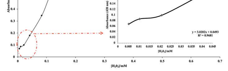
538                                   Cevahir Altinkaynak et al.

    The peroxidase-like catalytic activity of hemoglobin is slower and lower than the natural
peroxidase enzyme [19]. The natural hemoglobin molecule was therefore immobilized to different
types of nanocomposites for the production of H2O2 sensors such as Hb/Pluronic P123-nanografen
platelet [21], Hb/Collagen micro belt [22], and Hb/Ag sol films [23] and hemoglobin-DNA
conjugate on nanoporous gold thin film[18]. However, the difficult working principles of these
methods, their weak determination limits and their operation within a narrow linear range limit
their industrial use. For this reason, there is a need for the development of effective, reliable and
highly sensitive nanostructures, easy application to H2O2 determination, and rapid response. It is
known that the stabilities and activities of organic structures immobilized by organic-inorganic
hybrid nanoflowers method are higher than free molecules.
    In this area of research, by Gao et al. obtained two different biomolecules [24, 25]. The
hemoglobin-Mn3(PO4)2 hybrid nanoflowers were synthesized and found their opulent
electroactive [25]. At the same time, Gao et al. reported that the synthesized Hb-Cu3(PO4)2 HNFs
obtained an excellent stability and exhibited greater catalytic activity than free Hb against
Rhodamine 6G substrate [24]. As we know, an enzyme substrate is the material upon which an
enzyme acts, and enzymes exhibit different reactions against different substrates [26]. Inquiry
about the peroxidase-like activity of hemoglobin-inorganic hybrid nanoflowers against different
substrates is needed.
    The current work aims for a deeper understanding of the reaction of hemoglobin-based hybrid
materials against different substrates. So, it will be provided to forma hemoglobin-based hybrid
material (HbNFs@Cu) with a very low level of peroxidase mimic activity, commercially usable,
extremely cheap hemoglobin molecule (as a organic part) and inorganic part Cu(II) ion and
phosphate buffer. It is known that hemoglobin molecule shows structural changes at various pH
values in vitro conditions [27]. For this reason, hybrid materials were synthesized under different
experimental conditions, and the morphologies of hybrid materials were characterized using SEM,
EDX, XRD and FTIR. The enzymatic activity profiles of HbNFs@Cu against different substrates
were examined, and a minimum linear working range was detected for the determination of H2O2
under optimum conditions.
                                       EXPERIMENTAL

Chemicals and materials

Hemoglobin from bovine blood (H2625), bovine serum albumin (A7906), guaiacol and
phosphoric acid were purchased from Sigma-Aldrich. Copper(II) sulfate pentahydrate was
purchased from ISOLAB. ABTS was purchased from Roche. Coomassie plus protein assay
reagent was purchased from Thermo Scientific. KCl, NaCl, Na2HPO4, KH2PO4, NaH2PO4, NaOH,
H2O2, ethyl alcohol and HCl were purchased from Merck. All chemical reagents were of analytical
grade. Aqueous solutions were prepared using deionised water.

Fabrication of HbNFs@Cu

For the synthesis of HbNFs@Cu; hemoglobin and CuSO4.5H2O solution were used as the organic
and inorganic component, respectively. 0.8 mM CuSO4.5H2O solution adjusted phosphate buffer
saline buffer (10 mM, pH 5.0-9.0) containing various concentrations of hemoglobin (0.01-0.50
mg mL-1 of total solution) for each tube. After the solution was incubated for 3 days at 4 °C,
centrifuge separation produced a pellet. The pellet was washed three times with water to remove
unbounded protein molecules. To determine the encapsulated protein in the nanoflower, the
protein amount was applied to the supernatant after incubation via the Bradford protein assay.

                                Bull. Chem. Soc. Ethiop. 2021, 35(3)
Peroxidase-like activity of hemoglobin-based hybrid materials against different substrates   539

Characterization of HbNFs@Cu

Scanning electron microscopy (SEM) provides information on the surface morphology and
particle size of the material. The morphologies of the synthesized HbNFs@Cu were visualized
with SEM (LEO 440 Computer Controlled Digital). The presence of Cu, P, N, O, C and Cl metals
in HbNFs@Cu were detected by Energy dispersed X-ray analysis (EDX). The chemical structures
of the HbNFs@Cu hybrid structures were confirmed by FTIR analysis (Perkin Elmer 400 FT-IR
Spectrometer Spotlight 400 Imaging System) at a range of 4000-450 cm-1 wavenumbers. The
crystal structures of HbNFs@Cu were illuminated from 10° to 80° by XRD (X-ray diffraction)
analysis.

Measurement of peroxidase-like activities of HbNFs@Cu

The peroxidase-like activities of the HbNFs@Cu were examined in two different methods
compared to free hemoglobin. The first method [6] began by weighing 3 mg of HbNFs@Cu
synthesized in optimum pH condition. Each was taken into a 15 mL tube. 1 mL of PBS buffer in
different pHs, 1 mL of 22.5 mM H2O2 solution and finally 1 mL of 45 mM guaiacol solution were
added to the each tubes. The reaction mixtures were stirred and incubated at room temperature for
25 min. After centrifugation, the spectrophotometric absorbances at 470 nm were measured using
UV-spectrometer (HITACHI). The catalytic activity of free hemoglobin in equal amount was
performed with the same method.
    The second method [28] began by weighing 3 mg of HbNFs@Cu in optimum pH condition.
Each was taken into a 15 mL tube. 1 mL of PBS buffer in different pHs, 1 mL of 25 mM H2O2
solution and finally 1 mL of 1 mM ABTS solution were added to each tube. The reaction mixtures
were stirred and incubated at room temperature for 25 min. After centrifugation, the
spectrophotometric absorbances at 420 nm were measured using UV-spectrometer (HITACHI).
The catalytic activity of free hemoglobin in equal amount was performed with the same method.

Michaelis-Menten kinetic parameters of HbNFs@Cu

Kinetic parameters of HbNFs@Cu were determined by ABTS concentrations varying from 0.08-
2.7 mM mL-1 and evaluated for peroxidase-like activities in phosphate buffer solution (pH 5).
Michaelis-Menten constant (KM and Vmax of HbNFs@Cu) was calculated from Lineaweaver-Burk
plot (1/V and 1/S).

Reusability of HbNFs@Cu

The reusability of HbNFs@Cu was carried out under optimum activity conditions for 10 cycles
of reuse. After each cycle, the HbNFs@Cu was centrifuged for 5 min at 5000 g, washed with PBS
buffer and readied for the next cycle.

Colorimetric and spectrophotometric measurements of H2 O2 using HbNFs@Cu

The various concentrations of H2O2 solutions were prepared in water and the technical feasibility
of H2O2 colorimetric sensing was carried out in activity reaction system. Typically 3 mg of
HbNFs@Cu, 1 mL of PBS buffer (10 mM, pH 5), 1 mL of 4 mM ABTS solution and 1 mL of
various concentrations of H2O2 solution (0.008-2 mM) were added to the tubes. The reaction
mixture was stirred and incubated at room temperature for 25 min with stirring. After
centrifugation, the spectrophotometric absorbance at 420 nm and colorimetric response was
determined both using UV-spectrometer (HITACHI) and visually.

                              Bull. Chem. Soc. Ethiop. 2021, 35(3)
540                                  Cevahir Altinkaynak et al.

                                RESULTS AND DISCUSSION

Influence of synthesis conditions on the formation of HbNFs@Cu

Hemoglobin was used as an organic component and Cu(II) ions as an inorganic component in the
synthesis of the hybrid material. The experimental section describes the synthesis of HbNFs@Cu.
The effects of the synthesis medium pH (5-9) and hemoglobin amount (0.01-0.5 mg mL-1) on the
formation of HbNFs@Cu were examined. The scanned experimental parameters of HbNFs@Cu
show that the product could not be obtained when the synthesis medium pH was 5. We know the
isoelectric point (pI) of hemoglobin is 6.7 [24]; below this pH, no nanoflower precipitates form
[29]. The blue-brown colored pellet was observed in the other pH conditions.
    To determine the encapsulation and weight percentage of HbNFs@Cu, the protein assay was
applied on the supernatant after incubation. The encapsulation rate was found to be 0% due to no
product at pH 5. Weight percentage also could not be calculated. The HbNFs@Cu encapsulation
percentage using 0.05 mg mL-1 hemoglobin in the reaction solution at pH 7 was higher than under
other conditions; the amount of product was 15 mg and weight efficiency was 6.7%. In previous
studies Batule and co-workers [30] reported that the encapsulation efficiency of hybrid
nanostructures containing 0.1 mg mL-1 laccase was calculated as 77%. Zare et al. [31] indicated
that the encapsulation efficiency of laccase-Cu2+ hybrid nanostructures containing 0.1 mg mL-1 of
laccase enzyme was 64% and weight efficiency was 9%. Recently, Gao et al. [24] have reported
the encapsulation efficiency to drop from 87.75 to 58.47% with increasing of Hb concentration
from 0.05 to 0.5 mgmL-1 in the formation of hemoglobin-Cu3(PO4)2 organic/inorganic hybrid
nanoflowers. In another study, Gao [25] reported the hemoglobin weight ratio on the Hemoglobin-
Mn3(PO4)2 hybrid nanoflower to increase from 0 to 24.86%, while its encapsulation efficiency on
hybrid nanoflower decreased from 64.36 to 4.39%.
    Figure 1 presents the SEM images showing the morphologies of HbNFs@Cu formed with
0.01 mg mL-1 (Figure 1a), 0.02 mg mL-1 (Figure 1b), 0.05 mg mL-1 (Figure 1c), 0.1 mg mL-1
(Figure 1d), 0.2 mg mL-1(Figure 1e), and 0.5 mg mL-1 (Figure 1f) hemoglobin. All HbNFs@Cu
obtained had a flower-like morphology, and all nanoflowers were uniform. At low hemoglobin
amount (Figure 1a and Figure 1b), the petal density decreased and appeared to be thin. The product
amount of HbNFs@Cu containing 0.01 mg mL-1 and 0.02 mg mL-1 hemoglobin were 7.0 and 9.3,
respectively. At the 0.05 mg mL-1 concentration, the product amount was found 15 mg.
HbNFs@Cu containing 0.05 mg mL-1 hemoglobin has more petals than HbNFs@Cu containing
0.01 mg mL-1 and 0.02 mg mL-1 hemoglobin. At the 0.05 mg mL-1 concentration, this structure is
like to be a rose. The obtained product amount is more than HbNFs@Cu containing 0.01 mg
mL-1 and 0.02 mg mL-1 hemoglobin. But, at high hemoglobin amount (above 0.1 mg mL-1), the
petal density increased and became more stringent than under other synthesis conditions. With
the decreasing the pH value of PBS buffer; the surface of nanoflowers was rough and become
more compact (Figure 2) and the average particle size of the HbNFs@Cu increased. At pH 6 the
product amounts of HbNFs@Cu containing 0.01 mg mL-1, 0.02 mg mL-1 and 0.05 mg mL-1
hemoglobin were 3.2 mg, 4.5 mg and 4.9 mg. With the increasing the pH value of PBS, the surface
of nanoflowers was scattered (Figure 3 and 4) and the amount of HbNFs@Cu varied between 3.2
mg and 5.9 mg.
    Eventually, the optimum pH value and hemoglobin concentration for HbNFs@Cu were
determined to be 7 and 0.05 mg mL-1, respectively (Figure 1c). The average particle size of the
HbNFs@Cu was about 9-10 µm.

                               Bull. Chem. Soc. Ethiop. 2021, 35(3)
Peroxidase-like activity of hemoglobin-based hybrid materials against different substrates   541

Figure 1. SEM images of HbNFs@Cu formed with varying hemoglobin concentrations: (a) 0.01
          mgmL-1, (b) 0.02 mgmL-1, (c) 0.05 mgmL-1 (optimum condition), (d) 0.1 mgmL-1, (e)
          0.2 mgmL-1, and (f) 0.5 mgmL-1 [PBS (10 mM, pH 7), Cu2+ (0.8 mM), +4 °C and 3 days
          incubation].

Figure 2. SEM images of HbNFs@Cu formed with varying hemoglobin concentration: (a) 0.01
          mg mL-1, (b) 0.02 mg mL-1, and (c) 0.05 mg mL-1 [PBS (10 mM, pH 6), Cu2+ (0.8 mM),
          +4 °C, 3 days incubation].
                              Bull. Chem. Soc. Ethiop. 2021, 35(3)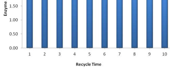
542                                  Cevahir Altinkaynak et al.

Figure 3. SEM images of HbNFs@Cu formed with varying hemoglobin concentration:(a) 0.01
          mg mL-1, (b) 0.02 mg mL-1, and (c) 0.05 mg mL-1 [PBS (10 mM, pH 8), Cu2+ (0.8 mM),
          +4 °C, 3 days incubation].

Figure 4. SEM images of HbNFs@Cu formed with varying hemoglobin concentration: (a) 0.01
          mg mL-1, (b) 0.02 mg mL-1, and (c) 0.05 mg mL-1 [PBS (10 mM, pH 9), Cu2+ (0.8 mM),
          +4 °C, 3 days incubation].

Characterization of HbNFs@Cu

The HbNFs@Cu synthesized under optimum conditions was characterized by EDX, XRD and
FTIR analyses. The presence of Cu, P, C, N and O in HbNFs@Cu was analyzed with EDX
technique. Corresponding peaks of Cu, P, and O arose from the copper phosphate crystal
structures. The presence of Cl was residual from the PBS buffer. The weight and atomic
percentage of HbNFs@Cu are 30.35 and 9.45, respectively. The given XRD spectrum shows the
peak positions and intensities of HbNFs@Cu. The diffraction peaks of HbNFs@Cu fit well with
the crystal pattern from Cu3(PO4)2.3H2O (JCPDS card (00-022-0548) [30–32]. The peaks at
8,925◦, 12,819◦, 20,543◦, 29,555◦, 33,797◦, 37,281◦, and 53,547◦ in the spectrum of HbNFs@Cu
belong to Cu3(PO4)2·3H2O nanostructures, indicating the inorganic composition of the hybrid
nanoflowers.
     The chemical structures of HbNFs@Cu and free hemoglobin were compared by FTIR
spectrums, where HbNFs@Cu accounts for all the peaks found in the free hemoglobin. These
results show the coordination between carboxyl/amino groups and copper ion and prove that the
hybrid structure was well crystallized. P-O group frequency observed around ~550 cm-1 has
shifted to frequencies of ~558 cm-1 and ~621 cm-1 in the immobilized structure. The specific bands
titled Amid III (~ 1384 cm-1), Amid II (~ 1523 cm-1), and Amid III (~ 1643 cm-1) were determined
at ~1400 cm-1, 1532 cm-1, and 1633 cm-1 in the hybrid material, respectively. The peaks in the
range of ~2940-3300 cm-1 were attributed to the -CH2 and -CH3 groups.

                               Bull. Chem. Soc. Ethiop. 2021, 35(3)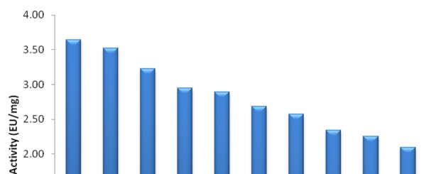
Peroxidase-like activity of hemoglobin-based hybrid materials against different substrates   543

Peroxidase-like activity profiles of HbNFs@Cu on different substrate and pH conditions

The enzymatic activity of the HbNFs@Cu was systematically investigated. The peroxidase-like
activities of the HbNFs@Cu were screened in the pH 5-9 range compared to free hemoglobin
using different substrates such as guaiacol and ABTS. The amount of enzyme catalyzing the
conversion of 1 mmol ABTS/Guaiacol substrate in one minute was calculated.
    Figure 5a and 5b show the peroxidase-like activities demonstrated by the free hemoglobin
protein to be very low in all screened pH parameters. When using guaiacol substrate, the highest
peroxidase-like activity was obtained at pH 9 and was 0.52 EU/mg (Figure 5a). The HbNFs@Cu
showed high activity against ABTS substrate in a wide pH range, but the highest activity was
calculated as 2.350 EU/mg at pH 5 (Figure 5b). According to the data, pH 5 and ABTS substrate
were determined optimal conditions for subsequent experiments. Unlike natural enzymes,
nanoflowers with peroxidase-like activity offer an increased enzymatic activity at extreme
conditions [33]. The HbNFs@Cu could act like a Fenton reagent in the presence of H2O2. In the
presence of HRP/peroxidase-like enzyme or material and ABTS, H2O2 could oxidize ABTS to
produce a pigmented ABTS·+ product [34]. The metal ions in the nanoflowers react with H2O2 to
produce metal1+ in the potential mechanism for the Fenton reaction, then metal1+ and H2O2 interact
to form a highly reactive hydroxyl radical (OH) [28, 34]. Some studies have demonstrated that
some copper compounds or iron compounds act as a Fenton-like reagent in an acidic environment,
which can catalyze the substrate to produce a pigmented product in the presence of H2O2 [28].

Michaelis-Menten kinetic parameters of HbNFs@Cu

To determine the optimal catalytic activities of HbNFs@Cu, the Lineaweaver-Burk plot was used
for different concentrations of ABTS (0.08-2.7 mM mL-1). The kinetic parameters of free
hemoglobin was not monitored because the peroxidase-like activity values were very low. KM and
Vmax values for HbNFs@Cu were 0.1357 mM/mL and 3.6995 EU/mg, respectively (Figure 6).
Both values were obtained at pH 5, and other conditions previously described.

                               Bull. Chem. Soc. Ethiop. 2021, 35(3)
544 Cevahir Altinkaynak et al.
Figure 5. Comparison of peroxidase-like activities at different pH values of HbNFs@Cu and free
hemoglobin using (a) guaiacol substrate and (b) ABTS substrate.
Figure 6. Lineweaver–Burk plot of the peroxidase-like activities of HbNFs@Cu at pH 5 using
ABTS substrate.
Bull. Chem. Soc. Ethiop. 2021, 35(3)Peroxidase-like activity of hemoglobin-based hybrid materials against different substrates 545
Reusability of HbNFs@Cu
As seen in Figure 7, HbNFs@Cu showed more than 57% of initial enzymatic activity after ten
uses. This result shows the durability of the material. Lee et al. [34] reported glutaraldehyde-
treated lipase nanoflowers exhibited more than 92% of their initial activity after 3 reuses. The
decrease of enzymatic activity may be attributed to the scattering of the patels in the centrifugation
process [29, 35].
Figure. 7. Reusability of HbNFs@Cu.
Colorimetric and spectrophotometric detection of H2O2 using HbNFs@Cu
The proposed biosensor system was based on colorimetric and spectrophotometric detection of
catalyzed H2O2 using HbNFs@Cu. After determining the optimum catalytic properties of the
HbNFs@Cu, varying concentrations of H2O2 from 0.003 mM to 0.667 mM were tested. Figure 8
shows the colorimetric color changes. The color of wells gradually changed from colorless to dark
green. As shown in Figure 9, a good linearity was determined in the range of 0.005-0.667 mM. It
is clear that H2O2 can be determined as a colorimetric sensor in the range of 0.005-0.667 mM in
UV spectrum. In addition, the limit of detection was 0.005 mM, colorimetrically. To assess the
specifity of biosensor system, the concentrations of H2O2 was decreased more. As shown in Figure
10, a good linearity was determined in the range of 0.003-0.0042 mM, spectrometrically. The
spectrometric limit of detection was determined to be 0.003 mM.
Lin et al. [14] investigated horseradish peroxidase (HRP)-inorganic hybrid nanoflowers as a
colorimetric platform for visual detection of hydrogen peroxide and found sensitive visual
detection of 20-500 µM linear range for hydrogen peroxide. Using Rhodamine 6G as a substrate
for detection of H2O2, Gao et al. [24] investigated organic/inorganic hybrid nanoflowers
containing hemoglobin as a biocatalyst to constructed colorimetric/fluorescent dual biosensors;
their colorimetric/fluorescent dual biosensors exhibited two linear responses in the range of 2-10
ppb and 20-100 ppb for H2O2. They also synthesized flower-like hemoglobin-Mn3(PO4)2 hybrid
Bull. Chem. Soc. Ethiop. 2021, 35(3)546 Cevahir Altinkaynak et al.
nanoflowers as an electrochemical sensing material to fabricate a H2O2 electrochemical biosensor.
Their biosensor displayed a linear response in the range of 20 nM–3.6 μM for H2O2 [25].
Figure. 8. Colorimetric detection of H2O2 using HbNFs@Cu.
Figure 9. (a) Absorbance values at different concentrations of H2O2 and (b) linear working range
of the colorimetric biosensor system at different concentrations of H2O2 (0.005-0.0042
mM).
In other previous studies, Xu et al. [22] fabricated a hemoglobin–silver sol in which the
hemoglobin showed direct electrochemistry on a glass carbon electrode with range of 1 × 10⁻⁶ to
2.5 × 10⁻² M for detection of H2O2. Liu et al. [36] synthesized TiO2 modified reduced graphene
oxide microspheres with immobilized hemoglobin to fabricate a mediator-free biosensor for
detection H2O2 within the linear range of 0.1-360 μM. Wang et al. [37] reported that tyrosine-
functionalized tetraphenylethene leads to fluorescence emission turn-on and fast detection of
H2O2 with high sensitivity and selectivity. The fluorogenic biosensor indicated a highly selective
enzymatic reactions with 0-50 µM linear range. Teodoro et al. [38] produced silver nanoparticles
that included polysaccharide (cellulose nanowhiskers), and tested as a colorimetric probe for
H2O2 detection. They determined the sensitivity of the colorimetric assay to lie within the range
of 0.01 μM to 600 μM H2O2. Qi et al. [39] designed a mediator-free H2 O2 biosensor by
immobilizing Hb on multiwalled carbon nanotubes modified with glassy carbon electrode with a
linear in the concentration range from 6.0 µM to 6.0 mM. Wan and co-workers [40] explained the
Bull. Chem. Soc. Ethiop. 2021, 35(3)Peroxidase-like activity of hemoglobin-based hybrid materials against different substrates 547
potential of nanozymes based on carbonaceous nanomaterials with their high stability and good
biocompatibility for a colorimetric and fluorescent dual modality platform capable of detecting
H2O2 and biomolecules (ascorbic acid-AA, acid phosphatase-ACP). Jamil and co-workers [41]
obtained a Cr2O3-TiO2-modified biomimetic paperbased-nanosensor to detect colorimetric
sensing of hydrogen peroxide. This paper-based colorimetric platform gave improved analytics
with a linear range of 0.005-100 μM and a 0.003 μM limit of detection. Jiang and co-workers [42]
synthesized the D-amino acid incorporating nanoflowers with peroxidase-like activity and a
diameter of 10-15 μm. The nanoflowers were used as a component for determining the level of
glutathione in the presence of TMB and H2O2.
Figure 10. (a) UV absorption spectrum values in the presence of different concentrations of H2O2
(0.003-0.667 mM and (b) linear working range of the spectrometric biosensor system
at varying concentrations of H2O2 (0.003-0.0042 mM).
CONCLUSION
There has been using a various hybrid materials obtained with organic-inorganic hybrid
nanoflowers preparation method for analytical assays in recently. In this article, HbNFs@Cu was
prepared at varying pH values and protein concentrations. The HbNFs@Cu product had a very
homogeneous structure and an average size of 9-10 µm under the optimized experimental
conditions. The encapsulation and weight efficiency was 100% and 6.7%, respectively.
Peroxidase-like activities were investigated against various substrates (ABTS and guaiacol) for
HbNFs@Cu. HbNFs@Cu performed to the enhanced peroxidase-like catalytic activity and
reusability. These peroxidase-like activities provided both colorimetric and spectrometric assays
for detection of H2O2. The linear operating range, detected colorimetrically as H2O2 sensor, was
0.005-0.0042 mM, while the linear operating range, detected spectrometrically, was 0.003-0.0042
mM. These results showed that hemoglobin-based organic-inorganic hybrid materials can be used
in potential applications to sense for H2O2.
Bull. Chem. Soc. Ethiop. 2021, 35(3)548 Cevahir Altinkaynak et al.
ACKNOWLEDGEMENTS
This study was supported by Scientific Research Projects Committee of Nevsehir Haci Bektas
Veli University (Project No: BAP18F10). We appreciate Yasin Polat at the Nevsehir Haci Bektas
Veli University for assistance with XRD analysis. We also appreciate the technical assistance
from Ihsan Aksit for SEM, EDX and FTIR analysis at Erciyes University. Please contact to the
corresponding author for supporting data.
REFERENCES
1. Gulmez, C.; Altinkaynak, C.; Özdemir, N.; Atakisi, O. Proteinase K hybrid nanoflowers (P-
HNFs) as a novel nanobiocatalytic detergent additive. Int. J. Biol. Macromol. 2018, 119, 803–
810.
2. Altinkaynak, C.; Kocazorbaz, E.; Özdemir, N.; Zihnioglu, F. Egg white hybrid nanoflower
(EW-HNF) with biomimetic polyphenol oxidase reactivity: Synthesis, characterization and
potential use in decolorization of synthetic dyes. Int. J. Biol. Macromol. 2018, 109, 205–211.
3. Al-Maqdi, K.A.; Bilal, M.; Alzamly, A.; Iqbal, H.M.N.; Shah, I.; Ashraf, S.S. Enzyme-loaded
flower-shaped nanomaterials: A versatile platform with biosensing, biocatalytic, and
environmental promise. Nanomaterials 2021, 11, 1460.
4. Eskin, A.; Ekremoglu, M.; Altinkaynak, C.; Özdemir, N. Effects of organic-inorganic hybrid
nanoflowers’ framework on hemocytes and enzymatic responses of the model organism,
Galleria mellonella (Lepidoptera: Pyralidae). Int. J. Trop. Insect. Sci. 2021, doi:
10.1007/s42690-021-00551-2.
5. Liu, Y.; Shao, X.; Kong, D.; Li, G.; Li, Q. Immobilization of thermophilic lipase in inorganic
hybrid nanoflower through biomimetic mineralization. Colloids Surf. B: Biointerfaces 2021,
197, 111450.
6. Özdemir, N.; Altinkaynak, C.; Türk, M.; Geçili, F.; Tavlaşoğlu, S. Amino acid-metal
phosphate hybrid nanoflowers (AaHNFs): Their preparation, characterization and anti-
oxidant capacities. Polym. Bull. 2021, DOI: https://doi.org/10.1007/s00289-021-03973-7.
7. Aydemir, D.; Gecili, F.; Özdemir, N.; Nuray Ulusu, N. Synthesis and characterization of a
triple enzyme-inorganic hybrid nanoflower (TrpE@ihNF) as a combination of three
pancreatic digestive enzymes amylase, protease and lipase. J. Biosci. Bioeng. 2020, 129, 679–
686.
8. Qiu, X.; Xiang, X.; Liu, T.; Huang, H.; Hu, Y. Fabrication of an organic-inorganic
nanocomposite carrier for enzyme immobilization based on metal-organic coordination.
Process Biochem. 2020, https://doi.org/10.1016/j.procbio.2020.05.007.
9. Shende, P.; Kasture, P.; Gaud, R.S. Nanoflowers: The future trend of nanotechnology for
multi-applications. Artif. Cells, Nanomed. Biotechnol. 2018, 46, 413–422.
10. Ahammad, A.J.S. Hydrogen peroxide biosensors based on horseradish peroxidase and
hemoglobin. J. Biosens. Bioelectron. 2012, s9, https://doi.org/10.4172/2155-6210.S9-001.
11. Lee, T.; Kim, T.-H.; Yoon, J.; Chung, Y.-H.; Lee, J.; Choi, J.-W. Investigation of
hemoglobin/gold nanoparticle heterolayer on micro-gap for electrochemical biosensor
application. Sensors 2016, 16, 660.
12. Mathew, M.; Sandhyarani, N.A Novel electrochemical sensor surface for the detection of
hydrogen peroxide using cyclic bisureas/gold nanoparticle composite. Biosens. Bioelectron.
2011, 28, 210–215.
13. Jv, Y.; Li, B.; Cao, R. Positively-charged gold nanoparticles as peroxidase mimic and their
application in hydrogen peroxide and glucose detection. Chem. Commun. (Camb.) 2010, 46,
8017–8019.
14. Lin, Z.; Xiao, Y.; Yin, Y.; Hu, W.; Liu, W.; Yang, H. Facile synthesis of enzyme-inorganic
hybrid nanoflowers and its application as a colorimetric platform for visual detection of
hydrogen peroxide and phenol. ACS Appl. Mater. Interfaces 2014, 6, 10775–10782.
Bull. Chem. Soc. Ethiop. 2021, 35(3)Peroxidase-like activity of hemoglobin-based hybrid materials against different substrates 549
15. Mu, J.; Zhang, L.; Zhao, M.; Wang, Y. Catalase mimic property of Co3O4 nanomaterials with
different morphology and its application as a calcium sensor. ACS Appl. Mater. Interfaces
2014, 6, 7090–7098.
16. Narwal, V.; Yadav, N.; Thakur, M.; Pundir, C.S. An amperometric H2O2 biosensor based on
hemoglobin nanoparticles immobilized on to a gold electrode. Biosci. Rep. 2017, 37,
https://doi.org/10.1042/BSR20170194.
17. Sezgintürk, M.K.; Dinçkaya, E. H2O2 Determination by a biosensor based on hemoglobin.
Prep. Biochem. Biotechnol. 2009, 39, 1–10. https://doi.org/10.1080/10826060802589361.
18. Jo, J.; Yoon, J.; Lee, T.; Cho, H.-Y.; Lee, J.-Y.; Choi, J.-W. H2O2 biosensor consisted of
hemoglobin-DNA conjugate on nanoporous gold thin film electrode with electrochemical
signal enhancement. Nano Convergence 2019, 6, 1. https://doi.org/10.1186/s40580-018-
0172-z.
19. Zhang, K.; Cai, R.; Chen, D.; Mao, L. Determination of hemoglobin based on its enzymatic
activity for the oxidation of O-phenylenediamine with hydrogen peroxide. Anal. Chim. Acta
2000, 413, 109–113.
20. Zhang, K.; Mao, L.; Cai, R. Stopped-flow spectrophotometric determination of hydrogen
peroxide with hemoglobin as catalyst. Talanta 2000, 51, 179–186.
21. Guo, F.; Xu, X.X.; Sun, Z.Z.; Zhang, J.X.; Meng, Z.X.; Zheng, W.; Zhou, H.M.; Wang, B.L.;
Zheng, Y.F. A novel amperometric hydrogen peroxide biosensor based on electrospun Hb–
collagen composite. Colloids Surf. B: Biointerfaces 2011, 86, 140–145.
22. Xu, Y.; Hu, C.; Hu, S. A hydrogen peroxide biosensor based on direct electrochemistry of
hemoglobin in Hb–Ag sol films. Sens. Actuators B: Chem. 2008, 130, 816–822.
23. Gao, F.; Yuan, R.; Chai, Y.; Tang, M.; Cao, S.; Chen, S. Amperometric third-generation
hydrogen peroxide biosensor based on immobilization of Hb on gold
nanoparticles/cysteine/poly(p-aminobenzene sulfonic acid)-modified platinum disk electrode.
Colloids Surf. A: Physicochem. Eng. Aspects 2007, 295, 223–227.
24. Gao, J.; Liu, H.; Pang, L.; Guo, K.; Li, J. A biocatalyst and colorimetric/fluorescent dual
biosensors of H2O2 constructed via hemoglobin-Cu3(PO4)2 organic/inorganic hybrid
nanoflowers. ACS Appl. Mater. Interfaces 2018, 10, https://doi.org/10.1021/acsami.8b10968.
25. Gao, J.; Liu, H.; Tong, C.; Pang, L.; Feng, Y.; Zuo, M.; Wei, Z.; Li, J. Hemoglobin-Mn3(PO4)2
hybrid nanoflower with opulent electroactive centers for high-performance hydrogen peroxide
electrochemical biosensor. Sens. Actuators B: Chem. 2020, 307, 127628.
26. Wu, D.; Feng, M.; Wang, Z.-X.; Qiao, K.; Tachibana, H.; Cheng, X.-J. Molecular and
biochemical characterization of key enzymes in the cysteine and serine metabolic pathways
of Acanthamoeba castellanii. Parasite Vectors 2018, 11, 604.
27. Wang, K.; Wang, J.; Hu, W.; Zhang, Y.; Zhi, F.; Zhou, Z.; Wu, J.; Hu, Y. Acid denaturation
inducing self-assembly of curcumin-loaded hemoglobin nanoparticles. Materials 2015, 8,
8701–8713.
28. Huang, Y.; Ran, X.; Lin, Y.; Ren, J.; Qu, X. Self-assembly of an organic–inorganic hybrid
nanoflower as an efficient biomimetic catalyst for self-activated tandem reactions. Chem.
Commun. 2015, 51, 4386–4389.
29. Nadar, S.S.; Gawas, S.D.; Rathod, V.K. Self-assembled organic-inorganic hybrid
glucoamylase nanoflowers with enhanced activity and stability. Int. J. Biol. Macromol. 2016,
92, 660–669.
30. Batule, B.S.; Park, K.S.; Kim, M.I.; Park, H.G. Ultrafast sonochemical synthesis of protein-
inorganic nanoflowers. Int. J. Nanomedicine 2015, 10, 137–142.
31. Ge, J.; Lei, J.; Zare, R.N. Protein-inorganic hybrid nanoflowers. Nat. Nanotechnol. 2012, 7,
428–432.
32. Somturk, B.; Hancer, M.; Ocsoy, I.; Özdemir, N. Synthesis of copper ion incorporated
horseradish peroxidase-based hybrid nanoflowers for enhanced catalytic activity and stability.
Dalton Trans. 2015, 44, 13845–13852.
Bull. Chem. Soc. Ethiop. 2021, 35(3)550 Cevahir Altinkaynak et al.
33. Singh, S. Nanomaterials exhibiting enzyme-like properties (nanozymes): Current advances
and future perspectives. Front. Chem. 2019, 7, 46.
34. Wei, H.; Wang, E. Fe3O4 magnetic nanoparticles as peroxidase mimetics and their applications
in H2O2 and glucose detection. Anal. Chem. 2008, 80, 2250–2254.
35. Lee, H.R.; Chung, M.; Kim, M.I.; Ha, S.H. Preparation of glutaraldehyde-treated lipase-
inorganic hybrid nanoflowers and their catalytic performance as immobilized enzymes.
Enzyme Microb. Technol. 2017, 105, 24–29.
36. Wang, X.; Shi, J.; Li, Z.; Zhang, S.; Wu, H.; Jiang, Z.; Yang, C.; Tian, C. Facile one-pot
preparation of chitosan/calcium pyrophosphate hybrid microflowers. ACS Appl. Mater.
Interfaces 2014, 6, 14522–14532.
37. Liu, H.; Guo, K.; Duan, C.; Dong, X.; Gao, J. Hollow TiO2 modified reduced graphene oxide
microspheres encapsulating hemoglobin for a mediator-free biosensor. Biosens. Bioelectron.
2017, 87, 473–479.
38. Wang, X.; Hu, J.; Zhang, G.; Liu, S. Highly selective fluorogenic multianalyte biosensors
constructed via enzyme-catalyzed coupling and aggregation-induced emission. J. Am. Chem.
Soc. 2014, 136, 9890–9893.
39. Teodoro, K.B.R.; Migliorini, F.L.; Christinelli, W.A.; Correa, D.S. Detection of hydrogen
peroxide (H2O2) using a colorimetric sensor based on cellulose nanowhiskers and silver
nanoparticles. Carbohydr. Polym. 2019, 212, 235–241.
40. Chen, S.; Yuan, R.; Chai, Y.; Zhang, L.; Wang, N.; Li, X. Amperometric third-generation
hydrogen peroxide biosensor based on the immobilization of hemoglobin on multiwall carbon
nanotubes and gold colloidal nanoparticles. Biosens. Bioelectron. 2007, 22, 1268–1274.
41. Wan, Y.; Zhao, J.; Deng, X.; Chen, J.; Xi, F.; Wang, X. Colorimetric and fluorescent dual-
modality sensing platform based on fluorescent nanozyme. Front. Chem. 2021, 9, 774486.
42. Jamil, S.; Nasir, M.; Ali, Y.; Nadeem, S.; Rashid, S.; Javed, M.Y.; Hayat, A. Cr2O3-TiO2-
modified filter paper-based portable nanosensors for optical and colorimetric detection of
hydrogen peroxide. ACS Omega 2021, 6, 23368–23377.
43. Jiang, N.; Zhang, C.; Li, M.; Li, S.; Hao, Z.; Li, Z.; Wu, Z.; Li, C. The fabrication of amino
acid incorporated nanoflowers with intrinsic peroxidase-like activity and its application for
efficiently determining glutathione with TMB radical cation as indicator. Micromachines
(Basel) 2021, 12, 1099.
Bull. Chem. Soc. Ethiop. 2021, 35(3)You can also read