Prevalence and characteristics of mitral valve prolapse in military young adults in Taiwan of the CHIEF Heart Study
←
→
Page content transcription
If your browser does not render page correctly, please read the page content below
www.nature.com/scientificreports
OPEN Prevalence and characteristics
of mitral valve prolapse
in military young adults in Taiwan
of the CHIEF Heart Study
Pang‑Yen Liu1, Kun‑Zhe Tsai2, Yen‑Po Lin3, Chin‑Sheng Lin1, Huan‑Chang Zeng2,4,
Eiki Takimoto5 & Gen‑Min Lin1,2,6*
The prevalence of mitral valve prolapse (MVP) among middle- and older-aged individuals is estimated
to be 2–4% in Western countries. However, few studies have been conducted among Asian individuals
and young adults. This study included a sample of 2442 consecutive military adults aged 18–39 years
in Hualien, Taiwan. MVP was defined as displacement of the anterior or posterior leaflet of the
mitral valve to the mid portion of the annular hinge point > 2 mm in the parasternal long-axis view of
echocardiography. Cardiac chamber size and wall thickness were measured based on the latest criteria
of the American Society of Echocardiography. The clinical features of participants with MVP and those
without MVP were compared using a two-sample t test, and the cardiac structures were compared
using analysis of covariance with adjustment for body surface area (BSA). Eighty-two participants
were diagnosed with MVP, and the prevalence was 3.36% in the overall population. Compared with
those without MVP, participants with MVP had a lower body mass index (kg/m2) (24.89 ± 3.70 vs.
23.91 ± 3.45, p = 0.02) and higher prevalence of somatic symptoms related to exercise (11.0% vs.
4.9%, p = 0.02) and systolic click in auscultation (18.3% vs. 0.6%, p < 0.01). In addition, participants
with MVP had greater left ventricular mass (gm) and smaller right ventricular wall thickness (mm)
and dimensions (mm) indexed by BSA than those without MVP (149.12 ± 35.76 vs. 155.38 ± 36.26;
4.66 ± 0.63 vs. 4.40 ± 0.68; 26.57 ± 3.99 vs. 25.41 ± 4.35, respectively, all p-values < 0.01). In conclusion,
the prevalence and clinical features of MVP in military young adults in Taiwan were in line with those
in Western countries. Whether the novel MVP phenotype found in this study has any pathological
meaning needs further investigation.
Mitral valve prolapse (MVP) is one of the most common valvular heart diseases, with a prevalence of 2.0–4.0%
in the general p opulation1–6. MVP is composed of heterogeneous types with varying clinical presentations. Syn-
dromic MVP commonly coexists with heritable connective tissue disorders such as Ehlers-Danlos syndrome and
Marfan syndrome7–9, and familial nonsyndromic MVP usually exhibits autosomal dominant inheritance and age-
and sex-dependent c haracteristics10. Other types are grouped as sporadic nonsyndromic MVP. A floppy mitral
VP11 and can be assessed by imaging and physical examinations.
valve plays a pivotal role in the pathogenesis of M
Systolic click in cardiac auscultation is a typical finding for floppy mitral valve, and the prevalence is estimated
from 10 to 70% in patients with MVP1,12. A specific heart sound has emerged in patients after 30 years of age,
and the intensity increases with more severe MVP and decreases with a higher grade of mitral r egurgitation11,13.
In addition, several somatic symptoms, also called MVP syndrome, such as chest pain, palpitation, and dyspnea,
appear early in adolescent and young adult patients, possibly due to low stroke volume and autonomic neural
feedback related to MVP11.
1
Division of Cardiology, Department of Internal Medicine, Tri-Service General Hospital and National Defense
Medical Center, Taipei, Taiwan. 2Department of Internal Medicine, Hualien Armed Forces General Hospital,
No. 100, Jinfeng St., Hualien City 970, Taiwan. 3Department of Critical Care Medicine, Taipei Tzu Chi General
Hospital, New Taipei City, Taiwan. 4GENEUS Medical Technology Co., New Taipei City, Taiwan. 5Department of
Cardiovascular Medicine, School of Medicine, University of Tokyo, Tokyo, Japan. 6Department of Preventive
Medicine, Northwestern University Feinberg School of Medicine, Chicago, IL 60611, USA. *email: farmer507@
yahoo.com.tw
Scientific Reports | (2021) 11:2719 | https://doi.org/10.1038/s41598-021-81648-z 1
Vol.:(0123456789)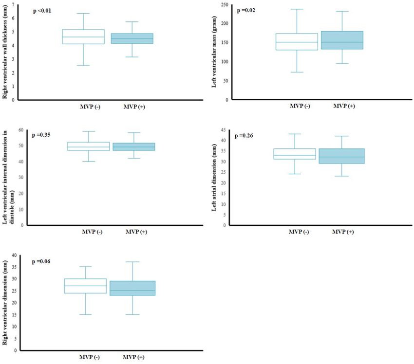
www.nature.com/scientificreports/
Although the Framingham Heart Study1 has shown that MVP is regarded as a benign condition with a low
incidence of complications in the general population, numerous studies have uncovered several adverse events,
such as ischemic s troke14, bacterial endocarditis15,16, severe mitral r egurgitation2,4,17, lethal a rrhythmia18,19 and
sudden cardiac d eath20 related to MVP. In addition to the Framingham Heart Study that mainly Caucasian
21,22
participants , the clinical characteristics of MVP were also investigated in a cohort of American Indians in
the Strong Heart Study2, and in a small sample of Canadian adolescents of European, South Asian and Chinese
descent in the Study of Health Assessment and Risk in Ethnic groups (SHARE)5. Notably, the SHARE study
revealed no ethnic differences in the prevalence and clinical features of MVP. However, there has been no large
population study carried out in Asia investigating the epidemiology of MVP. Therefore, the purpose of this
study was to determine the prevalence, clinical features and echocardiographic profiles of MVP in a large Asian
military cohort in T aiwan23,24.
Methods
Study population. The study used 2442 young male and female adults in the military, aged 18–39 years,
from the cardiorespiratory fitness and hospitalization events in armed forces (CHIEF) study performed in Tai-
wan during 2014–201825. All participants underwent a comprehensive physical examination, and completed a
self-reported questionnaire for their family history of inherited diseases and experience regarding toxic sub-
stance use, somatic symptoms such as easy palpitation, fatigue, or dyspnea during exercise and physical activ-
ity in the Hualien Armed Forces General Hospital of Taiwan. Of these, there were a total of 2442 participants
undergoing transthoracic echocardiography for their rank promotions and awards. The rationale and design of
the CHIEF study have been described in detail previously23,24,26,27.
Measurements of transthoracic echocardiography. All procedures of transthoracic echocardiogra-
phy using a 1–5 MHz transducer (iE33; Philips Medical Systems, Andover, MA, USA) were performed by the
same experienced and certified technician and verified by the cardiologist (Lin GM) at the Hualien-Armed
Forces General Hospital. All participants were examined using parasternal long-axis and short-axis approaches
and apical four- and two-chamber views in supine and left lateral positions. According to the suggestions of
the American Society of E chocardiography28, quantification of LV wall thickness (interventricular septal and
posterior walls) and chamber dimension were measured approximately at the onset of the QRS complex of end
diastole and tips of the mitral valve by M-mode and two-dimensional measurements in parasternal long axis
view. In the parasternal long-axis view, MVP was defined as a displacement of the anterior or posterior leaflet of
the mitral valve to the mid portions of the mitral annular hinge point > 2 mm1,14,29–31. LV mass was thus calcu-
lated according to the corrected formula proposed by Devereux et al.32. LV mass = 0.8 × {1.04 × [(left ventricular
internal diameter at end diastole (LVIDd) + end diastolic posterior wall thickness + end diastolic interventricular
septal thickness]3 − LVIDd3} + 0.6. Echocardiographic LV hypertrophy was defined as LV mass divided by body
surface area (BSA) based on the Dubois formula33, as the cutoff value ≥ 88 g/m2,34. Measurements of anterior
right ventricular (RV) wall thickness were performed by dimensional windows and M-mode at the onset of
the QRS complex of end diastole via parasternal long-axis a pproaches35. Echocardiographic RV hypertrophy
was defined as anterior RV wall thickness > 5.2 mm, which was the 95th percentile in our young adult cohort in
Taiwan36.
Statistical analysis. Characteristics and echocardiographic parameters of the participants with MVP and
those without MVP are expressed as the mean ± standard deviation (SD) for continuous variables and numbers
(percentage) for categorical variables, respectively. Continuous variables were compared by two-sample t test
and categorical variables were compared by the chi-square or Fisher’s exact test. Dimensions of aortic root, left
atrial dimension, LV chamber, RV outflow tract, and thickness of LV and RV wall, and LV mass were compared
using analysis of covariance (ANCOVA) with adjustment for BSA. A two-tailed value of P < 0.05 was considered
significant. All analyses were performed using SAS version 9.4 (SAS Institute Inc, Cary, NC, USA).
Ethics statement. This study was reviewed and approved by the Institutional Review Board of Mennonite
Christian Hospital (No. 16-05-008) in Taiwan, and written informed consent was obtained from all participants.
All methods were performed in accordance with the relevant guidelines and regulations.
Ethics approval and consent to participate. Since there was no research ethics committee regularly
held in the Hualien Armed Forces General Hospital, this study applied to the Institutional Review Board (IRB)
of the Mennonite Christian Hospital (No. 16-05-008) in Hualien, Taiwan, to approve access to the data for the
CHIEF Heart study, and written informed consent was obtained from all participants.
Results
Clinical features and laboratory findings. Of the participants in the CHIEF study, there were 2154
males (88.2%) and 288 females (11.8%) participants (Table 1). Eighty-two participants (3.3%) had MVP, which
was more prevalent in males (77 of 2154 [3.6%] in males vs. 5 of 288 [1.7%] in females). Participants with MVP
had lower levels of body mass index (23.78 kg/m2 vs. 24.65 kg/m2, P = 0.03) and fasting plasma glucose (90.3 mg/
dL vs. 93.63 mg/dL, P = 0.01), and had a higher prevalence of intolerable somatic symptoms including chest
pain, dyspnea or easy palpitation during exercise (MVP syndrome, 9 of 82 [11.0%]) than those without MVP.
In addition, 7 of the 82 participants with MVP (8.5%) were found to have mild pectus excavatum, a kind of
chest wall abnormality, which was a higher percentage than among those without MVP (0.6%, p < 0.01). There
Scientific Reports | (2021) 11:2719 | https://doi.org/10.1038/s41598-021-81648-z 2
Vol:.(1234567890)www.nature.com/scientificreports/
Total (n = 2442) Males (n = 254) Females (n = 288)
MVP (−) MVP (+) MVP (−) MVP (+) MVP (−) MVP (+)
(n = 2360) (n = 82) p-value (n = 2077) (n = 77) p-value (n = 283) (n = 5) p-value
Age 27.36 ± 5.62 26.51 ± 5.82 0.18 27.51 ± 5.70 26.48 ± 5.89 0.12 26.29 ± 4.88 27.00 ± 5.15 0.74
Height
170.78 ± 6.75 171.58 ± 5.97 0.28 172.12 ± 5.79 172.34 ± 5.19 0.74 160.93 ± 4.90 159.98 ± 5.50 0.66
(cm)
Weight
72.12 ± 12.79 70.17 ± 11.68 0.17 73.83 ± 12.24 71.11 ± 11.38 0.05 59.52 ± 9.27 55.66 ± 4.75 0.35
(kg)
Body
mass
24.65 ± 3.69 23.78 ± 3.43 0.03 24.89 ± 3.70 23.91 ± 3.45 0.02 22.94 ± 3.16 21.81 ± 2.53 0.42
index
(kg/m2)
Body
surface
1.84 ± 0.19 1.82 ± 0.17 0.33 1.87 ± 0.17 1.84 ± 0.16 0.09 1.62 ± 0.14 1.57 ± 0.07 0.37
area
(m2)
Waist
circum-
82.57 ± 9.99 81.08 ± 10.03 0.18 83.59 ± 9.80 81.51 ± 10.05 0.06 75.10 ± 8.04 74.40 ± 7.47 0.84
ference
(cm)
Systolic
blood
pressure 118.07 ± 13.72 116.50 ± 15.64 0.31 119.65 ± 13.30 117.32 ± 15.43 0.13 106.41 ± 10.90 103.80 ± 14.62 0.59
(mm
Hg)
Dias-
tolic
blood
70.04 ± 10.40 70.03 ± 12.02 0.97 70.78 ± 10.43 71.01 ± 11.35 0.85 64.52 ± 8.31 54.40 ± 12.44 < 0.01
pressure
(mm
Hg)
Heart
rate
66.72 ± 10.83 65.44 ± 10.87 0.29 66.53 ± 10.89 65.44 ± 11.09 0.39 68.17 ± 10.26 65.40 ± 7.40 0.54
(beats/
minute)
Somatic
symp- 116 [4.9] 9 [11.0] 0.02 96 [4.6] 7 [9.1] 0.07 20 [7.0] 2 [40.0] 0.06
toms
Pectus
excava- 13 [0.6] 7 [8.5] < 0.01 12 [0.6] 6 [7.8] < 0.01 1 [0.4] 1 [20.0] < 0.01
tum
Current
alcohol 1048 [44.4] 43 [52.4] 0.15 995 [47.9] 43 [55.8] 0.17 53 [18.7] 0 [0.0] 0.28
intake
Current
cigarette
967 [41.0] 30 [36.6] 0.42 912 [43.9] 30 [39.0] 0.39 55 [19.4] 0 [0.0] 0.27
smok-
ing
Serum
creati-
nine 0.92 ± 0.14 0.94 ± 0.13 0.36 0.96 ± 0.12 0.96 ± 0.12 0.98 0.68 ± 0.09 0.66 ± 0.06 0.51
(mg/
dL)
Total
choles-
terol 171.00 ± 33.36 167.43 ± 29.08 0.33 171.62 ± 33.87 167.56 ± 29.76 0.29 166.40 ± 29.02 165.40 ± 17.01 0.93
(mg/
dL)
HDL-C
(mg/ 49.78 ± 10.69 50.50 ± 10.60 0.54 48.57 ± 9.97 50.12 ± 10.80 0.18 58.67 ± 11.61 56.40 ± 3.78 0.66
dL)
LDL-C
(mg/ 103.85 ± 28.93 99.71 ± 25.83 0.20 105.49 ± 29.23 100.13 ± 26.34 0.11 91.80 ± 23.33 93.20 ± 16.56 0.89
dL)
Serum
triglyc-
eride 104.01 ± 84.10 91.16 ± 56.49 0.17 108.95 ± 87.48 91.86 ± 57.46 0.08 67.69 ± 36.24 80.40 ± 41.72 0.43
(mg/
dL)
Fasting
plasma
glucose 93.63 ± 11.89 90.30 ± 9.76 0.01 94.01 ± 12.34 90.25 ± 9.77 < 0.01 90.85 ± 7.22 91.20 ± 10.52 0.91
(mg/
dL)
Hemo-
globin 14.97 ± 1.25 15.12 ± 1.24 0.28 15.25 ± 0.96 15.28 ± 0.80 0.82 12.87 ± 1.10 12.62 ± 1.11 0.61
(g/dL)
Scientific Reports | (2021) 11:2719 | https://doi.org/10.1038/s41598-021-81648-z 3
Vol.:(0123456789)www.nature.com/scientificreports/
Table 1. Characteristics of young male and female adult participants with mitral valve prolapse and those
without. Continuous variables are expressed as mean ± SD (standard deviation), and categorical variables as N
[%]. HDL-C high-density lipoprotein cholesterol, LDL-C low-density lipoprotein cholesterol, MVP mitral valve
prolapse.
Total (n = 242) Males (n = 254) Females (n = 288)
MVP (−) MVP (+) MVP (−) MVP (+) MVP (−) MVP (+)
(n = 2360) (n = 82) p-value (n = 2077) (n = 77) p-value (n = 283) (n = 5) p-value
Cardiac geometry*
Aortic valve open (mm), PLAX 19.84 ± 2.04 19.46 ± 2.08 0.15 20.13 ± 1.91 19.68 ± 1.96 0.10 17.75 ± 1.67 16.20 ± 0.45 0.05
Aortic root dimension (mm), PALX 29.53 ± 3.53 29.48 ± 2.67 0.87 29.95 ± 3.44 29.74 ± 2.53 0.78 26.40 ± 2.34 25.40 ± 1.14 0.55
LV mass (gm), PLAX 149.12 ± 35.76 155.38 ± 36.26 < 0.01 154.82 ± 33.55 158.70 ± 34.82 0.02 107.26 ± 20.44 104.21 ± 9.73 0.83
LV posterior wall (mm), PLAX 8.53 ± 1.11 8.73 ± 1.13 0.01 8.69 ± 1.05 8.84 ± 1.08 0.03 7.36 ± 0.79 7.00 ± 0.00 0.47
LV internal dimension in diastole (mm),
48.78 ± 3.71 49.10 ± 3.40 0.16 49.26 ± 3.51 49.35 ± 3.32 0.35 45.22 ± 3.16 45.20 ± 2.28 0.69
PLAX
LV internal dimension in systole (mm),
30.56 ± 3.44 30.64 ± 3.39 0.74 30.94 ± 3.38 30.73 ± 3.45 0.93 27.87 ± 2.55 29.00 ± 1.72 0.37
PLAX
Interventricular septum, (mm), PLAX 8.74 ± 1.21 8.95 ± 1.13 0.02 8.91 ± 1.15 9.04 ± 1.11 0.08 7.48 ± 0.85 7.60 ± 0.55 0.45
RV wall thickness (mm)a, PLAX 4.66 ± 0.63 4.40 ± 0.68 < 0.01 4.70 ± 0.62 4.42 ± 0.69 < 0.01 4.33 ± 0.58 4.07 ± 0.50 0.50
RV outflow tract dimension in diastole
26.57 ± 3.99 25.41 ± 4.35 0.01 26.79 ± 3.97 25.68 ± 4.35 0.06 24.96 ± 3.76 21.40 ± 1.14 0.05
(mm), PLAX
Left atrial dimension (mm), PLAX 32.88 ± 4.06 32.10 ± 4.17 0.15 33.22 ± 4.06 32.32 ± 4.18 0.26 30.38 ± 3.03 28.60 ± 2.07 0.33
LV hypertrophy 114 [4.8] 3 [3.7] 0.62 102 [4.9] 3 [3.9] 0.68 12 [4.2] 0 [00.0] 0.63
RV hypertrophya 206 [10.1] 2 [2.9] 0.04 187 [10.5] 2 [3.1] 0.05 19 [7.5] 0 [00.0] 0.52
Cardiac functional assessment
LV ejection fraction (%), PLAX 61.90 ± 5.06 62.61 ± 4.23 0.21 61.97 ± 5.13 62.64 ± 4.32 0.26 61.43 ± 4.44 62.20 ± 2.59 0.70
Tricuspid valve prolapse, PSAX 388 [16.4] 17 [20.7] 0.30 350 [16.9] 16 [20.8] 0.36 38 [13.4] 1 [20.0] 0.67
Aortic regurgitation ≥ mild grade 38 [1.6] 1 [1.2] 0.78 34 [1.6] 1 [1.3] 0.81 4 [1.4] 0 [0.0] 0.78
Mitral regurgitation ≥ mild grade 1760 [74.6] 69 [84.1] 0.04 1544 [74.3] 65 [84.4] 0.04 216 [76.3] 4 [80.0] 0.84
Pulmonary regurgitation ≥ mild grade 1442 [61.1] 53 [64.6] 0.51 1293 [62.3] 50 [64.9] 0.63 149 [52.7] 3 [60.0] 0.74
Tricuspid regurgitation ≥ mild grade 1884 [79.8] 62 [75.6] 0.35 1657 [79.8] 60 [77.9] 0.69 227 [80.2] 2 [40.0] 0.02
RV systolic pressure (mmHg) 27.31 ± 4.67 26.63 ± 4.62 0.19 27.39 ± 4.70 26.81 ± 4.66 0.29 26.73 ± 4.35 23.80 ± 2.78 0.13
Peak E velocity (cm/s) 85.84 ± 15.49 86.26 ± 14.70 0.81 84.69 ± 15.02 86.02 ± 15.03 0.44 93.91 ± 17.17 89.90 ± 8.41 0.60
Peak A velocity (cm/s) 49.46 ± 10.72 45.87 ± 9.83 < 0.01 49.28 ± 10.67 46.01 ± 10.06 < 0.01 50.80 ± 11.04 43.78 ± 5.41 0.15
E/A ratio 1.81 ± 0.50 1.95 ± 0.46 0.01 1.79 ± 0.49 1.94 ± 0.48 0.01 1.94 ± 0.57 2.06 ± 0.10 0.65
Peak E′ velocity (cm/s) 16.73 ± 5.72 15.98 ± 3.19 0.24 16.56 ± 5.93 15.94 ± 3.23 0.37 17.99 ± 3.70 16.54 ± 2.80 0.38
Peal A′ velocity (cm/s) 8.30 ± 3.17 7.34 ± 1.98 < 0.01 8.28 ± 3.27 7.34 ± 2.01 0.01 8.47 ± 2.27 7.31 ± 1.48 0.25
E′/A′ ratio 2.19 ± 1.08 2.34 ± 0.84 0.21 2.18 ± 1.12 2.34 ± 0.85 0.21 2.26 ± 0.69 2.35 ± 0.64 0.76
Table 2. Echocardiographic findings of young male and female participants with mitral valve prolapse and
those without. Continuous variables are expressed as mean ± SD (standard deviation), and categorical variables
as N [%]. BSA body surface area, LV left ventricle, RV right ventricle, PLAX echocardiographic parasternal
long axis view, PSAX echocardiographic parasternal short axis view a N = 2102 in total (N = 1842 for males and
N = 260 for females). *indicates the p-value for the echocardiographic geometry variables with adjustment for
body surface area.
were no significant differences in waist circumference, BSA, heart rate, systolic blood pressure, diastolic blood
pressure, alcohol consumption, tobacco smoking, serum creatinine, total cholesterol, high-density lipoprotein,
low-density lipoprotein, or hemoglobin concentrations. There was a sex difference in diastolic blood pressure.
Females with MVP had a lower diastolic blood pressure than those without MVP (54.4 mmHg vs. 64.5 mmHg,
P < 0.01), while this difference did not appear in males.
Echocardiographic findings in all participants. Table 2 showed the results of a comparison of the car-
diac geometry and function assessment between groups. With regard to the cardiac geometry adjusted for BSA,
there were no differences in the aortic root, LV chamber, or the left atrial dimension between groups. However,
the participants with MVP had lower levels of RV outflow tract dimension in diastole (25.41 ± 4.35 mm vs.
26.57 ± 3.99 mm, P = 0.01) and RV wall thickness (4.40 ± 0.68 mm vs. 4.66 ± 0.63 mm, P = 0.001) than those with-
out MVP. In contrast, there were greater levels of LV mass (155.38 ± 36.26 gm vs. 149.12 ± 35.76 gm, P < 0.01),
Scientific Reports | (2021) 11:2719 | https://doi.org/10.1038/s41598-021-81648-z 4
Vol:.(1234567890)www.nature.com/scientificreports/
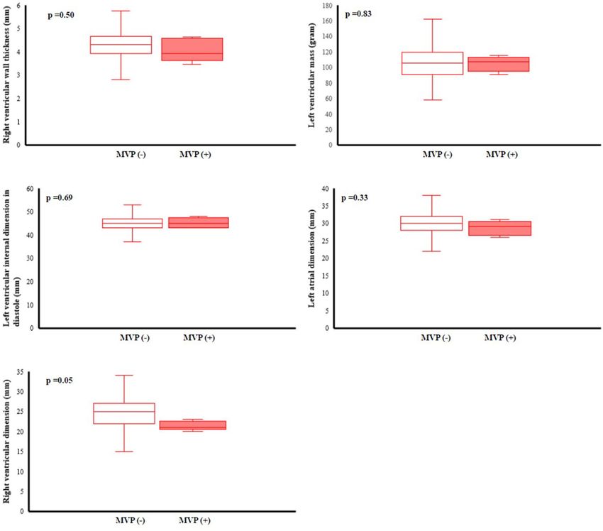
Figure 1. Echocardiographic left atrial dimension, left ventricular internal dimension in diastole, left
ventricular mass, right ventricular outflow tract dimension in diastole and right ventricular wall thickness in the
overall population were compared using analysis of covariance with adjustment for body surface area.
interventricular septal thickness (8.95 ± 1.13 mm vs. 8.74 ± 1.21 mm, P = 0.02) and LV posterior wall thickness
(8.73 ± 1.13 mm vs. 8.53 ± 1.11 mm, P = 0.01) in the participants with MVP than those without MVP. It is also
notable that the pattern of RV geometry adjusted for BSA in MVP in the overall participants (Fig. 1) were con-
sistent in both males (Fig. 2) and females (Fig. 3), whereas the pattern of a greater LV mass in MVP was only pre-
sented in males but not females. Participants with MVP had a higher prevalence of mitral regurgitation, at least
mild grade, than those without MVP (84.1% vs. 74.6%, P = 0.04), but showed no differences in the prevalence of
aortic, pulmonary or tricuspid regurgitation or tricuspid valve prolapse. In addition, there was no difference in
LV ejection fraction between groups. With regard to the diastolic filling, the participants with MVP had a higher
E/A ratio (1.95 ± 0.46 vs. 1.81 ± 0.50, P = 0.01) and a lower late diastolic peak A velocity (45.87 ± 9.83 cm/s vs.
49.46 ± 10.72 cm/s, P < 0.01) but no difference in early diastolic peak E velocity.
Echocardiographic findings in participants without mitral regurgitation. Since the LV and RV
geometry could be influenced by the presence of mitral regurgitation, the echocardiographic characteristics in
those free of mitral regurgitation were also analyzed (n = 613) in Table 3. The results showed that the participants
with MVP had similar levels of LV mass, LV chamber size and anterior RV wall thickness indexed by BSA com-
pared to those without MVP. Participants with MVP had a smaller RV outflow tract dimension indexed by BSA
Scientific Reports | (2021) 11:2719 | https://doi.org/10.1038/s41598-021-81648-z 5
Vol.:(0123456789)www.nature.com/scientificreports/
Figure 2. Echocardiographic left atrial dimension, left ventricular internal dimension in diastole, left
ventricular mass, right ventricular outflow tract dimension in diastole and right ventricular wall thickness in
males were compared using analysis of covariance with adjustment for body surface area.
(25.23 ± 5.21 mm vs. 27.16 ± 4.04 mm, P = 0.05) and a lower late diastolic peak A velocity (50.80 ± 11.38 cm/s vs.
43.19 ± 9.11 cm/s, P = 0.01) than those without MVP.
Discussion
The principal findings in this population-based study were that the prevalence of MVP in an unselected young-
aged military cohort in Taiwan was 3.36%, and the clinical features of those with MVP included a higher preva-
lence of somatic symptoms related to exercise and manifested with both lower body mass index and fasting
plasma glucose concentration. In addition, a new phenotype of MVP in cardiac geometry presented by greater
LV mass and smaller RV wall thickness and RV dimension adjusted to BSA was observed in military individu-
als in Taiwan.
This was the second study following a Japanese population study37 to provide information on the prevalence
and correlates of MVP in a large population in Asia, specifically in Taiwan. The prevalence of MVP reported in a
previous case–control study in Taiwan was estimated to be 1.92% in a population of ethnic Taiwanese individuals
with a mean age of 49.2 years36. However, their study subjects were retrieved from the Nationwide Health Insur-
ance Database in Taiwan, and the cases were selected merely on the basis of the International Classification of
Diseases, 9th Revision, Clinical Modification code for M VP38, which might underestimate the prevalence of MVP.
In the present study, 3.3% of young adults had MVP, falling toward the higher end of the range among previous
population-based studies1,2,5,6,21. This might be attributed in part to using a displacement over 2 mm of mitral
valve as the only criterion for defining MVP, regardless of considering the thickness of leaflets1,29 and possibly
increasing the sensitivity in detection. In the Coronary Artery Risk Development in Young Adults (CARDIA)
Scientific Reports | (2021) 11:2719 | https://doi.org/10.1038/s41598-021-81648-z 6
Vol:.(1234567890)www.nature.com/scientificreports/
Figure 3. Echocardiographic left atrial dimension, left ventricular internal dimension in diastole, left
ventricular mass, right ventricular outflow tract dimension in diastole and right ventricular wall thickness in
females were compared using analysis of covariance with adjustment for body surface area.
study, the prevalence of MVP was estimated to be 0.6% using the same echocardiographic criterion in a similar
age group3 and is lower than that in our study population. While the CARDIA study comprised biethnic samples
of black and white individuals3, the ethnic group in this study was Asian adults. Therefore, the discrepancy may
arise from the different ethnic groups.
This was consistent with previous study findings that those with MVP presented prevalent somatic symptoms
associated with exercise and favorable metabolic features, including leaner body mass index and lower fasting
Scientific Reports | (2021) 11:2719 | https://doi.org/10.1038/s41598-021-81648-z 7
Vol.:(0123456789)www.nature.com/scientificreports/
Participants without mitral regurgitation
(n = 613)
MVP (−) MVP (+)
(n = 600) (n = 13) p-value
Cardiac geometry*
Aortic valve open (mm), PLAX 19.61 ± 2.11 19.54 ± 1.76 0.89
Aortic root dimension (mm), PALX 28.95 ± 4.18 29.85 ± 3.36 0.38
LV mass (gm), PLAX 153.43 ± 34.79 144.73 ± 40.72 0.26
LV posterior wall (mm), PLAX 8.70 ± 1.12 8.53 ± 1.33 0.55
LV internal dimension in diastole (mm), PLAX 48.83 ± 3.66 47.38 ± 2.95 0.11
LV internal dimension in systole (mm), PLAX 30.55 ± 3.72 28.75 ± 2.55 0.13
Interventricular septum, (mm), PLAX 8.94 ± 1.19 8.92 ± 1.65 0.96
RV wall thickness (mm)a, PLAX 4.57 ± 0.86 4.42 ± 0.75 0.52
RV outflow tract dimension in diastole (mm), PLAX 27.16 ± 4.04 25.23 ± 5.21 0.05
Left atrial dimension (mm), PLAX 33.11 ± 4.00 32.23 ± 4.69 0.34
LV hypertrophy 46 [7.7] 0 [0.0] 0.29
RV hypertrophya 53 [10.6] 0 [0.0] 0.21
Cardiac functional assessment
LV ejection fraction (%), PLAX 61.31 ± 4.86 62.85 ± 4.86 0.25
Tricuspid valve prolapse, PSAX 70 [11.7] 2 [15.4] 0.68
Aortic regurgitation ≥ mild grade 1 [0.2] 0 [0.0] 0.88
Pulmonary regurgitation ≥ mild grade 263 [43.8] 6 [46.2] 0.86
Tricuspid regurgitation ≥ mild grade 334 [55.7] 5 [38.5] 0.21
RV systolic pressure (mmHg) 26.18 ± 4.44 24.87 ± 3.81 0.29
Peak E velocity (cm/s) 85.42 ± 14.91 82.96 ± 14.73 0.55
Peak A velocity (cm/s) 50.80 ± 11.38 43.19 ± 9.11 0.01
E/A ratio 1.76 ± 0.51 1.96 ± 0.34 0.16
Peak E′ velocity (cm/s) 15.86 ± 3.38 15.04 ± 2.72 0.38
Peal A′ velocity (cm/s) 8.00 ± 2.24 7.28 ± 2.06 0.25
E′/A′ ratio 2.13 ± 0.74 2.26 ± 0.94 0.55
Table 3. Comaprisons of cardiac structure between participants with mitral valve prolapse and those without
in absence of mitral regurgitation. Continuous variables are expressed as mean ± SD (standard deviation),
and categorical variables as N [%]. BSA body surface area, LV left ventricle, RV right ventricle, PLAX
echocardiographic parasternal long axis view, PSAX echocardiographic parasternal short axis view a N = 512.
*indicates the p-value for the echocardiographic geometry variables with adjustment for body surface area.
plasma glucose1,2,5. Most importantly, the present study showed new associations, independent of BSA, between
MVP and biventricular geometry, including greater LV mass and reduced anterior RV wall thickness and dimen-
sion. Previous studies on the cardiac geometry of MVP have mainly focused on the left ventricle, and the results
have been i nconsistent2,3. While the CARDIA study showed no difference in LV mass indexed by BSA between
the echocardiographic MVP group and the no MVP group in young adults3, Devereux et al. displayed lower LV
mass indexed by BSA in those with M VP2 LV mass growth might be an unfavorable index for cardiovascular
disease in MVP19,39,40. It is likely that greater LV mass in this cohort is related to underlying myopathy with focal
basal inferolateral hypertrophy in the setting of annular curling or, when present, to significant volume load from
mitral regurgitation. The mechanism was supported by the finding that those with MVP had similar levels of
LV mass compared with those without MVP in the absence of mitral regurgitation in this study. Furthermore,
the finding of a lower peak A wave velocity, possibly indicating a status of early left atrial systolic dysfunction
in MVP, was consistent between those with mitral regurgitation and those without. With regard to the smaller
RV wall thickness and dimension in MVP, the mechanisms are not clear. Whether the specific RV geometry
of MVP originates in an inherited manner or is compensated by a lower stroke volume related to MVP needs
further investigation11,41.
Another interesting finding in this study is that females with MVP presented favorable ventricular geometry,
while males with MVP had greater LV mass indexed by BSA and smaller RV wall thickness and dimension.
Females with MVP were unlikely to have more mitral regurgitation than males. This anatomical difference in
MVP by sex was consistent with the study conducted by Avierinos et al., who documented that more females
harbored benign forms of MVP in anatomy (more anterior and bileaflet prolapse, more thickened mitral leaflets,
fewer flail leaflets) and physiology (less mitral regurgitation)42. However, inconsistent with a previous r eport43, the
prevalence of MVP in females was lower than that in males in this study, possibly because of the small number
of females (N = 288) included in the present study.
Scientific Reports | (2021) 11:2719 | https://doi.org/10.1038/s41598-021-81648-z 8
Vol:.(1234567890)www.nature.com/scientificreports/
Study limitations. Our study had some limitations. First, 88% (72/82) of the MVP morphology was pre-
sented by elongations of the anterior mitral leaflet (32/82), posterior mitral leaflet (22/82) or both leaflets (18/82)
without significant leaflet thickening, and the other 10% (10/82) had focal leaflet thickening, probably due to
myxomatous degeneration. Moreover, there were no cases of chordae tendineae ruptures or coexisting congeni-
tal heart diseases, such as bicuspid aortic valves. It is possible that young people with familial syndromic MVP
were initially excluded at enlistment, leaving most of the cases in the military as familial or sporadic nonsyndro-
mic MVP. Second, our participants were recruited from among military personnel who might not be completely
representative of the general population regarding their physically active behavior, which could result in bias
such as a greater LV mass in MVP.
Conclusion
The prevalence and clinical features of MVP in military young adults in Taiwan were in line with those in Western
countries. Those with MVP in Taiwan were found to have smaller levels of RV chamber size and RV wall thick-
ness, left atrial systolic dysfunction, and a greater LV mass index. Whether there were any pathological meanings
or sequelae for the novel phenotype of MVP found in this study needs further investigation.
Data availability
Since the study materials were obtained from the military in Taiwan, the data are confidential and not allowed
to be opened in public. If there are any questions for clarification, the readers can contact Dr. Gen-Min Lin, the
corresponding author, for sharing the data.
Received: 20 August 2020; Accepted: 8 January 2021
References
1. Freed, L. A. et al. Prevalence and clinical outcome of mitral-valve prolapse. N. Engl. J. Med. 341, 1–7 (1999).
2. Devereux, R. B. et al. Prevalence and correlates of mitral valve prolapse in a population-based sample of American Indians: the
Strong Heart Study. Am. J. Med. 111, 679–685 (2001).
3. Flack, J. M. et al. Anthropometric and physiologic correlates of mitral valve prolapse in a biethnic cohort of young adults: the
CARDIA study. Am. Heart. J. 138(3 Pt 1), 486–492 (1999).
4. Devereux, R. B. et al. Complications of mitral valve prolapse. Disproportionate occurrence in men and older patients. Am. J. Med.
81, 751–758 (1986).
5. Theal, M. et al. Prevalence of mitral valve prolapse in ethnic groups. Can. J. Cardiol. 20, 511–515 (2004).
6. Savage, D. D. et al. Mitral valve prolapse in the general population 1. Epidemiologic features: the Framingham Study. Am. Heart.
J. 106, 571–576 (1983).
7. Levine, R. A. et al. Mitral valve disease—morphology and mechanisms. Nat. Rev. Cardiol. 12, 689–710 (2015).
8. Leier, C. V. et al. The spectrum of cardiac defects in the Ehlers–Dnlos syndrome, types I and III. Ann. Intern. Med. 92, 171–178
(1980).
9. Hirata, K., Triposkiadis, F., Sparks, E., Wooley, C. F. & Boudoulas, H. The Marfan syndrome: cardiovascular physical findings and
diagnosis correlates. Am. Heart. J. 123, 743–752 (1992).
10. Devereux, R. B. et al. Inheritance of mitral valve prolapse: effect of age and sex on gene expression. Ann. Intern. Med. 97, 826–832
(1982).
11. Boudoulas, K. D. et al. Floppy mitral valve/mitral valve prolapse: a complex entity with multiple genotypes and phenotypes. Prog.
Cardiovasc. Dis. 63, 308–326 (2020).
12. Chandraratna, P. A. On the frequency of early systolic clicks in mitral valve prolapse. Cardiology 62, 315–321 (1977).
13. Fukuda, N. et al. Systolic clicks in mitral valve prolapse: their pathophysiological relationship to the grade and causes of prolapse.
J. Cardiol. Suppl. 18, 45–52 (1988).
14. Nishimura, R. A. et al. Echocardiographically documented mitral-valve prolapse. Long-term follow-up of 237 patients. N. Engl.
J. Med. 313, 1305–1309 (1985).
15. Danchin, N. et al. Mitral valve prolapse as a risk factor for infective endocarditis. Lancet 1, 743–745 (1989).
16. MacMahon, S. W. et al. Mitral valve prolapse and infective endocarditis. Am. Heart. J. 113, 1291–1298 (1987).
17. Singh, R. G. et al. Severe mitral regurgitation due to mitral valve prolapse: risk factors for development, progression, and need for
mitral valve surgery. Am. J. Cardiol. 85, 193–198 (2000).
18. Yontar, O. C. et al. Assessment of ventricular repolarization inhomogeneity in patients with mitral valve prolapse: value of T wave
peak to end interval. Int. J. Clin. Exp. Med. 7, 2173–2178 (2014).
19. Turker, Y. et al. Predictors of ventricular arrhythmias in patients with mitral valve prolapse. Int. J. Cardiovasc. Imaging. 26, 139–145
(2010).
20. Nalliah, C. J. et al. Mitral valve prolapse and sudden cardiac death: a systematic review and meta-analysis. Heart 105, 144–151
(2019).
21. Delling, F. N. et al. Evolution of mitral valve prolapse: insights from the Framingham Heart Study. Circulation 133, 1688–1695
(2016).
22. Delling, F. N. & Vasan, R. S. Epidemiology and pathophysiology of mitral valve prolapse: new insights into disease progression,
genetics, and molecular basis. Circulation 129, 2158–2170 (2014).
23. Tsai, K. Z. et al. Association between mild anemia and physical fitness in a military male cohort: the CHIEF study. Sci. Rep. 9,
11165 (2019).
24. Chen, K. W. et al. Sex-specific association between metabolic abnormalities and elevated alanine aminotransferase levels in a
Military Cohort: the CHIEF Study. Int. J. Environ. Res. Public Health. 15, 545 (2018).
25. Lin, G. M. et al. Rationale and design of the cardiorespiratory fitness and hospitalization events in armed forces study in Eastern
Taiwan. World J. Cardiol. 8, 464–471 (2016).
26. Chao, W. H., Su, F. Y., Lin, F., Yu, Y. S. & Lin, G. M. Association of electrocardiographic left and right ventricular hypertrophy with
physical fitness of military males: the CHIEF study. Eur. J. Sport. Sci. 19, 1214–1220 (2019).
27. Lin, J. W. et al. Sex-specific association between serum uric acid and elevated alanine aminotransferase in a Military Cohort: the
CHIEF Study. Endocr. Metab. Immune. Disord. Drug. Targets 19, 333–340 (2019).
28. Sahn, D. J., DeMaria, A., Kisslo, J. & Weyman, A. Recommendations regarding quantitation in M-mode echocardiography: results
of a survey of echocardiographic measurements. Circulation 58, 1072–1083 (1978).
Scientific Reports | (2021) 11:2719 | https://doi.org/10.1038/s41598-021-81648-z 9
Vol.:(0123456789)www.nature.com/scientificreports/
29. Levine, R. A., Stathogiannis, E., Newell, J. B., Harrigan, P. & Weyman, A. E. Reconsideration of echocardiographic standards for
mitral valve prolapse: lack of association between leaflet displacement isolated to the apical four chamber view and independent
echocardiographic evidence of abnormality. J. Am. Coll. Cardiol. 11, 1010–1019 (1988).
30. Marks, A. R., Choong, C. Y., Sanfilippo, A. J., Ferre, M. & Weyman, A. E. Identification of high-risk and low-risk subgroups of
patients with mitral-valve prolapse. N. Engl. J. Med. 320, 1031–1036 (1989).
31. Perloff, J. K. & Child, J. S. Mitral valve prolapse. Evolution and refinement of diagnostic techniques. Circulation 80, 710–711 (1989).
32. Devereux, R. B. et al. Echocardiographic assessment of left ventricular hypertrophy: comparison to necropsy findings. Am. J.
Cardiol. 57, 450–458 (1986).
33. Du Bois, D. & Du Bois, E. F. A formula to estimate the approximate surface area if height and weight be known. Nutrition 5, 303–311
(1989).
34. Lin, G. M. & Liu, K. An electrocardiographic system with anthropometrics via machine learning to screen left ventricular hyper-
trophy among young adults. IEEE J. Transl. Eng. Health Med. 8, 1800111 (2020).
35. Rudski, L. G. et al. Guidelines for the echocardiographic assessment of the right heart in adults: a report from the American Society
of Echocardiography endorsed by the European Association of Echocardiography, a registered branch of the European Society of
Cardiology, and the Canadian Society of Echocardiography. J. Am. Soc. Echocardiogr. 23, 685–713 (2010).
36. Lin, G. M. & Lu, H. H. A 12-lead ECG-based system with physiological parameters and machine learning to identify right ven-
tricular hypertrophy in young adults. IEEE J. Transl. Eng. Health Med. 8, 1900510 (2020).
37. Sakamoto, T. Phonocardiographic assessment of the prevalence of mitral valve prolapse in the prospective survey of heart disease
in schoolchildren: a seven year cumulative study. Acta Cardiol. 38, 261–262 (1983).
38. Cheng, Y. F., Yang, T. H., Wu, C. S., Huang, C. C. & Lin, H. C. Association between mitral valve prolapse and sudden sensorineural
hearing loss: a case–control population-based study. PLoS ONE 13, e0205199 (2018).
39. Koren, M. J., Devereux, R. B., Casale, P. N., Savage, D. D. & Laragh, J. H. Relation of left ventricular mass and geometry to morbidity
and mortality in uncomplicated essential hypertension. Ann. Intern. Med. 114, 345–352 (1991).
40. Zia, M. I. et al. Relation of mitral valve prolapse to basal left ventricular hypertrophy as determined by cardiac magnetic resonance
imaging. Am. J. Cardiol. 109, 1321–1325 (2012).
41. Santos, A. D., Mathew, P. K. & Hilal, H. Orthostatic hypotension: a commonly unrecognized cause of symptoms in mitral prolapse.
Am. J. Med. 71, 746–750 (1981).
42. Avierinos, J. F. et al. Sex differences in morphology and outcomes of mitral valve prolapse. Ann. Intern. Med. 149, 787–795 (2008).
43. Wilcken, D. E. Genes, gender and geometry and the prolapsing mitral valve. Aust. N. Z. J. Med. 22(5 Suppl), 556–561 (1992).
Acknowledgments
None.
Authors’ contributions
P.-Y.L. wrote the paper; G.-M.L., Y.-P.L. and C.-S.L. collected the data; K.-Z.T. performed the analyses; E.T. raised
critical comments and edited the manuscript; H.-C.Z. made a second revision for the paper; G.-M.L. was the
principal investigator and conceived the design.
Funding
The CHIEF Heart study was supported by grants from the Medical Affairs Bureau Ministry of National Defense
(No. MND-MAB-110-148) in Taiwan, which was the main place involved in the study design, data collection,
analyses and writing of this research.
Competing interests
The authors declare no competing interests.
Additional information
Correspondence and requests for materials should be addressed to G.-M.L.
Reprints and permissions information is available at www.nature.com/reprints.
Publisher’s note Springer Nature remains neutral with regard to jurisdictional claims in published maps and
institutional affiliations.
Open Access This article is licensed under a Creative Commons Attribution 4.0 International
License, which permits use, sharing, adaptation, distribution and reproduction in any medium or
format, as long as you give appropriate credit to the original author(s) and the source, provide a link to the
Creative Commons licence, and indicate if changes were made. The images or other third party material in this
article are included in the article’s Creative Commons licence, unless indicated otherwise in a credit line to the
material. If material is not included in the article’s Creative Commons licence and your intended use is not
permitted by statutory regulation or exceeds the permitted use, you will need to obtain permission directly from
the copyright holder. To view a copy of this licence, visit http://creativecommons.org/licenses/by/4.0/.
© The Author(s) 2021
Scientific Reports | (2021) 11:2719 | https://doi.org/10.1038/s41598-021-81648-z 10
Vol:.(1234567890)You can also read