Role of power Doppler and gray-scale ultrasound of the median nerve in the evaluation of carpal tunnel syndrome: A comparative analysis between ...
←
→
Page content transcription
If your browser does not render page correctly, please read the page content below
ORIGINAL ARTICLE                                                ASIAN JOURNAL OF MEDICAL SCIENCES

Role of power Doppler and gray-scale
ultrasound of the median nerve in the evaluation
of carpal tunnel syndrome: A comparative
analysis between sonographic and surgical
measurements of the median nerve
Mohammad Farooq Mir1, Shazia Bashir2, Hilal Ahmad Lone3, Tahir Ahmad Dar4
Associate Professor, 2Lecturer, 3PG Resident, Department of Radiology, 4Associate Professor, Department of
1

Orthopedics, Sher-i-Kashmir Institute of Medical Sciences, JVC, Bemina Srinagar, India

Submission: 22-06-2022                                      Revision: 25-08-2022                                        Publication: 01-10-2022

ABSTRACT
Background: Carpal tunnel syndrome (CTS) is the prevalent cause of pain, numbness,                                 Access this article online
tingling, and weakness. It is known that whenever the median nerve, a major peripheral
                                                                                                                Website:
nerve of the upper limb, originating from the forearm into the palm of the hand, becomes
                                                                                                                http://nepjol.info/index.php/AJMS
pressed or squeezed at the wrist, it leads to a condition commonly known as CTS.
Aims and Objectives: The present study has been objectively conducted to assess the role                        DOI: 10.3126/ajms.v13i10.46013
of grey scale and power Doppler ultrasound of the median nerve at the wrist in evaluating                       E-ISSN: 2091-0576
                                                                                                                P-ISSN: 2467-9100
CTS. The comparative analysis between USG findings and surgical findings of median
nerve in CTS will also be made. Materials and Methods: The present prospective study has
been conducted at the SKIMS-MC, Srinagar for a period of 6 months from January 2022                           Copyright (c) 2022 Asian Journal of
to June 2022 in the Department of Radio diagnosis and Imaging in association with the                         Medical Sciences
Department of Orthopedics. A total of 30 patients have been included in the study. The
cases were referred from Department of Orthopedics with clinically characterized CTS.
Results: Out of total 30 patients; 03 (10%) were males and 27 (90%) were females with
a male to female ratio of 1:9. We observe that vascularity of median nerve was present in
(56.7%) and absent in (43.3%) patients. The median nerve thinning and indentation were
reflected by (80%) patients and the same was absent among (20%) patients, we did not                          This work is licensed under a Creative
find any agreement between surgical and sonographic parameters with the mean difference                       Commons Attribution-NonCommercial
                                                                                                              4.0 International License.
of 2.527 mm between sonographic and intraoperative perimeters, the difference was
statistically significant (P
Mir, et al.: Role of power Doppler and gray-scale ultrasound of the median nerve in the evaluation of carpal tunnel syndrome
thyroid, autoimmune disorder, and pregnancy trimesters Department of Orthopedics with clinically characterized
can also be its causal factors.2 In the literature, population CTS. Following inclusion and exclusion criteria were
studies have reported a difference in the prevalence of adopted to randomly select the patients:
CTS between male and females; for women, it has been
reported around 9% in women and 0.6% among males.3 Ethical consideration
The prompt and timely diagnosis always leads to good A properly written informed consent was taken from each
outcomes and restricts permanent and further affliction participant/guardians. To maintain the confidentiality of
of CTS; however, unattended management and delaying participants, covertness, and dignity, the details were not
diagnosis often results in the permanent damage of median disclosed. Moreover, the ethical approval of the study was
nerve. The symptomatic features of CTS may be apparently taken from the Institute of Ethical Committee, SKIMS,
different from patient to patient but swollen hands, tingling, Medical College, Srinagar.
or numbness in palm and fingers are some commonest
symptomatic characteristics. In spite of the fact, CTS is Inclusion criteria
commonly diagnosed with the help of clinical history and Patients with clinically characterized CTS were included
physical examination but at times, it is very difficult to in the study.
diagnose such a thenar muscle atrophy by merely clinical
Exclusion criteria
and physical examination. Electomyography has been
The following criteria were excluded from the study:
treated as a gold standard for the complicated diagnosis
1) Pregnant patients, patients with the previous wrist
of CTS but given the fact that it produces false negatives
surgery or injury, clinical suspicion of any other
with a plausibility rate varying from 10% to 20% and lacks
neuropathies, for example cervical spondylosis were
information specific to median nerve itself surrounding
excluded from the study.
tissues.4,5 MRI and ultrasonography has been exploited
2) All patients with suspected underlying disorders
by a good corpus of radiologists over the last layers
that might be associated with neuropathies diabetes
to diagnose possible CTN with satisfactory precision.6
mellitus, thyroid disease, connective tissue disorders,
Because apart from the fact that USG is a simple, non-
and renal and hepatic diseases.
invasive, and valuable tool for confirming the diagnosis
of CTS, it can specifically detect the features of median Imaging
nerve compression and space occupying lesions such as All imaging examinations were performed using a linear
ganglia, neural tumors, and tenosynovitis.7 Moreover, to array transducer of 9–13 mega-Hertz (MHz) frequency
assess the degree of severity of CTS before operational connected to a real-time ultrasound machine with pulsed
management, the power Doppler ultrasound might be and color Doppler options. The patient was seated supine
a useful imaging method. The present study has been with the forearm extended and the fingers semi extended
objectively conducted to assess the role of grey scale and on a flat surface facing the examiner. Undue excess pressure
power Doppler ultrasound of the median nerve at the wrist of the transducer over the wrist was avoided to minimize
in evaluating CTS. The comparative analysis between USG sampling errors. The median nerve was examined axially
findings and surgical findings of median nerve in CTS will and longitudinally along the carpal tunnel by gray-scale and
also be made. power Doppler US. The gray-scale was used to detect the
compression criteria of the median nerve including median
Aims and objectives
nerve edema, swelling, and flattening as well as increased
The present study has been objectively conducted to assess
bowing of the flexor retinaculum. The power Doppler
the role of grey scale and power Doppler ultrasound of
was used to detect intra neural hyper vascularization of
the median nerve at the wrist in evaluating CTS. The
the median nerve. The normal median nerve is formed of
comparative analysis between USG findings and surgical
hypoechoic bundle of fascicles surrounded by hyperechoic
findings of median nerve in CTS will also be made.
epineuria connective tissue; all are enclosed in a hyperechoic
nerve sheath. Nerve edema causes altered signals of the
MATERIALS AND METHODS nerve structures resulting in an increase in the hypoechoic
nerve signal. Nerve swelling was defined as an enlargement
The present prospective study has been conducted at of the cross-sectional area (CSA) of the median nerve to
the SKIMS-MC, Srinagar for a period of 6 months from 11 mm 2 or more within the carpal tunnel. The CSA of
January 01, 2022, to June 22, 2022, in the Department the nerve was defined as the area of the nerve bundles in
of Radio diagnosis and Imaging in association with the the perineural fibrous tissue. Axial images of the median
Department of Orthopedics. A total of 30 patients have nerve were taken at three levels. Level (1), located just
been included in the study. The cases were referred from 10 cm proximal to pisiform bone in forearm (in the
Asian Journal of Medical Sciences | Oct 2022 | Vol 13 | Issue 10 249Mir, et al.: Role of power Doppler and gray-scale ultrasound of the median nerve in the evaluation of carpal tunnel syndrome
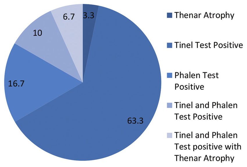
distal forearm at the level of proximal third of pronator thenar atropy. However, (6.7%) patients with thenar atropy
quadratus) (CSA1). Level (2) located at the pisiform bone had both tinel and phalen test positive (Figure 1).
level (CSA2). Level (3) located at the hamate bone level.
A CSA of the median nerve was measured by means of The mean cross-sectional area of median nerve at forearm
direct tracing with electronic calipers around the margin level (CSA1) and carpal tunnel inlet (CSA2) was 7 mm²
of the nerve on sonograms at the first and second levels. and 15.33 mm², respectively. The areal difference (DCSA)
The cross-sectional area difference (DCSA) was obtained between CSA1 and CSA2 was 8.27 mm². The mean
by subtracting CSA1 from CSA2. Level (3) was used to flattening ratio in our study was 2.5 ranging from 1.8 to
calculate the flattening degree by calculating the ratio 3.8, with flattening ratio of 2 or more was observed in
(the flattening ratio) between the largest diameter and the 83.3% of patients. Moreover, the mean bowing of flexor
diameter perpendicular to this finding. The presence of retinaculum in our study was observed as 2.5 mm (Table 2).
increased palmar bowing of the flexor retinaculum was
detected when the palmar apex of the retinaculum was We observe that vascularity of median nerve was present
displaced 2 mm or more from the straight line between the in (56.7%) and absent in (43.3%) patients (Table 3).
trapezium tubercle and the hamate bone. Power Doppler
Table 4, displays the median nerve thinning and indentation
US was done after the gray-scale US to calculate the
were reflected by (80%) patients and the same was absent
number of vessels in the median nerve along the carpal
among (20%) patients.
tunnel. Standardized machine settings were selected to
reach the highest degree of sensitivity of detection of low We assessed hyperemic and edematous median nerve
velocity and low blood flow volume. The color box of the among patients and found that it is present in (63.3%) and
power Doppler was limited to the region of interest. The absent in (36.7%) patients (Table 5).
pulsed wave spectral Doppler imaging was performed
after visualization of power-flow signals, using the lowest
Table 1: The distribution of symptoms among
filter setting (125 Hz). Moreover, to confirm the power
patients
Doppler signals that represent true arterial or venous flow,
Symptoms Frequency Percent
a spectral Doppler tracing was acquired. The sonographic
Paresthesia only 17 56.6
perimeter of the median nerve was obtained by drawing a
Paresthesia and Nocturnal Pain 5 16.7
continuous line around the boundary of the nerve. A single Noctural Pain only 5 16.7
surgeon operated all the patients to standardize the surgical Noctural Pain and Weakness 2 6.7
technique. Median nerve surgical perimeter was measured Weakness only 1 3.3
TOTAL 30 100
by means of a suture thread placed around the nerve
in the same location of the sonographic measurement.
Thinning and indentation of the median nerve were noted. Table 2: Flattening ratio of patients
Hyperemia of the median nerve was noted. FR Frequency Percent
2 or More 25 83.3
Less than 2 5 16.7
RESULTS Total 30 100
Mean 2.5 Range: (1.8–3.8)
In this section, the results of the study will be described
in tabular and graphical form: Out of total 30 patients;
03 (10%) were males and 27 (90%) were females with a
male to female ratio of 1:9. The mean age of patients in
our study was 43 years with majority of patients belonging
to the age group of (40–49) years.
We observe that “paresthesia only” was the predominant
symptom accounting for (56.6%) cases followed by
“paresthesia with nocturnal pain” observed in (16.7%) cases.
Noctural pain only was observed in 6.7% cases (Table 1).
We observe that the most of the patients accounting for
(63.3%) had tinel test positive followed by (16.7%) patients
with phalen test positive. Around (10%) patients tested
positive for both tinel and phalen, while as (3.3%) had Figure 1: Showing distribution of patients as per physical signs
250 Asian Journal of Medical Sciences | Oct 2022 | Vol 13 | Issue 10Mir, et al.: Role of power Doppler and gray-scale ultrasound of the median nerve in the evaluation of carpal tunnel syndrome
The patient showed mean sonographic perimeter of years.10 The most common symptom among studied
16.250 mm with upper and lower values of 17.1201 mm subjects was found to be “paresthesia only” accounting
and 15.3799 mm (95% CI). Same patients show mean for (56.6%) cases followed by “paresthesia with nocturnal
surgical perimeter of 18.777 mm with the upper and lower pain” observed in (16.7%) cases. Noctural pain only was
values of 19.6327 mm and 17.9206 mm (95% CI) and the observed in 6.7% cases. Contemporary to the literature;
difference was significant (Table 6). CTS has been reported to be commonly accompanied
with paresthesia, pain, and less commonly associated with
weakness.11 In Kendall’s series on 327 patients, around
DISCUSSION
(95.7%) had paresthesia, (38%) had nocturnal symptoms
In the present study on the assessment of the role of grey only, (58%) reported mild symptoms through day and night,
scale and power Doppler USG on the median nerve at but severe at night, however, (5%) had symptoms during
the wrist in evaluating CTS and to compare sonographic the day only.12 The present study revealed that the most
findings with surgical findings, we have comprehensively of the patients accounting for (63.3%) had only Tinel test
analyzed patients’ data on the basis of demographic, positive followed by (16.7%) patients with only Phalen
clinical, radiological, and surgical data. We observed that test positive. Around (10%) patients tested positive for
out of 30 studied patients; (90%) were females and only both Tinel and Phalen, while as (3.3%) had thenar atropy.
(10%) were males. In consonance to the literature, the However, (6.7%) patients with thenar atropy had both Tinel
predominance of females with CTS has been reported and Phalen test positive, these results are comparable with
by a good corpus of medicos.3,8,9 In the present study, we the studies of numerous authors.3,8 It is known that CTS is
found that the average age of patients was 43 years, with largely diagnosed with the presence of clinical symptoms
majority of them belonging to the age interval group of and nerve conduction, albeit it has been reported that
(40–49) years, consistent with this; CTS among individuals some patients around 13–27% patients have false normal
has been reported to occur in the age group of (40–60) nerve conduction studies; therefore, alternative diagnosis
with the help USG is useful. Buchberger et al., were first
to quantifiably enumerate the anatomic changes in CTS
Table 3: Vascularity distribution of median nerve with the help of sonography; they showed that an rise in
on Doppler the proximal or middle CSA of median nerve to 10 mm²,
Status Frequency Percent a distal flattening ratio above 3, or a displacement of the
Present 17 56.7 flexor retinaculum above 4 mm are essentially suggestive
Absent 13 43.3
Total 30 100
sonographic signs of CTS.13 It is known that there is a
sudden change in the diameter of the median nerve in
the longitudinal view, especially at the entry of the tunnel
Table 4: Median nerve thinning and indentation in cases of CTS. Therefore, the measurement of the
distribution nerve diameter just before the tunnel inlet in addition
Status Frequency Percent to measurement at the carpel tunnel might contribute
Present 24 80 to sonographic diagnosis of CTS.14 If the degree of the
Absent 06 20 nerve swelling in the carpal tunnel is compared with the
Total 30 100
proximal CSA of the nerve, a DCSA measurement (area
difference between CSA1 and CSA2) will compensate for
Table 5: Hyperemic and edematous median the inter individual variability in the CSA of the median
nerve distribution nerve and yield a more accurate diagnosis of CTS and
Status Frequency Percent provide additional confirmatory criterion for the diagnosis
Present 19 63.3 of CTS.7 With this in mind, we measured the CSA at two
Absent 11 36.7 levels and obtained CSA1 and CSA2 values. The mean
Total 30 100 cross-sectional area of median nerve at forearm level
(CSA1) and carpal tunnel inlet (CSA2) was 7 mm² and 15.33
Table 6: Sonographic versus surgical perimeter mm², respectively.15 The areal difference (DCSA) between
of median nerve CSA1 and CSA2 was 8.27 mm². CSA1 values ranged from
N Mean Min Max P‑value
4 to 12 mm², CSA2 values ranged from 12 to 20mm² and
Sonographic 30 16.250±2.330 15.37 17.12 0.001
DCSA ranged from 7 to 13 mm. Likewise to our study,
parameter the authors like Mallouhi et al., and Sarría et al., reported
Surgical 30 18.777±2.29 17.92 19.63 comparable measurements on CSA and DCSA.16,17 Multiple
parameter
studies have proposed a cutoff range of values for CSA
Asian Journal of Medical Sciences | Oct 2022 | Vol 13 | Issue 10 251Mir, et al.: Role of power Doppler and gray-scale ultrasound of the median nerve in the evaluation of carpal tunnel syndrome
of median nerve and reported thresholds for the CTS CONCLUSION
diagnosis in the literature vary from 9 to 15 mm2. Median
CSA measurement is reliably accepted diagnostic method The present study demonstrated that ultrasonography
but at times, only CSA measurement may vary with other aided with power Doppler precisely detected CTS cases.
patient’s characteristics factors. The mean flattening ratio Evidently, we find a significant difference between surgical
in our study was 2.5 ranging from 1.8 to 3.8, with flattening and sonographic parameters with the mean difference
ratio of 2 or more was observed in 83.3% of patients. of 2.527 mm between sonographic and intraoperative
Moreover, the mean bowing of flexor retinaculum in our perimeters. We recommend large sample studies to
study was observed as 2.67 mm. However, mean flattening evaluate median nerve measurements in other diseases
ratio has been reported by some authors as poor predictive different from CTS, objectively to verify whether changes
of CTS because of its high variability.18-20 The presence in median nerve perimeter are accompanied with the post-
of increased palmar bowing of the flexor retinaculum carpal tunnel release among patients without compressive
was determined to be displacement of the palmar apex derangement of the nerve in the wrist.
of the retinaculum 2 mm or more from the straight line
between its attachments to the trapezium tubercle and the
hamate hook. Determination of its degree was seldom ACKNOWLEDGMENT
done in the previous studies due to its low sensitivity and
Authors are highly thankful to the unknown reviewer(s)
specificity of sonographic diagnosis. In our study, patients
suggestions that lead to the significant improvement of
of CTS showed palmar bowing of flexor retinaculum
the paper.
varying from 1.5 to 4.6 mm with an average value of
2.67 mm. Power Doppler US is the most sensitive to flow
and is used to document diminished blood flow in the REFERENCES
median nerve as a result of high sensitivity to slow flow,
no angle dependency, and no aliasing.21,22 We observed 1. Stecco C and Aldegheri R. Historical review of carpal tunnel
syndrome. Chir Organi Mov. 2008;92(1):7-10.
that vascularity with arterial waveform of median nerve
https://doi.org/10.1007/s12306-008-0033-8
was present in (56.7%) and absent in (43.3%) patients.
2. Watts RA, Conaghan P, Denton C, Isaacs J, Müller-Ladner U,
Nerve vascularity was found associated with symptoms Foster H, et al. Oxford Textbook of Rheumatology. 4th ed. Oxford:
such as paresthesia and pain. We found that 80% of our Oxford University Press; 2013. p. 1316.
patients had thinning and indentation of the median nerve 3. Mondelli M, Giannini F and Giacchi M. Carpal tunnel
while as hyperemic and swollen median nerve was seen in syndrome incidence in a general population. Neurology.
2002;58(2):289-294.
63.3%. The study evaluated the correlation between the
https://doi.org/10.1212/wnl.58.2.289
sonographic and surgical perimeters in CTS. The surgical
4. Parisi DM and Trumble TE. Wrist and hand reconstruction. In:
perimeter was considered as an actual of the median AAOS Orthopaedic Knowledge Update, 9400 West Higgins
nerve, since the nerve was measured during surgery and Road, Rosemont, Illinois., USA; 2005. p. 351.
confirmed with a certified ruler. In our study, we observed 5. Trumble TE, Diao E, Abrams RA and Gilbert-Anderson MM.
that generally, the sonographic perimeter is smaller than the Single portal endoscopic carpal tunnel release compared with
open release: A prospective, randomized trial. J Bone Joint Surg
surgical perimeter. The patient showed mean sonographic Am. 2002;84(7):1107-1115.
perimeter of 16.250 mm with the upper and lower values https://doi.org/10.2106/00004623-200207000-00003
of 17.1201 mm and 15.3799 mm (95% CI). Same patients 6. Alves MP, MoraesNeto GP and Tzirulnik M. Clinical-
show mean surgical perimeter of 18.777 mm with the ultrasonographic evaluation of de Quervain’s stenosing
upper and lower values of 19.6327 mm and 17.9206 mm tenosynovitis. Rev Bras Ortop. 2000;35(4):118-122
(95% CI). After the application of paired t-test, we did 7. Klauser AS, Halpern EJ, De Zordo T, Feuchtner GM, Arora R,
not find any agreement between surgical and sonographic Gruber J, et al. Carpal tunnel syndrome assessment with US:
Value of additional cross-sectional area measurements of the
parameters with the mean difference of 2.527 mm between median nerve in patients versus healthy volunteers. Radiology.
sonographic and intraoperative perimeters, the difference 2009;250(1):171-177.
was statistically significant (PMir, et al.: Role of power Doppler and gray-scale ultrasound of the median nerve in the evaluation of carpal tunnel syndrome
10. Hybbinette CH and Mannerfelt L. The carpal tunnel syndrome: 17. Sarría L, Cabada T, Cozcolluela R, Martínez-Berganza T and
A retrospective study of iOQ operated patients. Acta Orthop García S. Carpal tunnel syndrome: Usefulness of sonography.
Scand. 1975;46(4):610-620. Eur Radiol. 2000;10(12):1920-1925.
https://doi.org/10.3109/17453677508989243 https://doi.org/10.1007/s003300000502
11. Chammas M, Boretto J, Burmann LM, Ramos RM, Neto FC and 18. Mesgarzadeh M, Schneck CD and Bonakdarpour A. Carpal
Silva JB. Carpal tunnel syndrome part I (anatomy, physiology, tunnel: MR imaging. Part I. Normal anatomy. Radiology.
etiology and diagnosis). Rev Bras Ortop. 2014;49(5):429-436. 1989;171(3):743-748.
https://doi.org/10.1016/j.rboe.2014.08.001 https://doi.org/10.1148/radiology.171.3.2717746
12. Kendall WW. Results of treatment of severe carpal tunnel 19. Mesgarzadeh M, Schneck CD, Bonakdarpour A, Mitra A and
syndrome without internal neurolysis of the median nerve. Conaway D. Carpal tunnel: MR imaging. Part II. Carpal tunnel
J Bone Joint Surg Am. 1988;70(1):151. syndrome. Radiology. 1989;171(3):749-754.
https://doi.org/10.2106/00004623-198870010-00032 https://doi.org/10.1148/radiology.171.3.2541464
13. Buchberger W, Judmaier W, Birbamer G, Lener M and 20. Monagle K, Dai G, Chu A, Burnham RS and Snyder RE.
Schmidauer C. Carpal tunnel syndrome: Diagnosis with Quantitative MR imaging of carpal tunnel syndrome. AJR Am J
high-resolution sonography. AJR Am J Roentgenol. Roentgenol. 1999;172(6):1581-1586.
1992;159(4):793-798. https://doi.org/10.2214/ajr.172.6.10350293
https://doi.org/10.2214/ajr.159.4.1529845 21. Ghasemi-Esfe AR, Khalilzadeh O, Vaziri-Bozorg SM, Jajroudi M,
14. Fu T, Cao M, Liu F, Zhu J, Ye D, Feng X, et al. Carpal tunnel Shakiba M, Mazloumi M, et al. Color and power Doppler US
syndrome assessment with ultrasonography: Value of inlet- for diagnosing carpal tunnel syndrome and determining its
to-outlet median nerve area ratio in patients versus healthy severity: A quantitative image processing method. Radiology.
volunteers. PLoS One. 2015;10(1):e0116777. 2011;261(2):499-506.
https://doi.org/10.1371/journal.pone.0116777 https://doi.org/10.1148/radiol.11110150
15. Roquelaure Y, Chazelle E, Gautier L, Plaine J, Descatha A, 22. Mohammadi A, Ghasemi-Rad M, Mladkova-Suchy N and
Evanoff B, et al. Time trends in incidence and prevalence of Ansari S. Correlation between the severity of carpal tunnel
carpal tunnel syndrome over eight years according to multiple syndrome and color Doppler sonography findings. Am J
data sources: Pays de la Loire study. Scand J Work Environ Roentgenol. 2012;198(2):W181-W184.
Health. 2017;43(1):75-85. https://doi.org/10.2214/AJR.11.7012
https://doi.org/10.5271/sjweh.3594 23. Tajika T, Kobayashi T, Yamamoto A, Kaneko T and Takagishi K.
16. Mallouhi A, Pülzl P, Trieb T, Piza H and Bodner G. Predictors of Diagnostic utility of sonography and correlation between
carpal tunnel syndrome: Accuracy of gray-scale and color Doppler sonographic and clinical findings in patients with carpal tunnel
sonography. AJR Am J Roentgenol. 2006;186(5):1240-1245. syndrome. J Ultrasound Med. 2013;32(11):1987-1993.
https://doi.org/10.2214/AJR.04.1715 https://doi.org/10.7863/ultra.32.11.1987
Authors Contribution:
MF- Designed the study and prepared the draft of manuscript; SB- Performed review literature and wrote discussion; HAL- Did the cases and performed
statistical analysis and interpreted the same; and TAD- contributed in writing orthopedic terminology and disease status
Work attributed to:
SKIMS, JVC, Bemina Srinagar
Orcid ID:
Dr. Shazia Bashir - https://orcid.org/0000-0003-1750-8313
Source of Funding: Nil, Conflicts of Interest: None declared.
Asian Journal of Medical Sciences | Oct 2022 | Vol 13 | Issue 10 253You can also read