Scientific RepoRt 2019 - de Duve Institute & Ludwig Cancer Research - Brussels
←
→
Page content transcription
If your browser does not render page correctly, please read the page content below
Scientific
Report
2019
de Duve Institute
&
Ludwig Cancer Research - Brussels
Avenue Hippocrate 75
B-1200 Brussels, BELGIUM
[T] + 32-2-764 75 50
[F] + 32-2-764 75 73
[E] deduve_institute@uclouvain.be
[W] www.deduveinstitute.be
Directors Prof. Benoît Van den Eynde
Prof. Jean-François Collet
Prof. Sophie Lucas
Prof. Miikka Vikkula
Finance Manager Serge Thibaut
External Relations Isabelle de Duve
Photographs Jacky Delorme & Hugues Depasse
Research Highlights Francisca Voermans
For a copy of this report, please contact: nathalie.krack@uclouvain.be
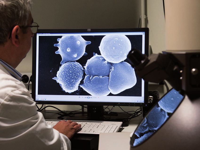
Picture: Scanning electron microscopy picture of red blood cells (imaging platform, Tyteca’s group)
CONTENTS
                                                            CANCER                                                            15
2 . . . . . . . . . . . . . . . . . . . . KEY NUMBERS       Stefan Constantinescu . . . . . . . . . . . . . . . 16
                                                            Jean-Baptiste Demoulin . . . . . . . . . . . . . . 17
                                                            Pierre Coulie . . . . . . . . . . . . . . . . . . . . . . . . . 18
3 . . . . . . . . . . . . . . FROM THE DIRECTOR
                                                            Sophie Lucas . . . . . . . . . . . . . . . . . . . . . . . . 19
                                                            Benoît Van den Eynde . . . . . . . . . . . . . . . . 20
4 . . . . . . . . . . . . . ABOUT THE INSTITUTE             Pierre van der Bruggen . . . . . . . . . . . . . . . 21

5 . . . . . . . . . . . . RESEARCH HIGHLIGHTS
                                                            GENETICS & DEVELOPMENT                                            22
                                                            Donatienne Tyteca . . . . . . . . . . . . . . . . . . . 23
                                                            Nisha Limaye . . . . . . . . . . . . . . . . . . . . . . . . 24
14 . . . . . . . . . . . . . .   RESEARCH GROUPS            Miikka Vikkula . . . . . . . . . . . . . . . . . . . . . . . 25
                                                            Charles De Smet . . . . . . . . . . . . . . . . . . . . . 26
44 . . . . . . . . . . SELECTED PUBLICATIONS                Anabelle Decottignies. . . . . . . . . . . . . . . . 27
                                                            Frédéric Lemaigre
                                                            & Patrick Jacquemin . . . . . . . . . . . . . . . . . 28
54 . . . . . . . . TECHNOLOGY PLATFORMS                     Wen-Hui Lien . . . . . . . . . . . . . . . . . . . . . . . . 29
                                                            Christophe Pierreux . . . . . . . . . . . . . . . . . . 30
55 . . . . .   PRIZES, AWARDS & HONORS
                                                            INFECTIONS & INFLAMMATION                                         31
55 . . . . . . . . . . . . . . . . . . . .   PHD THESES     Jean-François Collet . . . . . . . . . . . . . . . . . . 32
                                                            Géraldine Laloux . . . . . . . . . . . . . . . . . . . . . 33
                                                            Jean-Paul Coutelier . . . . . . . . . . . . . . . . . . 34
57 . . . . . . . . . . . . . . . . . . . . . . . LECTURES   Thomas Michiels . . . . . . . . . . . . . . . . . . . . . 35
                             & SCIENTIFIC EVENTS            Jean-Christophe Renauld
                                                            & Laure Dumoutier . . . . . . . . . . . . . . . . . . . 36

61 . . . . . . . . . . . . . . . . . . . MANAGEMENT
                                                            METABOLISM & HORMONES                                             37
                                                            Emile Van Schaftingen
62 . . . . . SUPPORTING ORGANIZATIONS                       & Maria Veiga-da-Cunha. . . . . . . . . . . . . . 38
                                                            Guido Bommer . . . . . . . . . . . . . . . . . . . . . . 39
63 . . . . . . . . . . . PRIVATE & CORPORATE                Mark Rider . . . . . . . . . . . . . . . . . . . . . . . . . . . 40
                                                            Etienne Marbaix
                                               FUNDING      & Patrick Henriet . . . . . . . . . . . . . . . . . . . . . 41

64 . . . . . . . . . . . . ACKNOWLEDGEMENTS                 COMPUTATIONAL BIOLOGY                                             42
                                                            Laurent Gatto . . . . . . . . . . . . . . . . . . . . . . . . 43

                                                                                                           Introduction           |1
41 nationalities

                                       297 members

                           45%                             55%

                    94 PhD students                     37 postdoctoral students

                    95 publications                     36 international lectures

                    6 technology platforms              8000 m2

2|   Introduction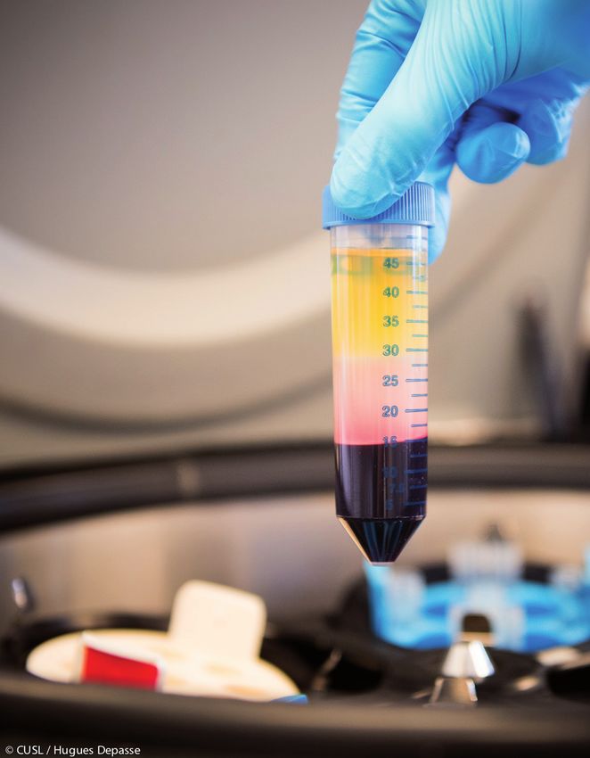
FROM THE DIRECTOR

T    he year 2019 was marked with important changes in the management of our Institute. The Directorate
     was renewed following the retirement of Professor Emile Van Schaftingen after 15 years at the head
of the Directorate. In its meeting held in May 2019, the Board of Directors of the de Duve Institute
appointed a new Directorate, composed of Professors Jean-François Collet, Benoît Van den Eynde and
Miikka Vikkula. As a new addition, Jean-François Collet will bring fresh blood in the management team,
while Van den Eynde and Vikkula will ensure strategic continuity as they have already been part of the
Directorate during the last 15 years. The new Directorate is presided by Benoît Van den Eynde. This trio
is a balanced representation of the different research areas explored at the Institute, with expertise in
bacteriology and biochemistry (Collet), in immunology and cancer (Van den Eynde) and in genetics of
human diseases (Vikkula). The mission of the Directorate is to organize, coordinate and support scientific
activities and collaborations within the Institute in a manner that will best promote excellence and achieve
the goal expressed in our motto: ‘Deeper knowledge for better cures’.

De Duve Institute has close links with university UCLouvain. University matters pertaining to our activities
are managed by DDUV, a university structure (also called an ‘Institute’) that has replaced the previous
departments. DDUV is headed by a President: the mandate of Professor Frédéric Lemaigre, who held this
position in the last six years, came to an end in September 2019. He was replaced by Professor Sophie
Lucas. To ensure optimal efficiency, these two structures work in close collaboration, and all management
meetings are held jointly with the Directorate and the DDUV President.

We express our deepest gratitude to Emile Van Schaftingen and Frédéric Lemaigre for their dedicated
involvement in the management of the Institute(s) in recent years.

The year 2019 was filled with a number of important discoveries, ranging from a new mechanism of
telomere maintenance, which may represent a therapeutic target in pediatric cancers (see page 27), to the
understanding of the pathogenesis of an orphan neutrophilic disease, which led to the identification of
a drug acting on this pathogenic process to treat the disease (see pages 6-7 and page 38). These are only
two examples, and there are many other biomedical discoveries that you will find detailed in this report.

Our research activities are supported in large part by research grants, but also, increasingly, through
donations from our sponsors coming in largely on the occasion of our annual gala dinner. In terms of
grants, 2019 was a successful year as our scientists performed remarkably well in the last call for WELBIO
grants, which are the most prestigious public research grants available in the French-speaking part of
Belgium. In this highly competitive call, our Institute obtained five new grants and three renewals.

One of the challenges of the Institute is the renewal of the research teams, as a number of professors will
retire in the coming years. To this end, a young promising scientist named Nick van Gastel was invited to
settle in the Institute and create a new group focusing on leukemia research. Of Belgian origin, Nick was
trained at KULeuven and at Harvard University. He will join us in the early summer 2020.

                                                                                    Benoît Van den Eynde

                                                                                                Introduction   |3
ABOUT THE INSTITUTE

      O    riginally named International Institute of Cellular and Molecular Pathology (ICP), the de Duve Institute
           was founded in 1974 by Professor Christian de Duve († 4th of May 2013) to develop basic biomedical
      research with potential medical applications.
      Excellence and freedom of the researchers to choose their line of research are our core values as defined
      by de Duve. We attract excellent researchers from Belgium and from abroad, and give them the liberty
      to develop their original ideas in an inspiring environment. Discovery is the endpoint of their efforts and
      the only element taken into account for their evaluation.
      We value collaborative work and interdisciplinary research. The Institute functions in symbiosis with
      the Faculty of Medicine of the University of Louvain (UCLouvain) and many of its senior members hold
      a Faculty position and have teaching appointments. The influx of doctoral students and postdoctoral
      fellows from the University is key to the success, as well as the close collaborations with clinicians of the
      University Hospital (Cliniques universitaires) Saint-Luc, located within walking distance. In addition we
      have good contacts and many joint research projects with other research institutions in Belgium and all
      over the world.
      In 1978 the Ludwig Institute for Cancer Research decided to base its Belgian branch within the walls of
      the de Duve Institute. A fruitful collaboration between the two institutions has been pursued ever since.
      Even though the two institutes are completely independent, the collaboration between their scientists
      is extremely close and the sharing of resources considerable.
      The de Duve Institute has the ambition of pursuing research projects of high quality under conditions that
      allow original, long-term projects. Research is funded by public bodies, national and international, as well
      as by private donations. Most funds are awarded on a competitive basis. The Institute has an endowment,
      which is a source of key financing for priority issues, such as the creation of new laboratories for promising
      young researchers. The quality of our researchers, supported by sound organisational approaches, will
      enable the de Duve Institute to remain at the forefront of European research. We are extremely grateful
      to all those who support the Institute.

4|   Introduction
Research Highlights

             Research Highlights   |5
UNCOVERING THE SECRETS OF METABOLISM
                Maria Veiga-da-Cunha elucidated, in collaboration with Emile Van Schaftingen,
              how a deficiency of two proteins leads to neutropenia. It has led to a new treatment
               for patients. The motto of the Institute “Deeper knowledge for better cures” could
                                            not be illustrated better.

Lisbon-born Maria Veiga-da-Cunha is a longtime collabo-       stand what was intoxicating and killing these neutrophils”,
rator of Emile Van Schaftingen. Their partnership started     says Maria Veiga-da-Cunha.
in 1991 when she joined his team as a postdoctoral
researcher. They form a perfect team: she is the experi-      The research to find this missing link illustrates the
mental wizard, he has a tremendous biochemical know-          strength of their collaboration. Maria Veiga-da-Cunha
ledge. Together, they have elucidated various metabolic       developed various experimental methods to be able to
puzzles and the last one is a beautiful story that explains   perform reliable experiments on the membrane-bound
the origin of the very handicapping neutropenia in two        proteins. But when all techniques worked, she could not
rare metabolic diseases. In the more common disease,          find the substrate that they were looking for. “We knew
patients are deficient in a transporter called G6PT, which    that the substrate must look like glucose 6-phosphate, but
allows specific sugar-phosphates to get in a compartment      we could not find a compound that met all requirements.
in the cell called the endoplasmic reticulum. In the rarer    We were stuck.” In the previous years, the team had done
one, patients are deficient in the phosphatase G6PC3,         much work on the specificity of metabolic enzymes and
which is inside the endoplasmic reticulum. Patients with      discovered that cells need dedicated metabolite-repair
these diseases have very low levels of the most abundant      enzymes to prevent diseases. This concept helped them
white blood cells (the neutrophils) in the bloodstream,       to come up with the solution. It was Emile Van Schaftingen
a condition called neutropenia. Since the function of         who remembered a paper from 1954 by Sols and Crane,
neutrophils is to kill invading bacteria, they suffer from    who purified and carefully studied the substrate specificity
frequent infections: they develop chronic inflammation        of hexokinase 1. Sols showed that hexokinases have side
that resembles Crohn’s disease, have aphthous lesions in      activities that allow them to phosphorylate other sub-
the mouth and various mucosal and skin abscesses.             strates that look like glucose, if these are present in the
                                                              cell. It was this lack of specificity of hexokinase, together
Twenty years ago, the team had identified the genetic         with the finding that in blood we all have a molecule that
cause of the deficiency in G6PT. The missing link required    looks very much like glucose (called 1,5-anhydroglucitol
to understand the neutropenia in these two rare genetic       or 1,5-AG) that pointed them to the solution: the molecule
disorders had stayed in their minds ever since. “We knew      intoxicating the neutrophils could be 1,5-AG-6-phosphate,
the genetic causes of both deficiencies, we knew that it      formed by side activities of glucose-phosphorylating
impacted neutrophil’s metabolism, but we did not under-       enzymes acting on 1,5-AG.
6|   Research Highlights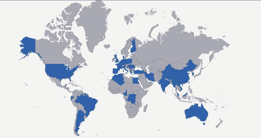
“  When the girl started to take
        the antidiabetic, her situation
           improved after a week.
       She can, for the first time, live a
                normal life.
                                     ”

This hypothesis proved to be the answer they were               hypothesis on patients that were particularly crippled by
looking for. With all the experimental tools at hand, and       the disease. They first tested it on a 20-year-old girl, who
with the help of Guido Bommer, the team was able to             was in a desperate condition. “She did not respond to her
demonstrate that cells defective in G6PT or G6PC3 accu-         treatment anymore and was in terrible pain. She had many
mulated 1,5-AG-6-phosphate when 1,5-AG was added to             infections and severe intestinal problems. When the girl
the culture media. 1,5-AG, a food derivative that resembles     started to take the antidiabetic, her situation improved
glucose, can be erroneously phosphorylated by enzymes           after a week. Her intestines now function almost normal
present in neutrophils that normally act on glucose. As         again, the ulcers in her mouth and the abscesses on her
patients with deficiencies of G6PC3 or G6PT cannot undo         skin healed. She can, for the first time, live a normal life.”
this reaction, phosphorylated 1,5-AG accumulates in the
patient’s neutrophils and inhibits the first step of glycol-    The trials are now extended to more patients, to adults but
ysis, depriving these cells of energy. This is particularly     also young children in Belgium and across the world. Maria
detrimental for neutrophils, because glycolysis is their only   Veiga-da-Cunha: “I believe it will become the standard
source of energy and explains the neutropenia.                  treatment for these rare diseases. The current treatment is
                                                                injection of a cytokine that accelerates the development
Though this finding was of great scientific importance, the     of neutrophils in the bone marrow. This becomes very
researchers went on to answer the next question: Could          painful after a while and some patients become resistant.
they help the patients? Reducing the 1,5-AG concentra-          And it increases their risk of developing a type of a myelo-
tion in the blood should restore the neutrophil levels. A       dysplastic syndrome, which is a group of cancers affecting
new class of antidiabetics, which prevents the re-uptake        the immature blood cells in the bone marrow. The antidia-
of glucose back into the blood circulation in the kidney,       betic is much cheaper, painless and can be taken at home.”
might do the trick. Indeed, these antidiabetics were also
shown to lower blood 1,5-AG. Tests in a mouse model of
G6PC3 deficiency showed that the drug indeed reduced            Reference
1,5-AG levels in blood and, by doing so, treated the            Veiga-da-Cunha M, Chevalier N, Stephenne X, Dufour JP, Paczia N,
neutropenia in these mice. It then all went very quickly.       Ferster A, Achouri Y, Dewulf JP, Linster CL, Bommer GT, Van Schaftingen
                                                                E. Failure to eliminate a phosphorylated glucose-analog leads to
Soon after they had published their results in PNAS, they       neutropenia in patients with G6PT and G6PC3 deficiency. Proc Natl
were contacted by clinicians that were eager to test their      Acad Sci USA. 2019;116:1241-50.
                                                                                                       Research Highlights         |7
TRACING THE ORIGIN OF CANCER
              Frédéric Lemaigre and Patrick Jacquemin try to understand how a normal cell turns
               into a cancer cell. They recently made breakthroughs on the mechanisms causing
                                            liver and pancreatic cancer.

How is the development of the liver and pancreas in an          dent in Brussels, Jacquemin as a PhD student in Liège.
embryo controlled? Why are specific genes expressed in          Later, after they both switched to studying developmental
these cells and others not? Frédéric Lemaigre and Patrick       biology, Lemaigre thought of Jacquemin to set up his new
Jacquemin try to answer these fundamental questions             group at ICP (now de Duve Institute). Then a postdoc in
about the early phases of life. Their work leads to interest-   France, Jacquemin happily agreed to return to Belgium.
ing new knowledge on how we are created, but also – and         They have been working together ever since.
that is their main goal – to a better understanding of how
cancer springs. “In cancer, embryonic development is in a       Jacquemin’s group recently made a discovery on the
way reversed. Cells regain numerous embryonic charac-           earliest phase of pancreatic cancer, one of the deadliest
teristics and start proliferating again. If we know how cells   cancers with a survival rate at 5 years of only 6 to 7%. The
differentiate in a normal embryo, we also know how they         disease starts with precancerous (‘neoplastic’) lesions that
can dedifferentiate in an adult. Our research thus helps        may or may not become malignant. “We had the idea in
to understand mechanisms that initiate cancer”, explains        mind that the initiation of pancreas cancer is related to
Lemaigre. The groups of the two researchers work closely        perturbation in cell differentiation. The intention of our
together. Lemaigre’s group studies the liver, Jacquemin’s       research was to identify the pancreas cell type that gives
the pancreas. Two organs that have a lot in common: they        rise to precancerous lesions”, says Jacquemin. Based on
have the same embryonic origin and architecture, similar        publications by other labs, his group was able to develop
active genes and proteins, and overlapping mechanisms           a good mouse model of the disease. “With the model we
of development. The organs are thus studied in similar          showed that ductal cells can be at the origin of the lesions.
ways and the two groups share tools and know-how. And,          These are the cells that line the pancreatic duct and that
more importantly, the two researchers habitually discuss        drive the digestive secretions from the pancreatic cells to
new ideas and results. “A new proposal never leaves the         the duodenum, the first part of the intestine.” They also
group before the other has reviewed it”, says Lemaigre.         identified the signaling pathway that drives the devel-
                                                                opment of the lesions. “Mutations in one gene, of the
The collaboration between Frédéric Lemaigre and Patrick         so-called β-catenin pathway, lead to the precancerous
Jacquemin goes back a long way. They got acquainted in          lesions. We found that these mutations are responsible for
’89 when they were both pioneering control mechanisms           a significant part of the development of pancreas cancer
of tissue-specific gene expression, Lemaigre as a PhD stu-      in our mouse model. Not for all cases though, another
8|   Research Highlights
“ In cancer, embryonic
development is in a way
reversed.
”
part is independent of this pathway.” There are inhibitors human cells in culture and human data from literature.
of this pathway available, but unfortunately they cannot “Thanks to computational biology we could study such a
be used clinically due to side effects. Jacquemin: “There gene network. Using tools like transgenic mice, one can
are no direct clinical applications of the discovery. It is a only study two or maximum three genes at a time”, says
contribution to obtain the picture of how cancer develops. Lemaigre.
Each contribution is important to find a cure one day.”
The model predicts how one gene influences all other
The group of Frédéric Lemaigre studies hepatocellular genes of the network. It helps to better understand the
carcinoma, the most common type of liver cancer. It results development of the cancer and it can be used to predict
from a variety of conditions such as viral infections or cir- the efficacy of a drug in individual patients. The model is
rhosis, giving rise to a significant heterogeneity among available to other researchers through the web, who can
patients and variable responses to therapeutic agents. use it on their cancer samples to find the best target to
“It is known for many years that the β-catenin pathway is modulate the network dynamics. Mathematical modeling
involved in the development of hepatocellular carcinoma. of gene networks has become a regularly used tool in
The question was: Does the pathway influence the dedif- their research, says Jacquemin: “It’s very useful to make
ferentiation of cells, so they lose their adult properties and predictions and set up a good hypothesis, which can then
start proliferating again?” The answer was not straightfor- be tested in experiments.” Though it’s a great tool, one has
ward. “We found that dedifferentiation is indeed caused by to be careful with the results, adds Lemaigre: “Biological
mutations in the pathway. But it is not an individual gene know-how is indispensable to interpret and validate the
that is responsible, instead it’s a number of genes acting results. The mix of both areas of expertise is one of the
in concert. These genes function as a dynamic network, strengths of our groups.”
some activating the expression of other genes, others
acting as suppressors.” Using a combination of computa- References
tional biology and experiments, the researchers identified Collet L et al. Kras and Lkb1 mutations synergistically induce intraductal
a network comprising seven genes, which regulates the papillary mucinous neoplasm derived from pancreatic duct cells. Gut.
2019; epub Jun 1.
progression of a subset of liver cancer with poor prognosis.
Gérard C et al. Dynamics and predicted drug response of a gene
They made a mathematical model of how the genes inter- network linking dedifferentiation with beta-catenin dysfunction in
act. The model was validated with experiments on primary hepatocellular carcinoma. J Hepatol. 2019;71:323-32.
Research Highlights |9THE FUNDAMENTALS OF A BACTERIUM’S LIFE
Bdellovibrio bacteriovorus predates on other bacteria and might therefore be
used as a new type of antibiotic. The team of Géraldine Laloux studies how this
bacterium orchestrates its eccentric life cycle.
A small bag containing a fluid and randomly distributed She continued with a doctoral study in the lab of Xavier
molecules. That’s how a bacterium was for long envi- De Bolle in Namur, where she first started to work on bac-
sioned. But, as scientists study the unicellular organisms teria. After her postdoc at Yale, she came to the de Duve
in detail, they discover their well-organized and diverse Institute to join the lab of Jean-François Collet. Here, the
ways to survive. “Bacteria do not have organelles, the spe- at Yale acquired knowledge on fluorescence microscopy
cialized intracellular structures that animal and plant cells proved of great value. “The lab studies the responses of
have. But they are still able to localize activities within the bacteria to stress. They use techniques from molecular
cell. During thousands of years of evolving, each bacte- biology, genetics and biochemistry to investigate the
rium developed its own way to reproduce”, says Géraldine signaling proteins in the cell envelope. When I started to
Laloux, a microbiologist who studies the life cycles of bac- study the stress responses with the microscope, I could
teria. see weird shapes of dividing cells. This led us to identify
an ensemble of proteins that monitor the status of the
Her passion for the odd little creatures was born when cell wall. Before, this system was thought to have another
she worked as a postdoc at Yale University. Her subject function! A discovery by serendipity.” They also discovered
was Caulobacter crescentus, a harmless organism with a the function of a protein associated with this stress-sens-
conspicuous stem-like extension. “It divides into two dif- ing system: it turns on the alarm when there is something
ferent daughter cells: one goes swimming around, the wrong with the trafficking of specific molecules of the
other stays attached to the surface. It has a very fancy bacterial envelope.
life cycle.” Using fluorescence microscopy to study the
processes on a single cell level, she could see how proteins In 2016, Géraldine Laloux started to develop her own
move in the bacterium and at the same time watch its research path at the Institute. Which put her for the big
shape changing. “During these 3.5 years at Yale, I fell in question: What topic to choose? After careful consider-
love with cell biology”, she says. ation, she decided to focus her research on Bdellovibrio
bacteriovorus, a predatory bacterium. Again an organism
Géraldine Laloux is one of the young group leaders at the with a curious life cycle, she explains: “B. bacteriovorus
de Duve Institute. She studied biology in Namur, during attacks Gram-negative bacteria, which are characterized
which she did her practical work at Harvard University in by a double cell membrane. It does so by invading and
the group of the Belgian genetics professor Marc Vidal. nesting in the periplasm, the space between the inner and
10 | Research Highlights“ We see the predator entering
the prey cell, eating the prey
content, growing, dividing and
finally attacking another prey.
”
outer membrane. From here, it eats the contents of the The group of Géraldine Laloux had a kick-start in 2018.
host bacterium and grows to form a long spaghetti-like After being appointed as FNRS researcher, she obtained
filament before dividing into small daughter cells. Finally, an ERC Starting grant and an FNRS-MIS grant. To make the
the host cell lyses and the new B. bacteriovorus cells go year even more hectic, she also gave birth to her second
searching for a next victim.” child. Thanks to the ERC grant she could buy an advanced
microscope that is crucial to the research. With a high
The bacterium is a promising alternative for current antibi- resolution, it is able to detect the faint signals within liv-
otics to which more and more bacteria become resistant. ing cells of one micrometer (1000 times smaller than a
“Many antibiotic-resistant pathogens are Gram-negative millimeter). “We can visualize the full predator cycle. We
and could thus be attacked by B. bacteriovorus, which itself see the predator entering the prey cell, eating the prey
is harmless to humans. Many labs study the bacterium for content, growing, dividing and finally attacking another
this purpose”, says Géraldine Laloux. She herself is more prey”, she says.
interested in the fundamental questions about the bac-
terium’s life cycle. “The bacterium divides into a variable Her team now consists of one postdoc, two PhD students
number of daughter cells: from 2 up to 7 or even more pro- and a technician. “They are working very well together and
vided that the prey bacterium is large enough. We want are really motivated to give our young lab a good start”,
to find out how these processes are orchestrated. How is she says proudly. Though the research is not directly aimed
the chromosome – a bacterium has only one – copied a at clinical application, the results will be important for the
certain number of times? How does the segregation of development of B. bacteriovorus as a new antibiotic, says
these chromosomes take place?” The team uses a com- Géraldine Laloux: “If you want to use the bacterium in a
bination of disciplines to answer these questions. “From clinical context, you have to know how it behaves, how it
genome sequencing data, we know which proteins are can be engineered. We must be able to control what we
present in the bacterium. We make interesting proteins put in a patient.”
fluorescent by modifying the genes, so we can follow them
in the living cell by microscopy. We also edit genes, to
see what the bacterium does without certain proteins.
And we use biochemistry to investigate the interactions
between proteins.”
Research Highlights | 11FROM MILLIONS OF DATA TO A NEW THERAPY
The genetic research of Miikka Vikkula led to the first molecular therapy for several
types of vascular anomalies. He was awarded the prestigious Generet Prize for
his work.
In 1993, young MD Miikka Vikkula left Helsinki for Boston The couple had found their mission in helping patients
as a postdoc, to study the role that collagens play in vas- with vascular anomalies. Laurence Boon went to work as
cular biology. His intention was to return to Finland after a plastic surgeon in the University Hospital Saint-Luc in
a few years to become a cardiologist. It all went differ- Brussels, where she coordinates the Center for Vascular
ently. In Boston, he was introduced to Dr Laurence Boon, Anomalies. Miikka Vikkula also searched a position in
a plastic surgeon in training, who had just arrived from Belgium, which he found at the de Duve Institute, then
Brussels for a fellowship at the Children’s hospital. Her called ICP. Here he set up his own research group and
chief had recognized a big Italian family with multiple continued his work on vascular anomalies. “Vascular
cases of vascular anomalies. The two fellows were asked to anomalies are a group of over forty disorders, most of
identify the genetic cause. A challenge that Vikkula eagerly which are relatively rare, and in which the blood or lymph
accepted. “I was interested in the cardiovascular system vessels develop in an uncontrolled way”, explains Vikkula.
and in genetics. In this research, it all came together.” “Patients have dark red to purple sponge- or cyst-like
lesions on their bodies. People with severe forms have
That first meeting in Boston changed both the career and chronic pain. Some have headaches, others have trouble
life of Miikka Vikkula. Together with Laurence Boon, he with breathing, eating or moving their limbs.”
threw himself into the laborious work of collecting and
analyzing samples of the patients. “At that time, we used Building on the successful approach in Boston, the cou-
big gels to run genetic tests, and after they were labelled ple started the collection of samples of vascular anomaly
radioactively, we developed films and scored bands on patients. That biobank, today containing thousands of
them. We could do one genetic marker per gel. Today we samples, became the basis for Miikka Vikkula’s research.
run 50,000 markers at a time”, the Finnish researcher says. His group performs genetic analyses on the samples to
The two fellows succeeded to find the genetic mutation find the mutations that cause the disease. In a next step,
responsible for the disease of the Italian family. It would they characterize the changes induced by these mutations
be the first of many discoveries on vascular anomalies within the cells. Once they know what goes wrong in the
by Vikkula. In these years in Boston, something beautiful cell, they try to find a way to interfere. “The beauty is that
grew between the two fellows and four years later they we do not have any idea of the molecular dysfunction a
got married. priori. We don’t need it. Because the disease is inherited,
we know that behind the cause there must lie a mutated
12 | Research Highlights“ A treatment like this was
unthinkable in 1993, when we
started this research.
”
gene.” The research group has found the genetic causes of molecular therapy for a venous malformation, which has
multiple vascular anomalies. They for example identified provoked a revolution in the field. “A treatment like this
the mutation responsible for a potentially deadly malfor- was unthinkable in 1993, when we started this research.
mation of the vein of Galen in the brain. Besides increasing Now everybody is talking about it”, says Vikkula.
the understanding of the diseases, this knowledge has
allowed to achieve better and faster diagnoses. The discovery of the second mutation made the lab also
look at tissular mutations as explanation for vascular
A major breakthrough came in 2006. The lab was still lesions in patients with no family history of such. Thanks
researching the venous malformation of the Italian family, to the high-quality samples of the biobank, they were
because, though they had found the inherited mutation, able to identify the first somatic mutations explaining
they could not explain why the lesions were localized and non-inherited lesions with regular Sanger sequencing.
other veins were normal. The group then discovered a This opened the doors for the whole world to look for
second mutation in the cells of the malformations them- similar explanations for other diseases. Now, roughly 70%
selves, a so-called somatic mutation. Only cells with both of non-inherited vascular malformations is explained by
mutations develop the disease. These mutations were a somatic mutation. Armed with the newest NGS tools
thereafter found to make vascular cells to over-activate and Highlander, an in-house developed software tool to
a specific signaling pathway. An existing drug, called analyze the enormous amounts of genetic data, and a
Rapamycin, inhibits this pathway. The researchers investi- large data cluster, the Vikkula lab continues its search for
gated the effects of the drug in in vitro and in vivo models genetic explanations for human diseases. Next to vas-
and showed that it indeed stopped lesion development. cular anomalies, they also investigate lymphedema and
Because the drug was already approved for use in humans cleft lips. “We believe many more diseases are caused by
for transplantations, the group could quickly start clinical genetic mutations. Finding these mutations can lead to a
trials, in collaboration with Prof. Laurence Boon. In two more effective treatment for patients and greatly improve
small groups of patients, Rapamycin drastically relieved their quality of life.” That is what drives Miikka Vikkula who,
patients from their painful symptoms. One of them suf- though he became a researcher, also still is a doctor: “To
fered from chronic headaches and is, since the treatment, be able to go back to the patient is the most important
able to work full-time again. A large trial is now being reward of this work.”
run with over 100 patients already involved. It is the first
Research Highlights | 13Research Groups 14
A cancer starts when cells acquire the ability
       to grow fast and the body’s defence system
     cannot control them. We study the signaling
   mechanisms that regulate cell growth and how

                                                      CANCER
    genetic mutations lead to aberrations in these
mechanisms, inducing for example blood cancers
      or child tumors. Our institute proudly was at
       the basis of today’s cancer immunotherapy
 treatments. We now investigate how the efficacy
             of these treatments can be enhanced.
Stefan Constantinescu
CELL SIGNALING We study how cytokines regulate blood formation via their membrane
& MOLECULAR receptors, Janus kinases, STAT proteins and other signaling pathways. We
HEMATOLOGY decipher how blood cancers evolve and can be targeted by treatment.
F ormation of blood requires small proteins, denoted persistently activate the Tpo receptor (TpoR/MPL). This
as cytokines, such as erythropoietin, thrombopoietin, closes the circle of major driver mutations in MPNs. The
interleukins or interferons. These proteins induce survival, next challenge is the delineation of the pathway by which
growth and differentiation of blood precursors. They act MPNs evolve to leukemia. We also study myeloprolifera-
by binding on the surface of target cells to ‘receptors’, tion and myeloid leukemia in children, especially those
which function like ‘antennae’ that transmit a signal to being resistant to treatment. This work is supported by
the cell interior. We study how these specific receptors ‘Les avions de Sébastien’.
assemble on the membrane and couple at the cell interior
to other proteins, such as In order to pursue our aims,
Janus Kinases (JAKs), which we use molecular biology
are absolutely required to approaches like extensive
transmit a signal. We found mutagenesis as well as func-
that mutations in JAKs or tional assays as read-outs
in receptors themselves for structural determinants,
confuse the cells and make biophysical approaches, as
them grow indefinitely, well as in vivo transgenesis,
leading to blood cancers, microscopy and fraction-
specifically myeloprolifera- ation, mouse bone marrow
tive neoplasms (MPNs). Our Substitution of V617 to phenylalanine (V617F) in JAK2 pseudokinase transplantation, as well as
hypothesis is that a curative (JH2) domain creates an aromatic stacking interaction (left) that is investigation of primary
responsible for kinase domain activation; the same circuit is required
treatment needs a JAK2 for activation of wild-type JAK2 by the ligand stimulated IFN-γ receptor patient cells.
V617F-specific inhibitor. To tetramer (right).
this end, we delineated the
circuit of JAK2 kinase activation by the V617F acquired
mutation in the pseudokinase domain that could be a
target for specific inhibition. The same circuit is required
for signaling by the tetrameric interferon (IFN)-γ receptor
(Figure, left), suggesting that a common set of molecules
might inhibit both IFN-γ signaling and JAK2 V617F (Figure, Staff members
right). The same strategy is taken for mutants in the Tpo Clinical Investigator: Jean-Philippe Defour • Senior
receptor (TpoR/MPL) that we discovered in 2006, where Investigators: Didier Colau, Christian Pecquet • Guest
Investigator: Pierre De Meyts • Postdoctoral Fellows: Audrey
mutations in W515 just after the transmembrane domain de Rocca Serra, Anita Roy, Leila Varghese • PhD Students:
activate the receptor. Alina Alexandru, Thomas Balligand, Harsh Goyal, Gabriel
Levy, Florian Perrin, Gaëlle Vertenoeil • Undergraduate
More recently, we discovered that mutations can endow Students: Nicolas Papadopoulos, Elisabeth Pop • Research
Assistants: Lidvine Genet, Céline Mouton, Yacine Rahmani,
a chaperone protein, calreticulin, the oncogenic ability to Madeleine Swinarska • Technical Assistant: Florin Militaru
16 | ResearchJean-Baptiste Demoulin
Our team analyzes the signaling pathways that promote cancer cell
proliferation. Recently, we made significant progress in understanding
infantile myofibromatosis. We showed that these life-threatening tumors CANCER SIGNALING
are caused by PDGFRB gene mutations and identified a treatment.
W e have a long-standing interest in platelet-derived
growth factors (PDGF), which act via two recep-
tor-tyrosine kinases, namely PDGFRA and PDGFRB. These
suffering from rare congenital disorders, such as Kosaki
overgrowth syndrome or Penttinen syndrome. In a pre-
clinical study, we showed that these mutants are sensitive
proteins play important roles in the development of the to a drug named imatinib, which potently blocks PDGF
embryo, as well as in cancer and fibrosis. We analyze sig- receptors. Based on our results, this drug was tested suc-
naling cascades activated by these receptors, with a partic- cessfully in a child harboring a germline PDGFRB mutation.
ular interest for transcription factors, such as STAT, FOXO,
HBP1 and SREBP. Recently, the role Finally, dominant PDGFRB muta-
of micro-RNA (miR), as modulators tions were also associated with
of gene expression and cell prolif- familial brain calcification (Fahr dis-
eration, was also investigated. ease). In this case, patients do not
develop tumors, but suffer from
PDGF receptors can be aberrantly severe neurological symptoms. We
activated by gene mutation or showed that these mutations cause
gene fusion in cancer. Gene fusions a partial loss of receptor function
involving PDGF receptors cause a (in sharp contrast to the mutations
rare type of leukemia character- described above).
ized by proliferation of eosinophils
(a blood cell type). Our team has In conclusion, we have shown that
studied the mechanism whereby alterations of PDGF receptors cause
these fusion products stimulate several human diseases. Our prom-
cell growth and differentiation ising results suggest that some
into eosinophils by introducing patients may benefit from PDGF
mutated receptors in hematopoi- Disease-causing mutations (indicated by colored receptor inhibitors. We now aim to
etic progenitors. In collaboration spheres) in the PDGF receptor kinase domain. understand these diseases in more
with the hematology unit of the detail and validate treatments.
University Hospital Saint-Luc, we also discovered new
fusion genes.
Recently, using deep sequencing, we identified mutations
in PDGFRB as a cause of childhood soft tissue tumors
Staff members
(infantile myofibromatosis). The disease is characterized
Clinical Investigator: Violaine Havelange • Postdoctoral
by the presence of multiple tumor masses, which can be Fellow: Emeline Bollaert • PhD Students: Guillaume Dachy,
life-threatening, particularly in young children. The muta- Emilie Guérit, Ariane Sablon • Undergraduate Student:
tions aberrantly activate the kinase domain of the receptor Boutaina Boulouadnine • Research Assistants: Sandrine
Lenglez, Virginie Vandewalle • Administrative Support:
(Figure). Similar mutations were also found in patients Geneviève Schoonheydt
Research | 17Pierre Coulie
Cancer immunotherapy is a breakthrough but only for a minority of
HUMAN TUMOR cancer patients. We explore important parameters of the very early
IMMUNOLOGY stages of the development of anti-tumor T cell immune responses,
notably antigenicity and inflammation.
O ur immune system protects us by destroying foreign cellular stress. Inflammation depends on soluble factors,
substances like bacteria and viruses. T cells, a type of notably cytokines including IL-1β. Immune responses start
white blood cells, are the effectors of this process as they only in the presence of some level of local inflammation,
recognize and destroy foreign cells. T cells can also recog- which acts as a danger signal. In tumors, and particularly
nize tumor cells. T. Boon and colleagues at the de Duve in small ones, inflammatory signals are faint and proba-
Institute discovered the specific markers, called antigens, bly often absent. Increasing them locally could induce or
that are recognized on cancer cells by T cells. It paved the increase anti-tumor T cell responses. In this context, we
way for clinical applications and remarkable clinical results study the secretion of IL-1β by monocytes, another type
have been obtained since 2010 with immunostimulatory of white blood cells and an important source of IL-1β. The
antibodies that enhance the activity process of secretion of IL-1β is com-
of anti-tumor T cells. However, many pletely different from that of most
“
patients do not respond to currently
Analyzing human tumors other secreted proteins, and is partly
available immunotherapies. We try unknown. Monocytes can secrete
to understand the mechanisms
at a very early stage, which IL-1β while they die, through a pro-
of these limitations to eventually is possible for example with cess called pyroptosis, thus releasing
improve cancer immunotherapy. in situ breast carcinomas, is their intracellular content. However
key to understand why anti- we have observed that under certain
One project focuses on T cells pres- tumor immunity is present or conditions monocytes secrete IL-1β
ent within human breast tumors. absent in a given patient.
We observed that anti-tumor T
cells were often absent from these
” but do not die. We try to understand
the mechanism of this secretion.
Specifically blocking or increasing
tumors, the simplest explanation this secretion could have important
being that T cells have nothing to recognize on breast medical applications in chronic inflammatory diseases or
tumor cells. Indeed, in a tumor that contained many anti- cancer, respectively.
gens we did find anti-tumor T cells, indicating that the
local environment of a breast tumor does not prevent the
development of anti-tumor immunity. Now we study the
earliest stage of breast cancer, the so-called in situ carci-
nomas. We wonder whether anti-tumor immunity would
not be stronger there than in more advanced tumors. If
this is true, immunotherapy should be tried in patients
with breast cancer at a much earlier stage than what is
Staff members
done now. Senior Investigators: Nicolas Dauguet (Platform Manager),
Tiphanie Gomard, Nicolas van Baren • PhD Students: Orian
Another project deals with inflammation, which is nor- Bricard, Walther Brochier, Alix Devaux • Research Assistants:
Gérald Hames, Catherine Muller, Nathalie Remy, Athina-Elena
mally a local response to microbes or various types of Somerhausen • Administrative Support: Suzanne Depelchin
18 | ResearchSophie Lucas
We study how regulatory T cells (Tregs) suppress immune responses.
Our long-term goal is to design therapeutic approaches to manipulate REGULATORY T CELLS
immune responses in patients suffering from diseases associated with Treg
dysfunction. These include cancer, chronic infections and auto-immunity.
& TGF-β
O ur immune system protects us against infections and
cancer, notably because immune cells are able to kill
and eliminate microbial pathogens, infected cells, and
cancer immunotherapy known as PD1/PD-L1 blockade.
The latter is the best currently available immunotherapeu-
tic approach, but it is still insufficiently efficient when used
tumor cells. But immune cells need to be kept under tight on its own. Our data indicate that anti-GARP antibodies
control to avoid aberrant destruction of healthy tissues. could improve the anti-tumor efficacy of PD1/PD-L1 block-
Tregs are specialized in the control of immune cells, which ade. Our antibodies were licensed to the biotech com-
they suppress to prevent pany argenx, then to the
auto-destructive reactions. pharmaceutical company
Patients with insufficient AbbVie, for further devel-
Tregs suffer from autoim- opment towards the clinic.
mune diseases. In contrast, A first clinical trial to test
excessive Treg function is these antibodies in can-
associated with cancer and cer patients was initiated
chronic infections. in March 2019. We used
x-ray crystallography to
We try to identify the solve the 3D structure
mechanisms by which of a protein assembly
Tregs suppress immune comprising GARP, TGF-β1
responses. We found that and a blocking antibody
Tregs produce a protein (Figure). This allowed us
called TGF-β1, which acts to understand how GARP
as an inhibitory messenger presents TGF-β1 for activa-
on immune cells. We also 3D structure of TGF-β1 presented by GARP on Tregs, blocked by an anti- tion on Tregs, and how our
GARP antibody which could serve for the immunotherapy of cancer.
found that production of antibody actually blocks
the immunosuppressive TGF-β1 requires another Treg pro- this process. We are now studying whether and when cell
tein called GARP. In all cell types, production of TGF-β1 is types other than Tregs also produce TGF-β1 via GARP. This
highly regulated and often requires accessory proteins. is important to predict potential adverse effects of drugs
Tregs are among the very few cell types to produce TGF- targeting GARP, but it will also serve to identify non-can-
β1 in a manner that depends on GARP. We developed cerous diseases, such as autoimmune diseases or chronic
tools (e.g. monoclonal antibodies) that bind GARP and infections, in which these drugs could be beneficial.
block TGF-β1 production by Tregs. We explore in mouse Staff members
models whether these tools could be used as drugs to Postdoctoral Fellows: Clément Barjon, Mélanie Gaignage,
Stéphanie Liénart • PhD Students: Charlotte Bertrand,
block Treg immunosuppression in patients suffering from Grégoire de Streel, Julien Devreux, Fanny Lambert,
cancer. Recently, we obtained encouraging results indi- Sara Lecomte, Pierre Van Meerbeeck, Xuhao Zhang •
cating that anti-GARP antibodies favor the elimination of Undergraduate Student: Pierre Maus • Research Assistants:
Noora Bleeckx, Nicolas Chalon • Administrative Support:
tumors when they are combined to another approach of Suzanne Depelchin
Research | 19Benoît Van den Eynde
Cancer immunotherapy is showing clinical benefit in a subset of cancer
IMPROVING CANCER patients. Our work studies the basic mechanisms of immune recognition
IMMUNOTHERAPY and rejection of cancers, aiming to increase the fraction of cancer
patients who respond to immunotherapy.
C ancer immunotherapy works by helping the immune
system to fight cancer. Its cornerstone is the notion,
pioneered at the de Duve Institute, that tumor cells
losing expression of classical tumor antigens, unmask
other antigens that we are characterizing. We also dis-
covered a new function of the proteasome, which enables
express markers, called ‘tumor antigens’, which are absent the splicing of peptides, i.e. the production of peptides
on normal cells and allow the immune system to iden- from noncontiguous fragments in the parental protein,
tify and destroy cancer cells. These tumor antigens are following a ‘cut and paste’ process.
recognized by cytolytic T lympho-
cytes, which have the capacity to In addition, we are also researching
kill tumor cells. However, many the immunosuppressive mecha-
tumors manage to resist immune nisms acting in the tumor microen-
rejection. This can be linked to two vironment. We recently observed
mechanisms: they can either lose that tumors can selectively induce
expression of the tumor antigen, the death of T lymphocytes by
or they can produce immunosup- apoptosis. Furthermore, our pre-
pressive factors that paralyze the vious work showed that tumors
immune system. Our group stud- are able to paralyze lymphocytes
ies these mechanisms, hoping to by starving them of a key amino
devise therapeutic strategies able acid, tryptophan. They do so by
to counteract resistance to immu- expressing an enzyme, called
notherapy. indoleamine dioxygenase (IDO),
which degrades tryptophan.
A T lymphocyte (green) in the tumor microenviron-
Tumor antigens are made of ment (blue) is undergoing apoptosis and disappearing Several pharmaceutical compa-
small protein fragments, named (red). nies, including our spin-off iTeos
peptides, which are presented Therapeutics, are developing IDO
at the cell surface by class I molecules of the Major inhibitors, some of which are currently in Phase III clinical
Histocompatibility Complex (MHC, also named HLA in trials in combination with immunotherapy.
human). These peptides generally come from the deg-
radation of intracellular proteins by the proteasome, a
Staff members
proteolytic particle localized in the cytoplasm and the Senior Investigators: Christophe Lurquin, Vincent Stroobant
nucleus. We have characterized different types of prote- • Postdoctoral Fellows: Joanna Abi Habib, Veronica
asomes, which differ in their ability to produce peptides Finisguerra, Wenbin Ma, Stefan Naulaerts, Nathalie Vigneron,
corresponding to tumor antigens. This means that the Jingjing Zhu • PhD Students: Raphaële Bombart, Tereza
Dvorakova, Violette Ferrari, Simon Klaessens, Julie Lesenfants,
antigens presented at the surface of cancer cells partly Pierre-Florent Petit, Marie Solvay, Jiali Yang • Undergraduate
depend on the proteasome composition of these cells, a Students: Célina Nielsen, Charlotte Wautelet • Research
notion that can explain the variability in tumor antigen Assistants: Thérèse Aerts, Loubna Boudhan, Rui Cheng,
Martin Doyen, Matteo Formenti, Luc Pilotte, Bénédicte Tollet •
expression. We further study how some cancers, while Administrative Support: Auriane Sibille
20 | ResearchPierre van der Bruggen
We study the interactions between cancer cells and the human immune
system. We ask why tumor-infiltrating lymphocytes (TILs) are often T LYMPHOCYTE
unable to eliminate tumors, and aim to find strategies to overcome this
challenge.
DYSFUNCTION
M ost tumors are not ignored by the immune system of
cancer patients. They contain immune cells, particu-
larly T cells directed against tumor antigens.
TILs become less functional in tumors, a phenomenon
often named exhaustion. We are characterizing in-depth
CD8 T cells infiltrating human ovarian carcinomas func-
tionally, phenotypically and molecularly. We abrogate or
In the ‘90s we identified the gene MAGE-1, which encodes increase the expression of specific transcription factors to
the first known antigen that is expressed by many tumors study how they contribute to or prevent TIL exhaustion.
but not by normal tissues, and that is recognized by cyto- The results may help to improve adoptive transfer thera-
lytic T lymphocytes. We subsequently identified genes pies with engineered CAR T cells.
with the same expression profiles (e.g. gene families MAGE,
BAGE, and GAGE). Antigenic pep- Finally, we ask how MDSCs impede T
“
tides encoded by these genes can cell functions. While rare in healthy
be recognized by both CD4 and CD8
T cells are described as individuals, MDSCs are found in
T cells. We designed approaches to ‘exhausted’ in mouse tumors greater numbers in patients with
identify the antigenic peptides and but this concept of T cell some chronic diseases or cancer. We
measure patients’ T cell responses to exhaustion remains abstract assess the suppressive functions of
vaccines.
Today, we remain dedicated to
in human tumor biology.
” MDSCs from blood and tumors from
ovarian carcinoma patients in T cell
co-cultures and by transcriptomic
studying T cells and the different approaches.
immunosuppressive mechanisms that operate in human
tumors. Tumor-infiltrating T cells (TILs) are often dysfunc-
tional, and we focus on three factors that might limit
TIL function: extracellular galectins, TIL exhaustion and
myeloid-derived suppressor cells (MDSCs).
We discovered that extracellular galectin-3 secreted by
tumor cells and macrophages binds glycoproteins at the T
cell surface, which blocks human TIL functions. Galectin-3
binds both glycans decorating IFN-γ and the extracellu-
lar matrix. T cell recruitment towards tumors requires a
chemokine gradient of IFN-γ-induced chemokines; the
Staff members
presence of galectin-3 reduces IFN-γ diffusion through the
Postdoctoral Fellow: Annika Bruger • PhD Students: Thibault
matrix, and prevents the establishment of a chemokine Hirsch, Mathieu Luyckx, Shufeng Nan, Damien Neyens,
gradient by tumor cells, T cell infiltration into tumors and Christophe Vanhaver • Undergraduate Student: Antoine
control of tumor growth. Huaux • Research Assistants: Alexandre Bayard, Quentin
D’Hondt, Claude Wildmann • Administrative Support: Julie
Klein
Research | 213D imaging of the close association of blood vessels
(in green) with pancreatic epithelial cells (in red)
(imaging platform, Pierreux’s group)
All cells of a human body originate from one
cell that, directed by the genetic information,
divides, grows and differentiates into a fully
functional organism. How do our cells develop
GENETICS AND in the embryo? Which genetic mutations lead
to diseases? How do cells maintain themselves
DEVELOPMENT during a lifetime and how do they age? How is
the expression of genes regulated? Our groups in
genetics and development try to elucidate these
secrets of life.Donatienne Tyteca
Cell deformation is critical for numerous pathophysiological processes.
Our group explores how plasma membrane biophysical properties
contribute with the cytoskeleton and membrane bending proteins to cell MEMBRANE BIOLOGY
deformation and how this interplay is deregulated in diseases.
I n their environment, cells face a variety of stimuli and
stresses inducing cell deformation. Typical examples are
shear stress by squeezing of red blood cells (RBCs) in the
elliptocytosis, two genetic RBC deformability disorders
(coll. B. Brichard, C. Lambert & C. Vermylen, University
Hospital Saint-Luc).
narrow pores of spleen sinusoïds, stretching of muscle
cells during contraction or pressure exerted by tumors on Some lipid domains are lost upon RBC storage at 4°C,
surrounding cells. Cell deformation is generally attributed suggesting they could represent sites susceptible to
to a dynamic cytoskeleton vesiculation, with poten-
and membrane bending tial implication for blood
proteins but the contribu- storage before transfusion.
tion of plasma membrane To test this hypothesis, we
biophysical properties is isolate and purify extracel-
not understood. We aim lular vesicles released by
at elucidating how plasma stored RBCs to determine
membrane contributes to their composition, biogen-
cell deformation, as a pre- esis and pathophysiolog-
requisite towards under- ical implications. Similar
standing diseases. approaches are developed
to determine the bio-
We mainly use RBCs, as the genesis and significance
simplest and best-charac- of erythroid cell-derived
terized human cell model with remarkable deformability. extracellular vesicles in erythroleukemia, a rare type of
Using high-resolution confocal imaging and atomic force acute myeloid leukemia with poor prognosis (coll. V.
microscopy (coll. D. Alsteens, UCLouvain), we discovered Havelange).
the existence of stable submicrometric lipid domains at
the living RBC plasma membrane. Three types of domains We recently started to explore the importance of plasma
coexist, showing differential composition, membrane cur- membrane organization and biophysical properties for
vature association and lipid order (Figure, left). One type (i) muscular cell migration and fusion into myotubes and
of domains contributes to RBC deformation through their Duchenne myopathy; and (ii) breast cancer cell invasion.
gathering in highly curved membrane areas. The two oth-
ers increase in abundance upon calcium influx and efflux
respectively, suggesting they could provide platforms for
Staff members
the recruitment and/or activation of proteins involved
Senior Investigator: Patrick Van Der Smissen (Platform
in calcium exchanges (Figure, right). These hypotheses Manager) • PhD Students: Anne-Sophie Cloos, Louise
are under investigation. Moreover, we found that plasma Conrard, Mauriane Maja, Hélène Pollet, Amaury Stommen,
membrane organization and properties are deregulated Juliette Vanderroost, Sandrine Verstraeten • Undergraduate
Student: Marine Ghodsi • Research Assistant: Maxime
in RBCs from patients suffering from spherocytosis and Lingurski • Administrative Support: Aimée-Lys Rusesabagina
Research | 23You can also read