Vertebral plate and ligament composite laminoplasty in spinal cord tumor surgery: Analysis of 94 patients
←
→
Page content transcription
If your browser does not render page correctly, please read the page content below
Translational Neuroscience 2021; 12: 40–45
Research Article
Xu Hao, Wang Lin*
Vertebral plate and ligament composite
laminoplasty in spinal cord tumor surgery:
Analysis of 94 patients
https://doi.org/10.1515/tnsci-2021-0007 there is significant difference (P < 0.05) in the spinal defor-
received May 30, 2020; accepted December 18, 2020 mity. For the patients with incision vertebral segments >3,
Abstract there is no significant difference in the spinal deformity
Objectives ‒ The aim of this study was to evaluate the (P > 0.05). Bone fusion was achieved in 7 (20%) patients in
value and long-term effect of laminectomy or lamino- the laminoplasty group. Laminoplasty vs laminectomy
plasty in spinal cord tumor surgery. was associated with a similar risk of progressive deformity.
Patients and methods ‒ Patients with spinal cord tumor Conclusion ‒ Vertebral plate and ligament composite
treated in Department of Neurosurgery from January 2016 replant is a simple and practical method in spinal cord
to October 2019 were included in this study. Posterior medi- tumor surgery. Neither every case got bone fusion nor
an approach tumor resection was preceded in 94 cases. positive results turned out in survival analysis, but it is
Vertebral plate and ligament composite replant (lamino- still valuable in reducing spinal deformity, especially in
plasty group) was proceeded in 34 cases, and vertebral cervical vertebra spinal cord tumor surgery.
plate resection (laminectomy group) was proceeded in
60 cases. All patients were followed up and neurological
function imagings were conducted 1 week, 3 months, and
6 months postsurgery to evaluate the surgical efficiency and 1 Introduction
spinal stability.
Results ‒ Total resection was achieved in 84 patients Intradural spinal tumor is the most common spinal
(89.0%); subtotal resection was achieved in 10 patients tumor, accounting for 4.3–10.4% in central nervous
(11%). There was no significant difference between the- system tumor [1]. While posterior median approach is
laminectomy group and laminoplasty group in terms of the most common method, both laminectomy and lamino-
operative time, surgical site, infection rate, cerebro- plasty were well used in the procedure. Previous research
spinal fluid (CSF) infection, CSF leak, and length of indicated that the progressive spinal deformity would
hospitalization (P > 0.05). The incidence of postopera- deteriorate the long-term function in many cases. The
tive spinal deformity was 15.0% in the laminectomy group deformity rate is 7–20%, according to different studies
and 11.7% in the laminoplasty group (P > 0.05). Lamino- [2–5], and for children the deformity rate could be
plasty vs laminectomy was associated with a similar risk of 20–100% [5–8].
progressive deformity. However, for the cervical patients, The technique of cervical laminoplasty was devel-
oped to decompress the spinal canal in patients with
compression caused by ossification of the posterior long-
itudinal ligament or cervical spondylosis [9].
Theoretically, laminoplasty is considered as a prac-
tical method to reconstruct normal anatomical structure,
preserve spinal stability, and prevent kyphosis compared
to laminectomy. But there is no convincing clinical evi-
* Corresponding author: Wang Lin, Department of Neurosurgery, dence to be recommended widely [10]. The aim of this
The First Affiliated Hospital of University of Science and Technology
study is to explore the difference between laminoplasty
of China, Hefei, Anhui, China, e-mail: 001doctor@sina.com,
tel: +86-0551-62283114, fax: +86-0551-62283114
and laminectomy in short-term prognosis, neurofunction
Xu Hao: Department of Neurosurgery, The First Affiliated Hospital of recovery, and the incidence and time of spinal deformity
University of Science and Technology of China, Hefei, Anhui, China occurrence.
Open Access. © 2021 Xu Hao and Wang Lin, published by De Gruyter. This work is licensed under the Creative Commons Attribution 4.0
International License.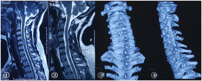
Laminoplasty in spinal cord tumor surgery 41
Figure 1: The technique of vertebral plate and ligament composite laminoplasty. ① Exposure of the spinous process; ⇨: the complete
supraspinous ligament. ② Using abrasion drill and milling cutter complete resection the vertebral plate and ligament composite; ➡: bone
fracture surface; ⇨: exposure of spinal dura mater. ③ Vertebral plate and ligament composite ➡: bone fracture surface; ⇨: vertebral plate
and ligament composite. ④ Screws and connectors were used for fixation of the spinous process ligament complex; ➡: restore ligament
integrity.
2 Patients and methods calcification), and patients younger than 14 years. All patients
from January 2016 to January 2018 were selected for lami-
nectomy; patients presenting from February 2018 to October
2.1 Patient population
2019 were selected for either laminectomy or laminoplasty
on the basis of surgeons’ preference. Patients underwent pre-
Patients with spinal cord tumor treated in Department of
operative and postoperative magnetic resonance imaging
Neurosurgery from January 2016 to October 2019 were
(MRI) in all cases. All patients were followed up with serial
included in this study. Inclusion criteria were patients MRI and targeted CT to assess sagittal alignment 1 week, 3
with spinal cord tumor including intramedullar tumors months, and 6 months postsurgery, then annually. The neuro-
(such as ependymoma and astrocytoma); extramedullar, logical functions were also assessed accordingly.
intradural tumors (such as neurinoma and meningioma); Functional status was graded according to a modified
and epidural tumors (chordomas, teratomas, hemangiomas, McCormick scale both preoperatively and at the follow-
and carcinomas metastases). up [11]. Preoperative radiographs were assessed for
Exclusion criteria were metastasis tumor, any other spinal deformity which was defined as progression of
cases in which decompression is required (such as chiari scoliotic or kyphotic curves by at least 10°. The onset
malformation, spinal stenosis, and ligamentum flavum time of progressive deformity was recorded.42 Xu Hao and Wang Lin
Informed consent: Informed consent has been obtained 2.3 Statistical analysis
from all individuals included in this study.
For intergroup comparison, the Student t test was used
Ethical approval: The research related to human use has for parametric data and the Mann–Whitney U test for
been complied with all the relevant national regula- nonparametric data. Percentages were compared via χ2
tions, institutional policies, and in accordance the tests. The absolute incidence of postoperative deformity
tenets of the Helsinki Declaration and has been approved was compared by χ2 tests. The incidence of deformity was
by the authors’ institutional review board or equivalent analyzed by the Kaplan–Meier method and then com-
committee. pared between laminoplasty and laminectomy groups
via the log-rank test.
2.2 Surgical techniques
3 Results
The decision to perform laminoplasty was based on sur-
geons’ preference. For laminoplasty, only medial facet
3.1 Patient population
joint exposure is performed by subperiosteal paraspinal
muscle dissection. An effort was made to preserve the
facet joint capsules in all cases. Abrasion drill and milling A total of 94 patients underwent surgical resection of
cutter were used to resect the spinous process ligament an intradural spinal tumor during the reviewed time
complex completely. The spinous processes of the planned period. Of them, 60 (64%) underwent laminectomy
laminoplasty segment were left intact to preserve the inter- and 34 (36%) underwent laminoplasty. The tumor loca-
spinous ligaments and ligamentum flavum [12]. Vertebral tion included cervical vertebra in 36 cases, thoracic ver-
plate and ligament composite was installed, then screws tebra in 21 cases, and lumbar vertebra in 37 cases. Total
and connectors were used for fixation (Figure 1). Patients resection was achieved in 84 patients (89.0%); subtotal
who underwent laminectomy had laminae removal above resection was achieved in 10 patients (11%). The tumor
the entire length of the tumor. Sensory-evoked and location included cervical vertebra in 36 cases, thoracic
motor-evoked potentials were used in all cases. All vertebra in 21 cases, and lumbar vertebra in 37 cases.
patients including cervical, thoracic, and lumbar ver- Total resection was achieved in 84 patients (89.0%);
tebrae received external fixation postsurgery for at least subtotal resection was achieved in 10 patients (11%)
3 months (Figure 2). (Table 1).
Figure 2: The MRI and CT imaging pre and postsurgery. ① T2 MRI before surgery. Postoperative pathology confirmed that it is an
ependymocytoma. ② T2 MRI in 1 year postsurgery, there is no recurrence of the tumor and no progressive spinal deformity. ③ and ④ CT
three-dimensional reconstruction showed that vertebral plate and ligament complex were in a suitable location.Laminoplasty in spinal cord tumor surgery 43
Table 1: Patient population Table 3: Laminoplasty vs laminectomy spinal stability in 12-month
follow-up
All patients Laminectomy Laminoplasty
(60) (34) All cases Total Laminectomy Laminoplasty P value
Mean age 45 47 44 94 60 34
M/F 58/36 38/22 20/14 Deformity cases 13 9 4 0.6624
Duration of 3 4 (1–17) 3 (1–16) Cervical 36 21 15
symptoms patients
(months) Deformity cases 9 8 1 0.0387
Levels ≤ 2 57 33 24 Levels > 3 33 19 14
Levels > 3 37 19 14 Deformity cases 9 6 3 0.5176
Cervical 36 21 13
Thoracic 21 14 7
Lumber 37 25 12
Then, we subdivided the patients into two subgroups,
including the cervical patient group and the group of
Pathology included ependymoma in 11 (12%), low- patients with incision vertebral segments >3. We analyzed
grade astrocytoma in 9 (10%), malignant astrocytoma the deformity rate in the two groups in 12 months post-
in 2 (2%), cavernoma in 3 (3%), schwannoma in 31 surgery. For the cervical patients, there is significant dif-
(33%), angiolipoma in 11 (12%), meningioma in 11 (12%), ference (P < 0.05) in the spinal deformity. For the patients
neurofibroma in 7 (7%), epidermoid cyst in 5 (5%), enter- with incision vertebral segments >3, there is no significant
ogenous cysts in 3 (3%), and epidural simple cyst in difference in the spinal deformity (P > 0.05) (Table 3).
2 (2%). Subsequently, the incidence of progressive deformity
The patients were followed up for a total of 17 months. after surgery was analyzed via the Kaplan–Meier method.
Surgical site infection occurred in one patient (1%), whereas In the cervical patient group, laminoplasty vs lami-
cerebrospinal fluid (CSF) infection occurred in five patients nectomy was associated with a similar risk of progres-
(6%). The incisional CSF leakage occurred in eight patients sive deformity (relative risk, 1.871; 95% CI, 0.4720 to
(9%). Mean length of hospitalization was 11 ± 4 days. There 7.415, P = 0.3726) (Figure 3). In the group of patients
was no significant difference between the two methods in with incision vertebral segments >3, laminoplasty vs
terms of operative time, surgical site, infection rate, CSF laminectomy was associated with a similar risk of pro-
infection, incisional CSF leak, and length of hospitalization. gressive deformity (relative risk, 2.018; 95% CI, 0.3201 to
In terms of neurological function, the McCormick score 12.72, P = 0.4549) (Figure 4).
change also showed no difference between the two groups
(P > 0.05) (Table 2). In two cases of laminoplasty, screw
loose and slip off were detected in the follow-ups, but
the patients showed no symptoms at that time. Intensive 4 Discussion
follow-ups were conducted accordingly.
Thirteen (13.9%) patients developed progressive radio- In this study, we assessed whether introducing lamino-
graphic deformity, most of them (11) occurred in 1 year plasty into our practice would influence the incidence
postsurgery. Nine patients developed progressive cervical of postoperative short-term prognosis, neuro-functional
deformity, two patients in lumbar, and two in thoracic recovery, and the spinal deformity occurrence following
vertebra, respectively. intradural tumor resection.
Table 2: Laminoplasty vs laminectomy short-term prognosis
All patients Laminectomy Laminoplasty P value
Operative time (min) 135 124 141 0.382
Surgical site infection 1 0 1
CSF infection 5 (5.32%) 3 (5.00%) 2 (5.88%) 0.987
Incisional CSF leakage 8 4 1 0.742
Length of hospitalization 11 11 11 0.997
McCormick score change 1.53 1.58 1.50 0.86444 Xu Hao and Wang Lin
critical segments, such as C2, C7 [5,15]; the surgery
incision more than three segments [16,17] and zygapo-
physis excision [17,18].
Due to a greater degree of cervical vertebra motion
compared with thoracolumbar vertebra, coupled with the
overuse of smart phones leading to a prolonged time of
cervical anteflexion, we believed that cervical surgery is
more likely to lead to spinal deformity and more attention
should be paid to those patients. In terms of stability of
the spine, the chi-square test in laminoplasty showed some
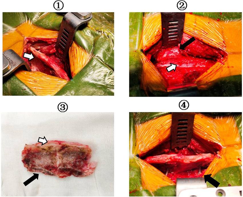
Figure 3: Kaplan–Meier curve of the incidence of progressive advantages in cervical patients during 1-year follow-up.
deformity after surgery of laminoplasty vs laminectomy in the Even though there is no significant difference in subse-
cervical patient group.
quent survival analysis curve, we still believe that lamino-
plasty could be helpful for the spinal stability. At the same
time, we also found that persistently wearing a neck brace
for at least 3 months could reduce the risk of cervical spinal
deformity.
For patients whose intraoperative diagnosis consid-
ered malignant tumor, we routinely performed lami-
nectomy. However, we also found that for benign tumors,
there were five patients who needed secondary surgery in
this study. We also found that laminoplasty can reduce
soft tissue hyperplasia and scar formation, restore anato-
mical layers, and make secondary surgery safer.
Figure 4: Kaplan–Meier curve of the incidence of progressive In addition, hemilaminectomy can better reduce the
deformity after surgery of laminoplasty vs laminectomy in the group biomechanical damage, but on the other hand, hemila-
of patients with incision vertebral segments >3.
minectomy will increase the risk of spinal cord injury
because of the limited operating field [19]. Internal fixa-
Laminectomy has long been considered as the tradi- tion, however, can better guarantee the stability of the
tional approach for intradural tumor resection because it is spine, but at the same time also brought some unique
well established, creating a relatively wide exposure of the complications and disadvantages. For example, screwing
spinal cord, and can easily be extended in sagittal directions. the pedicle could damage the vessel and nerve; fixation
Adults undergoing laminectomy for intradural tumor could destroy the original spine flexion, rotation, and
resection developed postoperative progressive spinal other physiological functions; and pathological changes
deformity in 7–20% of all cases. Laminoplasty has there- of segmental line could accelerate adjacent segment
fore been advocated to avoid such complications because degeneration [20].
the posterior elements of the spinal cord are replaced This study has some limitations because bias can be
[13,14]. This replacement is presumed to leave the pos- introduced in a retrospective review that does not have
terior element intact, theoretically stabilizing the spine randomized, prospectively matched groups. Second,
and preventing instability. bias can also be caused by different surgery techni-
According to our research, there is no significant dif- ques. Despite the use of Kaplan–Meier methods to
ference between the two methods in terms of complica- adjust for varying follow-up, our lack of long-term
tion rate and short-term prognosis. Vertebral plate and follow-up disallows any conclusions on long-term
ligament composite reduction cannot reduce CSF leakage, deformity is also a major limitation of this study.
which is different from previous studies [10]. Although
screw loose and slip off were found in two cases of lami-
noplasty and the surgery time may be prolonged to some
extent, it will not deteriorate the prognosis and can be 5 Conclusion
avoided with more proficient surgical techniques.
Previous research indicate that in the occurrence of Vertebral plate and ligament composite replant is a
instability important factors included destroying of simple and practical method in spinal cord tumorLaminoplasty in spinal cord tumor surgery 45
surgery. Considering both strengths and weaknesses, it is [7] De Jonge T, Slullitel H, Dubousset J, Miladi L, Wicart P, Illés T.
still valuable to conduct in spinal cord tumor surgery Late onset spinal deformities in children treated by lami-
especially in cervical vertebra surgery. nectomy and radiation therapy for malignant tumours. Eur
Spine. 2005;14(8):765–71.
[8] Lunardi P, Licastro G, Missori P, Ferrante L, Fortuna A.
Author contributions: Xu Hao: Conceptualization, Management of intramedullary tumours in children. Acta
Methodology, Software, Data curation, and Writing – Neurochir (Wick). 1993;120(1–2):59–65.
Original draft preparation. Wang Lin: Visualization, [9] Raimondi AJ, Gutierrez FA, Di Rocco C. Laminotomy and total
Investigation, and Writing – Reviewing and Editing. reconstruction of the posterior spinal arch for spinal canal
surgery in childhood. J Neurosurg. 1976;45(5):555–60.
[10] Ratliff JK, Cooper PR. Cervical laminoplasty: a critical review.
Conflict of interest: The authors state no conflict of J Neurosurg. 2003;98(suppl 3):230–8.
interest. [11] McCormick PC, Torres R, Post KD, Stein BM. Intramedullary
ependymoma of the spinal cord. J Neurosurg.
Data availability statement: All data generated or ana- 1990;72(4):523–32.
[12] Derenda M, Kowalina I. Cervical laminoplasty – review of sur-
lyzed during this study are included in this published
gical techniques, indications, methods of efficacy evaluation,
article.
and complications. Neurol Neurochir Pol. 2006;40(5):422–33.
[13] Hukuda S, Ogata M, Mochizuki T, Shichikawa K. Laminectomy
versus laminoplasty for cervical myelopathy: brief report.
J Bone Joint Surg Br. 1988;70(2):325–6.
[14] Abbott R, Feldstein N, Wisoff JH, Epstein FJ. Osteoplastic
References laminotomy in children. Pediatr Neurosurg.
1992;18(3):153–6.
[1] Herman JHI, Sonntag VKH. Cervical corpectomy and plate [15] Asazuma T, Nakamura M, Matsumoto M, Chibo K, Toyama Y.
fixation for postlaminectomykyphosis. J Neurosurg. Postoperative changes of spinal curvature and range of motion
1994;80:963–70. in adult patients with cervical spinal cord tumors: analysis of
[2] Seppälä MT, Haltia MJJ, Sankila RJ, Jääskeläinen JE, 51 cases and review of the literature. J Spinal Disord.
Heiskanen O. Long-term outcome after removal of spinal 2004;17:178–82.
schwannoma: a clinicopathological study of 187 cases. [16] Bresnahan L, Ogden AT, Natarajan RN, Fessler RG. A biome-
J Neurosurg. 1995;83(4):621–6. chanical evaluation of graded posterior element removal for
[3] Papagelopoulos PJ, Peterson HA, Ebersold MJ, Emmanuel RP, treatment of lumbar stenosis: comparison of a minimally
Choudhury SN, Quast LM. Spinal column deformity and invasive approach with two standard laminectomy techniques.
instability after lumbar or thoracolumbar laminectomy for Spine. 2009;34(1):17–23.
intraspinal tumors in children and young adults. Spine. [17] Sharrna M, Langrana NA, Rodriguez J. Role of ligaments and
1997;22(4):442–51. facets in lumbar spinal stability. Spine. 1995;20(8):887–900.
[4] Cristante L, Herrmann HD. Surgical management of intra- [18] Shirazi Adl A. Finite-element evaluation of contact loads on
medullary spinal cord: functional outcome and sources of facets of an L2–L3 lumbar segment in complex loads. Spine.
morbidity. Neurosurgery. 1994;35(1):69–76. 1991;16(5):533–41.
[5] Katsumi Y, Honma T, Nakamura T. Analysis of cervical [19] Spetzger U, Bertalanffy H, Reinges MHT, Gilsbach JM.
instability resulting from laminectomies for removal of spinal Unilateral laminotomy for bilateral decompression of lumbar
cord tumor. Spine. 1989;14(11):1171–6. spinal stenosis. Part II: clinical experiences. Acta Neuroehir.
[6] Yeh JS, Sgouros S, Walsh AR, Hockley AD. Spinal sagittal 1997;139:397–403.
malalignment following surgery for primary intramedullary [20] Hicks JM, Singla A, Shen FH, Arlet V. Complications of pedicle
tumours in children. Pediatr Neurosurg. screw fixation in scoliosis surgery: a systematic review. Spine.
2001;35(6):318–24. 2010;35(11):E465–70.You can also read