XSSJS inhibits hepatic fibrosis by promoting the miR-29b-3p/VEGFA axis in vitro and in vivo
←
→
Page content transcription
If your browser does not render page correctly, please read the page content below
Bioscience Reports (2022) 42 BSR20212241
https://doi.org/10.1042/BSR20212241

Research Article

XSSJS inhibits hepatic fibrosis by promoting the
miR-29b-3p/VEGFA axis in vitro and in vivo
    Tianyao Zhang1 , Yu Yang1 ,                          Baojia Wang1 , Long Wang1 , Dong Wang2 , Ning Cao2 and Jinyu Shi1
1 Department of Fundamental Medical Science, Chengdu University of Traditional Chinese Medicine, Chengdu, Sichuan, China; 2 Department of Fundamental Medical Science,
Shaanxi University of Traditional Chinese Medicine, Xianyang, Shaanxi, China
Correspondence: Baojia Wang (wangbaojia@cdutcm.edu.cn)

                                                                                                                                                                                           Downloaded from http://portlandpress.com/bioscirep/article-pdf/42/2/BSR20212241/929411/bsr-2021-2241.pdf by guest on 30 June 2022
                                           Hepatic pathological angiogenesis (HPA) is the key event of hepatic fibrosis (HF). Xueshisan-
                                           jia powder (XSSJS), a Chinese herbal compound, is beneficial for alleviating pathological an-
                                           giogenesis of hepatic tissue. The present study attempts to reveal the effect and mechanism
                                           of XSSJS via regulating miR-29b-3p/VEGFA axis against pathological angiogenesis in HF. In
                                           in vitro model, human embryonic kidney 293T cells were transfected with miR-29b-3p mim-
                                           ics, whereby the expression of miR-29b-3p was tested by real-time quantitative polymerase
                                           chain reaction (RT-qPCR), ensued by Luciferase assay determining the relationship between
                                           miR-29b-3p and vascular endothelial cell growth factor A (VEGFA). In addition, miR-29b-3p
                                           mimic transfected into the activated hepatic stellate cell T6 (HSC-T6). The Cell-Counting-Kit
                                           8 (CCK8) and 5-Bromodeoxyuridine (BrdU) staining were first utilized to detect the an-
                                           tiproliferative efficiency of XSSJS following the XSSJS compound serum intervention, and
                                           then used to observe the expression of transforming growth factor-β (TGF-β), VEGFA,
                                           platelet-derived growth factor (PDGF) via RT-PCR, Western blot (WB), and Immunofluores-
                                           cence (IF) methods. During the in vivo model, XSSJS with boil-free granules were fed to
                                           Wistar rats with liver fibrosis caused by intraperitoneal injection of pig serum followed by the
                                           transfection of miR-29b-3p adeno-associated virus (AAV). Hematoxylin–Eosin (HE) staining
                                           was used for histopathology assessment. The expression of miR-29b-3p, VEGFA, PDGF,
                                           TGF-β have been investigated in liver tissue using RT-PCR, WB, IF. The results verified that
                                           XSSJS could up-regulate miR-29b-3p and suppress the expression of VEGFA, PDGA, and
                                           TGF-β. In mechanism, miR-29b-3p primarily targeted the 3 UTR of VEGFA. In conclusion,
                                           XSSJS could modulate miR-29b-3p/VEGFA axis to inhibit the pathological angiogenesis of
                                           HF.

                                         Introduction
                                         Hepatic fibrosis (HF) is a common pathological hallmark of chronic liver diseases (CLDs) [1]. Hepatic
                                         pathological angiogenesis (HPA) associates strong implication in the development of HF, which is rec-
                                         ognized as a central event in hepatic stellate cells (HSCs) activation [2,3], inflammatory response [4–6]
                                         and hepatic vascular resistance (IHVR) [7,8]. In the progression of HPA, vascular endothelial growth
Received: 24 September 2021
                                         factor (VEGFA) contributes to the proliferation, migration, differentiation of vascular endothelial cells
Revised: 21 January 2022                 and the formation of vascular lumen [9–11], which eventually leads to intrahepatic angiogenesis and
Accepted: 02 February 2022               hepatic sinusoidal capillarization [12,13]. Therefore, it is of great significance to restrain pathological
                                         angiogenesis for liver disease deterioration, especially targeting the excessive VEGF [14,15]. However,
Accepted Manuscript online:
04 February 2022                         it is already known that the western drug of anti-angiogenesis such as Sorafenib, Bevacizumab and
Version of Record published:             Sunitinib, present the obvious adverse effects, and were forced to interrupt treatment [16–18]. Notice-
25 February 2022                         ably, accumulated evidence have demonstrated that the use of traditional Chinese medicines (TCMs)

© 2022 The Author(s). This is an open access article published by Portland Press Limited on behalf of the Biochemical Society and distributed under the Creative Commons Attribution   1
License 4.0 (CC BY).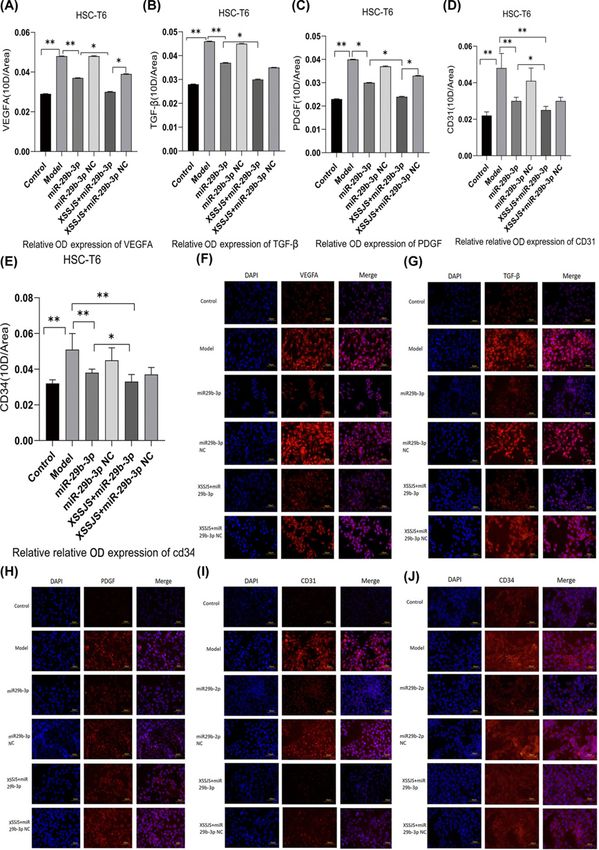
Bioscience Reports (2022) 42 BSR20212241
                                                                                                                                         https://doi.org/10.1042/BSR20212241

                                                                                                                                                                                           Downloaded from http://portlandpress.com/bioscirep/article-pdf/42/2/BSR20212241/929411/bsr-2021-2241.pdf by guest on 30 June 2022
           Figure 1. A schematic diagram with XSSJS promoting miR-29b-3p/VEGFA axis
           The schematic diagram representing the subject of the article shows that the XSSJS could down-regulate the expression of VEGFA
           and inhibit the PA of HF.

            have increased worldwide due to their exclusive properties, that activates blood circulation to dissipate stasis, which
           could prevent liver fibrosis via reducing liver tissue inflammation [19,20] and inhibiting pathological angiogenesis
           [21,22]. From TCM perspective, the pathology of liver fibrosis is the obstruction of collaterals by phlegm and blood
           stasis, so the therapy relies on its virtue of clearing phlegm, assisting blood circulation and dredging the collateral
           vessels. The extensive use of Xueshisanjia (XSSJS) for clinical treatment of CLD is owing to its significant clinical effi-
           cacy and inherent virtue of removing blood stasis and clearing the collateral vessels. Modern studies have shown that
           the anti-HF mechanisms of XSSJS include the inhibition of rat HSC-T6I, III collagen biosynthesis [23], as well as the
           pathological angiogenesis, accompanied by reducing microvascular density in fibrotic liver tissue and promoting the
           expression of VEGFA mRNA and protein [24–26]. Since, microRNAs are enriched in the liver tissue and involved in
           regulating the physiological and pathological processes, miR-29b is particularly expressed 100-times higher in HSCs
           than in other liver parenchymal cells [27]. miR-29b-3p is affiliated to miR-29b family, which bears high in-group
           similarity of mature sequences for humans, rats, and mice [28,29]. Some studies indicated that the overexpression of
           miR-29b is inversely proportional to the occurrence of liver fibrosis [30], and the main mechanism of miR-29b-3p
           to ease liver fibrosis is to inhibit the activation of HSCs [31,32], thus up-regulating miR-29b-3p is an effective strat-
           egy against HF. Previous researchers have been mostly absorbed in studying the physiological roles of miR-29b-3p
           on HF, yielding a general absence of the report on the mechanisms or target genes that cause the PA phenotypes.
           Therefore, we interest ourselves in the therapeutic effect and mechanisms of XSSJS on anti-fibrosis by regulating with
           miR-29b-3p/VEGFA axis, and the schematic presentation of the XSSJS inhibit PA via regulating miR-29b-3p/VEGFA
           axis is shown in Figure 1.

           Materials and methods
           Cell cultured and modeling
           Human embryonic kidney cells (HEK-293T cells) were acquired from Procell Biotech Solutions Co. Ltd. (Shanghai,
           China), and cultured with 55% DMEM+40% fetal bovine serum (FBS)+5% DMSO. The rat hepatic stellate cell beads
           (HSCs-T6), obtained from Fusheng Industrial Co., Ltd (Shanghai, China), were cultured with DMEM+10% FBS+1%
           (penicillin/streptomycin solution). Transforming growth factor-β1 (TGF-β1) were purchased from PeproTech China

2   © 2022 The Author(s). This is an open access article published by Portland Press Limited on behalf of the Biochemical Society and distributed under the Creative Commons Attribution
    License 4.0 (CC BY).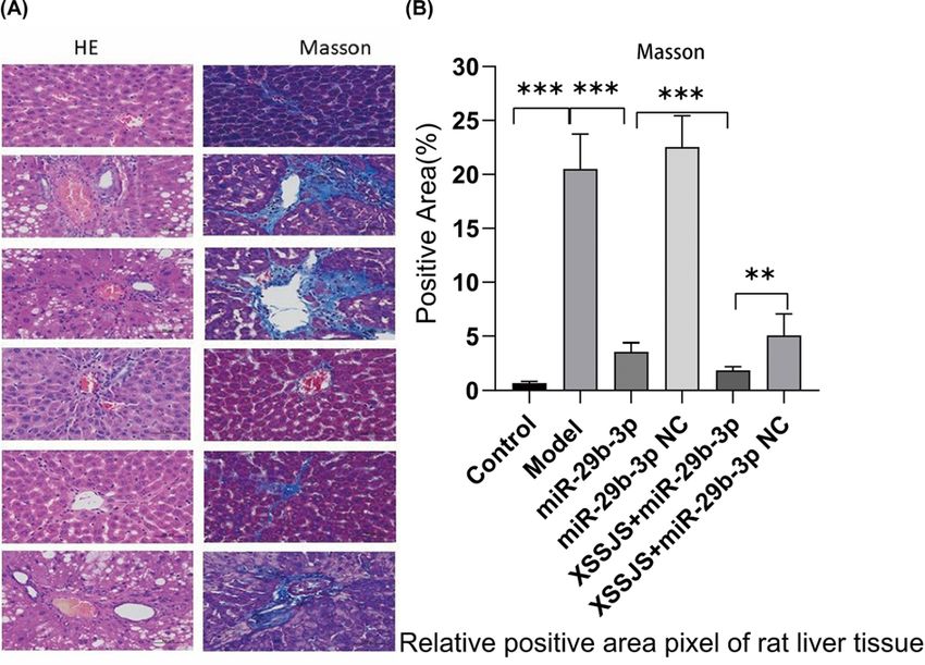
Bioscience Reports (2022) 42 BSR20212241
https://doi.org/10.1042/BSR20212241

Table 1 The primer sequences used in real-time quantitative polymerase chain reaction analysis
  Gene                                                           Primer                                                          Sequences (5 –3 )

  R-Actin                                                        R-Actin-F                                                       TGTCACCAACTGGGACGATA
                                                                 R-Actin-R                                                       GGGGTGTTGAAGGTCTCAAA
  R-TGF-β                                                        R-TGF-β-F                                                       GACCGCAACAACGCAATCTATGAC
                                                                 R-TGF-β-R                                                       CTGGCACTGCTTCCCGAATGTC
  R-VEGFA                                                        R-VEGFA-F                                                       CACGACAGAAGGGGAGCAGAAAG
                                                                 R-VEGFA-R                                                       GGCACACAGGACGGCTTGAAG
  R-PDGF                                                         R-PDGF-F                                                        TCTCTGCTGCTACCTGCGTCTG
                                                                 R-PDGF-R                                                        AAGGAGCGGATGGAGTGGTCAC
  U6                                                             Sn-U6-F                                                         CTCGCTTCGGCAGCACA
                                                                 Sn-U6-R                                                         AACGCTTCACGAATTTGCGT

                                                                                                                                                                                           Downloaded from http://portlandpress.com/bioscirep/article-pdf/42/2/BSR20212241/929411/bsr-2021-2241.pdf by guest on 30 June 2022
  miR-29b-3p                                                     mi-29b-F                                                        CGCGTAGCACCATTTGAAATC
                                                                 mi-29b-R                                                        AGTGCAGGGTCCGAGGTATT
  RT-primer                                                      GTCGTATCCAGTGCAGGGTCCGAGGTATTCGCACTGGATACGACAACACT

(Suzhou, China), when HSC-T6 was proliferating at logarithmic rate, the digestion of cells was achieved with 0.25%
trypsin, and 105 cells/well were inoculated in 96-well plates after counting, TGF-β1 (10 ng/ml) was instilled in the
model wells but not in those control ones. After 24 h, the supernatants and cells were subject to collection.

Reagents and antibodies
TRIzol reagent was purchased from Invitrogen (Invitrogen, U.S.A.). The miR-29b-3p mimics and the negative control
vectors (miR-29b-3p mimics NC) were synthesized and designed by Integrated Biotech Solutions Co. Ltd. (Shang-
hai, China IBS-01). U6 serves as an internal reference for miR-29b-3p and its sequence is shown in Table 1. The se-
quence of rno-miR-29b-3p is 3 uugugacuaaaguuuaccacgau, and NCBI Gene ID is 104797378. VEGFA-overexpressing
plasmid and the negative control vectors were synthesized and designed by SanGon Biotech Co., Ltd (Shanghai,
China), which gene sequence is VEGFA 3 UTR (1634–1641): 5 aucauuuauuuauuggugcuac, and NCBI Gene ID is
83785. Lipofectamine™ 2000 transfection reagent (Invitrogen, Thermo Fisher Scentific, China) was applied for cell
transfection. The miR-29b-3p adeno-associated virus (AAV) and the negative control vectors (the miR-29b-3p NC
AAV) were designed and synthesized from SanGon Biotech Co., Ltd (Shanghai, China). The primary antibodies,
such as anti-VEGFA (bs-1665R, Rabbit, 1:1000), anti-TGF-β1 (bs-0086, Rabbit, 1:1000) were bought from Boaosen
Biotechnology Co., Ltd (Beijing, China); anti-platelet-derived growth factor (PDGF; Gb-11261, Rabbit, 1:1000)
were purchased from Xavier Biotechnology Co., Ltd (Wuhan, China); anti-CD31 (platelet endothelial cell adhesion
molecule-1; 130 kDa, Rabbit, 1:1000), anti-CD34 (Gb-4000, Rabbit, 1:1000) and anti-β-Actin (Gb13001-1, Rabbit,
1:1000) were bought from Wuhan Google Biotechnology Co., Ltd (Wuhan, China); anti-glyceraldehyde-3-phosphate
dehydrogenase (GAPDH; Mab5465, Mouse, 1:2000) were bought from Multi Sciences Biotechnology Co., Ltd
(Hangzhou, China); dual-luciferase gene kit was bought from Beyotime Biotechnology Co., Ltd (Shanghai, China);
luc-3 -UTR-WT/MUT were designed by SanGon Biotech Co., Ltd (Shanghai, China). Cell-Counting-Kit-8 (CCK8)
and 5-Bromodeoxyuridine (BrdU) detection reagents were purchased from SanGon Biotech Co., Ltd (Shanghai,
China).

miR-29b-3p mimics transfection and XSSJS intervention in vitro
To overexpress the miR-29b-3p gene, HEK-293T cells were transfected with miR-29b-3p mimic and miR-29b-3p in-
hibitor via Lipofectamine 2000 transfection reagent (Invitrogen, Thermo Fisher Scentific, China) with reference to
the instructions of the manufacturer. Real-time PCR analysis was employed to assess the expression of miR-29b-3p
for 48 h of cell transfection. Thereafter, the mechanism of miR-29b-3p and VEGFA on the molecules via luciferase
assay was confirmed. The target promoter fragment was cloned from genomic DNA employed PCR technology and
inserted into luciferase reporter gene plasmid PGL3-BASIC by double digestion assay of restriction enzyme SacI and
XbaI, the luciferase activity of VEGFA-WT was tested following 24 h of cell con-transfection with mimic-miR-29b-3p
and VEGFA plasmids (firefly luciferase was used as a reporter gene and Renilla kidney luciferase was used as an
internal reference gene). In order to further reveal the regulatory relationship between miR-29b-3p/VEGFA axis
and XSSJS intervention in HF, and XSSJS containing serum was used for intervention following being transfected
with an miR-29b-3p mimic and miR-29b-3p mimic NC in activated HSCs [33]. Except for the control and model
groups, which were transfection-free, the miR29b-3P group and XSSJS+miR-29b-3p group were transfected with the

© 2022 The Author(s). This is an open access article published by Portland Press Limited on behalf of the Biochemical Society and distributed under the Creative Commons Attribution   3
License 4.0 (CC BY).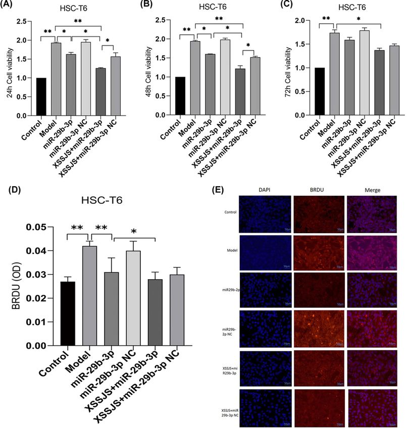
Bioscience Reports (2022) 42 BSR20212241
                                                                                                                                         https://doi.org/10.1042/BSR20212241

           miR-29b-3p mimics; miR-29b-3p NC group and XSSJS+ miR-29b-3p NC group were transfected with the miR-29b-3p
           mimics NC (negative control vectors) according to the instructions of the manufacturer and relative reference [34].
           The transfection efficiency was confirmed by RT-PCR after 24 h.

           Cell proliferation efficiency analysis
           Cellular proliferation was assessed using CCK8 and BrdU cell proliferation detection kits. Following the activation of
           HSC-T6 were the transfection with miR-29b-3p mimics and mimic NC, the cells were digested with 0.25% trypsin
           following HSC-T6’s logarithmic period, and 105 cells/well were inoculated in 96-well plates after counting and were
           washed three-times in PBS. Twelve hours later, the original medium was changed to the medium with XSSJS contain-
           ing serum when fusion rate of the cells reaches 80–95%. After 24 h of culturing, the biocompatibility and cytotoxicity
           of HSCs-T6 were assayed using a CCK8. According to the manufacturer’s instructions, 10 μl CCK8 reagent was ad-
           ministered to each well before harvesting, and the optical densities (OD) value at 450 nm was measured after 4 h of

                                                                                                                                                                                           Downloaded from http://portlandpress.com/bioscirep/article-pdf/42/2/BSR20212241/929411/bsr-2021-2241.pdf by guest on 30 June 2022
           incubation. Each experiment was repeated three times.
              Cell proliferation was also assessed by the number of nuclei that were 4 ,6-diamidino-2-phenylindole (DAPI)
           stained and marked with BrdU. During HSC-T6’s in logarithmic period, BrdU with concentration of 20 μmol/ml
           was added into the culture medium. The secondary antibody for BrdU was labeled TRITC. Images were obtained
           using an optical microscope (400×; Olympus Corporation, Tokyo, Japan).

           XSSJS no-decoction granule prescription
           XSSJS no-decoction granules contained turtle shell 15 g, pangolin 3 g, batryticated silkworm 10 g, ground beetle 10 g,
           peach kernel 10 g, bupleurum 5 g (total 53g). The same batch was purchased from Sichuan New green Pharmaceutical
           Science and Technology Development Co. Ltd. The Qualification Certificate No.: 15013726.

           Experimental animals
           Adult (8–10 weeks old) male Wistar rats weighing 180 +   − 20 g were obtained from Chengdu Dasuo Biotechnol-
           ogy Co., Ltd (License No.: SCXK(Sichuan), 2013-24, Certificate No.: 0002715). All animal experiments took place
           at Experimental Animal Center of Chengdu University of Traditional Chinese Medicine. A total of 80 Wistar rats
           were accommodated under a temperature-controlled condition (22 +         ◦
                                                                                − 2 C). Sixty rats were randomly and evenly
           spread into six groups (n=10 each) including control group, model group, miR-29b-3p group, miR-29b-3p NC group,
           XSSJS+miR29b-3P group, XSSJS+miR-29b-3p NC group. All of the animals were provided with day/night shift every
           12 h, and unlimited access to water and rodent chow.

           Models, transfection and specimen collection in vivo
           After a week of routine feeding, except for the control group via gavage with the same volume of 9 g/l normal saline,
           the remaining groups were fed with XSSJS granule prescription once a day on a regular time basis for 22 weeks.
           XSSJS granules prescription were dissolved with 9 g/l normal saline into liquid with 0.4 g/ml of concentration, and
           the volume of gastric capacity was 0.67 ml/100 g. we chose a small dose of porcine serum (Thermo Fisher, 26250084)
           (0.5 ml/kg, twice a week), which is managed with intraperitoneal injection methods for modeling [35]. At the 18th
           week of modeling, the sequence of miR-29b-3p was inserted into AAV expression vector (provided by Sangon Biotech
           Co., Ltd. Shanghai, China) and was injected into the tail vein, with an injection volume of 200 μl and an injection
           volume of 50 μl for each injection (reference: the in vivo transfection instructions). In the 22nd week of modeling,
           1% pentobarbital was applied to anesthetizing rats intraperitoneally. Then rats were killed by exsanguination, and the
           specimens were collected and forwarded to the later testing stage.

           XSSJS drug-contained serum intervention
           Next, we randomly split the 20 Wistar rats into the control group and drug-contained serum group. XSSJS
           no-decoction granules were directly infused into the stomachs of Wistar rat (twice a day at a fixed time) for
           drug-contained serum group, with other conditions (eg. capacity, concentration) unaltered as indicated above, while
           the control group was established via gavage with the same volume of 9 g/l normal saline. In the seventh day of intra-
           gastric administration, 1% pentobarbital was applied to anesthetize rats intraperitoneally, and blood was taken from
           the posterior abdominal aorta in sterile environment, centrifuged at 2000 rpm (r = 13.5 cm) for 20 min, the serum
           was separated 56◦ C water bath for 30 min, 0.22 μm membrane filtration, and finally the FBS in HSC-T6 medium
           after miR-29b-3p transfection was replaced by XSSJS containing serum and cultured for 24 h.

4   © 2022 The Author(s). This is an open access article published by Portland Press Limited on behalf of the Biochemical Society and distributed under the Creative Commons Attribution
    License 4.0 (CC BY).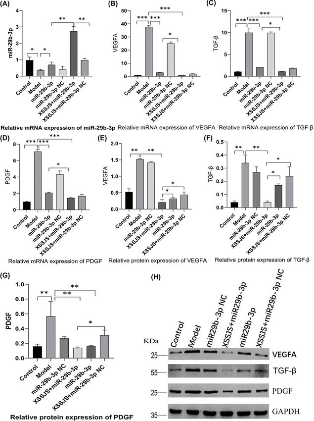
Bioscience Reports (2022) 42 BSR20212241
https://doi.org/10.1042/BSR20212241

Real-time quantitative polymerase chain reaction analysis
TRIzol reagent was used to extract the complete RNA from cells, and then detected the integrity of RNA with 1%
agarose electrophoresis via calculating with the ratio of A 260 nm/A280 nm, when the purity of RNA in 1.8–2.2, the
cDNA was synthesized using a HyPure™ Molecular Biology Grade Water 5× All-In-One MasterMix (with AccuRT
Genomic DNA Removal Kit, HyClone). After that, the cDNA was served as the substrate for real-time quantitative
polymerase chain reaction (RT-qPCR) with EvaGreen Express 2× PCR MasterMix-No Dye (ABM Goodchina Inc.),
the primer sequences used in RT-qPCR analysis are shown in Table 1. 2−Ct method makes the calculation for the
relative levels of mRNAs, β-Actin is the reference gene. The whole progress of manipulation follows the manufac-
turer’s reagent instructions.

Western blotting assays
The sodium dodecyl sulfate/polyacrylamide gel electrophoresis (SDS/PAGE) was prepared to separate from each

                                                                                                                                                                                           Downloaded from http://portlandpress.com/bioscirep/article-pdf/42/2/BSR20212241/929411/bsr-2021-2241.pdf by guest on 30 June 2022
group after treatment, the protein samples of liver tissue and cells (Wuhan Google Biotechnology Co., Ltd.), which
would be subsequently transferred to 0.45-μm PVDF membrane (Millipore Company, MA, U.S.A.) according to
different molecular weight of each protein at low temperature. After that, the cell membrane was incubated with
the indicated primary antibody at 4◦ C overnight. The dilution ratio of primary antibody is 1:1000. Subsequently,
the membranes were moved into the secondary antibody incubation box with a dilution ratio of 1:5000 for 60 min
at room temperature, after which the protein expression was tested with ECL Kit (Hangzhou Lianke Biotechnology
Co. Ltd, China). GAPDH was set as an internal reference. Chemiscope analysis software was used for gray analysis
(Shanghai Qinxiang Scientific Instrument Co., Ltd), whose relative expression was calculated with respect to the
protein of interest and GAPDH in the same sample and expressed graphically.

Luciferase gene reporter assay
The mechanism of miR-29b-3p and VEGFA on the molecules via luciferase assay was confirmed. The target pro-
moter fragment, cloned from genomic DNA using PCR technology, was inserted into luciferase reporter gene plasmid
PGL3-BASIC by double digestion assay of restriction enzyme SacI and XbaI. Luciferase reporter plasmids including
the 3 -UTR of VEGFA (called as VEGFA-WT) and the empty luciferase vector were purchased from the Shanghai
Integrated Biotech Solutions Co.,Ltd. (Shanghai, China, IBS-01). Mutations were made at the predicted target sites
between miR-29b-3p and VEGFA, therefore the name, VEGFA-MUT. HEK 293T cell were seeded in 96-well plates
and co-transfected with the VEGFA-WT/VEGFA-MUT and mimic-NC/mimic-miR-29b-3p. Lipofectamine™ 2000
transfection reagent was applied for cell transfection based on the specification. After 48 h of the con-transfection
with mimic-miR-29b-3p and VEGFA plasmids, the luciferase activity in each well was measured by using Luciferase
reporter gene detection kit (shanghai Beyotime Biotechnology) (firefly luciferase was used as a reporter gene and sea
kidney luciferase was used as an internal reference gene).

Histopathology assay
The hepatic tissues containing fixed 4% concentration of paraformaldehyde were embedded in paraffin. The 5-μm
sections were sliced and stained with Hematoxylin–Eosin (HE) and Masson. Slides were scanned and images were
taken under an optical microscope (400×; Shanghai Qinxiang Scientific Instrument Co., Ltd. China, Chemiscope
6100).

Immunofluorescence staining
The evenly distributed cells in the middle of the cover glass were marked with a histochemical circle, and then 50–100
μl working solution was added. After 0.5 h of incubation, cells were washed twice with PBS for 5 min each time.
The tissue was uniformly covered with 3%BSA for 30 min, thereafter PBS was added to the cell well plate, entailing
an incubation period overnight at 4◦ C. The cellular porous plates were processed by the decolorization shaker for
three-times, 5 min each time. Shortly after a momentary drying, the tissue was covered with secondary antibody
of the corresponding species in the histochemistry kit and incubated at room temperature for 1 h. The slide was
placed in PBS, and then shaken on the decolorization shaker for three-times, 5 min each time. After the sections
slightly dried, DAPI dying solution was dropped in the circle to redye the nuclei, and the cells were incubated at room
temperature for 10 min in dark, and then sealed with anti-fluorescence quenching sealing tablets. The sections were
observed under fluorescence microscope, from which images were collected. The immunofluorescent intensity ratio
was calculated between the target protein and endogenous protein.

© 2022 The Author(s). This is an open access article published by Portland Press Limited on behalf of the Biochemical Society and distributed under the Creative Commons Attribution   5
License 4.0 (CC BY).
Bioscience Reports (2022) 42 BSR20212241
                                                                                                                                         https://doi.org/10.1042/BSR20212241

                                                                                                                                                                                           Downloaded from http://portlandpress.com/bioscirep/article-pdf/42/2/BSR20212241/929411/bsr-2021-2241.pdf by guest on 30 June 2022
           Figure 2. miR-29b-3p targeted VEGFA in vitro
           (A) The binding sites between miR-29b-3p and the 3 UTR of VEGFA. (B) RT-PCR tested the expression of miR-29b-3p following
           24 h of cell transfection with miR-29b-3p mimics and miR-29b-3p inhibitor into HEK 293T cells. (C) A target gene of interest is
           transfected with a luc gene that expresses the enzyme luciferase at the target site, and the luciferase activity of VEGFA-WT was
           tested following 48 h of cell con-transfection (x +
                                                             − s, n=3, **P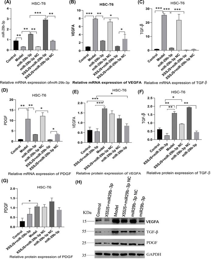
Bioscience Reports (2022) 42 BSR20212241
https://doi.org/10.1042/BSR20212241
XXSJS intervention inhibited PA via promotion miR-29b-3p/VEGFA axis in
vitro
Effect of XSSJS compound serum on proliferation of HSCT6 Cells
The CCK8 assay results (taken at 24, 48, 72 h, respectively) showed that HSC-T6 cells were pre-treated with TGF-β (20
ng/ml) followed by treatment with XSSJS compound serum for 24 h. The inhibitory effect of XSSJS and miR-29b-3p
on cell proliferation were assessed. The results were found that, compared with the ineffectiveness on cell prolifera-
tion of the control group, cell has increased in the model group, while it decreased in the three groups of miR-29b-3p,
XSSJS + miR-29b-3p and XSSJS + miR-29b-3p NC. Moreover, the decreased amount of XSSJS + miR-29b-3p group
was more significant than the amount of the other two (Figure 3A–C), which indicated that XSSJS + miR-29b-3p
could more significantly inhibit the HSC proliferation than using XSSJS or miR-29b-3p intervention alone. Cell pro-
liferation was also assessed using BrdU staining, which showed that XSSJS + miR-29b-3p group could better suppress
the proliferation of HSC compared with miR-29b-3p and XSSJS group alone. No significant difference in cell prolif-
Downloaded from http://portlandpress.com/bioscirep/article-pdf/42/2/BSR20212241/929411/bsr-2021-2241.pdf by guest on 30 June 2022
eration was observed between the miR-29b-3p and XSSJS + miR-29b-3p NC group (Figure 3D,E).
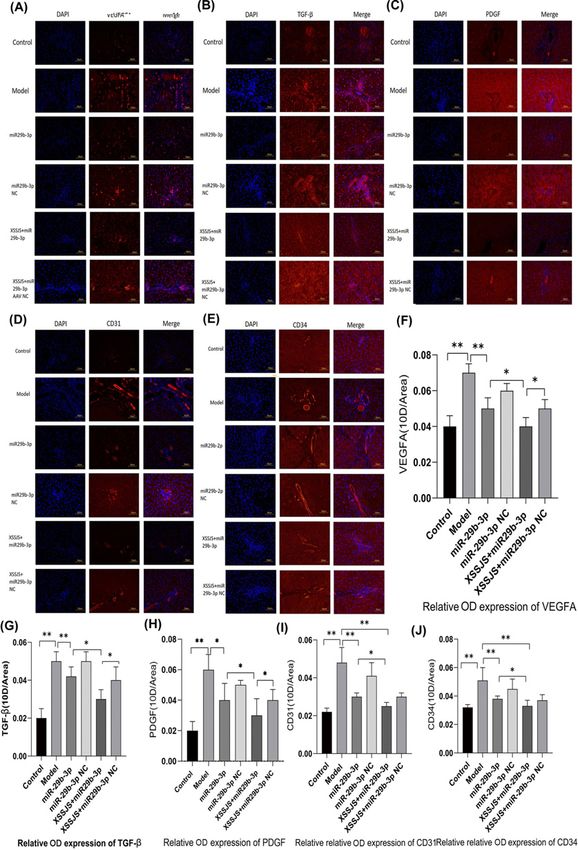
Effect of XSSJS compound serum on the expression of miR29b-3p, VEGFA, TGF-β, PDGF in
HSCT6 cells
Subsequently, we explored whether miR-29b-3p/VEGFA axis was involved in XSSJS-mediated PA improvement in
vitro. Firstly, XSSJS + miR-29b-3p promoted the miR-29b-3p expression more significantly than either miR-29b-3p
or XSSJS+ miR-29b-3pNC group (Figure 4A). Accordingly, XSSJS + miR-29b-3p more significantly curbed VEGFA
expression than miR-29b-3p or XSSJS (XSSJS+miR-29b-3p NC) (Figure 4B,E,H). Moreover, the level of other angio-
genesis factors, such as TGF-β, PDGF, were significantly reduced by SXXJS+miR-29b-3p (Figure 4C,D,F–H). How-
ever, we found the relative mRNA expression of PDGF inconsistent with the protein expression (Figure 4D,G). Via
analyzing the relevant literature and tests [36,37], this peculiar result of our experiment may either arise from the
time lag in the expression profile between protein and mRNA, or the modification issue after the protein translation.
Protein expression of VEGFA, TGF, PDGF, CD31, CD34 in HSCT6 cells
IF results indicated that XSSJS+miR-29b-3p is the most effective in decreasing the protein expression of VEGFA, as
well as other pathological angiogenesis-related factors (TGF-β, PDGF, CD31, CD34) (Figure 5A–J). These results
suggested that the XXSJS intervention with miR-29b-3p transfection could more significantly inhibit the protein
expression of VEGFA in HF. In addition, XXSJS also could reduce the protein expression of other PA-related markers
of liver fibrosis.
XXSJS intervention inhibited PA via promotion miR-29b-3p/VEGFA axis in
vivo
XSSJS intervention alleviated HF with reduced pathological course in HF rats
Compared with the control group, the liver tissue appeared swollen and many of the cells contained vacuoles in the
model group. Liver lobules were distorted by the irregular zones of parenchymal nodules that surrounded it. While the
inflammatory cell infiltration was observed. All the above-mentioned pathological reactions are mitigated under the
intervention of the following XSSJS, miR-29b-3p, and XSSJS+ miR-29b-3p, respectively, among those intervention
the XSSJS+miR-29b-3p significantly reduced pathological response compared with the other groups (Figure 6A).
The quantitative analysis of positive pixel area (%) in MASSON staining also exhibited the same effect (Figure 6B).
Therefore, we can conclude that XSSJS intervention following miR-29b-3p transfection can reduce the degree of liver
fibrosis, such as clear demarcation of hepatic lobules, insignificant proliferation of interlobular connective tissue, and
less infiltration of inflammatory cells around hepatic sinusoids.
Effect of XSSJS on the expression of miR29b-3p, VEGFA, TGF-β, PDGF in HF rats
In quest for further proof of miR-29b-3p/VEGFA axis being involved in XSSJS-mediated PA alleviation in vivo, we
investigated the effectiveness of XSSJS intervention on the expression of miR-29b-3p and VEGFA in liver fibrosis rats.
We found that the expression of VEGFA, PDGF, TGF-β remained relatively low in normal liver tissues, and VEGFA,
PDGF, TGF-β were significantly up-regulated in HF rats. However, on the basis of miR-29b-3p AVV transfection in
vivo, XSSJS treatment further increased the expression of miR-29b-3p in mRNA and reduced the expression values
of VEGFA in mRNA and protein levels as well as in other pathological angiogenesis-related factors (TGF-β, PDGF)
(Figure 7A–H). Those results suggested that XSSJS could promote the expression of miR-29b-3p, and its combination
© 2022 The Author(s). This is an open access article published by Portland Press Limited on behalf of the Biochemical Society and distributed under the Creative Commons Attribution 7
License 4.0 (CC BY).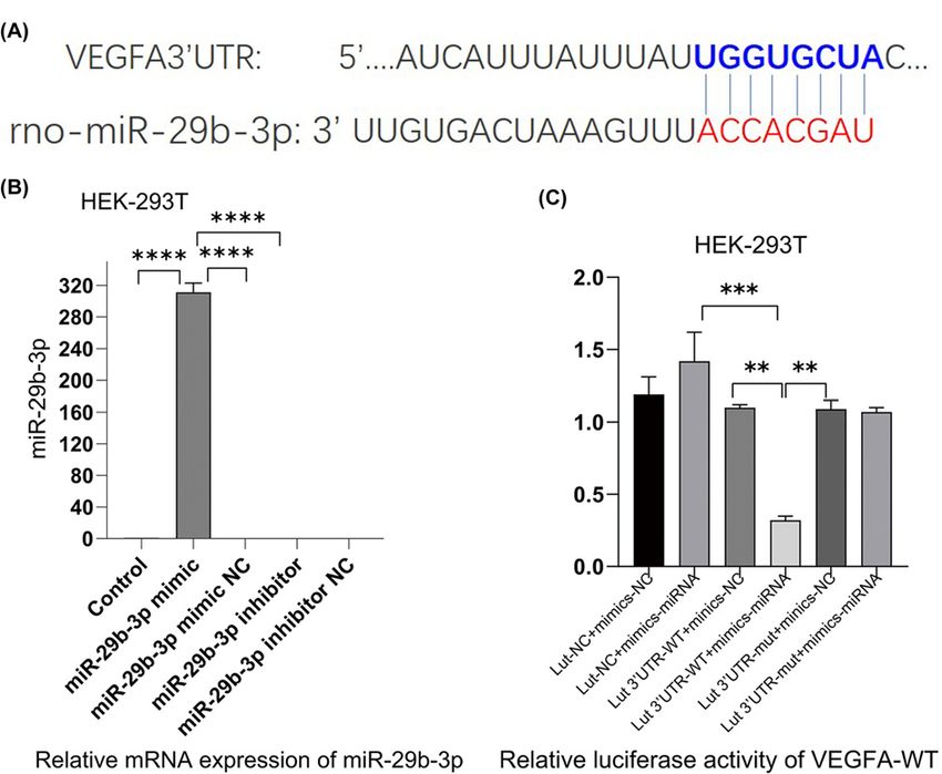
Bioscience Reports (2022) 42 BSR20212241
                                                                                                                                         https://doi.org/10.1042/BSR20212241

                                                                                                                                                                                           Downloaded from http://portlandpress.com/bioscirep/article-pdf/42/2/BSR20212241/929411/bsr-2021-2241.pdf by guest on 30 June 2022
           Figure 3. The cell proliferation was tested by using CCK8 and BrdU IF
           (A–C) The CCK8 proliferation assays (taken at 24, 48, 72 h, respectively) showed that XSSJS+miR-29b-3p better prevented the
           proliferation of HSC induced by TGF-β than XSSJS and miR-29b-3p at 24 and 48 h. There is little difference between their cell
           viability at 24 and 48 h, while the viability at 72 h is less effective than the viability at 24 or 48 h. (D) BrdU images showing
           that intervention with XSSJS+miR-29b-3p could more significantly decrease the number of BrdU-positive cells than XSSJS or
           miR-29b-3p administration. Bars = 50 μm. (E) Graphs of BrdU-positive ratios. All the experiments were repeated three times and
           represented as the means +         +
                                    − SEM (x − s, n=3, *P
Bioscience Reports (2022) 42 BSR20212241
https://doi.org/10.1042/BSR20212241
Downloaded from http://portlandpress.com/bioscirep/article-pdf/42/2/BSR20212241/929411/bsr-2021-2241.pdf by guest on 30 June 2022
Figure 4. The expression of miR29b-3p, VEGFA, TGF-β, PDGF were tested following XSSJS compound serum intervention
in HSCT6 cells
(A–D) The mRNA levels of miR-29b-3p, VEGFA, PDGF, TGF-β were tested by RT-PCR. XSSJS intervention following miR-29b-3p
transfection (XSSJS+miR-29b-3p group) more significantly promote the expression of miR-29b-3p and decrease the expression of
VEGFA, PDGF, TGF-β. (E–H) The protein level of VEGFA, PDGF, TGF-β were tested by WB; the protein expression of VEGFA, TGF-β
with XSSJS intervention following miR-29b-3p transfection (XSSJS+miR-29b-3p group) could be more significantly decreased than
using XSSJS or miR-29b-3p alone (x + − s, n=3, *PBioscience Reports (2022) 42 BSR20212241
https://doi.org/10.1042/BSR20212241
Downloaded from http://portlandpress.com/bioscirep/article-pdf/42/2/BSR20212241/929411/bsr-2021-2241.pdf by guest on 30 June 2022
Figure 5. Protein expression of VEGFA, TGF, PDGF, CD31, CD34 in HSCT6 cells
The protein expression of angiogenesis-related factors (VEGFA, TGF, PDGF, CD31, CD34) were tested by IF. (A–E) Left column,
DAPI channel showing blue fluorescence, middle column biomarkers channel showing red fluorescence from the liposomes, right
column is DAPI with biomarkers channels merged. Bars = 50 μm. The quantitative analysis of average fluorescence intensity
obtained from ImageJ software (F–J) (400×, x +
− s, n=3, *PBioscience Reports (2022) 42 BSR20212241
https://doi.org/10.1042/BSR20212241
Downloaded from http://portlandpress.com/bioscirep/article-pdf/42/2/BSR20212241/929411/bsr-2021-2241.pdf by guest on 30 June 2022
Figure 6. XSSJS intervention alleviated HF with reduced pathological course in HF rats
(A) Comparison of pathological stains of each group after HE and Masson staining (400×). (B) Positive pixel area (%) of Masson
are expressed (x +
− s, n=3, **PBioscience Reports (2022) 42 BSR20212241
https://doi.org/10.1042/BSR20212241
Downloaded from http://portlandpress.com/bioscirep/article-pdf/42/2/BSR20212241/929411/bsr-2021-2241.pdf by guest on 30 June 2022
Figure 7. The expression of miR29b-3p, VEGFA, TGF-β, PDGF were tested following XSSJS intervention in HF rats
(A–D) The levels of miR-29b-3p, VEGFA, PDGF, TGF-β mRNA were tested by using RT-PCR following XSSJS intervention in HF
rats. (E–H) The protein levels of VEGFA, PDGF, TGF-β were tested by WB following XSSJS intervention in HF rats (x +
− s, n=3,
*PBioscience Reports (2022) 42 BSR20212241
https://doi.org/10.1042/BSR20212241
Downloaded from http://portlandpress.com/bioscirep/article-pdf/42/2/BSR20212241/929411/bsr-2021-2241.pdf by guest on 30 June 2022
Figure 8. The protein expression of VEGFA, TGF-β, PDGF, CD31, CD34 in HF rats
The protein expression of angiogenesis-related factors (VEGFA, TGF, PDGF, CD31, CD34) were tested by IF. (A–E) Left column DAPI
channel showing blue fluorescence, middle column biomarkers channel showing red fluorescence from the liposomes, right column
is DAPI with biomarkers channels merged. Bars = 50 μm. The quantitative analysis of average fluorescence intensity obtained from
ImageJ software (F–J) (400×, x +
− s, n=3, *PBioscience Reports (2022) 42 BSR20212241
https://doi.org/10.1042/BSR20212241
multiple targets [44] (XSSJS is a compound of multiple herb ingredients based on TCM and on the molecular level,
its formulations typically feature multiple targets, multiple paths and multiple components).
Considering that the activation of HSCs is a key factor for HF [45], and TGF-β1 as a main profibrotic cytokines
of HF [46–48] could induce VEGFA expression in HSCs [49], HSC-T6 cells were used in this study, which were
activated by exposing the cells to TGF-β1. In vitro, the results demonstrated that XSSJS compound serum could
up-regulate miR-29b-3p and decrease the expression of VEGFA and other pathological angiogenesis related factors
(PDGF, TGF-β). The reasons are: (i) compared with the model group, it was found that cell viability was significantly
decreased after XSSJS compound serum treatment following miR29b-3p transfection (Figure 3A–E); (ii) the expres-
sion of miR29b-3p was up-regulated and the production of VEGFA, PDGF, TGF-β were down-regulated after XSSJS
compound serum intervention following miR-29b-3p transfection and its corresponding effectiveness gets the better
of those of using XSSJS or miR29b-3p alone. In vivo, we observed that oral administration of XSSJS in rats with HF
(once a day for 12 weeks) could promote the expression of miR-29b-3p and inhibit the expression of VEGFA, PDGF,
Downloaded from http://portlandpress.com/bioscirep/article-pdf/42/2/BSR20212241/929411/bsr-2021-2241.pdf by guest on 30 June 2022
and TGF-β in rats with liver fibrosis (Figure 7A–H). Furthermore, it could result in significant improvements of the
conditions of liver injury, inflammation, fibrosis, and angiogenesis (Figure 6). In addition, the stability and homo-
geneity of animal models are essential to the study of HF, the present study was performed on a rat model induced
by intraperitoneal injection of pig serum. The following two points: (i) the pattern of pig serum-induced immune
HF is remarkably similar to that of the clinical pathogeny in human CLD [50,51]; (ii) unlike other chemical model-
ing methods, such as CCI4, the rat death rate is low and liver dysfunction is insignificant, could explain the reason
why this approach has long since been extensively used and known as immune HF modeling [52]. Even so does the
model conforms to the pathological characteristics of liver fibrosis in modern medicine, it does not accurately reflect
the characteristics of collateral stasis involved in TCM syndrome typing. Therefore, in the future research, the model
taking account of both the disease and syndrome will be the subject of study as to support the brand-new theory of
TCM syndromes in more convincing manner, via which a prospective goal is to be attained, the combination of the
TCM strengths and the multitarget, multichannel holistic treatment of Chinese medicine.
There is an issue in our study that needs some clarification: we found in those results concerning PA-related fac-
tors, the expression of mRNA of the in-vivo model denotes that the difference between XSSJS+miR29b-3p and XSSJS
+ miR29b-3p NC group does not emerge in a statistically significant fashion. Quite contrastingly, the protein result
of the in-vivo model shows a significant difference between the XSSJS+miR29b-3p group and XSSJS+miR29b-3p
NC group. Meanwhile, the results of the in-vitro models unanimously point to a significant difference between
XSSJS+miR29b-3p group and XSSJS+miR29b-3p NC group, regardless it was with protein or mRNA (Figures 4 and
7). Therefore, via comprehensive overall data analysis, the aforementioned issue might be driven by factors such as
the time lag between the tests on protein and nucleic acid, or the modification issue after the protein translation
and potentially inappropriate handling during the experiment. Naturally, we will take this issue as one of the chief
considerations in our future studies.
In addition, from a theoretical perspective, the different indicators that inhibit the same phenotype should increase
or decrease homogeneously. However, CD31, CD34, as the most sensitive vessel endothelial markers, its abundance of
expression fell short of the expected level (Figures 5D,E and 8I,J). Its reasons may have a tie-in to certain effect brought
about by its intricate medical network (multitargeted), and the necessitated preparation for the direct verification
experiment is underway. As a matter of course, these data of a kind out of the ordinary would merit more incisive
studies in the future.
Conclusion and future work
In the present study, the result demonstrated that XSSJS intervention could up-regulate miR-29b-3p and
down-regulate VEGFA for both in-vitro and in-vivo cases. Therefore, we conclude that miR-29b-3p/VEGFA axis
have implications in the role XSSJS played in combating PA of HF, which furnishes us the handle for explaining
from the molecular level how XSSJS inhibits PA in the event of liver fibrosis. The PA test results of our experiment,
especially regarding CD31, CD34, on angiogenesis had not been able to incisively reveal much detailed informa-
tion due to the changes induced by using XSSJS with miR-29b being not as significant as other PA results. In the
light of this, it comes to our decision that we will enter upon a second phase of exploration right on the back of
our present one: first, we will introduce a bidirectional regulatory mechanism (activation and inhibition) to further
determine the relationship between XSSJS and miR-29b-3p/VEGFA axis. Meanwhile, it is proved fact that the com-
bination of the ‘medicines–targets’ network [53,54] and biological system network can reveal the complex Chinese
medicine pharmacological mechanisms on the ‘multiple-targets’ level. Therefore, we will explore the pharmacological
mechanisms of XSSJS for inhibiting PA following down this route. Subsequently, we will further explore the in-depth
14 © 2022 The Author(s). This is an open access article published by Portland Press Limited on behalf of the Biochemical Society and distributed under the Creative Commons
Attribution License 4.0 (CC BY).Bioscience Reports (2022) 42 BSR20212241
https://doi.org/10.1042/BSR20212241
insight behind this phenomenon. Besides, studies on the direct mechanisms of XSSJS for tackling angiogenesis (e.g.
three-dimensional assays of vascular morphogenesis) will also be covered in the future.
Data Availability
All the relevant data are contained in the main article and its supplementary files.
Competing Interests
The authors declare that there are no competing interests associated with the manuscript.
Funding
This work was supported by the National Natural Science Foundation of China [grant number 81904080].
Downloaded from http://portlandpress.com/bioscirep/article-pdf/42/2/BSR20212241/929411/bsr-2021-2241.pdf by guest on 30 June 2022
CRediT Author Contribution
Tianyao Zhang: Formal analysis, Investigation, Methodology, Writing—original draft, Writing—review & editing. Yu Yang: Supervi-
sion, Validation. BaojiaWang: Supervision. Long Wang: Software, Formal analysis. Dong Wang: Resources, Data curation. Ning
Cao: Resources, Visualization, Methodology, Project administration. Jinyu Shi: Data curation, Software.
Ethics Approval
Our study was ratified by the institutional Ethics Review Board of the Chengdou University of TCM. Relevant activities of this re-
search project were carried out in accordance with the Chengdou University of TCM Institutional Guidelines and Clinical Reg-
ulations. The research animals in our experiments stayed in line with the ethical requirements. The proof of ethical conduct on
laboratory animals was attached to the supplementary materials.
Abbreviations
AAV, adeno-associated virus; BrdU, bromodeoxyuridine; CCK8, cell-counting-kit-8; CD31, platelet endothelial cell ad-
hesion molecule-1; CLD, chronic liver disease; DAPI, 4 ,6-diamidino-2-phenylindole; FBS, fetal bovine serum; GAPDH,
glyceraldehyde-3-phosphate dehydrogenase; HE, Hematoxylin–Eosin; HF, hepatic fibrosis; HPA, hepatic pathological angio-
genesis; HSC-T6, hepatic stellate cell T6; IF, immunofluorescence; PDGF, platelet-derived growth factor; RT-qPCR, real-time
quantitative polymerase chain reaction; TCM, traditional Chinese medicine; TGF-β, transforming growth factor-β; VEGFA, vas-
cular endothelial growth factor; XSSJS, Xueshisanjia.
References
1 Bataller, R. and Brenner, D.A. (2005) Liver fibrosis. J. Clin. Invest. 115, 209–218, https://doi.org/10.1172/JCI24282
2 Yang, L. et al. (2014) Vascular endothelial growth factor promotes fibrosis resolution and repair in mice. Gastroenterology 146, 1339.e1–1350.e1,
https://doi.org/10.1053/j.gastro.2014.01.061
3 Sahin, H. et al. (2012) Chemokine Cxcl9 attenuates liver fibrosis-associated angiogenesis in mice. Hepatology 55, 1610–1619,
https://doi.org/10.1002/hep.25545
4 Keeley, E.C., Mehrad, B. and Strieter, R.M. (2008) Chemokines as mediators of neovascularization. Arterioscler. Thromb. Vasc. Biol. 28, 1928–1936,
https://doi.org/10.1161/ATVBAHA.108.162925
5 Strieter, R.M. et al. (2005) CXC chemokines in angiogenesis. Cytokine Growth Factor Rev. 16, 593–609, https://doi.org/10.1016/j.cytogfr.2005.04.007
6 Sulpice, E. et al. (2004) Platelet factor 4 disrupts the intracellular signalling cascade induced by vascular endothelial growth factor by both KDR
dependent and independent mechanisms. Eur. J. Biochem. 271, 3310–3318, https://doi.org/10.1111/j.1432-1033.2004.04263.x
7 Kitade, M. et al. (2006) Leptin-mediated neovascularization is a prerequisite for progression of nonalcoholic steatohepatitis in rats. Hepatology 44,
983–991, https://doi.org/10.1002/hep.21338
8 Adya, R. et al. (2008) Visfatin induces human endothelial VEGF and MMP-2/9 production via MAPK and PI3K/Akt signalling pathways: novel insights into
visfatin-induced angiogenesis. Cardiovasc. Res. 78, 356–365, https://doi.org/10.1093/cvr/cvm111
9 Vizio, B. et al. (2021) Cooperative role of thrombopoietin and vascular endothelial growth factor-A in the progression of liver cirrhosis to hepatocellular
carcinoma. Int. J. Mol. Sci. 4, 1818, https://doi.org/10.3390/ijms22041818
10 Salcedo, X. et al. (2005) The potential of angiogenesis soluble markers in chronic hepatitis C. Hepatology 42, 696–701,
https://doi.org/10.1002/hep.20828
11 Salcedo, X. et al. (2005) Review article: angiogenesis soluble factors as liver disease markers. Aliment. Pharmacol. Ther. 22, 23–30,
https://doi.org/10.1111/j.1365-2036.2005.02532.x
12 Friedman, S.L. (1993) Seminars in medicine of the Beth Israel Hospital, Boston. The cellular basis of hepatic fibrosis. mechanisms and treatment
strategies. N. Engl. J. Med. 328, 1828–1835, https://doi.org/10.1056/NEJM199306243282508
13 Lee, K.S. et al. (1995) Activation of hepatic stellate cells by TGF alpha and collagen type I is mediated by oxidative stress through c-myb expression. J.
Clin. Invest. 96, 2461–2468, https://doi.org/10.1172/JCI118304
14 Iwakiri, Y. (2014) Pathophysiology of portal hypertension. Clin. Liver Dis. 18, 281–291, https://doi.org/10.1016/j.cld.2013.12.001
© 2022 The Author(s). This is an open access article published by Portland Press Limited on behalf of the Biochemical Society and distributed under the Creative Commons 15
Attribution License 4.0 (CC BY).Bioscience Reports (2022) 42 BSR20212241
https://doi.org/10.1042/BSR20212241
15 Iwakiri, Y. (2012) Endothelial dysfunction in the regulation of cirrhosis and portal hypertension. Liver Int. 32, 199–213,
https://doi.org/10.1111/j.1478-3231.2011.02579.x
16 Llovet, J.M. et al. (2008) Sorafenib in advanced hepatocellular carcinoma. N. Engl. J. Med. 359, 378–390, https://doi.org/10.1056/NEJMoa0708857
17 Shah, V.H. and Bruix, J. (2009) Antiangiogenic therapy: not just for cancer anymore? Hepatology 49, 1066–1068, https://doi.org/10.1002/hep.22872
18 Siegel, A.B. et al. (2008) Phase II trial evaluating the clinical and biologic effects of bevacizumab in unresectable hepatocellular carcinoma. J. Clin.
Oncol. 26, 2992–2998, https://doi.org/10.1200/JCO.2007.15.9947
19 Mu, Y. et al. (2006) The inhibitory effect of the Xiayuxue decoction on liver fibrosis in rats during the progression period and its dialectical discussion. J.
Tradit. Chin. Med. 47, 215–218
20 Yao, F., Lau, P., Sun, J. et al. (2018) Clinical study on the reversal of liver fibrosis and its regulation of immune function by “the healthy and soft liver
blood flow side of the spleen”. Jiangsu Tradit. Chin. Med. 50, 42–44
21 Lu, J., Tan, W., Zhao, Z. et al. (2015) The anti-hepatic fibrosis mechanism of the complex component of fuzheng Huayu inhibits the rebirth of blood
vessels. World Chin. Med. 2, 186–191
22 Zhao, P. (2011) Advances in the study of curcumin anti-angiogenic rebirth. Med. Clin. Res. 28, 1407–1409
Downloaded from http://portlandpress.com/bioscirep/article-pdf/42/2/BSR20212241/929411/bsr-2021-2241.pdf by guest on 30 June 2022
23 Zheng, X., Hui, Y., Yu, S. et al. (2014) Effects of triamcinolone on the expression of HSC-T6 cells α-SMA, TIMP-1, and ROCK-I proteins. J. Tianjin Univ.
Trad. Chin. Med. 6, 359–361
24 Wang Ju, Based on the theory of “main passenger communication” the study on the effect of addition and subtraction of triacetate on the signaling
pathway of angiogenesis in liver fibrosis rats. Doctoral Dissertation, Chengdu University of Traditional Chinese Medicine,
https://kreader.cnki.net/Kreader/CatalogViewPage.aspxdbCode=CDFD&filename=1016045289.nh&tablename=CDFDLAST2016&compose=&first=1&uid=
25 Li, Y., Sun, Y., Fenghua, L. et al. (2013) The prevention and treatment effect of Huayu meidicine on liver fibrosis rats and their effect on the TGF-beta
1/Samd signaling pathway. The 25th National Conference on Chinese and Western Medicine Combined with Digestive Diseases,
https://d.wanfangdata.com.cn/conference/8256024
26 Wang, B., Kang, Y., Xu, Y. et al. (2016) Effects of XSSJS on liver microvascular density and VEGF-α expression in immun opastic fibrosis rats. Chinese
J. Tradit. Chin. Med. 5, 1705–1709
27 Roderburg, C., Trautwein, C. and Luedde, T. (2011) MicroRNA-199a/b-3p: a new star in the liver microcosmos. Hepatology 54, 729–731,
https://doi.org/10.1002/hep.24456
28 Tuomi, T. et al. (2014) The many faces of diabetes: a disease with increasing heterogeneity. Lancet 383, 1084–1094,
https://doi.org/10.1016/S0140-6736(13)62219-9
29 Sciarretta, S., Volpe, M. and Sadoshima, J. (2014) Mammalian target of rapamycin signaling in cardiac physiology and disease. Circ. Res. 114,
549–564, https://doi.org/10.1161/CIRCRESAHA.114.302022
30 Friedman, S.L. et al. (1989) Maintenance of differentiated phenotype of cultured rat hepatic lipocytes by basement membrane matrix. J. Biol. Chem.
264, 10756–10762, https://doi.org/10.1016/S0021-9258(18)81686-6
31 Koyama, Y. et al. (2016) New developments on the treatment of liver fibrosis. Dig. Dis. 34, 589–596, https://doi.org/10.1159/000445269
32 Koyama, Y. and Brenner, D.A. (2015) New therapies for hepatic fibrosis. Clin. Res. Hepatol. Gastroenterol. 39, S75–S79,
https://doi.org/10.1016/j.clinre.2015.06.011
33 Dong, P., Zheng, J., Lu, Z. et al. (2013) Experimental study of nano-shell polysaccharides carrying miR-29b transfected liver star cells. China J. Health
Insp. 23, 332–334
34 Li, X., Yang, H., Xiang, Y. et al. (2014) Rat CTGF miRNA plasmid vector construction and the establishment of stable transfection of liver astrocyte cell
lines. The 25th National Conference on Chinese and Western Medicine Combined with Digestive Diseases,
https://d.wanfangdata.com.cn/conference/8717989
35 Liu, X., Hu, Y., Hu, Y. et al. (2004) Comparison of tissue pathology between carbon tetrachloride and serum liver fibrosis models of pigs. World J. Chin.
Diges. 12, 1875–1879
36 Wen, J.D. et al. (2008) Following translation by single ribosomes one codon at a time. Nature 452, 598–603, https://doi.org/10.1038/nature06716
37 Zheng, Q. et al. (2010) Genome-wide double-stranded RNA sequencing reveals the functional significance of base-paired RNAs in Arabidopsis. PLoS
Genet. 6, e1001141, https://doi.org/10.1371/journal.pgen.1001141
38 Zhou, W.C., Zhang, Q.B. and Qiao, L. (2014) Pathogenesis of liver cirrhosis. World J. Gastroenterol. 20, 7312–7324,
https://doi.org/10.3748/wjg.v20.i23.7312
39 Melincovici, C.S. et al. (2018) Vascular endothelial growth factor (VEGF) - key factor in normal and pathological angiogenesis. Rom. J. Morphol.
Embryol. 59, 455–467
40 Ferrara, N., Gerber, H.P. and LeCouter, J. (2003) The biology of VEGF and its receptors. Nat. Med. 9, 669–676, https://doi.org/10.1038/nm0603-669
41 Hoeben, A. et al. (2004) Vascular endothelial growth factor and angiogenesis. Pharmacol. Rev. 56, 549–580, https://doi.org/10.1124/pr.56.4.3
42 Mu, D., Wang, B., Wang, J. et al. (2017) Effects of addition and subtraction of triacetate on liver microvascular density and PDGF-B expression in liver
fibrosis rats. Liaoning J. Tradit. Chin. Med. 7, 170–173, https://doi.org/10.13192/j.issn.1000-1719.2017.07.057
43 Ge, Y. et al. (2013) Circulating CD31+ leukocyte frequency is associated with cardiovascular risk factors. Atherosclerosis 229, 228–233,
https://doi.org/10.1016/j.atherosclerosis.2013.04.017
44 Zhou, S. et al. (2020) Deciphering the pharmacological mechanisms of taohe-chengqi decoction extract against renal fibrosis through integrating
network pharmacology and experimental validation in vitro and in vivo. Front. Pharmacol. 11, 425, https://doi.org/10.3389/fphar.2020.00425
45 Deleve, L.D., Wang, X. and Guo, Y. (2008) Sinusoidal endothelial cells prevent rat stellate cell activation and promote reversion to quiescence.
Hepatology 48, 920–930, https://doi.org/10.1002/hep.22351
16 © 2022 The Author(s). This is an open access article published by Portland Press Limited on behalf of the Biochemical Society and distributed under the Creative Commons
Attribution License 4.0 (CC BY).Bioscience Reports (2022) 42 BSR20212241
https://doi.org/10.1042/BSR20212241
46 Inagaki, Y. and Okazaki, I. (2007) Emerging insights into Transforming growth factor beta Smad signal in hepatic fibrogenesis. Gut 56, 284–292,
https://doi.org/10.1136/gut.2005.088690
47 Saito, D. et al. (2013) Transforming growth factor-β1 induces epithelial-mesenchymal transition and integrin α3β1-mediated cell migration of HSC-4
human squamous cell carcinoma cells through Slug. J. Biochem. 153, 303–315, https://doi.org/10.1093/jb/mvs144
48 Xu, T. et al. (2016) NLRC5 regulates TGF-β1-induced proliferation and activation of hepatic stellate cells during hepatic fibrosis. Int. J. Biochem. Cell
Biol. 70, 92–104, https://doi.org/10.1016/j.biocel.2015.11.010
49 Mitchell, S.J. et al. (2011) Age-related pseudocapillarization of the liver sinusoidal endothelium impairs the hepatic clearance of acetaminophen in rats.
J. Gerontol. A Biol. Sci. Med. Sci. 66, 400–408, https://doi.org/10.1093/gerona/glq221
50 Huang, Z. (1997) The serum-induced model of liver fibrosis and its mechanism in rats of heterogeneous animals. J. Shanghai Med. Univ. 24, 351–354
51 Wu, J. et al. (2009) Pig serum joint con a builds a model of liver fibrosis in Balb/c mice and evaluates sage. J. Anat. 32, 546–548
52 Lin, D. and Jin, Z. (2006) Research and evaluation of commonly used animal models of liver fibrosis. Shizhen Natl. Med. 17, 487–488
53 Chatr-Aryamontri, A. et al. (2017) The BioGRID interaction database: 2017 update. Nucleic Acids Res. 45, D369–D379,
https://doi.org/10.1093/nar/gkw1102
Downloaded from http://portlandpress.com/bioscirep/article-pdf/42/2/BSR20212241/929411/bsr-2021-2241.pdf by guest on 30 June 2022
54 Wang, H. et al. (2020) A combined phytochemistry and network pharmacology approach to reveal the effective substances and mechanisms of
Wei-Fu-Chun tablet in the treatment of precancerous lesions of gastric cancer. Front. Pharmacol. 11, 558471,
https://doi.org/10.3389/fphar.2020.558471
© 2022 The Author(s). This is an open access article published by Portland Press Limited on behalf of the Biochemical Society and distributed under the Creative Commons Attribution 17
License 4.0 (CC BY).You can also read