A cerebrospinal fluid microRNA analysis: Progressive supranuclear palsy - Spandidos ...
←
→
Page content transcription
If your browser does not render page correctly, please read the page content below
Molecular Medicine REPORTS 25: 88, 2022

                          A cerebrospinal fluid microRNA analysis:
                               Progressive supranuclear palsy
          WAKAKO NONAKA1, TADAYUKI TAKATA1, HISAKAZU IWAMA2, SATOSHI KOMATSUBARA3,
              HIDEKI KOBARA4, MASAKI KAMADA1, KAZUSHI DEGUCHI1, TETSUO TOUGE5,
          OSAMU MIYAMOTO6, TAKEHIRO NAKAMURA7, TOSHIFUMI ITANO1 and TSUTOMU MASAKI4

     1
     Department of Neurology, Faculty of Medicine; 2Life Science Research Center; Departments of 3Orthopedic Surgery,
      4
        Gastroenterology and 5Health Sciences, Faculty of Medicine, Kagawa University, Miki‑Cho, Kagawa 761‑0793;
 6
   Department of Medical Engineering, Faculty of Health Science and Technology, Kawasaki University of Medical Welfare,
Kurashiki, Okayama 701‑0193; 7Department of Physiology 2, Kawasaki Medical School, Kurashiki, Okayama 701‑0192, Japan

                                          Received July 13, 2021; Accepted December 7, 2021

                                                   DOI: 10.3892/mmr.2022.12604

Abstract. Progressive supranuclear palsy (PSP) is a neuro‑            system and autophagy pathway. Furthermore, these miRNAs
degenerative tauopathy described as a syndrome of postural            were found to target genes that have been reported to have
instability, supranuclear vertical gaze palsy, dysarthria, dystonic   epigenetic changes following an epigenome‑wide association
rigidity of the neck and trunk, dementia, and pseudobulbar palsy.     study of brain tissues of patients with PSP. This suggested that
The clinical diagnosis of PSP is often difficult because there        these miRNAs and genes may have some involvement in the
are no established biomarkers, and diagnosis is currently based       pathogenesis of PSP. However, the sample size of the present
on clinical and imaging findings. Furthermore, the etiology           study was small; therefore, a greater number of patients with
and pathogenesis of PSP remain unknown. Dysregulation of              PSP should be examined in future studies.
microRNAs (miRNAs/miRs) has been reported to serve an
important role in neurodegenerative diseases. However, the            Introduction
miRNA profiles of patients with PSP are rarely reported. The
present study aimed to examine cerebrospinal fluid miRNAs,            Neurodegenerative diseases are generally progressive and
which are considered to be more sensitive indicators of changes       refractory because they involve irreversible degeneration of
in the brain, to elucidate the pathophysiology of PSP and to          brain neurons and many remain incurable. Treatments to
establish specific biomarkers for diagnosis. The present study        slow progression after disease onset do not compensate for
used a microarray chip containing 2,632 miRNAs to examine             neural tissues that have already been lost and do not result in a
cerebrospinal fluid miRNA expression levels in 11 patients with       retrograde recovery of symptoms. Therefore, it is important to
PSP aged 68‑82 years. A total of 8 age‑ and sex‑matched controls      implement therapeutic interventions and risk management as
were also included. A total of 38 miRNAs were significantly           early as possible in order to delay disease onset and progres‑
upregulated and one miRNA was significantly downregulated             sion and to extend healthy life expectancy (1). However, this is
in the cerebrospinal fluid of patients with PSP. The patients were    difficult because diagnosis is based on the detection of clinical
divided into two groups based on disease stage (early onset and       symptoms and neuroimaging abnormalities, which appear
advanced), and changes in miRNA expression were examined.             relatively late in the disease course. Nonetheless, molecular
The miRNAs that were most significantly upregulated or down‑          responses to genetic and environmental disorders begin much
regulated in the early onset group were miR‑204‑3p, miR‑873‑3p        earlier, and non‑coding RNA networks have been found to be
and miR‑6840‑5p. The target genes of these miRNAs were                involved in these cellular regulatory mechanisms. MicroRNAs
associated with molecules related to the ubiquitin‑proteasome         (miRNAs) are small non‑coding RNAs of approximately
                                                                      20‑25 nucleotides, and more than 2,000 different miRNAs
                                                                      have been suggested to exist in humans (2). MiRNAs repress
                                                                      the expression of various genes in vivo and exhibit unique
                                                                      disease‑specific expression patterns. In addition, miRNAs are
Correspondence to: Dr Wakako Nonaka, Department of
Neurology, Faculty of Medicine, Kagawa University, 1750‑1 Ikenobe,
                                                                      stable in body fluids such as blood, urine, and saliva, which
Miki‑Cho, Kita‑Gun, Kagawa 761‑0793, Japan                            are easy to collect from living organisms, and can be detected
E‑mail: nonaka.wakako@kagawa‑u.ac.jp                                  with high sensitivity (3). Thus, profiling the miRNA expression
                                                                      patterns may facilitate the detection of diseases at a prelimi‑
Key words: progressive supranuclear palsy, microRNA, cerebrospinal    nary stage. Indirect evaluation of pathological processes is
fluid, humans, Parkinson's and related diseases                       particularly important in neurological diseases, where direct
                                                                      access to tissues for molecular analysis is not possible. Living
                                                                      neurons and other cells of the central nervous system (CNS)
2                                NONAKA et al: microRNA IN PROGRESSIVE SUPRANUCLEAR PALSY

secrete miRNAs wrapped in exosomes, microvesicles, and               We assessed brain magnetic resonance imaging scans for find‑
lipoprotein complexes (4). Interactions between external stimuli     ings suggestive of PSP. Furthermore, we applied the magnetic
and pathological processes in the brain have been reported to        resonance parkinsonism index (MRPI) (19) and MRPI 2.0 (20).
be reflected in peripheral tissues, and their use as potential       The threshold for the MRPI and MRPI 2.0 was >13.88 and
diagnostic markers is of interest. Cerebrospinal fluid (CSF) has     >2.50, respectively (20). Patients with PSP were divided into
also been shown to contain circulating miRNAs. As the CSF            two subgroups: The ‘early disease stage’ subgroup included
is in direct contact with the brain, it is said to reflect changes   those who underwent neurological evaluation within 2 years
in the brain and may be suitable as a biomarker (5). Recently,       of symptom onset and the ‘advanced disease stage’ subgroup
specific miRNAs in the CSF have been reported in Alzheimer's         included patients who underwent evaluation >2 years after
disease (6) and Parkinson's disease (PD) (7) and are expected to     symptom onset. Patients with concurrent malignant tumors,
be potential diagnostic markers and therapeutic targets.             psychiatric disorders, collagen diseases, endocrine diseases,
    Progressive supranuclear palsy (PSP) is a progressive            or infections were excluded because these conditions may
neurodegenerative tauopathy characterized by postural                alter the expression profile of some miRNAs. Owing to the
instability with falls, vertical supranuclear gaze palsy, parkin‑    above‑mentioned reason, participants in the control group with
sonism with poor levodopa response, pseudobulbar palsy,              concurrent malignant tumors, psychiatric disorders, collagen
and frontal release signs (8). The mean age of onset of PSP          diseases, endocrine diseases, or infections were also excluded. In
is approximately 65 years and the condition leads to death           addition, participants with neurological diseases such as dementia
within approximately 6‑9 years after symptom onset (9,10).           and parkinsonism were excluded. The study was approved by
The factors involved in its pathogenesis are still unknown, and      the Medical Ethics Committee of Kagawa University (Kagawa,
no fundamental treatment has been established. In addition,          Japan) (Ethics approval Heisei 22‑002). Participants and controls
the diagnosis of PSP is currently dependent on clinical and          received a full explanation of the study purpose and protocol and
imaging findings, which makes early diagnosis difficult.             provided written informed consent before inclusion in the study.
    It is difficult to differentiate PSP from PD and the other
Parkinsonian syndromes such as multiple system atrophy               CSF miRNA expression profiling. CSF samples were obtained
(MSA) and corticobasal degeneration (CBD), especially in the         during lumbar puncture between the L3 and L5 vertebrae with the
early stages. The differences in the Parkinsonian syndrome,          participant in a lateral supine position. Lumbar punctures were
which has similar clinical symptoms to PSP, are presented in         performed between 9 and 12 am, using a sterile technique and
Table SI. To improve the accuracy of the clinical diagnosis of       a 21‑23G spinal needle in accordance with local guidelines. At
Parkinsonian syndrome, CSF markers have been explored (11).          least 4 ml of CSF was collected from each participant. CSF was
Therefore, we focused on miRNAs as one candidate that could          collected in polypropylene tubes and cytologically controlled to
lead to early diagnosis of PSP.                                      be free of erythrocytes. The sample was centrifuged (2,000 x g,
    At present, there are very few reports of miRNA expres‑          4˚C, 10 min), frozen immediately, and stored at ‑80˚C until
sion profiling in PSP. It has been reported that miR‑132 (12),       use. The CSF was at no time thawed/refrozen. Total RNA was
miR‑147a, and miR‑518e (13) are expressed in the brain tissues       extracted from 900 µl of frozen CSF using a 3D‑Gene® RNA
of patients with PSP, but these miRNAs are rarely reported in        extraction reagent (Toray) according to the manufacturer's spec‑
biofluids. We herein aimed to examine CSF miRNAs, which              ifications. The BioAnalyzer 2100 system (Agilent Technologies,
are thought to be a sensitive indicator of changes in the brain,     Inc.) was used to determine the RNA integrity number (RIN)
to elucidate the pathophysiology of PSP and establish specific       for RNA quality assessment. No difference in total RNA
biomarkers for diagnosis.                                            concentration or RIN was observed between the PSP group
                                                                     and controls. After labeling with a 3D‑Gene miRNA labeling
Materials and methods                                                kit (Toray), the extracted total RNA was hybridized onto the
                                                                     3D‑Gene® Human miRNA oligo chip Version 22.0 (Toray) with
Participants. Eleven patients with PSP (nine men and two             2,632 fluorescent probes. Hybridization signals were obtained
women; median age, 76 years; range, 68‑82 years) and eight           using a 3D‑Gene 3000 miRNA microarray scanner (Toray).
age‑ and sex‑matched controls (seven men and one woman;              The raw data of each detected signal were normalized by the
median age, 75.5 years; range, 63‑89 years) were recruited for       global normalization method, subtracting the mean intensity of
the present study between January 2017 and December 2018             the background signal in each microarray, and the median of
from the Department of Neurology, Kagawa University Hospital.        the detected signal intensity was adjusted to 25. Microarray data
All participants in this study were Japanese. We enrolled patients   have been deposited in NCBI's Gene Expression Omnibus (21)
fulfilling the Movement Disorder Society (MDS) clinical diag‑        and are accessible through GEO Series accession number
nostic criteria for PSP (MDS‑PSP criteria) (14) and for probable     GSE186921 (https://www.ncbi.nlm.nih.gov/geo/query/acc.
PSP, Richardson's syndrome (PSP‑RS) phenotype according to           cgi?acc= GSE186921).
their disease history and clinical information including neuroim‑
aging findings. All patients had sporadic PSP. Patient background    Heatmap. Hierarchical clustering was performed on the PSP
data (age, sex, disease duration, comorbidities, and clinical        and control groups, using a subset of miRNAs determined
parameters) at the time of sample collection were retrospectively    to be significantly differentially expressed between the two
obtained from medical records. Clinical parameters included          groups. The Ward agglomerative method was used to cluster
scores on the PSP Rating Scale (PSPRS) (15), Mini‑Mental             both the samples and miRNAs. The heatmap was color‑coded
State Examination (MMSE) (16), Frontal Assessment Battery            according to the expression levels. The center level of the
(FAB) (17), and Montreal Cognitive Assessment (MoCA) (18).           color code was set as the median value over all of the values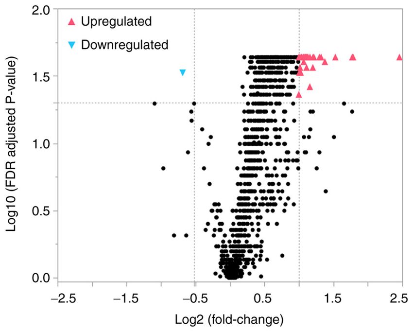
Molecular Medicine REPORTS 25: 88, 2022 3
Table I. General and clinical characteristics of the participants.
Variable Control (n=8) PSP early stage (n=5) PSP advanced stage (n=6) P‑value
Sex, n
Male 7 3 6 0.316a
Female 1 2 0
Median (IQR) age at test, years 75.5 (64.8‑79.0) 75.0 (69.0‑76.0) 76.5 (70.8‑79.8) 0.516b
Median (IQR) disease duration, years ND 2 (2) 5 (4‑6) 0.005c
Median (IQR) PSPRS score,/100 ND 26.0 (24.5‑31.0) 58.0 (51.8‑62.3) 0.008c
Median (IQR) MMSE score,/30 ND 26.0 (25.0‑28.0) 19.0 (11.5‑20.3) 0.008c
Median (IQR) FAB score,/18 ND 12.0 (10.5‑13.5) 6.0 (5.0‑9.3) 0.042c
Median (IQR) MoCA score,/30 ND 21.0 (19.0‑23.0) 8.5 (5.0‑12.8) 0.008c
Median (IQR) MRPI ND 30.6 (23.0‑31.2) 42.6 (37.9‑48.1) 0.014c
Median (IQR) MRPI 2.0 ND 6.8 (5.0‑7.9) 10.9 (9.7‑14.2) 0.008c
a
Fisher's Exact test; bKruskal‑Wallis test; cMann‑Whitney U test. IQR, interquartile range; ND, not determined; PSP, progressive supranuclear
palsy; PSPRS, progressive supranuclear palsy rating scale; MMSE, Mini‑Mental State Examination; FAB, Frontal Assessment Battery;
MoCA, Montreal Cognitive Assessment; MRPI, Magnetic Resonance Parkinsonism Index.
used in the heatmap. Gray color represented mean values,
red indicated an increase, and blue represented a decrease
in expression. The heatmap was created using JMP ® Pro
version 15.1.0 (SAS Institute Inc.).
miRNA target gene bioinformatics analysis. Using in silico
analysis, the target genes of miRNAs were predicted using
the miRDB (http://mirdb.org/miRDB/), Targetscan 7.2
(http://www.targetscan.org/), and mirTarBase version 8.0
(http://mirtarbase.mbc.nctu.edu.tw/) databases. To enhance
the reliability of the bioinformatics analysis, overlapping
target genes were identified in this analysis.
Statistical analysis. For comparisons of clinical characteris‑
tics between the groups, the Mann‑Whitney U test was used
to compare all variables except for sex and age. The Fisher's
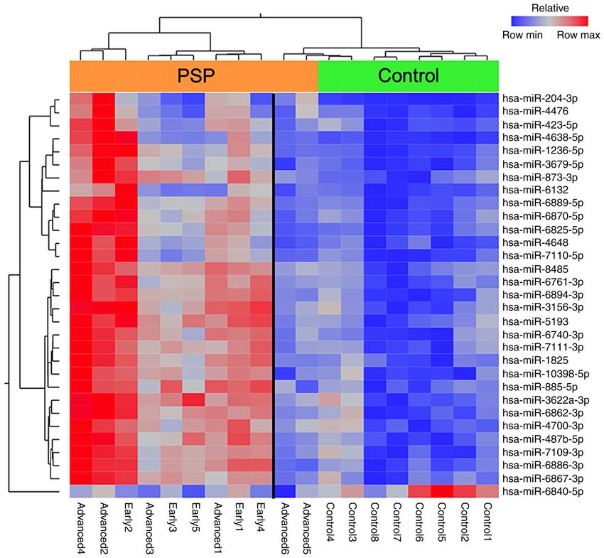
exact test and Kruskal‑Wallis test were used to compare sex Figure 1. Volcano plot of the miRNAs isolated from the cerebrospinal fluid of
and age, respectively. The median (interquartile range) values patients with progressive supranuclear palsy and controls. The volcano plot illus‑
trates the relationship between the fold change and significance of the difference
for each parameter are presented. Statistical analysis of between the two groups. The y‑axis depicts the negative log10 of FDR‑adjusted
miRNA expression levels in each group was conducted using a P‑values (the horizontal line at 1.3 corresponds to a P‑value of 0.05, a higher
non‑parametric Mann‑Whitney U test and the Kruskal‑Wallis value indicates greater significance) and the x‑axis is the difference in expres‑
test as appropriate. Benjamini‑Hochberg procedures for sion between the two experimental groups as log2 fold changes (the vertical
lines indicate that miRNAs are either upregulated or downregulated above a
adjusting the false discovery rate (FDR) in multiple compari‑ fold change of 2 and 0.7, respectively). The blue color indicates significantly
sons were also applied. Multiple comparisons of continuous downregulated miRNAs and the red color indicates significantly upregulated
variables among multiple groups were performed using the miRNAs. FDR, false discovery rate; miRNA, microRNA.
Kruskal‑Wallis test, followed by the Steel‑Dwass post‑hoc
test. P‑values of4 NONAKA et al: microRNA IN PROGRESSIVE SUPRANUCLEAR PALSY
Table II. Statistical results, chromosomal locations and known associated diseases of miRNAs which were significantly altered
in patients with progressive supranuclear palsy.
FDR adjusted Chromosomal
MiRNA Fold change P‑value P‑value localization miRNA‑disease association
Upregulated
hsa‑miR‑204‑3p 5.50 0.0003 0.0230 9q21.12 Retinal dystrophy and iris coloboma with or
without cataract (22)
hsa‑miR‑4476 3.46 0.0034 0.0230 9p13.2
hsa‑miR‑6132 3.42 0.0015 0.0230 7q31.2
hsa‑miR‑4638‑5p 2.89 0.0003 0.0230 5q35.3
hsa‑miR‑7110‑5p 2.60 0.0044 0.0249 3q21.1
hsa‑miR‑3679‑5p 2.50 0.0015 0.0230 2q21.2
hsa‑miR‑1236‑5p 2.46 0.0011 0.0230 6p21.33
hsa‑miR‑6867‑3p 2.33 0.0026 0.0230 17q21.1
hsa‑miR‑6761‑3p 2.31 0.0011 0.0230 12q24.12
hsa‑miR‑423‑5p 2.30 0.0057 0.0275 17q11.2
hsa‑miR‑7111‑3p 2.25 0.0020 0.0230 6p21.31
hsa‑miR‑3156‑3p 2.24 0.0015 0.0230 21q11.2
hsa‑miR‑12114 2.24 0.0118 0.0382 22q13.33
hsa‑miR‑6889‑5p 2.20 0.0026 0.0230 22q13.2
hsa‑miR‑6740‑3p 2.20 0.0011 0.0230 1q32.1
hsa‑miR‑885‑5p 2.18 0.0020 0.0230 3p25.3
hsa‑miR‑6894‑3p 2.18 0.0020 0.0230 Xp11.22
hsa‑miR‑487b‑5p 2.16 0.0015 0.0230 14q32.31 Neuroblastomas (23)
hsa‑miR‑6820‑5p 2.16 0.0057 0.0275 22q13.1
hsa‑miR‑873‑3p 2.15 0.0008 0.0230 9p21.1 Multiple sclerosis (24)
hsa‑miR‑7109‑3p 2.14 0.0026 0.0230 22q12.2
hsa‑miR‑5193 2.12 0.0015 0.0230 3p21.31
hsa‑miR‑4648 2.10 0.0034 0.0230 7p22.3
hsa‑miR‑10398‑5p 2.10 0.0044 0.0249 6p21.1
hsa‑miR‑1825 2.10 0.0015 0.0230 20q11.21
hsa‑miR‑6870‑5p 2.10 0.0026 0.0230 20p12.2
hsa‑miR‑6825‑5p 2.07 0.0034 0.0230 3q21.3
hsa‑miR‑4700‑3p 2.07 0.0020 0.0230 12q24.31
hsa‑miR‑3622a‑3p 2.05 0.0011 0.0230 8p21.1
hsa‑miR‑5001‑5p 2.04 0.0073 0.0300 2q37.1
hsa‑miR‑6510‑5p 2.04 0.0073 0.0300 17q21.2
hsa‑miR‑4505 2.04 0.0073 0.0300 14q24.3
hsa‑miR‑4665‑5p 2.03 0.0057 0.0275 9p24.1
hsa‑miR‑8485 2.02 0.0008 0.0230 2p16.3
hsa‑miR‑7110‑3p 2.01 0.0073 0.0300 3q21.1
hsa‑miR‑6862‑3p 2.00 0.0015 0.0230 16p12.1
hsa‑miR‑6886‑3p 2.00 0.0034 0.0230 19p13.2
hsa‑miR‑328‑5p 2.00 0.0149 0.0436 16q22.1 Myelogenous leukemia (25)
Downregulated
hsa‑miR‑6840‑5p 0.62 0.0073 0.0300 7q22.1
Significant differential miRNA expression was determined by a fold change ratio of >2 orMolecular Medicine REPORTS 25: 88, 2022 5 Figure 2. Heatmap representing the hierarchical clustering of aberrantly expressed miRNAs. Samples are arranged in columns and miRNAs in rows. The miRNA clustering tree is presented on the left and the sample clustering tree is present at the top of each heat map. The heat map depicts the relative expression intensity for each miRNA: Red cells indicate high levels of expression and blue cells indicate levels of low expression. The orange bar represents the PSP group and the green bar represents the control group. miRNA/miR, microRNA; PSP, progressive supranuclear palsy. FDR‑adjusted P‑value of
6                                     NONAKA et al: microRNA IN PROGRESSIVE SUPRANUCLEAR PALSY

Table III. Upregulated and downregulated miRNAs in the subgroups of patients with progressive supranuclear palsy.

MiRNA	                                                            P‑value (Kruskal‑Wallis)                                    Post hoc (Steel‑Dwass)

Upregulated
 hsa‑miR‑204‑3p
  Control vs. PSP early      0.0043 0.0120
  Control vs. PSP advanced   0.0024 0.0068
  PSP early vs. PSP advanced 0.0552 0.1338
 hsa‑miR‑873‑3p
  Control vs. PSP early      0.0043 0.0120
  Control vs. PSP advanced   0.0081 0.0222
  PSP early vs. PSP advanced 0.4113 0.6896
Downregulated
 hsa‑miR‑6840‑5p
  Control vs. PSP early      0.0338 0.0853
  Control vs. PSP advanced   0.0239 0.0617
  PSP early vs. PSP advanced 0.9273 0.9954

miRNA/miR, microRNA; PSP, progressive supranuclear palsy.

Figure 3. Upregulated and downregulated miRNAs in the subgroups of patients with progressive supranuclear palsy. (A) Upregulated miRNA that was
progressively elevated from early to advanced stages of disease. (B) Upregulated miRNA that was elevated from an early stage of illness and maintained that
trend into the advanced stage. (C) Downregulated miRNA in the early stage disease group. **P
Molecular Medicine REPORTS 25: 88, 2022                                                7

Table IV. Prediction of the genes targeted by the three miRNAs.

miRNA	                                                                   Target genes

miR‑204‑3p	UNG, NUP50, TBL1XR1, PIK3C2B, CNNM4, NFASC, KRAS, APOLD1, METTL1, ACAP2,
            MOB3A, TGFBR1, SEPT6, XKR7, AJAP1
miR‑873‑3p  MIDN, ZFX, USP36, CC2D2A, CD40LG, MXRA7
miR‑6840‑5p ENPP1, PFKFB3, ICAM5, IL1RAPL1, FBXL18, PITX1, MGRN1, PPP1R14B

miRNA/miR, microRNA.

significantly downregulated in patients with PSP relative to         cervical cancer (34). KHDRBS1 is also known as Sam68 or
controls (fold change ratio=0.76, P=0.03, data not shown), and       p62 and is widely known as the central regulator between
there was no significant difference in the expression between        the ubiquitin‑proteasome system and autophagy (35). The
the early and advanced stage groups. The profile of circulating      predicted target genes of miR‑204‑3p are listed in Table IV.
miRNAs in the CSF in our study differed slightly from that of        Among them, miR‑204‑3p was predicted to target adherens
miRNAs in the brain tissue reported in previous studies, indi‑       junctions associated protein 1 (AJAP1) (36). Interestingly,
cating that not all altered miRNAs in the tissue are released as     AJAP1 was found to be hypermethylated in patients with PSP
circulating miRNAs.                                                  in an epigenome‑wide association study (EWAS) (37). These
    In addition, miRNAs with altered expression levels were          data suggest that miR‑204‑3p may be involved in the patho‑
divided into early and advanced disease stages to understand         genesis of PSP.
the pathophysiology of the disease process by following the              miR‑873‑3p is reportedly downregulated in the CSF of
changes in miRNAs over the course of disease. We found               patients with PD, but upregulated miR‑873‑3p expression in the
that in the early stages of the disease, hsa‑miR‑204‑3p and          CSF of patients with other neurological disorders has not been
hsa‑miR‑873‑3p expression levels were significantly elevated         reported previously (7). In the present analysis, miR‑873‑3p
in the PSP group compared to those in the control group, and         was significantly elevated in the early stage group, suggesting
hsa‑miR‑6840‑5p expression was significantly lower in the            that miR‑873‑3p may be useful in differentiating PSP and
PSP group than in the control group. Genes that change from          PD, which can be difficult to distinguish in the early stages.
an early stage of PSP and show similar changes in the advanced       miR‑873‑3p was predicted to target midbrain nucleolar protein
stage were thought to be more involved in the pathogenesis           (midnolin, MIDN) (38‑40). MIDN has been confirmed as a
of PSP. Genes that show a gradual increase from early to             genetic risk factor for PD (41) and is suggested to be a novel
advanced stages may reflect the process of neurodegeneration.        regulator of parkin expression via the promotion of parkin E3
    In silico analysis was performed for each miRNA, and the         ubiquitin ligase expression (42). In this regard, hsa‑miR‑873‑3p
significance of miRNA expression changes was examined from           may be involved in the differences in the pathogenesis of PSP
the predicted target genes. The target genes were predicted          and PD.
using multiple databases, particularly for the miRNAs that               In this study, miR‑6840‑5p expression was downregulated
were significantly altered in the early stage group (miR‑204‑3p,     in patients with PSP. As shown in Table IV, miR‑6840‑5p
miR‑873‑3p, and miR‑6840‑5p). To further enhance the reli‑           was predicted to target genes such as the gene encoding
ability of the bioinformatics analysis, overlapping target genes     F‑Box and leucine‑rich repeat protein 18 (FBXL18) (43) and
were identified (Table IV).                                          Mahogunin ring finger‑1 (MGRN1) (43,44), as previously
    miR‑204‑3p was abundantly expressed in the PSP and               reported. Interestingly, FBXL18 was found to be hypo‑
control groups, and the expression level showed a small              methylated in patients with PSP in an EWAS (37). FBXL18
dispersion and equivalency in the control group. Further, the        has been reported to bind directly to leucine‑rich repeat
expression level in the PSP group increased from the early to        kinase 2 (LRRK2), regulate LRRK2 stability, and control
advanced stages of disease. miR‑204‑3p upregulation in the           LRRK2‑mediated toxicity (45). In contrast, MGRN1, an
CSF has not been reported previously, while miR‑204‑5p               E3 ubiquitin ligase of the really interesting new gene finger
expression level was demonstrated to be low in the CSF of            family, is a crucial component in autophagy and the ubiq‑
patients with frontotemporal dementia (26) or MSA (27). In this      uitin‑proteasome system and is involved in abnormal protein
study, miR‑204‑3p was significantly upregulated in the CSF of        degradation and quality control. Previous findings have
patients with PSP. Activating transcription factor 2 (ATF2) (28),    indicated that dysfunctional MGRN1 can cause neurodegen‑
regulator of G‑protein signaling 5 (RGS5) (29), protein phos‑        eration and mitochondrial dysfunction in mouse brains (46).
phatase, Mg2+/Mn2+ dependent 1K (30), and K homology                 In summary, we found that many of the genes targeted by
domain‑containing, RNA‑binding, signal transduction‑asso‑            miRNAs with significantly altered expression levels in this
ciated protein 1 (KHDRBS1) (31) are confirmed targets of             study were involved in the ubiquitin‑proteasome system and
miR‑204. miR‑204 has been reported to be involved in the             autophagy pathway.
process of autophagy (32). ATF2 is involved in the regulation            Recently, Ramaswamy's group proposed a role for
of cell proliferation, apoptosis, or autophagy of several cancers,   miRNAs in the expression of key pathological features of
such as glioblastoma (28), non‑small cell lung cancer (33), and      Parkinsonian movement disorders. Disruption of protein
8 NONAKA et al: microRNA IN PROGRESSIVE SUPRANUCLEAR PALSY
homeostasis mechanisms such as autophagy due to miRNA Availability of data and materials
dysregulation has been suggested to be involved in neurode‑
generation. According to them, miRNAs, such as miR‑204, The microarray datasets generated and/or analyzed
play a role in the regulation of the autophagy pathway (47). during the current study are available in the NCBI Gene
Interestingly, the expression of miRNAs, which may be Expression Omnibus repository (https://www.ncbi.nlm.nih.
involved in the autophagy pathway, was also found to be vari‑ gov/geo/query/acc.cgi?acc=GSE186921).
able in this study.
There were a number of limitations to this study. The Authors' contributions
miRNA expression levels could not be validated, and indi‑
vidual miRNA expression levels should be determined using WN, TTa, TI and TM conceived the study. WN, TTa, HI,
reverse transcription quantitative polymerase chain reaction in TI and TM curated data. WN, HI and TI performed formal
the future. In addition, it is necessary to investigate the expres‑ analysis. KD and TTo conceived the study and curated data.
sion of these miRNAs in serum, which is very accessible and WN, SK, MK and HK performed the experiments. WN, HI,
will make it easier to use as a biomarker. Furthermore, the OM and TN developed the methodology. KD, TTo, TI and TM
sample size was small as PSP is a rare disease; moreover, this were involved in project administration. SK, HK, MK, KD,
was a single‑center study and patients with concurrent malig‑ TTo and TM provided resources. HK, KD, TTo, OM, TN and
nant tumors, psychiatric diseases, collagen diseases, endocrine TM supervised the study. WN, TTa, KD, TTo, OM, TN and TI
diseases, or infectious diseases were excluded. In addition, were involved in validation. WN and HI were involved in visu‑
the collection of CSF samples is more invasive than that of alization. WN and TI wrote the original draft. TTa, HI, KD,
other body fluid samples such as blood; therefore, the number TTo, OM, TN, TI and TM reviewed and edited the manuscript.
of participants was limited, and the study was conducted All authors have read and approved the final manuscript. TTa
on a small sample size. There is only one previous report of and TI confirm the authenticity of all the raw data.
profiling data of CSF miRNAs in PSP patients. An miRNA
panel including 372 miRNAs was used (21). In this study, we Ethics approval and consent to participate
used a microarray chip containing 2632 miRNAs to perform
a comprehensive analysis that included many miRNAs that The procedures were approved by the ethics committee of
had not been analyzed in previous studies. Therefore, most of Kagawa University Faculty of Medicine (Miki‑cho, Kita‑gun,
the miRNAs that showed changes in this study were relatively Kagawa, Japan). All patients and/or their relatives provided
newly discovered, and their target genes and functions were written informed consent. All clinical investigations have
not fully understood. Thus, there was a limited number of been conducted according to the principles expressed in the
miRNAs for which in silico analysis was possible. Although Declaration of Helsinki.
this study is based on data from a small sample size, we
believe that the results of this analysis are significant in terms Patient consent for publication
of their novelty and originality. In future studies, we would
like to examine miRNA expression in a greater number of All patients and/or their relatives provided written informed
patients with PSP. consent.
In conclusion, we identified miRNAs that were
significantly altered in the CSF of patients with PSP using Competing interests
microarrays. Among them, some miRNAs (hsa‑miR‑204‑3p,
873‑3p, and 6840‑5p) were significantly altered from an early The authors declare that they have no competing interests.
stage of the disease. We performed bioinformatics analysis
of the genes associated with these miRNAs and found that References
they are involved in processes such as autophagy and the
1. Agrawal M and Biswas A: Molecular diagnostics of neurodegen‑
ubiquitin‑proteasome system, which are relevant to cellular erative disorders. Front Mol Biosci 2: 54, 2015.
quality control. Interestingly, we found altered expression 2. Lee RC, Feinbaum RL and Ambrost V: The C. elegans heter‑
of miRNAs targeting genes that have been reported to show ochronic gene lin‑4 encodes small RNAs with antisense
complementarity to lin‑14. Cell 75: 843‑854, 1993.
epigenetic changes following EWAS of DNA from brain 3. Quinn J F, Patel T, Wong D, Das S, Freedman J E,
tissues of PSP patients. This suggests that these miRNAs and Laurant LC, Carter BS, Hochberg F, van Keuren‑Jensen K,
genes may have some involvement in the pathogenesis of PSP. Huentelmann M, et al: Extracellular RNAs: Development as
biomarkers of human disease. J Extracell Vesicles 4: 27495, 2015.
In future studies, we will analyze how miRNAs interact with 4. Rao P, Benito E and Fischer A: MicroRNAs as biomarkers for
such molecules and how they contribute to the pathogenesis CNS disease. Front Mol Neurosci 6: 39, 2013.
of disease. 5. Roser AE, Caldi Gomes L, Schünemann J, Maass F and Lingor P:
Circulating miRNAs as diagnostic biomarkers for Parkinson's
disease. Front Neurosci 12: 625, 2018.
Acknowledgements 6. Denk J, Boelmans K, Siegismund C, Lassner D, Arlt S and Jahn H:
MicroRNA profiling of CSF reveals potential biomarkers to detect
Alzheimer's disease. PLoS One 10: e0126423, 2015.
Not applicable. 7. Burgos K, Malenica I, Metpally R, Courtright A, Rakela B,
Beach T, Shill H, Adler C, Sabbagh M, Villa S, et al: Profiles
Funding of extracellular miRNA in cerebrospinal fluid and serum from
patients with Alzheimer's and Parkinson's diseases correlate with
disease status and features of pathology. PLoS One 9: e94839,
No funding was received. 2014.Molecular Medicine REPORTS 25: 88, 2022 9
8. Steele JC, Richardson JC and Olszewski J: Progressive Supranuclear 28. Song S, Fajol A, Tu X, Ren B and Shi S: miR‑204 suppresses the
palsy: A heterogeneous degeneration involving the brainstem, basal development and progression of human glioblastoma by targeting
ganglia and cerebellum with vertical gaze and pseudobulbar palsy, ATF2. Oncotarget 7: 70058‑70065, 2016.
nuchal dystonia and dementia. Arch Neurol 10: 333‑359, 1964. 29. Banaei‑Esfahani A, Moazzeni H, Nosar PN, Amin S, Arefian E,
9. Coyle‑Gilchrist IT, Dick KM, Patterson K, Vázquez Rodríquez P, Soleimani M, Yazdani S and Elahi E: MicroRNAs that target
Wehmann E, Wilcox A, Lansdall CJ, Dawson KE, Wiggins J, RGS5. Iran J Basic Med Sci 18: 108‑114, 2015.
Mead S, et al: Prevalence, characteristics, and survival of frontotem‑ 30. Pan BF, Gao C, Ren SX, Wang YB, Sun HP and Zhou MY:
poral lobar degeneration syndromes. Neurology 86: 1736‑1743, 2016. Regulation of PP2Cm expression by miRNA‑204/211 and
10. Respondek G, Stamelou M, Kurz C, Ferguson LW, Rajput A, miRNA‑22 in mouse and human cells. Acta Pharmacol Sin 36:
Chiu WC, van Swieten JC, Troakes C, Al Sarraj S, Glepi E, et al: 1480‑1486, 2015.
The phenotypic spectrum of progressive supranuclear palsy: A 31. Wang L, Tian H, Yuan J, Wu H, Wu J and Zhu X: CONSORT: Sam68
retrospective multicenter study of 100 definite cases. Mov Disord 29: is directly regulated by miR‑204 and promotes the self‑renewal
1758‑1766, 2014. potential of breast cancer cells by activating the wnt/beta‑catenin
11. Magdalinou NK, Paterson RW, Schott JM, Fox NC, Mummery C, signaling pathway. Medicine (Baltimore) 94: e2228, 2015.
Blennow K, Bhatia K, Morris HR, Giunti P, Warner TT, et al: A 32. Yang Y and Liang C: MicroRNAs: An emerging player in
panel of nine cerebrospinal fluid biomarkers may identify patients autophagy. ScienceOpen Res: Dec 22, 2015 (Epub ahead of print).
with atypical parkinsonian syndromes. J Neurol Neurosurg doi: 10.14293/S2199-1006.1.SOR-LIFE.A181CU.v1.
Psychiatry 86: 1240‑1247, 2015. 33. Zhang S, Gao L, Thakur A, Shi P, Liu F, Feng J, Wang T, Liang Y,
12. Smith PY, Delay C, Girard J, Papon MA, Planel E, Sergeant N, Liu JJ, Chen M and Ren H: MiRNA‑204 suppresses human
Buée L and Hébert SS: MicroRNA‑132 loss is associated with non‑small cell lung cancer by targeting ATF2. Tumor Biol 37:
tau exon 10 inclusion in progressive supranuclear palsy. Hum Mol 11177‑11186, 2016.
Genet 20: 4016‑4024, 2011. 34. Li N, Guo X, Liu L, Wang L and Cheng R: Molecular mechanism
13. Tatura R, Buchholz M, Dickson DW, van Swieten J, McLean C, of miR‑204 regulates proliferation, apoptosis and autophagy of
Höglinger G and Müller U: MicroRNA profiling: Increased expres‑ cervical cancer cells by targeting ATF2. Artif Cells, Nanomed
sion of miR‑147a and miR‑518e in progressive supranuclear palsy Biotechnol 47: 2529‑2535, 2019.
(PSP). Neurogenetics 17: 165‑171, 2016. 35. Katsuragi Y, Ichimura Y and Komatsu M: P62/SQSTM1 func‑
14. Höglinger GU, Respondek G, Stamelou M, Kurz C, Josephs KA, tions as a signaling hub and an autophagy adaptor. FEBS J 282:
Lang AE, Mollenhauer B, Müller U, Nilsson C, Whitwell JL, et al: 4672‑4678, 2015.
Clinical diagnosis of progressive supranuclear palsy: The movement 36. Chi SW, Zang JB, Mele A and Darnell RB: Argonaute HITS‑CLIP
disorder society criteria. Mov Disord 32: 853‑864, 2017. decodes microRNA‑mRNA interaction maps. Nature 460:
15. Golbe LI and Ohman‑Strickland PA: A clinical rating scale for 479‑486, 2009.
progressive supranuclear palsy. Brain 130: 1552‑1565, 2007. 37. Weber A, Schwarz SC, Tost J, Trümbach D, Winter P,
16. Folstein MF, Folstein SE and McHugh PR: ‘Mini‑mental state’: A Busato F, Tacik P, Windhorst AC, Fagny M, Arzberger T, et al:
practical method for grading the cognitive state of patients for the Epigenome‑wide DNA methylation profiling in progressive supra‑
clinician. J Psychiatr Res 12: 189‑198, 1975. nuclear palsy reveals major changes at DLX1. Nat Commun 9:
17. Dubois B, Slachevsky A, Litvan I and Pillon B: The FAB: A Frontal 2929, 2018.
assessment battery at bedside. Neurology 55: 1621‑1626, 2000. 38. Hafner M, Landthaler M, Burger L, Khorshid M, Hausser J,
18. Nasreddine ZS, Phillips NA, Bédirian V, Charbonneau S, Berninger P, Rothballer A, Ascano M Jr, Jungkamp AC,
Whitehead V, Collin I, Cummings JL and Chertkow H: The Munschauer M, et al: Transcriptome‑wide identification of
montreal cognitive assessment, MoCA: A brief screening tool for RNA‑binding protein and microRNA target sites by PAR‑CLIP.
mild cognitive impairment. J Am Geriatr Soc 53: 695‑699, 2005. Cell 141: 129‑141, 2010.
19. Quattrone A, Morelli M, Williams DR, Vescio B, Arabia G, Nigro S, 39. Xue Y, Ouyang K, Huang J, Zhou Y, Ouyang H, Li H, Wang G,
Nicoletti G, Salsone M, Novellino F, Nisticò R, et al: MR parkin‑ Wu Q, Wei C, Bi Y, et al: Direct conversion of fibroblasts to neurons
sonism index predicts vertical supranuclear gaze palsy in patients by reprogramming PTB‑regulated microRNA circuits. Cell 152:
with PSP‑parkinsonism. Neurology 87: 1266‑1273, 2016. 82‑96, 2013.
20. Quattrone A, Morelli M, Nigro S, Quattrone A, Vescio B, Arabia G, 40. Krell J, Stebbing J, Carissimi C, Dabrowska AF, de Giorgio A,
Nicoletti G, Nisticò R, Salsone M, Novellino F, et al: A new MR Frampton AE, Harding V, Fulci V, Macino G, Colombo T and
imaging index for differentiation of progressive supranuclear Castellano L: TP53 regulates miRNA association with AGO2 to
palsy‑parkinsonism from Parkinson's disease. Parkinsonism Relat remodel the miRNA‑mRNA interaction network. Genome Res 26:
Disord 54: 3‑8, 2018. 331‑341, 2016.
21. Edgar R, Domrachev M and Lash AE: Gene expression omnibus: 41. Obara Y, Sato H, Nakayama T, Kato T and Ishii K: Midnolin is
NCBI gene expression and hybridization array data repository. a confirmed genetic risk factor for Parkinson's disease. Ann Clin
Nucleic Acids Res 30: 207‑210, 2002. Transl Neurol 6: 2205‑2211, 2019.
22. Conte I, Hadfield KD, Barbato S, Pizzo M, Bhat RS, Carissimo A, 42. Obara Y, Imai T, Sato H, Takeda Y, Kato T and Ishii K: Midnolin
Karali M, Porter LF, Urquhart J, Hateley S, et al: miR‑204 is is a novel regulator of parkin expression and is associated with
responsible for inherited retinal dystrophy associated with ocular Parkinson's disease. Sci Rep 7: 5885, 2017.
coloboma. Proc Natl Acad Sci USA 112: E3236‑E3245, 2015. 43. Karginov FV and Hannon GJ: Remodeling of Ago2‑mRNA inter‑
23. Gattolliat CH, Thomas L, Ciafrè SA, Meurice G, Le Teuff G, actions upon cellular stress reflects miRNA complementarity and
Job B, Richon C, Combaret V, Dessen P, Valteau‑Couanet D, et al: correlates with altered translation rates. Genes Dev 27: 1624‑1632,
Expression of miR‑487b and miR‑410 encoded by 14q32.31 locus is 2013.
a prognostic marker in neuroblastoma. Br J Cancer 105: 1352‑1361, 44. Spengler RM, Zhang X, Cheng C, McLendon JM, Skeie JM,
2011. Johnson FL, Davidson BL and Boudreau RL: Elucidation of tran‑
24. Liu X, He F, Pang R, Zhao D, Qiu W, Shan K, Zhang J, Lu Y, scriptome‑wide microRNA binding sites in human cardiac tissues
Li Y and Wang Y: Interleukin‑17 (IL‑17)‑induced MicroRNA by Ago2 HITS‑CLIP. Nucleic Acids Res 44: 7120‑7131, 2016.
873 (miR‑873) contributes to the pathogenesis of experimental 45. Ding X, Barodia SK, Ma L and Goldberg MS: Fbxl18 targets
autoimmune encephalomyelitis by targeting A20 Ubiquitin‑editing LRRK2 for proteasomal degradation and attenuates cell toxicity.
enzyme. J Biol Chem 289: 28971‑28986, 2014. Neurobiol Dis 98: 122‑136, 2017.
25. Eiring AM, Harb JG, Neviani P, Garton C, Oaks JJ, Spizzo R, Liu S, 46. Upadhyay A, Amanullah A, Chhangani D, Mishra R, Prasad A
Schwind S, Santhanam R, Hickey CJ, et al: miR‑328 Functions as an and Mishra A: Mahogunin ring finger‑1 (MGRN1), a multifaceted
RNA decoy to modulate hnRNP E2 Regulation of mRNA transla‑ ubiquitin ligase: Recent unraveling of neurobiological mechanisms.
tion in leukemic blasts NIH Public Access. Cell 140: 652‑665, 2010. Mol Neurobiol 53: 4484‑4496, 2016.
26. Schneider R, McKeever P, Kim TH, Graff C, van Swieten JC, 47. Ramaswamy P, Christopher R, Pal PK and Yadav R: MicroRNAs
Karydas A, Boxer A, Rosen H, Miller BL, Laforce R Jr, et al: to differentiate Parkinsonian disorders: Advances in biomarkers
Downregulation of exosomal miR‑204‑5p and miR‑632 as and therapeutics. J Neurol Sci 394: 26‑37, 2018.
a biomarker for FTD: A GENFI study. J Neurol Neurosurg
Psychiatry 89: 851‑858, 2018.
27. Starhof C, Hejl AM, Heegaard NHH, Carlsen AL, Burton M, Lilje B This work is licensed under a Creative Commons
and Winge K: The biomarker potential of cell‑free microRNA from Attribution-NonCommercial-NoDerivatives 4.0
cerebrospinal fluid in Parkinsonian syndromes. Mov Disord 34: International (CC BY-NC-ND 4.0) License.
246‑254, 2019.You can also read