Differential expression of COL4A3 and collagen in upward and downward progressing types of nasopharyngeal carcinoma
←
→
Page content transcription
If your browser does not render page correctly, please read the page content below
ONCOLOGY LETTERS 21: 223, 2021

   Differential expression of COL4A3 and collagen in upward and
    downward progressing types of nasopharyngeal carcinoma
                             XITING YANG*, QIUJI WU*, FENGYANG WU and YAHUA ZHONG

          Department of Radiation and Medical Oncology, Hubei Key Laboratory of Tumor Biological Behaviors,
       Hubei Cancer Clinical Study Center, Zhongnan Hospital of Wuhan University, Wuhan, Hubei 430071, P.R. China

                                         Received May 4, 2020; Accepted December 21, 2020

                                                     DOI: 10.3892/ol.2021.12484

Abstract. Upward (local growth and invasion of the base of         type (2.161±1.306 vs. 5.077±3.619; P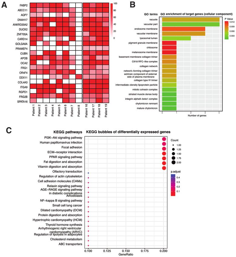
2                                   YANG et al: COL4A3 FOR NASOPHARYNGEAL CARCINOMA

and molecular mechanisms may lead to the identification of        concentration was measured using a Bradford protein assay kit
novel valuable biomarkers.                                        (Bio-Rad Laboratories, Inc.). Then, 60 µg per well of protein
    Type IV collagen, one of the main components of the           samples were separated using a 12% SDS-PAGE (Bio-Rad
extracellular matrix and basement membrane, has been found        Laboratories, Inc.) and transferred to a PVDF membrane
to interact with tumor cells and regulate tumor growth, prolif-   (EMD Millipore). After blocking with TBS-Tween-20 (TBST)
eration, differentiation, adhesion and metastasis (7-9), which    containing 5% skimmed milk for 1 h at room temperature,
is important for the study of metastasis of NPC. Type IV          the PVDF membrane was washed with TBST for 10 min,
collagen contains three highly similar collagen precursors        three times. The membrane was then incubated with primary
namely, (α1)2α2(IV), α3α4α5(IV) and (α5)2α6(IV) (10). The         antibodies diluted in TBST overnight at 4˚C. Antibody infor-
genes, COL4A1, COL4A2, COL4A3, COL4A4, COL4A5                     mation: COL4A3 (cat. no. PA5-39876, 1:500 dilution) and
and COL4A6, which synthesize α1, α2, α3, α4, α5 and α6            GAPDH (cat. no. MA5-15738-D800, 1:500 dilution) antibodies
peptide chains, respectively, are located in pairs on human       were from Thermo Fisher Scientific, Inc. Subsequently, the
chromosomes 13, 2 and X, respectively (11). In addition,          membrane was washed with TBST for 10 min thrice. Following
hypermethylation of the COL4A5/COL4A6 genes in colon              incubation with horseradish peroxidase (HRP)-conjugated
cancer led to a decrease in the expression level of the COL4A5/   goat anti-rabbit secondary antibody (cat. no. A32731; 1:10,000
COL4A6 chain and the destruction of the structural integrity      dilution; Thermo Fisher Scientific, Inc.) and HRP-conjugated
of the basement membrane (12). Similarly, it was found that       goat anti-mouse secondary antibody (cat. no. 35518; 1:10,000
the expression level of the COL4A5/COL4A6 genes was               dilution; Thermo Fisher Scientific, Inc.) for 2 h at room
decreased or deleted in the invasive stage of basal cell carci-   temperature, respectively. After another three washes with
noma and prostate cancer (13,14). However, the role of the        TBST for 10 min, the proteins were visualized using an ECL
collagen-encoding genes in different types of NPC remains         substrate (Pierce™ ECL Western Blotting Substrate; Thermo
undetermined.                                                     Fisher Scientific, Inc.). ImageJ software version 1.48; National
    In the present study, whole exon sequencing (WES)             Institutes of Health) was used to quantify the optical density
was used to detected gene mutations associated with NPC           of each protein. The relative optical density was calculated by
biological behaviors and to determine the key genes associated    comparing with that in GAPDH.
with the invasion and metastasis potential of NPC. The role of
COL4A3 gene expression and extracellular collagen, deposi-        Reverse transcription-quantitative (RT-q)PCR assay.
tion in both the upward and downward progressing types of         Total RNA was extracted from treated cells using TRIzol®
NPC, were also investigated. The inhibition of COL4A3 could       (Invitrogen; Thermo Fisher Scientific, Inc.) Then, 2.0 µg total
promote invasion and migration of the 5-8F NPC cell line.         RNA was used for RT using a TaqMan RT kit according
The present study may provide new information in identifying      to the manufacturer's instructions (Applied Biosystems;
novel biomarkers and establish a molecular classification of      Thermo Fisher Scientific, Inc.). Subsequently, the cDNA
NPC.                                                              was amplified using qPCR, in a 20 µl total reaction volume
                                                                  and the Go Tag Green Master Mix/Platinum SYBR Super
Materials and methods                                             mix (Invitrogen; Thermo Fisher Scientific, Inc.). qPCR was
                                                                  performed using the following conditions: Initial denaturation
Cell culture and transfection. The human 5-8F NPC cell            at 95˚C for 5 min, denaturation at 95˚C for 15 sec, 60˚C for
line was donated by Sun Yat-Sen University Cancer Center          30 sec, followed by 40 cycles at 72˚C for 30 sec and 72˚C for
and was cultured in RPMI-1640 medium (supplemented with           10 min. The following primers were used: COL4A3 forward,
10% fetal bovine serum, penicillin (100 U/ml) and strepto-        5'-GGACTCACGGGTTCCAAAGGT-3' and COL 4A3
mycin (100 mg‑ml) (all from Gibco; Thermo Fisher Scientific,      reverse, 5'-CCTGCTCACCCTTAGAACCACT-3'; GAPDH
Inc.) at 37˚C in a humidified incubator with 5% CO2 (Sanyo        forward, 5'-CAATGACCCCTTCATTGACC-3' and GAPDH
Eletcric Co., Ltd). Cells in the logarithmic growth phase were    reverse, 5'-GACAAGCTTCCCGTTCTCAG-3'. GAPDH was
placed in a 6-well plate and incubated until 70-90% conflu-       used as the internal control.
ence.
    Cells were transfected with specific short interfering        Transwell and Matrigel assays. For the Transwell assay, 24 h
(si)RNA targeting COL4A3 (designed and synthesized by             following siRNA transfection, the 5-8F cell line was harvested
Guangzhou Ribobio Co., Ltd.) using Lipofectamine® 2000            with trypsin and seeded into Transwell chambers (8 µm pore
(Invitrogen; Thermo Fisher Scientific, Inc.) according to         size; Costar; Corning, Inc.), at a density of 2x10⁴ cells per well.
manufacturer's instructions. The following siRNA was used:        The upper chambers contained 200 µl serum-free RPMI-1640
5'-GGGTAATCCTGGATTTCTA-3'. A scramble siRNA                       medium, while the lower chambers were filled with 600 µl
was also purchased from (Guangzhou Ribobio Co., Ltd) and          RPMI-1640 medium, containing 10% FBS. For the Matrigel
used as a control siRNA (siNC). The cells were assigned to        assay, 100 µl Matrigel (Sigma-Aldrich; Merck KGaA)
three experimental groups: Non-transfected group (control),       was added to the Transwell chamber, at a concentration of
siRNA negative control group (si-NC) and COL4A3 siRNA             200 µg/ml. The cells on the upper surface of the membrane
transfected group (si-COL4A3).                                    were removed using a cotton swab. The invaded cells were
                                                                  fixed with 4% paraformaldehyde for 30 min and stained
Western blot analysis. Cells were lysed in RIPA lysis buffer,     with 1% crystal violet for 30 min at room temperature. The
containing protease and phosphatase inhibitors (Sigma-            numbers of migrated cells were counted in five random fields
Aldrich; Merck KGaA) on ice for 30 min. The total protein         of view under a microscope at x100 magnification using a
ONCOLOGY LETTERS 21: 223, 2021                                                 3

Shunyu ICX41 inverted light microscope (Zhejiang Kangchen           The slides were visualized under x100 magnification using a
Biotech Co., Ltd.) in each membrane. Finally, cell counts were      light microscope. The compactness of cells between upward
determined using the ImageJ software (National Institutes of        and downward progressing types of NPC was observed.
Health). All experiments were repeated at least three times.
                                                                    Masson trichromatic staining. The expression level and distri-
Wound-healing assay. A wound-healing assay was performed            bution of collagen in the upward and downward progressing
to determine the effect of COL4A3 on cell migration. The            types of NPC specimens was detected using Masson trichrome
5-8F cell line was seeded into 6-well plates, at a density of       staining. The NPC specimens of the two groups were routinely
2x10⁵ cells/well, in RPMI-1640 medium, supplemented with            dewaxed with water by successively putting into xylene for
10% FBS, 24 h following transfection. When the cells had            20 min at room temperature, anhydrous ethanol for 10 min,
reached 90-100% confluence, a 200 µl pipette tip was used to        an ethanol series (95, 90, 80 and 70%), washed with distilled
scratch a wound on the cells. The medium was removed, and           water thrice, and then fixed with picric acid mixed fixation
the cells were gently washed twice with PBS. Next, the cells        solution for 10 min at room temperature. The nucleus was
were incubated with RPMI-1640 medium containing 1% FBS.             subsequently stained with H for 5-8 min, rinsed with distilled
Images were captured at 0 and 24 h under a light microscope         water thrice, and then stained with Lichun red acid Fuhong
at x40 magnification and the location and migration of the          solution for 5-7 min at room temperature. After washing with
cells was measured by determining the wound area. The               distilled water, specimens were stained with 1% phosphomo-
wound healing rate was quantified by using ImageJ software          lybdic acid solution for 5 min at room temperature and then
and calculated with the help of following formula (Aw - At)/        stained directly with bright green dye for 5 to 10 min at room
Aw x 100. Where Aw is an area of the wound at 0 h (control)         temperature. Subsequently, specimens were differentiated
and At is the area of the wound at 24 h after scratching. All the   with 1% glacial acetic acid for 30-60 sec, dehydrated with
experiments were repeated at least three times.                     95% ethanol for 3 min and then dehydrated with anhydrous
                                                                    ethanol for 5 min. Finally, all the specimens were made
Human specimens. The present study was approved by the              transparent with xylene and sealed with neutral gum. The
Medical Ethics Committee of Zhongnan Hospital of Wuhan              slides were scanned using a NanoZoomer S360 Digital slide
University (approval no. 2019084). Written informed consent         scanner (C13220-01; Hamamatsu Co., Ltd.) and analyzed
was obtained from each living patient or from their relatives       under x10 magnification using NDP.view2 Viewing software
for deceased patients in the study. Biopsies were collected         (U12388-01; Hamamatsu Co., Ltd.). The expression level of
from 20 patients with upward and downward progressing               collagen was quantified using Image-Pro Plus v6.0 software
types of NPC. In total, 17 patients were male and three were        (Media Cybernetics, Inc.).
female. The median age was 57 years (range, 42-83 years). The
following inclusion criteria were used: i) Newly diagnosed          Immunohistochemistry (IHC). For IHC staining, paraffin-
patients with NPC from January 2003 to December 2018;               embedded NPC tissue sections at 4-5 µm were deparaffinized
ii) between 18 and 85 years of age; iii) confirmed patho-           with xylene I for 20 min, xylene II for 20 min, and rehy-
logical diagnosis; and iv) stages of T3-4N0-1M0 (upward) and        drated with 100% ethanol for 10 min, and 95, 85 and 80%
Tis-1N2-3M0-1 (downward) (based on the 8th edition of American      ethanol for 5 min, each. Then the sections were incubated
Joint Committee on Cancer for nasopharyngeal carcinoma              with anti-COL4A3 antibody (cat. no. sc-52317; 1:100 dilu-
staging) (15). The following exclusion criteria were used: i)       tion; Santa Cruz Biotechnology, Inc.) overnight at 4˚C. The
Recurrent NPC; ii) no pathological diagnosis or pathological        next day, the slides were then incubated with a biotin-labeled
specimens; and iii) a history of other malignant tumors. At the     secondary antibody (cat. no. sc-2018; 1:100 dilutions; Santa
same time, the demographical and pathological information,          Cruz Biotechnology, Inc.) for 40 min at room temperature.
and imaging data were collected.                                    Subsequently, the secondary antibody was washed off with
                                                                    TBST (0.1% Tween-20). The cells were then visualized using
Hematoxylin and eosin (H&E) staining. The histopathological         a REALTM EnVisionTMDetection System, (Peroxidase/DAB+;
examination was performed by a senior pathologist at Zhongnan       Rabbit/Mouse; cat. no. K5007; Dako; Agilent Technologies,
Hospital. The biopsies of two groups of patients with NPC were      Inc.). The slides were scanned by NanoZoomer S360 Digital
stained with H&E, which was routinely used in the pathology         slide scanner and analysed under x300 magnification using
laboratory using the following procedure: Deparaffinization         NDP.view2 Viewing software. The expression level of
with xylene, twice for 10 min each; re-hydration with abso-         COL4A3 was quantified using Image-Pro Plus version 6.0
lute ethanol, twice, 5 min each; followed by 95% ethanol for        software (Media Cybernetics).
2 min and 70% ethanol for 2 min, followed by washing in
distilled water for 1 min; staining in H solution for 8 min at      WES. Sequencing and bioinformatic analysis of whole exons
room temperature, followed by washing in tap water for 5 min;       from the upward and downward progressing types of NPC
differentiation in acid ethanol for 30 sec, followed by washing     specimen were completed in cooperation with Shenzhen
in tap water for 1 min; bluing in 0.2% ammonia water for 1 min,     Chengqi Biotechnology Co., Ltd. DNA was fragmented and
followed by washing in tap water for 5 min. Finally, slides         hybridized using the SureSelect Human All Exome kit V5
were dehydrated with 85% ethanol and 95% ethanol for 5 min          (Agilent Technologies, Inc.). Exome shotgun libraries were
each and counterstaining with E Y solution for 5 min at room        sequenced on the SureSelect Human All Exon v.6 enrich-
temperature. Dehydration with absolute ethanol, three times,        ment, Illumina NovaSeq 6000 platform (Illumina, Inc.),
5 min each. Transparency with xylene, twice, 5 min each time.       generating paired end reads of 150 bp at each end. The cloud
4                                       YANG et al: COL4A3 FOR NASOPHARYNGEAL CARCINOMA

Table I. Clinical characteristic of patients with nasopharyngeal carcinoma.

A, Upward

ID                 WES              Sex           Age, years           T stagea          N stagea          M stagea           WHO typeb

Patient 1          Yes            Male                 66                  4                 0                 0                   II
Patient 2          Yes            Male                 42                  3                 1                 0                   II
Patient 3          Yes            Male                 59                  3                 1                 0                   II
Patient 4          Yes            Male                 55                  3                 1                 0                   II
Patient 5          Yes            Male                 68                  4                 1                 0                   II
Patient 6          Yes            Male                 53                  4                 1                 0                   II
Patient 7          Yes            Male                 54                  4                 0                 0                   II
Patient 8          Yes            Male                 67                  4                 1                 0                   II
Patient 9          No             Male                 49                  4                 1                 0                   II
Patient 10         No             Male                 78                  4                 1                 0                   II
Patient 11         No             Male                 62                  4                 1                 0                   II
Patient 12         No             Female               54                  3                 1                 0                   II
Patient 13         No             Male                 62                  3                 1                 0                   II
Patient 14         No             Female               44                  4                 1                 0                   I
Patient 15         No             Male                 73                  4                 0                 0                   II

B, Downward

ID                 WES            Sex             Age, years           T stagea          N stagea          M stagea           WHO typeb

Patient 16         Yes            Male                 63                  1                 2                 0                   II
Patient 17         Yes            Male                 53                  1                 3                 0                   II
Patient 18         Yes            Male                 54                  1                 3                 1                   II
Patient 19         Yes            Male                 83                  1                 3                 0                   II
Patient 20         No             Female               44                  1                 3                 0                   I
a
 Based on the 8th edition of AJCC nasopharyngeal carcinoma staging; bWHO types: I, keratinized squamous cell carcinoma; II, non-keratinized
carcinoma. WES, whole exon sequencing; T, tumor; N, node; M, metastasis.

analysis platform with FANSe algorithm (Chi-Cloud NGS                  which were visualized via R clusterProfiler package version
Analysis Platform) was applied for single-nucleotide variant           3.18.0 (18).
calling (16). Image analysis and base calling were performed
with CAVSAVR (Illumina, Inc.) using default parameters.                Survival analysis. COL4A3 gene expression, as determined
Adapter sequences were removed to obtain high-quality reads.           using RNA sequencing and survival data from patients with
These were aligned to the NCBI human reference genome hg19             head and neck squamous cell carcinoma were obtained and
using the Burrows-Wheeler Aligner alignment algorithm. The             analyzed using the Kaplan-Meier Plotter tool (http://kmplot.
high frequency mutant genes associated with metastasis of              com/analysis) (19). Due to a lack of NPC data, the data of
NPC were screened out. The associated amino acid replace-              head and neck squamous cell carcinoma (N=499) were used
ment events and the changes in protein coding sequence and             instead. Patients were divided by selecting the auto select
function were predicted.                                               best cut-off. When the auto select best cut-off is selected, all
                                                                       possible cut-off values between the lower and upper quartiles
Gene Ontology (GO) and Kyoto Encyclopedia of Genes and                 are computed, and the best performing threshold is used as
Genomes (KEGG) enrichment analysis of putative signaling               a cut-off. The auto select best cut-off value was 35. Patients
pathways. The designated genes were uploaded into the                  of both sexes, different ethnicities, disease stages and grades
function annotation portal of The Database for Annotation,             were included. Hazard ratio (HR) with 95% CI were analyzed
Visualization, and Integrated Discovery (DAVID), online                and the median survivals times were compared between the
bioinformatics resources for investigating the biological              high and low expression level groups.
signaling pathways enriched by input genes (17). GO (http://
geneontology.org/) and KEGG (https://www.kegg.jp/)                     Statistical analysis. Quantitative results are expressed as the
corpus were adopted to identify putative signaling pathways            mean ± SD. A Wilcoxon rank sum test was performed to analyze
ONCOLOGY LETTERS 21: 223, 2021 5
Table II. Whole exon sequencing results of patients with nasopharyngeal carcinoma.
Position Gene Original Mutation
Chromosome (1-based) name Original Mutation (peptide) (peptide) Annotation P-valuea
chr10 135440216 FRG2B G A H Y FSHD region gene 2
family member B 0.87870
chr10 17147521 CUBN G T P T Cubilin 0.01335
chr1 12939765 PRAMEF4 A G L P PRAME family member 4 0.00000
chr12 31242081 DDX11 G A R Q DEAD/H-box helicase 11 0.27880
chr12 8327016 ZNF705A T C A T zinc finger protein 705A 0.01544
chr15 102463117 OR4F4 T G H P Olfactory receptor family 4
subfamily F member 4 0.49690
chr15 28197037 OCA2 T C H R OCA2 melanosomaltrans-
membrane protein 0.01397
chr15 45392075 DUOX2 G A S L Dual oxidase 2 0.21210
chr15 74374822 GOLGA6A T G H P golgin A6 family member A 0.00665
chr16 48258198 ABCC11 C T G R ATP binding cassette
subfamily C member 11 0.07139
chr17 76433898 DNAH17 T C H R Dyneinaxonemal heavy
chain 17 0.20260
chr17 78178893 CARD14 C T R W Caspase recruitment
domain family member 14 0.07064
chr2 21231524 APOB G A P L Apolipoprotein B 0.30960
chr2 228135631 COL4A3 C T P L Collagen type IV
alpha 3 chain 0.00149
chr2 87214281 RGPD1 T G V G RANBP2-like and
GRIP domain containing 1 0.05126
chr3 172835082 SPATA16 C T G E Spermatogenesis associated 16 0.08735
chr3 37574951 ITGA9 G A G E Integrin subunit alpha 9 0.28760
chr4 120241902 FABP2 T C T A Fatty acid binding protein 2 0.03159
chr4 190881957 FRG1 G T D Y FSHD region gene 1 0.05745
chr9 33386465 AQP7 A G Y H Aquaporin 7 0.04698
chr9 42376286 ANKRD20A2 C T A V Ankyrin repeat domain 20
family member A2 0.08193
a
Single nucleotide variations between upward and downward progressing types of nasopharyngeal carcinoma were detected by whole exome
sequencing and Genome-Wide Association Studies were used to analyze differential gene expression. P6 YANG et al: COL4A3 FOR NASOPHARYNGEAL CARCINOMA cervical lymph node metastasis or distant metastasis occurred even when the primary tumor was in early stages. On the other hand, the upward progressing type of NPC (T3-T4) showed significant infiltration and destruction of the surrounding tissue by the primary tumor, with documented clinical manifesta- tions of space and nerve involvement associated with tumor invasions (Fig. 1D and F). Nonetheless, cervical lymph node metastasis of the upward progressing type was not significant (N0-N1) and no evidence of distant metastasis was detected. COL4A3 was identified as the target gene. A total of 8 upward NPC specimens and 4 downward NPC specimens were sequenced, from which 4,125 differential mutation sites were found, involving 2,511 genes. The molecular events such as single nucleotide variations, insertions/deletions, copy number variations and gene fusion, with significant differences in mutation frequency between the upward and downward progressing types of NPC were detected. After harmful predictive filtration, there were 21 residual differ- ential mutation sites, involving 21 genes (Table II; Fig. 2A). Single nucleotide variations between upward and downward progressing types of NPC, detected by WES and Genome- Wide Association Studies, were used to analyze differential gene expression. P
ONCOLOGY LETTERS 21: 223, 2021                                                                   7

Figure 2. Sequencing results of the whole exons in nasopharyngeal carcinoma. (A) The differentially mutated genes between the upward and downward
progressing types of NPC were identified using sequencing of the whole exons. The color block in the figure indicated the frequency of gene mutation. (B) GO
analysis showed that some of the differentially mutated genes were enriched in the genes associated with collagen expression. (C) KEGG bubbles signal
pathway analysis showed that the differentially mutated genes were involved in the extracellular matrix-associated signaling pathway. GO, Gene Ontology;
KEGG, Kyoto Encyclopedia of Genes and Genomes.

changes in the tumor microenvironment, particularly in the                     It is well known that the α3 (IV) chain encoded by COL4A3,
remodeling of extracellular matrix characterized by the degra-                 is lysed by matrix metalloproteinase-9 (MMP9) to produce
dation, deposition and cross-linking of type IV collagen (20-22).              the primary bioactive fragment, tumstatin, which can inhibit
The extracellular matrix is a barrier for tumor cell metastasis.               the formation of blood vessels in vivo and inhibit the prolif-
Type IV collagen, from the extracellular matrix, is the primary                eration and metastasis of tumors (24). In the early stage of
component of the basement membrane. The expression of                          tumorigenesis, the MMP-mediated mechanism promotes the
type IV collagen affects the deposition of the tumor extracel-                 release of tumstatin (also an endogenous angiogenic inhibitor),
lular matrix; thus, affecting the potential of tumor metastasis.               by separating it from the basement membrane, to permit its
The investigation into the effect of type IV collagen on extra-                anti-angiogenesis and antitumor activities (25). These two
cellular matrix deposition and its function is beneficial for                  antitumor properties have been found to be regulated by
understanding the molecular mechanism of tumor metastasis                      an ITGB3-mediated RGD-independent mechanism. Once
and provide novel ideas for identifying molecular markers and                  tumstatin has been dissociated from the basement membrane,
effective treatment for tumor metastasis.                                      it interacts with integrin αVβ3 in endothelial cells, which leads
    The COL4A3 domain binds and inhibits the proliferation                     to the arrest of the cell cycle or apoptosis (26), and has been
of melanoma and other epithelial tumor cell lines in vitro (23).               found to cause apoptosis (27,28) and block the proliferation
8 YANG et al: COL4A3 FOR NASOPHARYNGEAL CARCINOMA Figure 3. Expression of COL4A3 and the deposition of collagen fibers in NPC. (A and B) The expression of COL4A3 in the downward progressing type of NPC was significantly lower compared with that in upward progressing type of NPC. *PA, G1242D) in analysis, six associated signaling pathways were identified: exon 42 of COL4A3 played a causative role in Alport syndrome PI3K-Akt signaling pathway, focal adhesion, ECM-receptor and thin basement membrane nephropathy (31). Other mutations interaction, PPAR signaling pathway, cell adhesion molecules were also recently identified in a consanguineous family with and NF- κ B signaling pathway. Among these pathways,
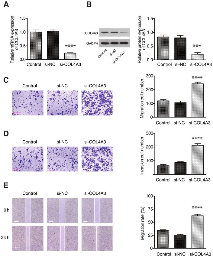
ONCOLOGY LETTERS 21: 223, 2021                                                                     9

Figure 4. Knockdown of COL4A3 promoted the migration and invasion of the 5-8F cell line in vitro. The relative COL4A3 (A) mRNA and (B) protein expres-
sion levels in each group as determined by reverse transcription-quantitative PCR and western blot assay, respectively. (C) Cell migration and (D) invasion
abilities were examined using Transwell assay in each group. (E) The migration rate of 5-8F cell line in each group, as detected by a wound-healing assay. Data
are presented as the mean ± SD from 3 independent experiments. NC, negative control; si, short interfering. ***P
10                                    YANG et al: COL4A3 FOR NASOPHARYNGEAL CARCINOMA

type of NPC was significantly decreased. The results indicated        collection, integration analysis and figure processing. All
that the knockdown of COL4A3 might promote the invasion and           authors have made substantial contributions to this manuscript.
migration of NPC cells. The low expression level of COL4A3            All authors confirmed and approved the final manuscript.
in the downward progressing type of NPC was associated with
increased risk of distant metastasis, suggesting that COL4A3          Ethics approval and consent to participate
expression might be a good biomarker for NPC metastasis.
    However, there are still some limitations in the present study.   The present study was approved by the Medical Ethics
Firstly, a small number of matched samples were collected             Committee of Zhongnan Hospital of Wuhan University
in this study. This was partly due to availability of eligible        (Ethical number: 2019084). Written informed consent was
samples, since samples that were suitable for WES should be           obtained from each living patient or from their relatives for
relatively recent to ensure DNA integrity. Therefore, the number      deceased patients in the study.
of samples was relatively small in the current study. A larger
number of samples should be performed to verify the results of        Patient consent for publication
the present study and further clarify the gene mutation spectrum
affecting the metastatic risk of NPC by WES. Next, the function       Not applicable.
of certain genetic mutation should be examined in detail. The
mutation status of the aforementioned genes should be veri-           Competing interests
fied by PCR and Sanger sequencing. Differentially expressed
genes between upward and downward progressing types of                The authors declare that they have no competing interests.
NPC could potentially be detected by performing transcrip-
tomic sequencing. Thirdly, in vivo experiments were lacking           References
in the present study, which help validate the hypothesis, prove
the essential role of COL4A3 in NPC metastasis and uncover             1. Ferlay J, Soerjomataram I, Dikshit R, Eser S, Mathers C,
                                                                          Rebelo M, Parkin DM, Forman D and Bray F: Cancer incidence
potential molecular mechanisms. A plasmid harboring targeted              and mortality worldwide: Sources, methods and major patterns
gene mutation will be constructed in the future for the detection         in GLOBOCAN 2012. Int J Cancer 136: E359-E386, 2015.
of protein expression. The in vivo effect of COL4A3 should also        2. Carioli G, Negri E, KawakitaD, Garavello W, La Vecchia C and
                                                                          Malvezzi M: Global trends in nasopharyngeal cancer mortality
be studied by manipulating its expression and functions.                  since 1970 and predictions for 2020: Focus on low-risk areas. Int
    In conclusion, although several limitations exist, the present        J Cancer 140: 2256-2264, 2017.
study showed that the COL4A3 gene was highly expressed in              3. Sun P, Chen C, Chen XL, Cheng YK, Zeng L, Zeng ZJ, Liu LZ,
                                                                          Su Y and Gu MF: Proposal of a clinical typing system and
the upward progressing type of NPC, but was low in the down-              generation of a prognostic model in patients with nasopharyngeal
ward progressing type. This was associated with the degree                carcinoma from Southern China. J BUON 19: 474-483, 2014.
of collagen fibrosis, thus affecting the migration and invasion        4. Liang WJ, Qiu F, Hong MH, Guo L, Qin HD, Liu QC, Zhang XS,
of NPC. COL4A3 may be a novel biomarker in the diagnosis                  Mai HQ, Xiang YQ, Min HQ, et al: Differentially expressed genes
                                                                          between upward and downward progressing types of nasopha-
and treatment of metastatic NPC, which could be beneficial to             ryngeal carcinoma. Chin J Cancer 27: 460-465, 2008 (In Chinese).
establish a potential molecular typing method and identify the         5. Mo L, Weng J, Zeng F, Li X, Liu B, Li Z and Kuang G: The
target for individualized therapy for metastatic NPC.                     relationship between extend types and distant metastasis of naso-
                                                                          pharyngeal carcinoma. Lin Chung Er Bi Yan Hou Tou Jing Wai
                                                                          Ke Za Zhi 24: 554-555, 558, 2010 (In Chinese).
Acknowledgements                                                       6. Chew MM, Gan SY, Khoo AS and Tan EL: Interleukins, laminin
                                                                          and Epstein - Barr virus latent membrane protein 1 (EBV LMP1)
                                                                          promote metastatic phenotype in nasopharyngeal carcinoma.
Not applicable.                                                           BMC Cancer 10: 574, 2010.
                                                                       7. Morrissey MA, Jayadev R, Miley GR, Blebea CA, Chi Q, Ihara S
Funding                                                                   and Sherwood DR: SPARC promotes cell invasion in vivo by
                                                                          decreasing type IV collagen levels in the basement membrane.
                                                                          PLoS Genet 12: e1005905, 2016.
The present study was supported by a grant from the Scientific         8. Tanjore H and Kalluri R: The role of type IV collagen and
Research Project of Hubei Provincial Health and Family                    basement membranes in cancer progression and metastasis. Am
                                                                          J Pathol 168: 715-717, 2006.
Planning Commission (grant no. WJ2019H064) and from                    9. Timpl R, Wiedemann H, van Delden V, Furthmayr H and Kühn K:
the National Natural Science Foundation of China (grant                   A network model for the organization of type IV collagen molecules
no. 81803061).                                                            in basement membranes. Eur J Biochem 120: 203-211, 1981.
                                                                      10. Mundel TM and Kalluri R: Type IV collagen-derived angio-
                                                                          genesis inhibitors. Microvasc Res 74: 85-89, 2007.
Availability of data and materials                                    11. Khoshnoodi J, Pedchenko V and Hudson BG: Mammalian
                                                                          collagen IV. Microsc Res Tech 71: 357-370, 2008.
                                                                      12. Ikeda K, Iyama K, Ishikawa N, Egami H, Nakao M, Sado Y, Ninomiya Y
The datasets used and/or analyzed during the current study are            and Baba H: Loss of expression of type IV collagen alpha5 and alpha6
available from the corresponding author on reasonable request.            chains in colorectal cancer associated with the hypermethylation of
                                                                          their promoter region. Am J Pathol 168: 856-865, 2006.
Authors' contributions                                                13. Tanaka K, Iyama K, Kitaoka M, Ninomiya Y, Oohashi T, Sado Y
                                                                          and Ono T: Differential expression of alpha 1(IV), alpha 2(IV),
                                                                          alpha 5(IV) and alpha 6(IV) collagen chains in the basement
XY, QW and YZ conceived the study and drafted the manu-                   membrane of basal cell carcinoma. Histochem J 29: 563-570, 1997.
script. XY collected the clinical tissues, was responsible for        14. Dehan P, Waltregny D, Beschin A, Noel A, Castronovo V,
                                                                          Tryggvason K, De Leval J and Foidart JM: Loss of type IV
the study design and provided the data. QW performed the                  collagen alpha 5 and alpha 6 chains in human invasive prostate
statistical analysis. FW performed the bioinformatic data                 carcinomas. Am J Pathol 151: 1097-1104, 1997.
ONCOLOGY LETTERS 21: 223, 2021 11
15. Pan XX, Tong LH, Chen YF, Li FL, Tang WB, Liu YJ and 28. Maeshima Y, Yerramalla UL, Dhanabal M, Holthaus KA,
Yang WA: simplified T classification based on the 8th edition of Barbashov S, Kharbanda S, Reimer C, Manfredi M,
the UICC/AJCC staging system for nasopharyngeal carcinoma. Dickerson WM and Kalluri R: Extracellular matrix-derived
Cancer Manag Res 11: 3163-3169, 2019. peptide binds to alphavbeta3 integrin and inhibits angiogenesis. J
16. Zhang G, Fedyunin I, Kirchner S, Xiao C, Valleriani A and Biol Chem 276: 31959-31968, 2001.
Ignatova Z: FANSe: An accurate algorithm for quantitative 29. Maeshima Y, Sudhakar A, Lively JC, Ueki K, Kharbanda S,
mapping of large scale sequencing reads. Nucleic Acids Res 40: Kahn CR, Sonenberg N, Hynes RO and Kalluri R: Tumstatin, an
e83, 2012. endothelial cell-specific inhibitor of protein synthesis. Science
17. Huang W, Sherman BT and Lempicki RA: Systematic and inte- 295: 140-143, 2002.
grative analysis of large gene lists using DAVID bioinformatics 30. Pasco S, Ramont L, Venteo L, Pluot M, Maquart FX and
resources. Nat Protoc 4: 44-57, 2009. Monboisse JC: In vivo overexpression of tumstatin domains
18. Yu G, Wang LG, Han Y and He QY: clusterProfiler: An R by tumor cells inhibits their invasive properties in a mouse
package for comparing biological themes among gene clusters. melanoma model. Exp Cell Res 301: 251-265, 2004.
OMICS 16: 284-287, 2012. 31. Hou P, Chen Y, Ding J, Li G and Zhang H: A novel mutation of
19. Nagy Á, Lánczky A, Menyhárt O and Győrffy B: Validation of COL4A3 presents a different contribution to Alport syndrome
miRNA prognostic power in hepatocellular carcinoma using and thin basement membrane nephropathy. Am J Nephrol 27:
expression data of independent datasets. Sci Rep 8: 9227, 2018. 538-544, 2007.
20. Wei X, Li S, He J, Du H, Liu Y, Yu W, Hu H, Han L, Wang C, Li H, 32. Rana K, Tonna S, Wang YY, Sin L, Lin T, Shaw E, Mookerjee I
et al: Tumor-secreted PAI-1 promotes breast cancer metastasis and Savige J: Nine novel COL4A3 and COL4A4 mutations
via the induction of adipocyte-derived collagen remodeling. Cell and polymorphisms identified in inherited membrane diseases.
Commun Signal 17: 58, 2019. Pediatr Nephrol 22: 652-657, 2007.
21. Guo S and Deng CX: Effect ofstromal cells in tumor microen- 33. Saravani R, Yari D, Saravani S and Hasanian-Langroudi F:
vironment on metastasis initiation. Int J Biol Sci 14: 2083-2093, Correlation between the COL4A3, MMP-9, and TIMP-1
2018. polymorphisms and risk of keratoconus. Jpn J Ophthalmol 61:
22. Kaur A, Ecker BL, Douglass SM, Kugel CH III, Webster MR, 218-222, 2017.
Almeida FV, Somasundaram R, Hayden J, Ban E, Ahmadzadeh H, 34. Saravani S, Yari D, Saravani R and Azadi Ahmadabadi C:
et al: Remodeling of the collagen matrix in aging skin promotes Association of COL4A3 (rs55703767), MMP-9 (rs17576)and
melanoma metastasis and affects immune cell motility. Cancer TIMP-1 (rs6609533) gene polymorphisms with susceptibility to
Discov 9: 64-81, 2019. type 2 diabetes. Biomed Rep 6: 329-334, 2017.
23. Chelberg MK, Tsilibary EC, Hauser AR and McCarthy JB: 35. Hao XD, Chen XN, Zhang YY, Chen P, Wei C, Shi WY and
Type IV collagen-mediated melanoma cell adhesion and Gao H: Multi-level consistent changes of the ECM pathway
migration: Involvement of multiple, distinct domains of the identified in a typical keratoconus twin's family by multi-omics
collagen molecule. Cancer Res 49: 4796-4802, 1989. analysis. Orphanet J Rare Dis 15: 227, 2020.
24. Hamano Y, Zeisberg M, Sugimoto H, Lively JC, Maeshima Y, 36. Jiang CP, Wu BH, Chen SP, Fu MY, Yang M, Liu F and Wang BQ:
Yang C, Hynes RO, Werb Z, Sudhakar A and Kalluri R: High COL4A3 expression correlates with poor prognosis after
Physiological levels of tumstatin, a fragment of collagen IV cisplatin plus gemcitabine chemotherapy in non-small cell lung
alpha3 chain, are generated by MMP-9 proteolysis and suppress cancer. Tumour Biol 34: 415-420, 2013.
angiogenesis via alphaV beta3 integrin. Cancer Cell 3: 589-601, 37. Nie XC, Wang JP, Zhu W, Xu XY, Xing YN, Yu M, Liu YP,
2003. Takano Y and Zheng HC: COL4A3 expression correlates with
25. Hamano Y and Kalluri R: Tumstatin, the NC1 domain of alpha3 pathogenesis, pathologic behaviors, and prognosis of gastric
chain of type IV collagen, is an endogenous inhibitor of patho- carcinomas. Hum Pathol 44: 77-86, 2013.
logical angiogenesis and suppresses tumor growth. Biochem This work is licensed under a Creative Commons
Biophys Res Commun 333: 292-298, 2005. Attribution-NonCommercial-NoDerivatives 4.0
26. Maeshima Y, Colorado PC and Kalluri R: Two RGD-independent International (CC BY-NC-ND 4.0) License.
alpha vbeta 3 integrin binding sites on tumstatin regulate distinct
anti-tumor properties. J Biol Chem 275: 23745-23750, 2000.
27. Zhang GM, Zhang YM, Fu SB, Liu XH, Fu X, Yu Y and
Zhang ZY: Effects of cloned tumstatin-related and angiogenesis-
inhibitory peptides on proliferation and apoptosis of endothelial
cells. Chin Med J (Engl) 121: 2324-2330, 2008.You can also read