Are Cyanobacteria an Ancestor of Chloroplasts or Just One of the Gene Donors for Plants and Algae? - MDPI
←
→
Page content transcription
If your browser does not render page correctly, please read the page content below
G C A T
T A C G
G C A T
               genes
Hypothesis
Are Cyanobacteria an Ancestor of Chloroplasts or Just One of
the Gene Donors for Plants and Algae?
Naoki Sato

                                          Graduate School of Arts and Sciences, University of Tokyo, Meguro-ku, Tokyo 153-8902, Japan;
                                          naokisat@bio.c.u-tokyo.ac.jp

                                          Abstract: Chloroplasts of plants and algae are currently believed to originate from a cyanobacterial
                                          endosymbiont, mainly based on the shared proteins involved in the oxygenic photosynthesis and
                                          gene expression system. The phylogenetic relationship between the chloroplast and cyanobacterial
                                          genomes was important evidence for the notion that chloroplasts originated from cyanobacterial en-
                                          dosymbiosis. However, studies in the post-genomic era revealed that various substances (glycolipids,
                                          peptidoglycan, etc.) shared by cyanobacteria and chloroplasts are synthesized by different pathways
                                          or phylogenetically unrelated enzymes. Membranes and genomes are essential components of a
                                          cell (or an organelle), but the origins of these turned out to be different. Besides, phylogenetic
                                          trees of chloroplast-encoded genes suggest an alternative possibility that chloroplast genes could be
                                          acquired from at least three different lineages of cyanobacteria. We have to seriously examine that the
                                          chloroplast genome might be chimeric due to various independent gene flows from cyanobacteria.
                                          Chloroplast formation could be more complex than a single event of cyanobacterial endosymbiosis.
                                          I present the “host-directed chloroplast formation” hypothesis, in which the eukaryotic host cell

                                   that had acquired glycolipid synthesis genes as an adaptation to phosphate limitation facilitated
                                          chloroplast formation by providing glycolipid-based membranes (pre-adaptation). The origins of the
Citation: Sato, N. Are Cyanobacteria
                                          membranes and the genome could be different, and the origin of the genome could be complex.
an Ancestor of Chloroplasts or Just
One of the Gene Donors for Plants
and Algae? Genes 2021, 12, 823.
                                          Keywords: chloroplast origin; cyanobacterial endosymbiosis; glycolipids; host-directed chloroplast
https://doi.org/10.3390/genes12060        formation; peptidoglycan; Paulinella chromatophore; phylogenetic analysis
823

Academic Editors: Denis Baurain
and Luc Cornet                            1. Introduction
                                                Cyanobacteria are a distinct group of bacteria that perform oxygenic photosynthesis.
Received: 26 March 2021                   They have specialized internal membranes called thylakoid membranes in which photo-
Accepted: 25 May 2021
                                          synthetic systems reside. Galactolipids and sulfolipids that are major constituents of the
Published: 27 May 2021
                                          thylakoid membranes have been believed to be necessary for the photosynthetic activ-
                                          ity. Cyanobacteria and chloroplasts of plants and algae share thylakoid membranes with
Publisher’s Note: MDPI stays neutral
                                          galactolipids and sulfolipids, various components of photosystems, and photosynthetic
with regard to jurisdictional claims in
                                          pigments. These traits of substance, structure, and function as a whole were taken as good
published maps and institutional affil-
                                          evidence for the cyanobacterial origin of chloroplasts. The endosymbiotic hypothesis of
iations.
                                          chloroplast origin was first proposed by C. Mereschkowsky in 1905 [1] who obtained a
                                          hint from a short note of A. Schimper in 1883 [2]. Since then, it remained a well-known but
                                          unsupported hypothesis. Various authors in the 1960s revitalized the hypothesis based on
                                          the discovery of chloroplast DNA, and the similarities of chloroplasts and cyanobacteria
Copyright: © 2021 by the author.          became evident thanks to the developments in cell biology and biochemistry of photo-
Licensee MDPI, Basel, Switzerland.
                                          synthetic organisms in the 1960s and 1970s. It was the phylogenetic analysis in the 1980s
This article is an open access article
                                          and later that provided convincing evidence for the cyanobacterial origin of chloroplasts.
distributed under the terms and
                                          Historical views on the endosymbiotic origin of chloroplasts are explained in some review
conditions of the Creative Commons
                                          articles [3–10].
Attribution (CC BY) license (https://
                                                All textbooks of biology explain the origin of chloroplasts by cyanobacterial endosym-
creativecommons.org/licenses/by/
4.0/).
                                          biosis that occurred 1 or 2 billion years ago. The origin of mitochondria is also explained

Genes 2021, 12, 823. https://doi.org/10.3390/genes12060823                                                    https://www.mdpi.com/journal/genes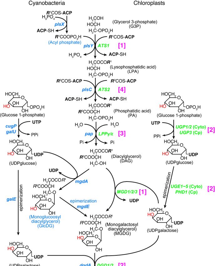
Genes 2021, 12, 823                                                                                           2 of 30

                      by the endosymbiosis of a proteobacterium. These origin stories are currently considered
                      as well-established facts that form the basis of the formation of eukaryotic cells. Many
                      phenomena of eukaryotic cells are currently understood in terms of the endosymbiotic
                      origin of organelles. Stiller [11] criticized this situation: “While many research articles and
                      reviews refer to a single primary origin of all plastids as an established fact, it remains a
                      working hypothesis that gained popularity under a set of assumptions that turn out to
                      have little empirical support in nature (p. 467)”.
                            There are still two major questions about the endosymbiotic theory or hypothesis.
                      First, what is exactly the decisive evidence that the chloroplasts and mitochondria origi-
                      nated from cyanobacterial and proteobacterial endosymbionts, respectively, and how and
                      when this theory was established? It is well known that, in the late 1960s and 1970s, Lynn
                      Margulis claimed her original version of the origin of eukaryotic cells, which involved
                      the endosymbiotic origin of chloroplasts, mitochondria, and mitotic apparatus or flag-
                      ella [12,13]. Nevertheless, it was the molecular phylogeneticists that presented convincing
                      evidence for the close relationship between chloroplasts and cyanobacteria, and between
                      mitochondria and α-proteobacteria. The accumulation of supporting data continued until
                      the early 2000s when the genomic sequences of major model organisms became available.
                      Under these situations, not only the organellar genes but also nuclear-encoded organelle
                      proteins were used for the phylogenetic analysis that finally confirmed the close relation-
                      ships, chloroplasts/cyanobacteria, and mitochondria/α-proteobacteria. Nevertheless, this
                      is just a story of organellar DNA. DNA origin is not enough to affirm the two endosym-
                      biotic events as illustrated in many books and review articles. Membranes and genomes
                      are essential components of a cell or an organelle such as chloroplasts and mitochondria.
                      Can we deduce an endosymbiotic image of the origin of an organelle directly from its
                      DNA origin?
                            Another question, related to this point, arises from more recent studies on the ex-
                      tensive phylogenetic studies on many enzymes involved in the characteristic, common
                      traits of chloroplasts and cyanobacteria. Most of the enzymes involved in the synthesis
                      of thylakoid membrane lipids in plants and algae do not originate from cyanobacteria.
                      How can we explain this? In mitochondria, systematic phylogenetic analysis of the full
                      complement of mitochondrial proteins also raised a comparable question: namely, how
                      can we explain that 90% of mitochondrial enzymes [14,15] existed in the eukaryotic cell
                      long before the formation of mitochondria? Keeling [16] pointed out the “cognitive inertia”
                      of endosymbiotic thinking, and suggested removing the historically biased view: “It is
                      interesting, therefore, to consider what kinds of conclusions we might draw if we learned
                      what we know today in a different order, or learned it all at once (p. 10)”. It is worth trying
                      to re-examine all available evidence for the endosymbiotic theory and to re-organize the
                      history of the formation of chloroplasts and mitochondria. Therefore, this is a discussion
                      or hypothesis article with a limited length on the historical and biological questions on
                      the endosymbiotic origins of organelles, but I focus mainly on the relationship between
                      cyanobacteria and chloroplasts.
                            Because many readers in biology might not be familiar with this kind of half-philosophy,
                      half-biology article, I first present the overall plan of the article. There are three major,
                      mutually related topics: (1) History section (Section 2) deals with the ambiguous meaning
                      of “endosymbiosis”. Endosymbiosis notion includes physical integration of cyanobacterial
                      cell into a eukaryotic cell, but this cannot be proven by cytological experiments. Only DNA
                      origin can be identified by phylogenetic analysis. Then, I discuss the visually identifiable
                      components (membranes) and genome in the following two sections. (2) The lipid and
                      peptidoglycan section (Section 3) deals with the problem that the structural elements of
                      chloroplasts did not originate from cyanobacteria. (3) Chloroplast gene section (Section 4)
                      deals with putative, multiple origins of chloroplast genome within cyanobacteria. Indeed,
                      this is mostly a re-interpretation of previous results. Section 5 also discusses the origins of
                      some other chloroplast enzymes. After these three parts, Section 6 is devoted to discussing
                      the Paulinella model of chloroplast formation, and I present a role of pre-adaptation that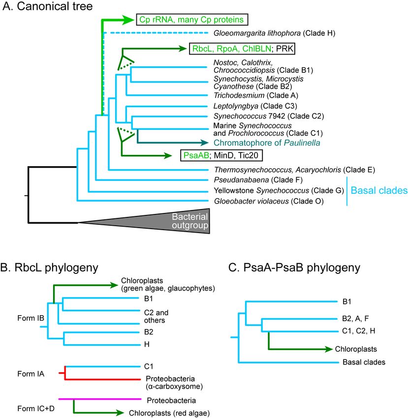
Genes 2021, 12, 823                                                                                          3 of 30

                      determined the fate of chloroplasts. This is not a simple rejection of the endosymbiotic
                      hypothesis, but a broadening of the hypothesis. I also have to mention that the topic of
                      the present article is limited to the formation of “primary” chloroplast. The secondary
                      endosymbiosis must be a different story.

                      2. Historical and Theoretical Problems on the Origin of Chloroplasts
                      2.1. Historical Demarcation
                            The relationship between cyanobacteria and chloroplasts has been repeatedly argued
                      since Mereschkowsky [1] (see also an English translation [3]). I already presented a detailed
                      historical analysis of the endosymbiotic theories [8,9]. Readers are referred to these as
                      well as [4–6,17] for the history of endosymbiotic theories. In the early 20th century, the
                      similarity of cyanobacteria and chloroplasts was restricted to the similarity in size, the
                      presence of photosynthetic pigments, and the activity of carbon fixation with oxygen
                      evolution (“photosynthetic oxygen production” has been conventionally called “oxygen
                      evolution”), all of which reflected the status of knowledge at the time. The history of
                      endosymbiotic arguments may be divided into three major phases: In the first phase
                      (1905–1961), the similarity of cyanobacteria and chloroplasts in terms of photosynthetic
                      properties as defined above was recognized by many researchers in plant and algal studies,
                      but the majority of top researchers, such as Pascher [18] and Buchner [19], clearly rejected
                      endosymbiotic origin of any eukaryotic organelle (chloroplasts or mitochondria). However,
                      Pascher [18] distinguished cyanelles from chloroplasts and described that the cyanelles of
                      Cyanophora paradoxa and Paulinella chromatophora were authentic cyanobacterial cells living
                      in a eukaryotic cell. Cyanelles were evidence of the cyanobacterial origin of chloroplasts
                      for some researchers but were distinct from chloroplasts for many others. Lederberg [20]
                      suggested later that the genetic system supposed to reside in chloroplasts was a kind
                      of plasmid, an exogenous genetic element. Symbiogenesis was also supported by some
                      speculative biologists or philosophers [21,22].
                            In the second phase (1962–1977), the presence of organellar DNA in chloroplasts and
                      mitochondria was taken as evidence for the bacterial origin of these organelles. In parallel,
                      cytological and biochemical characterization of chloroplasts and mitochondria enabled
                      precise comparison of these organelles with cyanobacteria and other aerobic bacteria.
                      The prokaryotic nature of the organellar ribosomes (70S sedimentation-based size, partial
                      rRNA sequence, sensitivity to antibiotics) was an important, novel fact that supported the
                      similarity of organelles and bacteria. Moreover, physical characterization of photosynthetic
                      mechanisms, biochemical identification of photosystem components, and chemical analysis
                      of lipid components of thylakoid membranes, all these data were additional evidence for
                      the similarity of chloroplasts and cyanobacteria. In 1966, Echlin presented a comprehensive
                      table comparing cyanobacteria and chloroplasts [23]. Hybridization of chloroplast rRNA
                      and cyanobacterial DNA was also innovative molecular evidence that favored the similarity
                      of cyanobacteria and chloroplasts. Nevertheless, all these similarities could be used in
                      both ways, namely, for and against the endosymbiotic origin of chloroplasts. In 1967,
                      Goksøyr published a short paper in Nature, illustrating the sequential endosymbiotic
                      origins of mitochondria and chloroplasts [24] (note that this preceded the 1967 paper of
                      Lynn Margulis (Lynn Sagan then) [12], and the Serial Endosymbiosis Theory (SET) of
                      Taylor published in 1974 [25]). The autogenous origin of chloroplasts could also explain
                      the data [26–28], although it was difficult to explain the origin of both chloroplasts and
                      mitochondria within a single scheme. Prudent opinions remained, while changes began to
                      emerge [29–32].
                            The third phase was marked by the development of phylogenetic analysis, which
                      was the breakthrough that changed the status of the endosymbiotic theory. An epoch-
                      making paper of Schwartz and Dayhoff appeared in 1978, reporting a reliable phylogenetic
                      relationship between cyanobacteria and chloroplasts [33]. Phylogenetic analysis was indeed
                      the only rational way of identifying the direct lineage from cyanobacteria to chloroplasts.
                      The methodology of phylogenetic inference became more and more accurate and reliable.
Genes 2021, 12, 823                                                                                           4 of 30

                      Innovations in both computer hardware and computational technology, and dramatic
                      speedup in DNA sequencing and assembly helped the progress of phylogenetic analysis.
                      From the 1980s onwards, chloroplasts became regarded as descendants of cyanobacteria.
                      Nevertheless, it is difficult to distinguish when the conceptual change occurred. The change
                      seemed to proceed gradually and slowly during the 1990s and 2000s. Phylogenetic data
                      accumulated, and all data tended to be interpreted in the light of the endosymbiotic theory,
                      namely, the cyanobacterial origin of chloroplasts. Then, no other way of thinking became
                      mentioned in the literature.

                      2.2. Genocentrism and the Changes in Thinking at the Beginning of the 21st Century
                           I have to point out curious changes in the thinking of researchers from the 1980s to the
                      early 2000s regarding the endosymbiotic origin of organelles, especially the cyanobacterial
                      origin of chloroplasts. The review article of Gray and Doolittle [34] affirmed the cyanobac-
                      terial origin of chloroplasts, based on various phylogenetic trees. Among them, the most
                      important was the phylogenetic tree of rRNA, because the ribosome is the core of the gene
                      expression system in any organism. Wallace [35] also discussed the origin of organelle
                      genomes and concluded:
                           “All of these data are consistent with the hypothesis that the chlDNA genes
                           diverged from the blue-green algae well after the divergence of the eubacterial
                           and eucaryotic nuclear lines. Therefore, chlDNAs must have originated by a
                           symbiotic event between members of the eubacteria and eucaryotic nuclear
                           lineages. Similarly, sequence data have indicated that the mtDNA rRNA genes
                           are more homologous to bacterial rRNA genes than to rRNA genes of eukaryotic
                           nuclei. They too probably also had an endosymbiotic origin.” ([35] p. 227)
                            The origin of organelle DNA was considered convincing evidence for the endosymbi-
                      otic origin of organelles because no other way of acquiring exogenous DNA was known in
                      the 1980s. The notion of horizontal gene transfer appeared later, but the phylogenetic data
                      supporting endosymbiosis were no longer examined in the light of horizontal gene transfer.
                      Delwiche et al. [36] reported the results of robust phylogenetic analyses of the tufA gene,
                      and concluded: “The finding from tufA that all plastids are derived from cyanobacteria
                      is compatible with analyses of 16S rRNA and atpB sequences” (p. 124). Moreira and
                      Philippe [37] wrote in 2001: “a wealth of data strongly suggests that all known photosyn-
                      thetic plastids are monophyletic and that they have a cyanobacterial origin” (p. 771) by
                      citing Delwiche [38], who, nevertheless, noted cautiously in the legend for the illustration
                      of endosymbiotic origin of plastids: “One hypothesis for endosymbiotic events in the
                      evolution of plastids. . . . This figure assumes a single origin of plastids, but this remains
                      uncertain, as does the precise relationship among the primary plastid lineages” (p. S168).
                      Looking back at the literature, the beginning of the 21st century was a turning point, in
                      which the cyanobacterial origin of chloroplasts became accepted as almost established,
                      although the debate on the monophyly of primary plastids continued [39,40]. Certainly,
                      the genome sequencing of the cyanobacterium Synechocystis sp. PCC 6803 [41] and other
                      cyanobacteria opened a way of directly comparing the genomes of cyanobacteria and
                      chloroplasts, and the accumulation of sequence data of both cyanobacteria and chloro-
                      plasts facilitated the comparison of the two entities. McFadden [42] listed various lines of
                      convincing evidence, and wrote also in 2001: “the endosymbiotic origin of plastids from
                      cyanobacterial ancestors thus seems confirmed.” (p. 953)
                            The similarity of RNA polymerase in cyanobacteria and chloroplasts was also known
                      in the 1990s. As described in biochemistry textbooks, bacterial RNA polymerases consist
                      of α, β, β’, and σ subunits. However, in the cyanobacterial counterpart, the β’ subunit
                      is split into two parts, called β’ and β”. This split is found at the level of the gene, as
                      well as protein. This strange situation is also found in RNA polymerases encoded by
                      the chloroplast genome (see the references cited in [36] for the genes that served as evi-
                      dence for the cyanobacterial origin of plastids). Essential components of gene expression
                      in the chloroplast were found almost cyanobacterial. The chloroplast genome was also
Genes 2021, 12, 823                                                                                            5 of 30

                      found to be similar to the cyanobacterial genomes, except for the large difference in sizes.
                      Almost all enzymes and RNA that are encoded by the chloroplast genome had clearly
                      identifiable cyanobacterial counterparts. The similarity of photosynthetic proteins was
                      evident: the components encoded by the chloroplast genome had orthologs in cyanobacte-
                      ria. However, in many cases, the photosynthesis-related components were only found in
                      cyanobacteria and chloroplasts, and it was not possible to identify the root; namely, it was
                      not possible to find where the evolution started. Another problem was unidentified DNA
                      polymerase (see Section 5.1) in chloroplasts. Nevertheless, the majority of opinions favored
                      the cyanobacterial origin of chloroplasts.
                           In retrospect, this was certainly motivated by the importance of genome and gene
                      expression systems in biology. The cyanobacterial nature of the genetic apparatus of
                      chloroplasts including DNA, RNA polymerase, and ribosomal components was taken as
                      evidence that the chloroplast was a descendant of cyanobacteria. I call this “genocentrism”
                      (gene-centered view), which was certainly the decisive impetus that changed the view on
                      the origin of chloroplasts. I have to point out, however, that the genocentrism has been hid-
                      den behind the illustrations clearly showing the membranes to explain the endosymbiotic
                      origin of organelles in many review articles [5,6,43–45] and textbooks (see for example [46]).
                      In contrast, only a vague relationship was known between the cyanobacterial membranes
                      and the chloroplast membranes. The presence of galactolipids and sulfolipids, as well as
                      various photosynthetic components, was known in both cyanobacteria and chloroplasts,
                      which was the evidence for the similarity of thylakoid membranes. The origin of the
                      chloroplast envelope remained ambiguous: it could originate from the cyanobacterial
                      plasma membrane, modified endoplasmic reticulum, phagosome, or peri-algal vacuole.
                      Any origin was possible.

                      2.3. Spread of Endosymbiotic Gene Transfers (EGT)
                            Many components of the chloroplast ribosome were found to be encoded by the
                      nuclear genome. Phylogenetic analysis suggested that these nuclear genes originated from
                      chloroplast genes. This was explained by the endosymbiotic gene transfer (EGT), namely,
                      chloroplast genes were somehow transferred to the nucleus [47]. This was supported by
                      artificial EGT experiments [48]. A fairly efficient gene flow from the chloroplast genome
                      (and the mitochondrial genome) to the nucleus was supposed to work during the long
                      history of plant evolution (for a review of the time, see [49]).
                            However, as the cases of EGT became abundant, many other nuclear genes encod-
                      ing chloroplast proteins have been simply considered to result from EGT, such as the
                      peptidoglycan synthesis enzymes that were identified in the nuclear genome of the moss
                      Physcomitrella patens. Various other chloroplast enzymes were also identified in plant nu-
                      clear genomes. In many cases, phylogenetic analysis was not performed. In other cases,
                      only cyanobacterial sequences and plant sequences were used in phylogenetic analysis.
                      Bacterial sequences were rarely used to identify the origin of the nuclear genes.

                      2.4. Currently Accepted Evidence for the Endosymbiotic, Cyanobacterial Origin of Chloroplasts
                            Currently, several different lines of evidence for the cyanobacterial origin of chloro-
                      plasts exist (Table 1. See also [9,50]). Photosynthesis with oxygen evolution (Item 1) is
                      the best trait that suggests a direct relationship between cyanobacteria and chloroplasts,
                      which also have structural and biochemical similarities. Structurally, both have internal,
                      flattened membranes called “thylakoid” (meaning a sac) membranes, which are the site of
                      light-driven reactions of photosynthesis. The chloroplast is surrounded by a set of limit-
                      ing membranes called envelope membranes, whereas the cyanobacterial cell is enclosed
                      by inner and outer membranes. Note that Gloeobacter lacks thylakoid membranes, and
                      photosynthetic reaction centers reside in the inner cell membrane. Additional and related
                      shared traits in cyanobacteria and chloroplasts include the presence of photosynthetic
                      pigments such as chlorophyll a and the presence of galactolipids in the membranes. The
                      presence of DNA and its associated genetic system (Item 2) is evidence for the autonomy
Genes 2021, 12, 823                                                                                                                6 of 30

                                    of chloroplasts, and therefore, evidence for the exogenous origin of chloroplasts, implying
                                    endosymbiotic origin. Reproduction by binary fission (Item 3) is another similarity between
                                    chloroplasts and cyanobacteria. Another classical evidence is the presence of peptidoglycan
                                    (Item 4) in some chloroplasts, which will be explained in detail later (Section 3.1). The
                                    presence of cyanelles, which were believed to be living cyanobacterial cells within a photo-
                                    synthetic eukaryote (see Section 2.1), is no longer evidence for the visual cyanobacterial
                                    endosymbiosis that can explain chloroplasts origin. The cyanelle of Cyanophora is now
                                    recognized as a chloroplast, rather than a cyanobacterium. The cyanelle of Paulinella is not
                                    an independent organism, which will be discussed later.

                                      Table 1. Evidence for the cyanobacterial origin of chloroplasts.

   Item                        Evidence                                                     Explanation
                                                            (a) Photosynthesis with oxygen evolution
                                                            (b) Presence of chlorophyll a and carotenoids
     1                Similarity of photosynthesis
                                                            (c) Similarity in the structure consisting of two limiting membranes
                                                            and internal thylakoid membranes containing galactolipids
                        Similarity of the genetic
     2                                                      Prokaryotic RNA polymerase and ribosome
                                 system
     3                 Similarity in reproduction           Binary fission
                                                            Glaucophytes, some green algae, charophytes, bryophytes, pteridophytes,
     4                Presence of peptidoglycan
                                                            some gymnosperms
                                                            (a) rRNA genes
                                                            (b) Ribosomal proteins and house-keeping enzymes
     5                 Phylogenetic relationship
                                                            (c) Photosynthetic components (orthologs found only in cyanobacteria and
                                                            chloroplasts)
                                                            (a) Ribosomal proteins
     6                 Synteny of gene clusters             (b) ATPase subunits
                                                            (c) RNA polymerase subunits

                                         Next, phylogenetic analysis of chloroplast-encoded genes (Item 5; see Section 4)
                                    showed their cyanobacterial origin. Nearly all protein-coding genes and RNA genes
                                    encoded by the chloroplast genomes of algae and plants originated from cyanobacteria.
                                    Rare exceptions will be explained in Section 4.
                                         Another line of evidence is the synteny of related genes in the chloroplast genomes
                                    and the cyanobacterial genomes (Item 6) [51,52]. The order of gene arrangement in the
                                    ribosomal protein cluster is highly conserved in bacteria, and the same order is found
                                    in the chloroplast genome if missing genes are allowed. The missing ribosomal proteins
                                    are supposed to be in the nuclear genome as a result of EGT. This idea was often verified
                                    by subsequent genomic analyses. Synteny is also found in the genes coding for ATPase
                                    subunits and RNA polymerase subunits, although there are many supposedly missing
                                    genes. Synteny is considered superior to phylogenetic trees because the latter could be
                                    affected by calculation methods and data sets, but there is no doubt about the order
                                    of genes.

                                    2.5. Archaeplastida and the Cyanobacterial Origin of Chloroplasts
                                          Many researchers argue that the monophyletic origin of the three lineages, namely,
                                    green algae/plants, red algae, and glaucophytes, is an important piece of evidence for the
                                    cyanobacterial origin of chloroplasts. Phylogenetic analysis of chloroplast genes invariably
                                    shows (with some exceptions as already mentioned) that the chloroplast genes diverge
                                    from the cyanobacterial clade as a single clade [53,54]. This is taken as evidence for the
                                    cyanobacterial origin of chloroplasts. Besides, extensive phylogenetic analysis of the 143
                                    nuclear genes supports the monophyly of the three lineages [55]. Additionally, we showed
                                    that the chimeric origins of carbon fixation cycle (also called Calvin-Benson cycle) enzymes
Genes 2021, 12, 823 7 of 30
are common in the red algae and the green plants [56], which was subsequently confirmed
in glaucophytes [57]. Phylogenetic analysis showed that four enzymes in the Calvin-Benson
cycle originated from cyanobacteria, but others are of eukaryotic origin, except rubisco,
whose origin is different in the green and red lineage. These chimeric origins are conserved
in the three lineages. This chimerism must have been established early in the chloroplast
formation before the diversification of the three lineages. In other words, this is another
evidence for the monophyly of the three lineages.
In contrast, Stiller [58] argued that the similarity of green and red algae (both chloro-
plasts and nuclear genomes) was a result of convergent evolution with a large influx of
plastid DNA into nuclear genomes, and rejected a rhodophyte-green plant sister relation-
ship. Using slowly evolving nuclear genes, Nozaki et al. [59] suggested that the three
lineages are not really monophyletic, because some non-photosynthetic clades are also
included in “Plantae” that they define. These two anti-monophyletic views were not
identical, but presented serious concerns about “primary endosymbiosis”. Nevertheless,
Nozaki et al. [59] still try to interpret their results “under the assumption of the single
plastid primary endosymbiosis”. The recent identification of non-photosynthetic predators
sister to red algae named Rhodelphis could also break the monophyly of Archaeplastida [60].
However, the majority of researchers seem to affirm that not only the chloroplasts but also
the entire cells of the three lineages are monophyletic.
The three lineages, green algae/plants, red algae, and glaucophytes, are classified
in a single phylum called Archaeplastida [61]. Curiously, the definition of Archaeplas-
tida involved the notion of cyanobacterial endosymbiosis: “Photosynthetic plastid with
chlorophyll a from an ancestral primary endosymbiosis with a cyanobacterium; plastid
secondarily lost or reduced in some; usually with cellulose cell wall; flat cristae; starch
storage product.” ([61], p. 420. My emphasis). In the most recent revision [62], this
was slightly changed: “Photosynthetic plastid with chlorophyll type-a from an ancestral
primary endosymbiosis with a cyanobacterium; plastid with two membranes without
periplastid endoplasmic reticulum; plastid reduced in some; usually with cell wall or other
extracellular covering; flat mitochondrial cristae; starch storage product (p. 35)”. That is all
that the authors gave as the criteria of classification of Archaeplastida.
We should distinguish two problems here: monophyly of the three lineages of al-
gae/plants on the one hand, and the cyanobacterial origin of their chloroplasts on the
other. The common origin of the three lineages of Archaeplastida (with or without non-
photosynthetic clades) does not imply that their chloroplasts are cyanobacterial descendants.
2.6. Recent Discussions on the Mitochondrial Origin
At this point, I briefly summarize the situation in mitochondria. The postgenomic era
opened a new way of understanding. About 1000 proteins were identified in the mitochon-
drial proteome. Large-scale phylogenetic analysis suggested that about 90% of them did
not originate from α-proteobacteria, which is supposed to be the origin of the mitochon-
drial ribosome and mitochondrial DNA. Gray [14,15] proposed the “pre-endosymbiont
hypothesis”. Pitis and Gabaldón [63] and Gabaldón [64] proposed the “pre-mitochondrial
symbioses” hypothesis. Harish and Kurland [65] clearly stated: “mitochondria are not
captive bacteria”. This could be a beginning of a new way of thinking of mitochondrial
origin. We can imagine various scenarios to explain the situation: (1) Many genes for
the future mitochondrial proteins had been acquired by the eukaryotic host before the
formation of mitochondria by the acquisition of a proteobacterial endosymbiont. The old
proteins changed their localization and function upon the formation of mitochondria. (2) A
different form of mitochondrion was initially present in the eukaryotic cell, and then the
mitochondrial genome was replaced by the one from a proteobacterium. At the same time,
some more proteobacterial enzymes were introduced. Various other scenarios are also
probable. Mitochondrial origin might not be a simple event of endosymbiosis, although
many researchers still believe that an endosymbiosis of a proteobacterium was essential inGenes 2021, 12, 823 8 of 30
establishing the mitochondria. We now focus on the situation in chloroplasts, which also
have similar problems to re-examine.
2.7. Phylogenetic Analysis of Chloroplast Enzymes
We consider here chloroplast enzymes that are encoded by both nuclear and chloro-
plast genomes and examine their phylogenetic relationships to the cyanobacterial ho-
mologs. Phylogenetic trees of the enzymes described in this section have been published
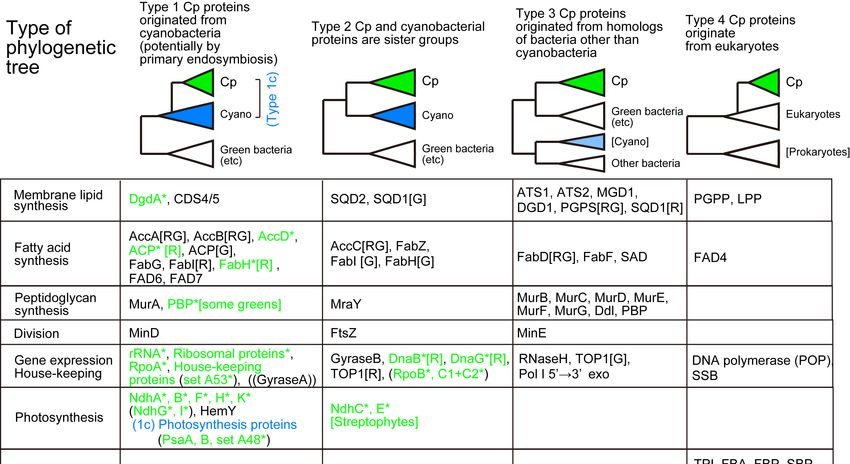
as Supplementary Materials of a previous paper [50]. Figure 1 presents four types of phy-
logenetic relationships between the cyanobacterial clade and the chloroplast clade. Type 1
tree indicates that the chloroplast gene originated from cyanobacteria, which is consistent
with the idea of chloroplast origin by cyanobacterial endosymbiosis. When homologs are
only found in cyanobacteria and chloroplasts, we cannot determine the position of the
root, and hence, the direction of evolution in a strict sense. In some special cases such
as PsaA and PsaB, we can root the phylogenetic tree, but in most cases, the root position
is undetermined. These cases are classified as Type 1c. In this case, Gloeobacter or basal
Synechococcus are often taken as the root. In the Type 2 tree, the chloroplast clade is sister
to the cyanobacterial clade. This means that the chloroplast originated from an ancestor
of all extant cyanobacteria, which seems unbelievable. However, we often obtain this
type of tree. This could be an artifact of phylogenetic reconstruction, but the tree form
is rather robust in most cases, even if taxon samplings and phylogenetic methods are
varied. We will have to find an explanation for this type of tree. Type 3 tree indicates that
the chloroplast enzymes originated from bacteria other than cyanobacteria. Type 4 tree
shows that eukaryotic enzymes are re-targeted to the chloroplast. Enzymes found only in
cyanobacteria are classified as Type 5.Genes 2021, 12, 823 9 of 30
Genes 2021, 12, x FOR PEER REVIEW 9 of 31
Figure 1. Four major types of phylogenetic relationships between cyanobacterial and chloroplast proteins. Chloroplast-
Figure 1. Four major types of phylogenetic relationships between cyanobacterial and chloroplast proteins. Chloroplast-
encoded proteins (or RNA) are marked with an asterisk and colored. [R] and [G] indicate red and green lineages, respec-
encoded proteins
tively. This (or RNA) are
is an extended, marked
revised with an
version asterisk
of the and in
original colored.
[9,50]. [R] and [G] indicate red and green lineages, respectively.
This is an extended, revised version of the original in [9,50].
3. Origin of Structural Elements of Chloroplasts
3.1. Peptidoglycan
3. Origin Synthesis
of Structural Enzymes
Elements of Chloroplasts
3.1. Peptidoglycan Synthesis Enzymes
Peptidoglycan is a meshwork structure that exists between the outer and inner mem-
branes of Gram-negative
Peptidoglycan bacteria including
is a meshwork structure thatcyanobacteria.
exists between In Gram-positive
the outer and inner bacteria,
mem- a
thick layer of peptidoglycan covers the whole cell. The presence
branes of Gram-negative bacteria including cyanobacteria. In Gram-positive bacteria, a of a wall-like layer in
chloroplasts
thick layer oforpeptidoglycan
similar photosynthetic
covers thestructures
whole cell. wasThe firstpresence
suggested of ainwall-like
P. chromatophora
layer in
and C. paradoxa
chloroplasts in the early
or similar 20th century.
photosynthetic The chromatophore
structures was first suggested of Paulinella is now consid-
in P. chromatophora and
ered a structure
C. paradoxa in thethat
earlyis phylogenetically
20th century. The distinct from the chloroplasts
chromatophore of Paulinellaof isplants and algae,
now considered
but in the 1920s,
a structure that isitphylogenetically
was identified asdistinct
a livingfromcyanobacterial cell within
the chloroplasts the eukaryotic
of plants and algae,cell.
but
in the
The 1920s, it surrounded
organelles was identified as awall-like
by the living cyanobacterial
layer in Paulinella cell within the eukaryotic
and Cyanophora were sub-cell.
The organelles
sequently calledsurrounded by the wall-like
cyanelles, meaning layer in Paulinella
cyanobacterial cells living andin Cyanophora
the cell as an were subse-
organelle
quently called cyanelles, meaning cyanobacterial cells living in the
[18] and were distinguished from the common chloroplasts. The identity of peptidoglycan cell as an organelle [18]
and
in were distinguished
Cyanophora cyanelles was fromestablished
the common chloroplasts.
by chemical The identity
analysis of peptidoglycan
[66]. Peptidoglycan has long in
Cyanophora
been cyanelles
considered was established
important evidence for by the
chemical analysis [66].
endosymbiotic origin Peptidoglycan has long
of not only cyanelles
beenalso
but considered important
chloroplasts (see a evidence for the
microbiology endosymbiotic
textbook [67]. seeorigin
also Item of not
4 ofonly cyanelles
Table 1). but
also chloroplasts
The presence(see a microbiologyintextbook
of peptidoglycan [67]. was
green plants see also Item 4by
suggested of the
Table 1).
genome analysis
of theThe
moss presence
P. patens of[68].
peptidoglycan
The complete in green plants was
set of enzymes suggested
involved bysynthesis
in the the genome anal-
of pepti-
ysis of the moss P. patens [68]. The complete set of enzymes involved
doglycan was identified in the nuclear genome of P. patens. The presence of peptidoglycan in the synthesis of
peptidoglycan was identified in the nuclear genome of P. patens.
was suggested by treatment with ampicillin, an inhibitor of peptidoglycan synthesis: The presence of pep-
tidoglycan
namely, was suggested
chloroplast division bywas
treatment
inhibited withby ampicillin,
ampicillin in antheinhibitor of peptidoglycan
moss. Although the role
synthesis: namely, chloroplast division was inhibited by ampicillin
of peptidoglycan in chloroplast division is still not understood, this was taken as evidence in the moss. Although
the role of peptidoglycan
suggesting that peptidoglycan in chloroplast
exists in thedivision is still not understood,
moss chloroplast. Genes for the this was taken
complete set
of peptidoglycan synthesis enzymes were also found in other organisms, such asfor
as evidence suggesting that peptidoglycan exists in the moss chloroplast. Genes the
green
complete
algae set of peptidoglycan
(Micromonas), charophytessynthesis enzymesliverworts
(Klebsormidium), were also(Marchantia),
found in other organisms,
pteridophytesGenes 2021, 12, 823 10 of 30
such as green algae (Micromonas), charophytes (Klebsormidium), liverworts (Marchantia),
pteridophytes (Selaginella), and even in some gymnosperms [69]. Arabidopsis thaliana also
possesses homologs of some of the peptidoglycan synthesis enzymes. Curiously, AtMurE
was shown to be involved in chloroplast development, rather than biosynthesis of a glycan-
like substance [70].
In contrast, all enzymes of peptidoglycan synthesis except MurF are encoded by the
chromatophore genome of P. chromatophora and Paulinella micropora. MurF was found to be
encoded by a nuclear gene, which originated from proteobacteria [9,50,71–74].
Peptidoglycan has not been observed by electron microscopy as a clearly defined
layer in plant chloroplasts. Nevertheless, a well-designed experiment using the ‘Click
chemistry’ was a breakthrough that identified peptidoglycan as a layer surrounding the
moss chloroplast [75]. Subsequently, peptidoglycan materials were located between the
outer and inner envelope membranes of moss chloroplasts by quantitative densitometry of
transmission electron micrographs [76].
Then, a question is asked: “Does the presence of peptidoglycan supports the en-
dosymbiotic origin of chloroplasts?” Recent phylogenetic analysis showed clearly that the
peptidoglycan synthesis enzymes in plants and algae are monophyletic, but the majority of
them originated from bacteria other than cyanobacteria [77,78]. Note that this conclusion
was obtained not only by our study [77] but also by an independent study [78]. Rare
examples of cyanobacteria-related enzymes include MurA and MraY (Figure 1). A class of
penicillin-binding proteins (PBP) encoded by some green algal chloroplast genomes also
diverged from cyanobacteria.
These results suggest that the peptidoglycan synthesis system was introduced into
green algae and glaucophytes from various bacteria. However, important questions remain.
First, when (or at which stage of evolution) was the peptidoglycan synthesis system
introduced? The timing could be either in the common origin of Archaeplastida, or in
the common ancestor of green algae and glaucophytes (if red algae diverged first), or
separately in green algae and glaucophytes. In the first case, red algae must have lost
the peptidoglycan synthesis system. Some Cyanophora enzymes are not closely related
to green algal homologs and could have different origins. We also have to consider that
many green algae such as Chlamydomonas do not have any peptidoglycan synthesis gene.
We have another question: How were the genes introduced into algal cells? If each gene
was introduced one by one from different bacteria, then the introduced genes could not
function in peptidoglycan synthesis until all necessary genes are provided. There is no
evolutionary advantage of keeping individual genes during the gene acquiring process.
A possible scenario might be like this: a complete system of peptidoglycan synthesis was
present at first in an organism, and each gene was replaced by horizontal gene transfer.
After gene exchanges, a new complete, functional set of genes was introduced into an
ancestor of algae (either of the three possibilities as above). Continued studies are necessary
on this topic.
3.2. Lipid Biosynthesis Enzymes
Chloroplast is the major site of fatty acid synthesis in plant cells. A large proportion of
fatty acids produced in the chloroplast are exported to the cytosol for the lipid synthesis in
the endoplasmic reticulum (ER), while the remaining fatty acids are used for the chloroplast
lipid synthesis. The proportion of the two metabolic flows depends on organisms and
tissues. All the enzymes of lipid biosynthesis in the ER of plants and algae are eukaryotic
enzymes that have closely related orthologs in other eukaryotes (animals and protists). To
discuss the cyanobacterial origin of chloroplasts, we have to examine fatty acid synthesis
and lipid synthesis within the chloroplasts.
3.2.1. Fatty Acid Synthesis
Two types of fatty acid synthases (FAS) are known: Type II FAS is found in bacte-
ria including cyanobacteria and consists of four separate subunits catalyzing the mainGenes 2021, 12, 823 11 of 30
cycle of chain elongation. Type I FAS is a large molecule containing the four enzymatic
activities (plus acyl carrier protein (ACP) and other related activities) within one or two
multifunctional polypeptide(s), which is found in the cytosol of fungi and animals. Fungal
and animal FAS are also different in the arrangement of enzymatic active centers within
the polypeptides. Because plant and algal chloroplasts contain Type II FAS, the fatty acid
synthesis activity in the chloroplasts has been compared with the cyanobacterial activity.
Type II FAS is also present in the mitochondria of eukaryotes including animals, plants,
and other organisms, but only limited production of fatty acids is ascribed to mitochondria.
Note that all enzymes involved in fatty acid synthesis (except some algal enzymes encoded
by the chloroplast genome, such as red algal ACP and FabH) are encoded by the nuclear
genome. Under these circumstances, fatty acid synthesis in the chloroplast was simply
believed to originate from cyanobacteria. Recent phylogenetic analysis showed indeed
cyanobacterial origin of many components of fatty acid synthesis, but not all (Figure 1). The
condensing enzyme called FabF that catalyzes the chain elongation reaction in the chloro-
plast originates from green sulfur bacteria, whereas the mitochondrial homolog originates
from proteobacteria [9,50]. Chloroplast FabD, which transfers malonyl CoA to ACP, is also
closely related to green sulfur bacteria and green non-sulfur bacteria. The green bacteria
(both sulfur and non-sulfur) are photosynthetic bacteria that do not produce oxygen but
utilize light energy for carbon fixation. Curiously, in many phylogenetic analyses, green
bacteria are often found to be the most related group of chloroplasts.
In the chloroplasts, the initial products of FAS are palmitic and stearic acids. In
the chloroplasts of green plants and algae, stearic acid is desaturated (introduction of
a double bond is called desaturation) to oleic acid by stearoyl ACP desaturase (SAD).
Therefore, the primary products of fatty acid synthesis in these chloroplasts are palmitic
and oleic acids. SAD is not present in red algae and glaucophytes, nor in cyanobacteria.
Nevertheless, diatoms have homologs of SAD. Desaturation in cyanobacteria, including
oleic acid synthesis, uses acyl lipids as the substrate rather than acyl ACP or acyl CoA.
3.2.2. Chloroplast Lipid Biosynthesis
Both chloroplasts and cyanobacteria contain two galactolipids, monogalactosyl di-
acylglycerol (MGDG) and digalactosyl diacylglycerol (DGDG), as well as a sulfolipid,
sulfoquinovosyl diacylglycerol (SQDG), and a phospholipid, phosphatidylglycerol (PG).
A trace amount of phosphatidylcholine is present in the chloroplast. Tri- and tetragalactosyl
diacylglycerols are also found in land plants depending on growth conditions. The set
of MGDG, DGDG, and SQDG, or ‘glycolipid trio’, is indeed a common trait of oxygen-
evolving photosynthetic organisms. A rare exception is Gloeobacter which lacks SQDG. The
lack of SQDG and thylakoid membrane in this group of cyanobacteria could be related
to each other, but little is known about this question. Photosynthetic bacteria without
oxygen evolution also contain some of these lipids, but not the complete trio. Therefore,
the glycolipid trio seemed to be good evidence for the cyanobacterial origin of chloro-
plasts. This prompted lipid biologists to call the lipid synthesis pathway in the chloroplast
“prokaryotic pathway” [79]. This name had a connotation that the lipid biosynthetic path-
way of chloroplast originated from cyanobacteria. However, this turned out to be false. The
entire pathways in cyanobacteria and chloroplasts are unrelated in terms of biochemistry
and phylogeny (Figure 2).Genes 2021, 12, 823 12 of 30
Genes 2021, 12, x FOR PEER REVIEW 12 of 31
Figure2.2. Glycolipid
Figure Glycolipid biosynthesis
biosynthesis pathways
pathwaysinincyanobacteria
cyanobacteriaand
and chloroplasts. TheThe
chloroplasts. distinction be-
distinction
tween galactose and glucose is shown by the 4’-OH group in red. Cyanobacterial genes are shown
between galactose and glucose is shown by the 4’-OH group in red. Cyanobacterial genes are shown in
in cyan, whereas eukaryotic or non-cyanobacterial genes are shown in green. Note that the bio-
cyan, whereas eukaryotic or non-cyanobacterial genes are shown in green. Note that the biochemical
chemical reactions are different in the two steps marked [1] in magenta. In other steps, biochemi-
reactions are different
cally identical in the
reactions aretwo steps marked
catalyzed [1] in magenta.
by structurally In other
unrelated steps, biochemically
(isofunctional identical
non-homologous)
reactions
enzymesare catalyzed
marked by structurally
[2]. Eukaryotic unrelated
paralogs (isofunctional non-homologous)
and phylogenetically distant orthologsenzymes marked
are marked [3]
[2].
and Eukaryotic paralogs and phylogenetically distant orthologs are marked [3] and [4], respectively.
[4], respectively.
Essential
Essentialdifferences
differencesininthe
thegalactolipid
galactolipidbiosynthetic
biosyntheticpathways
pathwaysinincyanobacteria
cyanobacteriaandand
chloroplasts
chloroplastswere
werefound byby
found labeling experiments
labeling experimentsalready in in
already thethe
1980s ([80,81]
1980s andand
([80,81] thethe
refer-
ref-
erences therein). Namely, a glucolipid, monoglucosyl diacylglycerol (GlcDG), acts as anGenes 2021, 12, 823 13 of 30
ences therein). Namely, a glucolipid, monoglucosyl diacylglycerol (GlcDG), acts as an inter-
mediate in producing MGDG in cyanobacteria. All genes encoding the galactolipid synthe-
sis enzymes were identified in chloroplasts by the end of the 20th century [82,83], whereas
the cyanobacterial genes for the synthesis of galactolipids were identified later [84–87].
The conversion of GlcDG to MGDG is an isomerization of sugar moiety called epimeriza-
tion [88,89]. The enzymes involved in the synthesis of UDP-glucose and UDP-galactose,
UDP-glucose pyrophosphorylase and UDP-glucose 4-epimerase, respectively, are both
different in cyanobacteria and chloroplasts: UDP-glucose pyrophosphorylase is encoded
by the galU or cugP genes in cyanobacteria [90], whereas it is encoded by the UGP1/2
gene (cytoplasmic enzyme) and UGP3 gene (chloroplast enzyme) in plants and algae (see
Table 4 in [91]). These are nonhomologous, isofunctional genes. UDP-glucose 4-epimerase
is encoded by the galE gene in cyanobacteria, while it is encoded by the UGE1~5 genes
(cytoplasmic enzyme) and PHD1 gene (chloroplast enzyme) in plants and algae. These
three kinds of genes are also nonhomologous, isofunctional genes.
Acyltransferases that produce lysophosphatidic acid and phosphatidic acid were also
identified in the chloroplast in the early days, but cyanobacterial acyltransferases were iden-
tified later. Escherichia coli acyltransferases encoded by the plsB and plsC genes were found
in the 1970s, but the acyltransferase for the first acylation was identified quite recently in
other bacteria including cyanobacteria. The bacterial system uses acyl phosphate as the acyl
donor of the first acylation step and two genes named plsX and plsY were identified [92].
Identification and phylogenetic analysis [93] and functional characterization [94] of the
cyanobacterial genes were performed recently. The cyanobacterial pathway of galactolipid
synthesis is, therefore, biochemically different from the corresponding pathway in chloro-
plasts in at least two points (see Figure 2). The second galactosylation step is catalyzed
by two different types of galactosyltransferases in cyanobacteria (DgdA) and chloroplasts
(DGD1). These two enzymes belong to the CAZy GT4 family, but their domain structures
are different [95]. Note that the chloroplast genome of some red algae in Cyanidiales
encodes a dgdA gene [96].
In addition, phylogenetic analysis of all the enzymes involved in the synthesis of galac-
tolipids revealed that none of the chloroplast enzymes originated from cyanobacteria [9,50].
In terms of biochemical reaction, the second acylation and the dephosphorylation are
common in chloroplasts and cyanobacteria. Nevertheless, phylogenetic analysis showed
that the chloroplast enzymes and cyanobacterial enzymes belong to distant clades. Figure 2
shows that all enzymes in the galactolipid synthesis pathway are biochemically or phyloge-
netically different in cyanobacteria and chloroplasts (see Figure 1 for the summary of phy-
logenetic analysis). They are classified into three biochemical categories and a phylogenetic
category (this is shown in magenta brackets in Figure 2). (1) Different reactions in cyanobac-
teria and chloroplasts: PlsX + PlsY vs. ATS1, MgdA + MgdE vs. MGD1/2/3. (2) Structurally
different (non-homologous, isofunctional) enzymes: CugP/GalU vs. UGP1/2/3, GalE
vs. UGE1-5/PHD1, DgdA vs. DGD1/2. (3) Chloroplast enzymes are paralogs of ER
enzymes: Pap vs. LPPγ/ε. Therefore, the galactolipid biosynthesis system is unrelated to
cyanobacteria and chloroplasts, and this is mostly supported by evidence that does not rely
on phylogenetic analysis. (4) Only the contrast PlsC vs. ATS2 was based on phylogenetic
analysis that robustly supported their distant relationship.
Biochemically, the biosynthetic pathways of SQDG and PG are both similar in cyanobac-
teria and chloroplasts, but the phylogenetic relationship of the enzymes is rather com-
plex [50]. In SQDG synthesis, the UDP-sulfoquinovose is synthesized from UDP-glucose
by SQD1/SqdB (chloroplasts/cyanobacteria, respectively [97]), and then sulfoquinovose is
transferred to diacylglycerol (DAG) by SQD2/SqdX (chloroplasts/cyanobacteria, respec-
tively [98]). SqdC replaces SqdX in some cyanobacteria. All enzymes of SQD1/SqdB are
homologous, but divided into two major clades, one containing the cyanobacterial clades
A-B1-B2 (see Figure 3A for the distinction between the major groups of cyanobacteria),
green non-sulfur bacteria, and green algae/plants as well as glaucophytes, and anotherGenes 2021, 12, 823 14 of 30
containing
Genes 2021, 12, x FOR PEER REVIEW the cyanobacterial clades C1-C2, α-proteobacteria, and red algae (Figure 1).31
14 of
Chloroplast SQD2 is a single clade, sister to the cyanobacterial SqdX.
Figure 3. Schematic phylogenetic trees showing different origins of chloroplast enzymes (or RNA)
Figure 3. Schematic phylogenetic trees showing different origins of chloroplast enzymes (or RNA)
within the cyanobacterial diversity. (A) Canonical phylogenetic tree of cyanobacteria and chloro-
within
plasts.the cyanobacterial
Various clades ofdiversity. (A) Canonical
cyanobacteria are shown phylogenetic
according tree of cyanobacteria
to [53,54]. and chloroplasts.
Chloroplast-encoded pro-
Various
teins and RNA are shown in green. Nuclear-encoded chloroplast proteins are shownproteins
clades of cyanobacteria are shown according to [53,54]. Chloroplast-encoded in black.and
The
RNA
cladeare shown
names arein green.
taken fromNuclear-encoded
[53]. According to chloroplast proteinsisare
[54], G. lithophora shown
sister to theinmain
black. The clade
chloroplast
names
clade are taken from
represented [53]. According
by chloroplast rRNA. to For G. lithophora
[54],simplicity, is sister
Clade to the
H is used formain chloroplast
G. lithophora. Theclade
posi-
tions of branching
represented of RbcLrRNA.
by chloroplast and PsaA/B are shown
For simplicity, withHdotted
Clade is usedlines because
for G. they The
lithophora. do not match of
positions
exactly with
branching the canonical
of RbcL and PsaA/B tree.are
(B)shown
Different
withorigins
dottedof chloroplast
lines and cyanobacterial
because they RbcL. (C)
do not match exactly with
Probable
the canonicalorigin
tree.of(B)
PsaA and PsaB.
Different As of
origins explained in the
chloroplast andtext and Supplementary
cyanobacterial RbcL. (C) Materials,
Probablethe phy-
origin
logeny of PsaA and PsaB is very difficult. This diagram, which is consistent with previous results,
of PsaA and PsaB. As explained in the text and Supplementary Materials, the phylogeny of PsaA
is still a plausible hypothesis among many other alternatives. Different line colors are used to
and PsaB is very difficult. This diagram, which is consistent with previous results, is still a plausible
show different lineages such as cyan for cyanobacteria and green for chloroplast. Colored protein
hypothesis
names areamongused tomany
showother alternatives. Different
chloroplast-encoded line colors are used to show different lineages
proteins.
such as cyan for cyanobacteria and green for chloroplast. Colored protein names are used to show
chloroplast-encoded
In PG synthesis, proteins.
there are two different enzymes of CDP diacylglycerol synthase
(CDS): the eukaryotic CDS is found in ER in plants (CDS1 and 2 in Arabidopsis), algae,
In PG synthesis, there are two different enzymes of CDP diacylglycerol synthase (CDS):
animals, and fungi, whereas the prokaryotic CDS is found in the chloroplast (CDS4 and 5) and
the eukaryotic CDS is found in ER in plants (CDS1 and 2 in Arabidopsis), algae, animals,
bacteria. Chloroplast CDS is likely to originate from cyanobacteria. This is indeed the rare
and fungi, whereas the prokaryotic CDS is found in the chloroplast (CDS4 and 5) and
case (with Cyanidiales DgdA), in which a direct phylogenetic relationship is demon-
bacteria. Chloroplast CDS is likely to originate from cyanobacteria. This is indeed the rare
strated in cyanobacterial and chloroplast orthologs (Type 1 in Figure 1).
In summary, chloroplasts and cyanobacteria are similar in the compositions of lipids
that make up thylakoid membranes, but they have essentially unrelated pathways of lipid
synthesis. This could be convergent evolution, rather than vertical inheritance, that sus-
tains the functioning of membrane-embedded components of photosynthetic machineryGenes 2021, 12, 823 15 of 30
case (with Cyanidiales DgdA), in which a direct phylogenetic relationship is demonstrated
in cyanobacterial and chloroplast orthologs (Type 1 in Figure 1).
In summary, chloroplasts and cyanobacteria are similar in the compositions of lipids
that make up thylakoid membranes, but they have essentially unrelated pathways of
lipid synthesis. This could be convergent evolution, rather than vertical inheritance, that
sustains the functioning of membrane-embedded components of photosynthetic machinery
(photosystems, electron transfer chain, and ATP synthase). Many of the enzymes acting
within the chloroplasts are eukaryotic enzymes or enzymes that are distantly related to
cyanobacterial counterparts. However, many components of the chloroplast fatty acid
synthesis system originate from cyanobacteria. This suggests a close relationship between
photosynthesis and fatty acid synthesis, which was discussed in the context of the “fatty
acid hypothesis” that explains the origin of eukaryotes [9].
4. Diverse Origins of Chloroplast-Encoded Genes
The chloroplast genome has been believed as a reduced cyanobacterial genome that
was introduced into the hypothetical algal ancestor. Phylogenetic analysis of the rRNAs
was the representative support for the cyanobacterial origin of the chloroplast. How-
ever, as pointed out in Section 2.2, this was strongly affected by the genocentric view of
the time. Phylogenetic analysis of chloroplast genome has been traditionally performed
with concatenated amino acid sequences of conserved genes [53,54] because chloroplast-
encoded proteins (e.g., ribosomal proteins) are generally small. In practice, we obtain
divergent phylogenetic trees for individual chloroplast ribosomal proteins, but a fairly
reliable phylogenetic tree of concatenated ribosomal sequences that resembles the tree
of rRNA [9,50]. In the phylogenetic trees of the chloroplast rRNA as well as the concate-
nated chloroplast-encoded proteins, the chloroplast clade branches from the basal clade
of cyanobacteria after the clade E, but before the split of the clade A-B1-B2 and the clade
C1-C2-C3 (Figure 3A) [9,34,35,53,54]. Recent work suggested that Gloeomargarita lithophora
(assigned “clade H” in the present article) is the closest cyanobacterium to the chloro-
plasts [54]. This is the canonical phylogenetic tree of chloroplasts and cyanobacteria that
supports the current notion of the cyanobacterial origin of chloroplasts (Figure 3A). It
should be noted, however, that the unity of the chloroplast genome should not be the first
assumption to make.
Each of the large proteins, such as the large subunit of rubisco, the RNA polymerase
subunits, and the two large subunits of Photosystem I reaction center P700, has sufficient
phylogenetic signals for constructing a reliable, individual phylogenetic tree. Examination
of these individual trees suggested that the relationship between the cyanobacteria and
the chloroplasts might not be straightforward as indicated by the canonical tree. The
carbon-fixation enzyme, rubisco, was the first to present evidence for the multiple origins
of the chloroplast genome. Let us examine the RbcL phylogeny.
The classification of the large subunit of rubisco, RbcL, is quite complicated. Tabita [99]
presented a classification of bacterial and chloroplast RbcL based on biochemical and
phylogenetic data. He showed that the red algal rubisco belongs to Form ID, whereas
the rubisco of the green plants and algae, as well as many cyanobacteria, belong to Form
IB. Form ID was closely related to Form IC in various proteobacteria (see Figure S1A).
Delwiche and Palmer [100] proposed that the chloroplast rbcL (and rbcS, too) gene, which
had been originally introduced by a cyanobacterial endosymbiont, was replaced by an
α-proteobacterial homolog in the red lineage. This was a novel idea that the chloroplast
genome can be a target of horizontal gene transfer. Non-cyanobacterial origin of chloroplast-
encoded genes was also found in the phylloquinone/menaquinone biosynthesis genes
(menA, B, C, D, E, F, G, and H) [101], which was interpreted as multiple horizontal gene
transfer events in the ancestral endosymbiont and cyanobacteria.
Further phylogenetic analysis of the RbcL proteins revealed that bacterial RbcL is
highly diversified and that the RbcL of some cyanobacteria, later classified as clade C1 [53],
is sister to a clade including various proteobacteria containing α-carboxysomes (Figure S1.You can also read