Function of Drosophila Synaptotagmins in membrane trafficking at synapses
←
→
Page content transcription
If your browser does not render page correctly, please read the page content below
Cellular and Molecular Life Sciences (2021) 78:4335–4364
https://doi.org/10.1007/s00018-021-03788-9 Cellular and Molecular Life Sciences
REVIEW
Function of Drosophila Synaptotagmins in membrane trafficking
at synapses
Mónica C. Quiñones‑Frías1,2,3 · J. Troy Littleton1,2
Received: 10 December 2020 / Revised: 29 January 2021 / Accepted: 9 February 2021 / Published online: 22 February 2021
© The Author(s) 2021
Abstract
The Synaptotagmin (SYT) family of proteins play key roles in regulating membrane trafficking at neuronal synapses. Using
both Ca2+-dependent and Ca2+-independent interactions, several SYT isoforms participate in synchronous and asynchronous
fusion of synaptic vesicles (SVs) while preventing spontaneous release that occurs in the absence of stimulation. Changes
in the function or abundance of the SYT1 and SYT7 isoforms alter the number and route by which SVs fuse at nerve termi-
nals. Several SYT family members also regulate trafficking of other subcellular organelles at synapses, including dense core
vesicles (DCV), exosomes, and postsynaptic vesicles. Although SYTs are linked to trafficking of multiple classes of synaptic
membrane compartments, how and when they interact with lipids, the SNARE machinery and other release effectors are still
being elucidated. Given mutations in the SYT family cause disorders in both the central and peripheral nervous system in
humans, ongoing efforts are defining how these proteins regulate vesicle trafficking within distinct neuronal compartments.
Here, we review the Drosophila SYT family and examine their role in synaptic communication. Studies in this invertebrate
model have revealed key similarities and several differences with the predicted activity of their mammalian counterparts.
In addition, we highlight the remaining areas of uncertainty in the field and describe outstanding questions on how the SYT
family regulates membrane trafficking at nerve terminals.
Keywords Drosophila · Synapse · Neurotransmitter release · Synaptic vesicle · Synaptotagmin · Exocytosis
Synaptic communication and the synaptic vesicles (SVs) to position the release machinery near clus-
vesicle release machinery tered neurotransmitter receptors in the postsynaptic mem-
brane [3–14]. During action potential propagation along
The nervous system relies on regulated secretion of neuro- the axon, membrane depolarization triggers the opening of
transmitters to meditate synaptic communication between voltage-gated Ca2+ channels and influx of extracellular C a2+
neurons [1, 2]. Synaptic transmission typically occurs at to trigger SV fusion at release sites [2, 15–20]. SV exocy-
specialized release sites in presynaptic terminals known tosis is a highly stochastic process at individual AZs, with
as active zones (AZs). Highly conserved AZ scaffold pro- the probability of an individual fusion event varying over a
a2+ channels and synaptic
teins concentrate voltage-gated C wide range depending on the neuronal population [21–23].
The evoked response recorded postsynaptically represents
the probability of SV fusion events that occur over a popula-
* J. Troy Littleton tion of individual release sites.
troy@mit.edu The timing for single SV fusion events can occur over
1
Department of Biology, The Picower Institute for Learning a relatively broad temporal window of ~ 1–200 ms at indi-
and Memory, Massachusetts Institute of Technology (MIT), vidual AZs following an action potential (Fig. 1a). Release
Bldg. 46‑3243, 43 Vassar St., Cambridge, MA 02139, USA kinetics have been loosely classified into two distinct phases
2
Department of Brain and Cognitive Sciences, The Picower termed synchronous and asynchronous release [24]. The
Institute for Learning and Memory, Massachusetts Institute synchronous phase accounts for the majority of neuro-
of Technology (MIT), Bldg. 46‑3243, 43 Vassar St., transmitter release at most synapses, with SV fusion events
Cambridge, MA 02139, USA
decaying within several milliseconds after presynaptic Ca2+
3
Department of Biology, Brandeis University, Waltham, MA, influx. The asynchronous phase (also known as the delayed
USA
13
Vol.:(0123456789)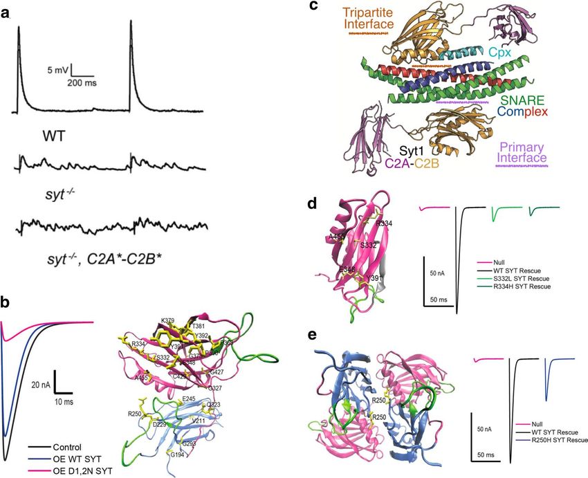
4336 M. C. Quiñones‑Frías, J. T. Littleton
Fig. 1 Regulation of synchronous and asynchronous SV fusion. a SYT1 acts as the Ca2+ sensor for synchronous release and resides on
Model depicting phases of synchronous and asynchronous release SVs. A slower asynchronous component can last for hundreds of mil-
after nerve stimulation (arrow). b Structure of the fusion machinery liseconds and include fusion of SVs farther from release sites. SYT7
that bridges the SV and plasma membrane, with SYT1 (grey) and the has emerged as a candidate for the asynchronous C a2+ sensor with
SNARE complex (Syntaxin—red; SNAP25—green; Synaptobrevin— a2+ binding. The role of SYT7 is still
higher affinity than SYT1 for C
blue). c Model of synchronous and asynchronous release in relation controversial, with studies in Drosophila suggesting it controls SV
to presynaptic Ca2+ entry (shaded). Following C a2+ influx, SVs fuse availability and fusogenicity
during a synchronous phase at AZs that occurs within milliseconds.
response) is less pronounced at most synapses but becomes at the SV-plasma membrane interface prior to fusion in a
more robust during high frequency nerve stimulation. In this cis-conformation where the transmembrane domains of Syn-
slower component of release, individual SV fusion events aptobrevin and Syntaxin remain separated in the SV and
occur within several hundred milliseconds across the AZ plasma membrane, respectively. The assembly and arrest
population [19, 25, 26]. In addition to the two phases of of SNARE zippering at this intermediate state is regulated
evoked release, single SVs can fuse spontaneously (termed by the cytosolic SNARE-binding protein Complexin (CPX),
minis) in the absence of nerve stimulation [23, 26–28]. which is hypothesized to provide a clamping brake on fusion
All three pathways for SV release (synchronous, asyn- until Ca2+ entry occurs [36–43]. The SYT1 family of SV
chronous, spontaneous) require a highly conserved fusion Ca2+ sensors binds Ca2+ ions through the action of a clus-
machinery that controls the regulated assembly of a four ter of negatively charged aspartate residues present in loop
stranded coiled-coil SNARE complex that bridges the SV structures that emerge from its two C2 domains (Fig. 1b).
and presynaptic membranes [29–31]. This complex includes The Ca2+-bound loops of SYT1 interact with negatively
the v-SNARE Synaptobrevin on the SV, and the t-SNAREs charged lipid headgroups in the presynaptic membrane. This
Syntaxin and SNAP-25 on the presynaptic plasma mem- interaction alters local lipid structure and helps trigger full
brane (Fig. 1b). Multiple SNARE chaperones, including the zippering of the SNARE complex into a trans-SNARE state
UNC13 and UNC18 families, position and regulate the tim- where the transmembrane domains of the SNARE proteins
ing of the highly energetic assembly of the SNARE alpha- reside together in the fused membrane [2, 40, 44–46]. In
helices during the SV cycle [2, 32–35]. Current models addition to activating release, SYT1 also inhibits spontane-
suggest the coiled-coil SNARE bundle partially assembles ous fusion through a clamping mechanism similar to the role
13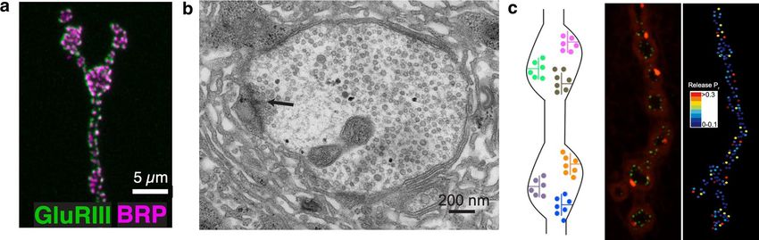
Function of Drosophila Synaptotagmins in membrane trafficking at synapses 4337
of CPX [47–55]. Together, these proteins act to drive full isoforms (SYT1, SYT4, and SYT7) that are found at most
collapse of the SV into the plasma membrane during presyn- synapses. Similar to Drosophila, the homologs of these three
aptic Ca2+ influx to exocytose neurotransmitters through the SYT isoforms are among the most abundant members in
fast synchronous pathway [24, 51, 56–64]. mammals [84]. SYT1 and SYT7 have been shown to regu-
The asynchronous release pathway uses a similar fusion late SV trafficking in both systems, while SYT4 has been
machinery with the major exception of not requiring SYT1 linked to presynaptic exosomes and postsynaptic retrograde
(Fig. 1c). Several mechanisms have been proposed that dif- signaling in Drosophila. Electrophysiology, imaging and
ferentiate asynchronous and synchronous release [16, 24, structure–function studies have provided insights into how
65–74], including distinct Ca2+ sensors, heterogeneity in SV SYT1, SYT4, and SYT7 regulate synaptic communication
protein content, SV distance from C a2+ channels, distinct in vivo. We review this data, as well as examine what is
Ca entry pathways, and regulation of Ca2+ extrusion and
2+
known about the remaining family members. Finally, we
buffering. Asynchronous release is enhanced in Syt1 mutants highlight recent studies identifying mutations in SYT fam-
[51, 57, 75], suggesting C a2+ can activate this slower path- ily proteins in human neurological disorders and describe
way through a distinct Ca2+ sensor(s). Manipulations of work on potential pathological mechanisms using Dros-
SYT7 change the amount of release occurring through the ophila models.
slower asynchronous SV fusion pathway, but whether the
protein functions as a Ca2+ sensor for the fusion process
itself or modifies Ca2+-dependent SV availability is unclear Analyzing synaptic transmission
[58, 76–84]. Following fusion of SVs through either path- at Drosophila neuromuscular junctions
way, α-SNAP binding to trans-SNARE complexes in the
plasma membrane recruits the AAA ATPase NSF to disas- Electrophysiological and imaging analysis at Drosophila
semble the complex and recharge individual SNAREs for 3rd instar larval neuromuscular junction (NMJ) synapses
additional rounds of release [29, 85–90]. NSF can also dis- has proven highly effective for dissecting the roles of SYTs
assemble trans-SNARE complexes present on the SV that in synapse biology. At this connection, motoneurons form
escape endocytosis control, releasing free v-SNAREs to glutamatergic synapses onto muscles, with motor axons con-
form productive cis-SNARE complexes required for fusion taining tens of en passant presynaptic boutons with hundreds
[91]. Following endocytosis, SNARE chaperones and the of individual AZs highlighted by a T-bar filamentous struc-
priming machinery reposition fusogenic SVs at AZs for ture at the center (Fig. 2a, b) [14]. SV release from motoneu-
additional cycles of release. rons can be measured using electrophysiological recordings
In the current review, we discuss models for how the of synaptic currents from the muscle or by optical imaging
SYT family regulates membrane trafficking at synapses. of postsynaptic fusion events. Individual AZs at the NMJ are
In particular, we focus on studies performed in Drosophila aligned to discrete postsynaptic glutamate receptor (GluR)
that examine the function of the three most abundant SYT fields. This arrangement facilitates use of modified GCaMPs
Fig. 2 The Drosophila larval NMJ as a model for synaptic function. GFP, green) and evoked SV release events (red) visualized with a
a Immunolabeling of the nerve terminal showing AZs (anti-BRP, modified jRGECO C a2+ indicator expressed postsynaptically. The
magenta) and post-synaptic densities (anti-Glutamate Receptor III right panel shows quantal imaging of evoked release probability at
staining, green) at a larval NMJ. b EM of a single synaptic bouton the same NMJ, revealing heterogeneity in AZ strength as noted on the
with an AZ T-bar denoted (arrow). c The left panel shows a model color-coded heat map. a Modified from [105], b modified from [82],
of synaptic boutons with multiple individual AZs (left). The middle and c modified from [22]
panel shows an NMJ expressing the presynaptic Ca2+ channel (Cac-
13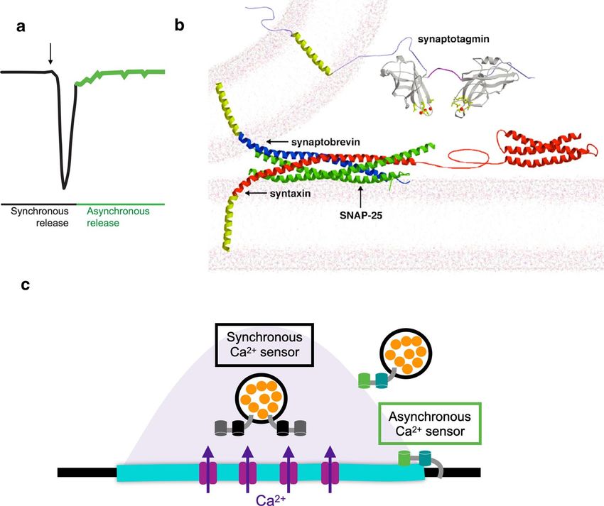
4338 M. C. Quiñones‑Frías, J. T. Littleton
expressed postsynaptically to image SV fusion events by C a 2+ binding aspartate residues (SYT12, SYT14) [99,
visualizing spatially localized Ca2+ influx following GluR 100]. Three Drosophila SYT isoforms are highly expressed
opening (Fig. 2c) [22, 23, 92–94]. This toolkit makes the in neurons (SYT1, SYT4, SYT7) based on in situ mRNA
Drosophila NMJ one of the only models where every SV expression and RNAseq analysis (Fig. 3b) [100]. SYT1 is
fusion event can be imaged and assigned to single AZs, homologous to mammalian SYT1, SYT2, and SYT9 pro-
greatly increasing the spatial resolution of SV release com- teins, all of which reside on SVs (Fig. 3c) and function as
pared to electrophysiology alone. These imaging approaches Ca2+ sensors driving fast synchronous SV fusion in specific
demonstrate single quanta are released at AZs following an neuronal populations [57, 60, 61, 63, 101]. Drosophila Syt1
action potential, as multi-vesicular release at individual sites null mutants generally die as embryos or during early larval
is rare [22, 23, 92–95]. Similar to other systems, AZs formed development [63, 102], though some can survive to adult-
by a single Drosophila motoneuron display heterogeneity hood at low frequency when cultured directly on food where
in SV release probability (Pr), with a small population of minimal movement is required [103]. These surviving adults
strong AZs present amongst many weaker ones (Fig. 2c). are completely ataxic and die within several days. SYT7 is
Highlighting the stochastic nature of the release process, encoded by single gene in both Drosophila and mammals
the average AZ Pr is ~ 0.07 at the NMJ, indicating most AZs and has been linked to asynchronous SV release [80, 82,
release a SV less than 10% of the time in response to single 84]. The Drosophila homolog resides on a tubular membrane
action potentials [22]. This quantal imaging approach has compartment present within the peri-AZ, a synaptic domain
been applied to Syt1 and Syt7 mutants to examine SV release implicated in endocytosis and protein sorting (Fig. 3d).
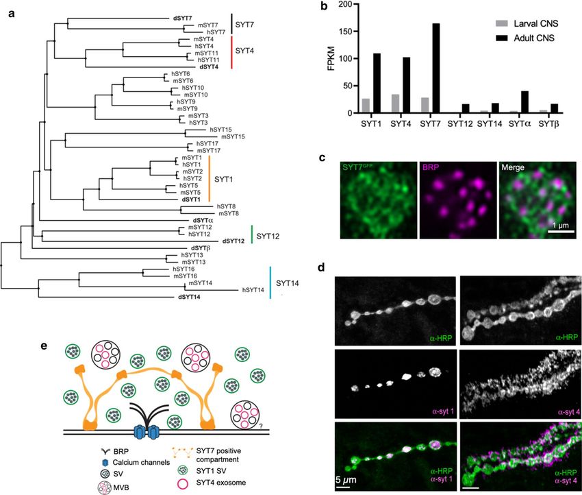
at individual AZs. Drosophila SYT4 is homologous to mammalian SYT4 and
SYT11 and is transferred to the postsynaptic compartment
from exosomes (Fig. 3b), where it functions in retrograde
The Synaptotagmin superfamily signaling [97, 100, 104–107]. Mutations in Drosophila Syt4
and Syt7 are viable and fertile as adults [82, 97, 104, 105],
SYTs are conserved family of membrane-trafficking pro- though no studies to date have examined potential roles in
teins containing a single transmembrane domain and two synaptic plasticity and learning in adult animals. SYT12 and
paired cytosolic C2 domains (Figs. 1b, 3). Their expres- SYT14 are more divergent in sequence, but appear ortholo-
sion is largely restricted to the nervous system, though a gous to mammalian SYT12 and SYT14 [99]. These isoforms
few isoforms participate in a smaller subset of trafficking are expressed at low levels in Drosophila and little is known
pathways in non-neuronal cells. Genes encoding SYT pro- about their function. SYTα and SYTβ are present on dense
teins are not found in bacterial or yeast genomes, indicat- core vesicles (DCVs) in neuroendocrine neurons and are
ing intracellular membrane fusion events like ER to Golgi presumed to function as DCV C a2+ sensors in Drosophila
trafficking do not require the function of this protein fam- [100, 108–110]. Their precise relationship to specific mam-
ily. The first members of the SYT family emerged during malian SYT isoforms is unclear. No mutants in these final
evolution in the placozoans, a primitive branch of multi- four SYT isoforms have been described. Beyond these seven
cellular metazoans that lack neurons. Homologs of SYT1 SYT family members in Drosophila, there are multiple genes
and SYT7 are encoded in the Trichoplax adhaerens genome, encoding proteins that contain C2 domains that are orthologs
suggesting a non-neuronal origin of SYTs during a period of other synaptic and non-synaptic proteins that will not be
when multicellular communication was emerging [96, 97]. discussed. These include the ER residents extended SYT
The SYT family expanded during invertebrate evolution, (ESYT) and multiple C2 domain protein (MCTP) family
and the Drosophila genome encodes seven distinct SYT members [111–114], homologs of the synaptic proteins
proteins. With genome duplications in vertebrates, greater Rabphilin, Rim, and Munc13 [99, 115, 116], as well as
diversification occurred and 17 SYT family members are homologs of mammalian Otoferlin (Misfire) [117] and
encoded in the human genome [96]. Many SYT proteins Granulophilin (Bitesize) [118]. In contrast to mammals, the
can be grouped into functional orthologs based on sequence Drosophila genome does not encode a homolog of the DOC2
similarity across evolution, while other isoforms are more family, a group of SYT-like proteins with two cytosolic C2
divergent without clear orthologs between invertebrate and domains lacking a transmembrane domain. The DOC2 fam-
vertebrate proteins (Fig. 3a). ily has been implicated in SV trafficking [119–122], but they
SYTs can be subdivided into whether or not they are appear to represent a novel adaptation to the process found
likely to bind C a2+ based on conservation of negatively only in vertebrates.
charged aspartate residues within their C2 domains that The domain structure of the SYT family is highly con-
mediate this interaction [98]. For the seven Drosophila served across all isoforms, with a single-pass transmem-
SYT homologs, five are predicted to bind Ca2+ (SYT1, brane domain, a variable linker and two tandem cytosolic
SYT4, SYT7, SYTα, SYTβ), while two lack conserved C2 domains, termed C2A and C2B [98–100, 123, 124]. C2
13
Function of Drosophila Synaptotagmins in membrane trafficking at synapses 4339
Fig. 3 Conservation, abundance and localization of Drosophila istry. The motor axon is stained with anti-HRP (green). e Model of
SYTs. a Phylogenetic tree of SYT homologs in Drosophila mela- the subcellular localization of Drosophila SYTs. SYT1 is attached
nogaster (d), Mus musculus (m), and Homo sapiens (h). The SYT1, to SVs and triggers synchronous SV fusion. SYT7 localizes to an
SYT4, SYT7, SYT12, and SYT14 subfamilies are highlighted. The internal peri-AZ compartment and negatively regulates SV re-entry
sequences of each SYT were extracted from NCBI and the tree was into the readily releasable pool and SV fusogenicity. SYT4 localizes
generated using neighbor clustering algorithm. b Expression level to presynaptic exosomes that are released from multi-vesicular bod-
of Drosophila Syt genes in larval and adult brain using RNAseq. c ies (MVBs) to transfer the protein to the postsynaptic compartment
Expression of endogenously CRISPR-tagged SYT7-GFP compared to where it mediates retrograde signaling. The graph in panel b was
the AZ protein BRP in a single larval NMJ bouton. SYT7 surrounds generated by plotting gene expression levels in the CNS of 7-day-old
AZs and localizes to an interconnected tubular membrane compart- males reported in [368], panel c was modified from [100], and panel
ment within the peri-AZ. d Localization of SYT1 to SVs and SYT4 d was modified from [82]
to postsynaptic puncta at the larval NMJ using immunocytochem-
domains are found in a wide array of proteins and often the plasma membrane in response to internal C a2+ elevation
function as Ca2+-dependent lipid binding modules. They through its lipid-binding properties. Such an evolutionarily
represent one of several C a2+ binding domains found among conserved role can be postulated for some SYT family mem-
proteins, with EF hands representing another prominent bers, which are tethered to intracellular membrane organelles
motif that translates intracellular Ca2+ rises into downstream like SVs via their transmembrane domains. This would allow
effector responses [125–128]. Within the protein kinase C the C2 domains of SYTs to bridge distinct membrane com-
(PKC) family, the C2 domain serves to bring the kinase to partments and bring two lipid bilayers in close proximity
13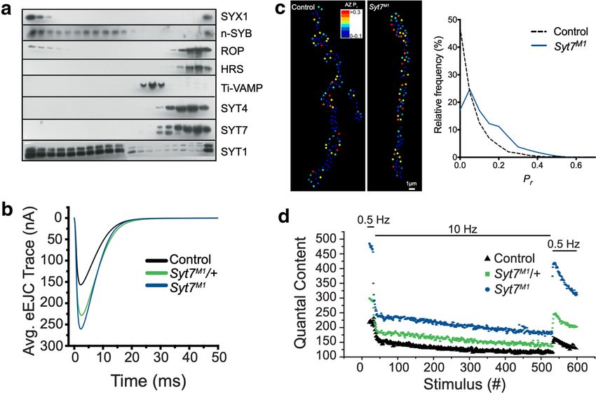
4340 M. C. Quiñones‑Frías, J. T. Littleton
for potential fusion or lipid mixing. Although C2 domains The multi-functional nature of SYT1 has made it chal-
are largely considered Ca2+-dependent lipid binding mod- lenging to separate its role for specific steps in SV traffick-
ules, they also mediate a host of Ca2+-independent interac- ing, as null mutations disrupt all its properties. This has
tions. As noted above, multiple SYT family members lack been partially addressed in Drosophila using point mutants
the required aspartate residues that coordinate Ca2+ bind- altering a single amino acid, or by rescuing null mutants
ing, suggesting these isoforms use the C2 domain as a pro- with SYT1 transgenes containing mutations in one or several
tein–protein interaction module instead. This is also the case residues [49, 51, 57, 62, 75, 131, 150, 152, 156, 164–168].
for SYTs that display C a2+-dependent lipid binding, as other Although these approaches have improved resolution, sev-
regions of the C2 domain interact with multiple effector pro- eral functions of SYT1 are likely to require similar interac-
teins, including the SNARE complex [38, 129]. In addition, tions and are difficult to separate. A second issue is that
a polybasic motif on the surface of the C2 domains of some some point mutants have dominant-negative activity and dis-
SYT proteins can interact with lipids in a C a2+-independent rupt release more than the complete absence of SYT1, con-
manner to facilitate vesicle docking [56, 98, 130–132]. founding interpretations for these alleles. This has been par-
Beyond the role of specific isoforms in SV fusion, SYT ticularly problematic for C2B domain Ca2+ binding mutants
family members also function as C a2+ sensors for DCV that act in a dominant-negative manner [49, 57, 62, 75, 156].
fusion that mediates release of neuropeptides and neuro- A final issue in interpreting defects in Syt1 mutants has been
modulators [108, 133–142]. Though C a2+ regulation of SV the predominant use of the 3rd instar NMJ preparation for
and DCV release by SYTs in presynaptic terminals repre- analysis. Although this synaptic connection is excellent for
sent the most-well characterized role, there is also evidence physiological studies, it requires animals to progress through
they function in postsynaptic vesicle trafficking [97, 104, earlier larval stages that last for days where compensation
105, 143–148]. Postsynaptic Ca2+ influx is required to regu- through homeostatic mechanisms could mask some pheno-
late membrane trafficking to support retrograde signaling types. In addition, reduced activity in Syt1 mutants may have
and postsynaptic neurotransmitter receptor cycling. Family consequences on synaptic development. A few studies have
members of the SYT4/SYT11 and SYT3 subgroups, along used the more difficult embryonic NMJ preparation where
with SYT1 and SYT7, have been implicated in these post- developmental defects and compensation are less likely to
synaptic processes. Finally, the group of SYT proteins with occur [57, 75, 151]. Acute inactivation approaches have also
degenerate Ca2+ binding sites within their C2 domains are been used to avoid these issues, though their specificity is
a2+-independent membrane
likely to participate in distinct C somewhat unclear [157, 169].
trafficking steps [98–100]. In general, little is known about The initial analysis of Drosophila Syt1 mutants revealed
these more obscure SYT isoforms. multiple defects in SV release at larval NMJs. The first
key observation was that evoked release was dramatically
reduced while spontaneous mini frequency was elevated
Synaptotagmin 1 functions as the major (Fig. 4a) [63, 154]. This result indicated SYT1 played dis-
Ca2+ sensor for triggering synchronous SV tinct roles in SV release by promoting evoked fusion and
fusion clamping Ca2+-independent spontaneous release. Although
reductions in evoked release can be secondary to many
SYT1 is the best characterized member of the SYT family. It potential defects in the SV cycle, the finding that the Ca2+
is found on SVs and functions as the primary Ca2+ sensor for dependence of release was altered in some Syt1 hypomor-
activating fast synchronous fusion in all species examined to phic mutants provided support for the protein acting as a
date. In mammals, SYT1 is the most highly expressed mem- Ca2+ trigger for fusion [51]. Subsequent work in mice identi-
ber of the SYT subgroup found on SVs and serves as the sole fied specific point mutants in SYT1 that shifted the Ca2+ sen-
Ca2+ sensor for synchronous fusion for most neurons in the sitivity of release, further pointing towards a release defect
CNS. SYT2 and SYT9 share largely redundant roles with tied to Ca2+ sensing [170]. Following the initial characteri-
SYT1 in regulating SV fusion for a smaller population of zation, additional studies of Drosophila Syt1 mutants indi-
CNS neurons and most PNS neurons [60, 101]. In Drosoph- cated its role in evoked release was twofold. Loss of SYT1
ila, a single gene encodes SYT1 and its loss disrupts evoked caused a dramatic reduction in fast synchronous release with
release as well [49, 51, 57, 63, 75, 149–156]. Although the enhanced release through the slower asynchronous fusion
role of SYT1 as a C a2+ sensor for fusion is widely accepted, pathway at both embryonic and larval NMJs [57, 75, 151].
the protein also functions in additional steps of the SV cycle, This work confirmed SYT1 was not the sole C a2+ sensor for
including docking, priming, and endocytosis [57, 150, 155, SV fusion, consistent with earlier studies on null mutants
157]. We focus our discussion primarily on SYT1 studies [102]. A two C a2+ sensor model for SV exocytosis emerged
performed in Drosophila, as multiple reviews describing in the field (Fig. 1c), with SYT1 driving the majority of
mammalian SYT1 are available [40, 44, 70, 86, 158–163]. rapid synchronous release and an unknown C a2+ sensor
13Function of Drosophila Synaptotagmins in membrane trafficking at synapses 4341
Fig. 4 Function of SYT1 in SV fusion. a Syt1 null mutants reduce primary and tripartite interface of the SYT1/SNARE/CPX complex.
synchronous fusion and enhance asynchronous release and mini fre- d Location of mutations disrupting the primary SNARE interface on
quency. Rescue with a Syt1 transgene with defective Ca2+ binding to the SYT1 C2B domain. The polybasic stretch is shown in grey and
C2A(*) and C2B(*) fails to support synchronous fusion and causes localizes to the opposite C2B surface. SNARE-binding mutations fail
higher rates of spontaneous release. b Overexpression of a C2B C a2+ to rescue release defects in Syt1 null mutants (right panel). e Location
binding mutant (D1,2N) suppresses release compared to overexpres- of the R250H mutation at the SYT1 dimer interface that disrupts oli-
sion of wildtype SYT1. Twenty essential residues mapping to C2A gomerization. This mutation reduces SV release (right panel), though
(blue) or C2B (magenta) were identified in an intragenic suppressor not as severely as SNARE-binding mutants. a Modified from [49], b,
screen that blocked the dominant-negative effects. c. Structure of the d, and e modified from [62], and c modified from [38]
activating asynchronous fusion. Candidates for the asyn- further demonstrated a profound reduction in release prob-
chronous Ca2+ sensor included other SYT family members, ability [22], confirming loss of SYT1 reduces synchronous
with SYT4 and SYT7 being the only attractive isoforms SV fusion and dramatically decreases the total number of
in Drosophila given their broad expression throughout the SVs released overall. The dual roles of SYT1 in activat-
nervous system [100]. However, electrophysiological stud- ing synchronous release and suppressing asynchronous
ies of Syt1/Syt4 and Syt1/Syt7 double mutants found these fusion were genetically separated and mapped to distinct
animals retain asynchronous release [153, 171], indicating C2 domains [75]. These observations supported a model
Ca2+-sensitive mechanisms outside of the SYT family con- whereby SYT1 actively inhibited the asynchronous path-
tribute to the slower release pathway at Drosophila synapses. way, versus the alternative where SVs normally destined
Initial models for SYT1 function in autaptic neuronal cul- for synchronous release were simply being released asyn-
tures from Syt1 mutant mice suggested loss of the protein chronously. Together, these data suggested some similarities
resulted in shifts in the time course of fusion without alter- between SYT1 and rapidly inactivating ion channels that
ing the actual number of SVs released. This view was not open to allow ion flow and undergo a second conformational
consistent with the dramatic reduction in the total number of change that inactivates the channel. For SYT1, Ca2+ bind-
SVs released in Drosophila Syt1 mutants. Subsequent stud- ing triggers a conformational state or set of interactions that
ies in mice confirmed observations from Drosophila that the increase the likelihood of a SV undergoing fusion within
total number of SVs released was dramatically reduced in several milliseconds. Shortly after, SYT1 undergoes another
the absence of SYT1 [53, 60, 64, 84, 101]. Quantal imaging conformational change, potentially triggered by C a 2+
of SV release at individual AZs in Drosophila Syt1 mutants unbinding, that actively inhibits SV release and reduces the
134342 M. C. Quiñones‑Frías, J. T. Littleton
probability of any slower asynchronous fusion events. This C2A Ca2+ binding appeared less important. However, these
dual activity of SYT1 helps ensure SVs are released within results have become more difficult to interpret with the
precise temporal windows that are needed for rapid neuronal observation that mutations in the C2B Ca2+ binding pocket
computations. cause dominant-negative phenotypes in Drosophila, mice,
Beyond a role in regulating fusion, a number of studies and humans [49, 57, 75, 152, 156, 164, 189–191]. Indeed,
also identified defects in SV docking, priming and endocy- release in Syt1 null mutants rescued with C2B D1,2N or
tosis, indicating SYT1 acts at multiple steps to regulate SV C2B D3,4N display less SV fusion that the null mutant itself
cycling in Drosophila [57, 150, 155, 157]. Although Syt1 [75, 156]. This has led to re-evaluation of how these sub-
mutants have defects in docking and endocytosis, the pro- stitutions are altering C2 domain function. One hypothesis
tein is not essential for either pathway as these processes is neutralization of the charged residues makes SYT1 more
continue with reduced efficacy in the absence of the pro- likely to undergo C a2+-independent membrane interactions
tein. Interactions between SYT1 and clathrin adapter pro- via the loops that potentially block fusion sites. As such, the
teins provide candidate mechanisms for its role in facilitating preferential role of C a2+ binding to C2B as the sole trigger
endocytosis [150, 172–179]. In contrast, how SYT1 supports for SV fusion requires re-evaluation. More recent mutagen-
SV docking is less clear. Ca2+-independent membrane inter- esis work using different amino acid substitutions within the
actions mediated by a polybasic amino acid stretch in the C2 domain loops that alter hydrophobicity of residues rather
two C2 domains, together with its SNARE-binding proper- than charge indicate C2A C a2+ binding also has an important
ties, represent candidate mechanisms. Overall, initial studies role in SYT1 [165, 192–195]. These observations suggest
of SYT1 provided models for how it regulates SV cycling. Ca2+ binding to both C2 domains is important for the full
Loss of SYT1 did not eliminate spontaneous or asynchro- activity of SYT1 in triggering synchronous SV fusion. Muta-
nous release and had no obvious effects on spontaneous tions disrupting C a2+ binding to both C2 domains of SYT1
SV fusion kinetics. As such, SYT1 does not appear to be still support SV docking and endocytosis, indicating these
essential for fusion or dynamics of the SV fusion pore itself, functions of the protein are largely Ca2+-independent [49],
but acts as a C a2+-dependent trigger to increase the likeli- though Ca2+ binding may enhance endocytosis rates [176].
hood SNARE-dependent fusion occurs and is limited to a Interestingly, C2A–C2B Ca2+ binding-defective SYT1 ani-
short temporal window. Although the kinetics of single SV mals have a far greater increase in spontaneous release com-
fusion events do not appear to be controlled by SYT1, there pared to null mutants (Fig. 4a), indicating restoring docking
is evidence the protein can regulate fusion pore kinetics of and endocytosis magnifies the role of SYT1 in clamping
DCVs and in reconstituted in vitro fusion systems [138, 139, spontaneous fusion that is less pronounced in nulls due to
180–188]. These early studies in Drosophila also highlighted reduced SV number [49].
differences between the mechanisms for fast synchronous SV One candidate mechanism for Ca2+-dependent membrane
release that require SYT1 versus asynchronous and sponta- binding by SYT1 is based on structural similarity of the
neous fusion that are negatively regulated by the protein. C2 domain loops to fusion peptides in viral fusion proteins
like hemagglutinin [196]. For viral fusion proteins, confor-
mational changes in the acidic environment of endosomes
Role of C2 domain C a2+ binding and SNARE expose the loops, allowing them to drive fusion between
interactions for Synaptotagmin 1 function viral and endosomal membranes and release viral content.
The fusion loops in SYT1 contain negatively charged aspar-
To directly characterize the role of the C2 domain Ca2+ tate residues nested within them, unlike the loops of viral
binding loops in Drosophila, a subset of the key aspartate fusion proteins. In the absence of Ca2+, these residues repel
residues in each loop (numbered D1–D5) were mutated to C2 domains from interacting with negatively charged phos-
neutralize the negative charge and prevent Ca2+ binding. The pholipid containing membranes. In the presence of Ca2+, the
most common approach was to generate D to N substitu- negative charge on these residues are neutralized and the C2
tions to create D1,2N and D3,4N mutants in either or both domains engage the lipid bilayer, potentially allowing the
C2 domains and use these transgenes to rescue Syt1 null loops to function similarly to those of viral fusion proteins.
mutants. This approach indicated the C2B domain had a Beyond the role of C a2+-dependent lipid binding by the
critical role in evoked release, with a near complete loss C2 domains, several other SYT1 interactions have been
of synchronous SV fusion in C2B D1,2N or C2B D3,4N implicated in SV release in Drosophila. Syt1 transgenes
mutants [49, 75, 156]. Similar transgenes disrupting the C2A containing mutations in the C2B Ca2+ binding pocket domi-
domain did not block evoked release, but rather failed to nantly disrupt release (Fig. 4b) and cause lethality even in
clamp asynchronous fusion [49, 75]. Together with stud- the presence of endogenous SYT1. Taking advantage of
ies in mice, these findings pointed towards C a2+ binding to this observation, chemical mutagenesis screens have been
the C2B domain as the critical trigger for exocytosis, while performed to identify intragenic SYT1 suppressors where
13Function of Drosophila Synaptotagmins in membrane trafficking at synapses 4343
randomly generated second-site mutations in the domi- suggest transmembrane tethering of SYT1 positions the
nant-negative transgene reduce the toxic effect [62]. This a2+ binding properties can
protein near release sites so its C
approach uncovered 20 essential residues within SYT1 that rapidly drive fusion. An additional study examined the rel-
mapped to distinct areas of the protein (Fig. 4b). The screen evance of the individual SYT1 C2 domains for release by
identified the five essential C2B domain residues (S332 generating otherwise wildtype SYT1 proteins with only C2A
(S279 in mammalian SYT1), R334 (R281), Y391 (Y338), or C2B alone, double C2A–C2A or C2B–C2B modules or
E348 (E295) and A455 (A402) that form the primary sur- reversed C2B–C2A order [49]. None of these manipulations
face interaction site that docks one side of the C2B domain rescued release defects in Syt1 null mutants, indicating both
onto the SNARE complex based on the elucidated structure C2 domains are uniquely required for release and must be
(Fig. 4c) [129]. Generation of several of these mutations into present in the correct sequence. Similar observations have
otherwise wildtype Syt1 transgenes demonstrated they failed been made for C2 domain swaps between mammalian SYT1
to rescue evoked release defects in Syt1 nulls (Fig. 4d). Dis- and SYT7, where chimeric proteins failed to rescue Syt1
ruption of the SYT1–SNARE complex interaction caused a mutant phenotypes in mice [207]. Although C2 domains
loss of synchronous release, enhanced asynchronous fusion, may have a generalized function, they have uniquely evolved
a reduction in the Ca2+ cooperativity curve, and elevated within each SYT family member, with the specific C2A and
spontaneous release. As such, these SYT1 functions require C2B domains serving distinct roles.
SNARE complex binding. SV docking and endocytosis
defects were not observed, suggesting these roles are inde-
pendent of SNARE binding or redundant with other SYT1 Open questions on the role
binding partners. of Synaptotagmin 1 in SV trafficking
Beyond SNARE interactions, mutations that disrupt and fusion
the C2B polybasic stretch on the opposite surface of the
C2B SNARE-binding domain were also identified in the It is widely accepted that the central role of SYT1 is centered
screen. In the absence of C a2+, SYT1 binds to PIP2 pre- around Ca2+-dependent lipid binding, but questions about
sent in the plasma membrane through a polybasic stretch additional interactions and their role in fusion are still being
of amino acids lining this surface of the C2B domain. This debated. For example, the precise role of SNARE binding is
interaction is predicted to occur before fusion as part of less clear. Studies in Drosophila provide strong in vivo evi-
the docking/priming process, helping to increase mem- dence this interaction is required for SYT1 to promote syn-
brane penetration of the C2 loops during Ca2+ entry by chronous fusion and suppress asynchronous release, but it is
prepositioning SYT1 at fusion sites [44, 56, 130, 132, 165, unknown when the interaction occurs during the SV cycle.
197–202]. Similar to prior structure function-studies, disrup- Part of this confusion is tied to the lack of a precise under-
a2+-independent lipid binding surface reduced
tion of this C standing of the structure of the primed SNARE complex
evoked release, but caused far milder phenotypes than loss before fusion in vivo. Are SNARE complexes partially zip-
of Ca2+-dependent lipid binding or SNARE interactions [62, pered before fusion as most models suggest, and can SYT1
131]. Together, these data suggest a model where SYT1 is bind to partially assembled SNAREs in this state? If so, this
prepositioned at the SV-plasma membrane interface in part would place the interaction at a pre-fusion point to position
through its C2B polybasic stretch and more fully by its C2B SYT1 for future membrane insertion and/or regulate SNARE
SNARE-binding interaction. Upon Ca2+ entry, the C2A and zippering. The interaction could also contribute to multim-
C2B Ca2+ binding loops pivot into the plasma membrane to erization of SNARE complexes through the oligomeriza-
drive lipid instability and facilitate complete SNARE zip- tion properties of SYT1. Alternatively, the interaction might
pering to activate fusion [44, 56, 203–206]. An alternative require fully assembled trans-SNARE complexes that are
model includes two separate pools of SYT1 on the SV, one predicted to only form during fusion. This would place the
bound to SNARE complexes to support docking/priming interaction during the fusion process itself or after fusion has
and another pool not associated with SNAREs that mediate been completed. Although early biochemical work suggested
Ca2+-dependent lipid binding and membrane insertion of SYT1 binding to individual t-SNAREs and the t-SNARE
the protein. complex was C a2+-dependent, current data suggest the pri-
The role of SYT1 tethering to SVs has also been exam- mary binding mode is Ca2+-independent. As such, SNARE
ined by generating transgenes lacking the transmembrane binding could act before, during or after Ca2+ entry.
domain or replacing the transmembrane domain with a Several other binding modes between SYT1 and the
myristoylation motif [152]. These transgenes could not sup- SNARE complex have also been described beyond the pri-
port fast synchronous fusion in Syt1 null mutants, though mary interface on the C2B surface that has genetic support
cytosolic C2 domains enhanced asynchronous release even in Drosophila. One model based on X-ray crystal structure
more than that observed in Syt1 nulls. These observations suggests formation of an alpha helix partially contributed by
134344 M. C. Quiñones‑Frías, J. T. Littleton
a piece of the SYT1 C2B domain and part of the SNARE- activates fusion? How specifically do SYT1 proteins prevent
binding protein CPX (Fig. 4c) [38]. This “tripartite” inter- asynchronous and spontaneous release; are these shared or
action mode brings in another key component for fusion distinct mechanisms from its positive role in activating syn-
regulation in CPX, but no genetic data has implicated this chronous release? Defining the timing and precise role for
region of SYT1 in Drosophila SV trafficking. However, stud- each of SYT1’s interactions in the various routes of SV traf-
ies in Drosophila indicate the activity of SYT1 and CPX ficking and fusion will require future studies with additional
are indeed intimately tied together in controlling SV release temporal and spatial resolution.
dynamics [37]. Cpx null mutants share similar phenotypes Additional questions in the field center on the role of
with milder Syt1 loss-of-function alleles. Both mutants have oligomerization in SYT1 function. Although SYT1 forms
decreased synchronous release, enhanced asynchronous oligomers in vitro [49–51, 219–224], the importance, sta-
fusion and elevated rates of spontaneous release [37, 41, bility, and timing of SYT1 oligomerization for SV release
208–210]. Although SYT1 is the primary candidate for a in vivo is unclear. Distinct SYT1 oligomerization states may
fusion clamp to prevent spontaneous release in mammals, also exist, as multimerization can occur via Ca2+-dependent
that role is mediated more prominently by CPX at inverte- and Ca2+-independent mechanisms and through interactions
brate synapses. Syt1 null mutants and animals with mutated mediated by the linker domain, residues on the C2A surface
C2A/C2B Ca2+ binding sites display a two–tenfold increase or via the C2B polybasic region [49, 82, 177, 219, 221–227].
in mini frequency compared to controls [37, 49, 51], while In the intragenic suppressor screen described above, an oli-
Cpx null mutants have a far greater increase that can exceed gomerization-defective C2A mutant (R250H) was identi-
100-fold [36, 37, 41, 95, 208–213]. As such, both proteins fied [62]. This mutant reduced evoked release but did cause
negatively regulate spontaneous release by clamping fusion, disruptions to the synchronicity of fusion (Fig. 4e). As such,
but the balance of their effects have shifted during evolution. the R250H mutant suggests a potential role for oligomeriza-
SYT1 and CPX also display genetic interactions in double tion in enhancing the number of SVs that fuse, with release
mutants or following co-overexpression, suggesting CPX is timing and suppression of asynchronous fusion independent
likely to exert many of its effects by regulating the timing of oligomerization. Although oligomerization is defective in
and activity of SYT1 during SV priming and fusion in Dros- R250H, this mutation also disrupts C2A–C2B intradomain
ophila [37]. An attractive hypothesis involves CPX bind- interactions within individual SYT1 monomers, suggesting
ing to the partially assembled SNARE complex to regulate either interaction could contribute to defects observed in this
when and how SYT1 interacts, allowing additional spatial mutant. It is also possible that other oligomerization states
and temporal control of SYT1-SNARE interactions during function independently of the C2A R250 residue to drive
priming and fusion. SYT1 activity. Defining the role of SYT1 multimers in SV
Another question remaining in the field is how many trafficking requires future studies in the field.
SNARE complexes and SYT proteins are required for
fusion in vivo. Most estimates suggest 3–5 SNARE com-
plexes drive evoked fusion, while the assembly of only
a single SNARE complex might be sufficient to trigger a2+ sensor
Is Synaptotagmin 7 the C
spontaneous release [214–217]. Increasing the number of for asynchronous release?
assembling SNARE complexes has been shown to increase
release probability for SV fusion in vivo [31]. In terms of The discovery that SYT1 mediates synchronous release
the number of SYTs required, estimates suggest ~ 15 SYT but does not abolish asynchronous fusion raised the pos-
proteins are found on a single SV [218]. Do SYTs have to sibility that other members of the SYT family might func-
bind every SNARE complex at the fusion site or is bind- tion as the asynchronous C a2+ sensor. SYT7 emerged as
ing to only one sufficient? Beyond the number of SYTs that a promising candidate due to its higher affinity for Ca2+,
bind SNAREs, how many SYTs are required overall to trig- strong lipid binding properties, slower dissociation from
ger an evoked fusion event and inhibit asynchronous and membranes, and widespread expression within the nervous
spontaneous release? If more than one SYT is required, do system [228–233]. These properties could potentially allow
they need to form an oligomeric complex, or can they act SYT7 to activate SV fusion farther away from AZs and after
independently? Do the number of SYT proteins activated the initial rise in Ca2+ influx (Fig. 1c). This would provide
during Ca2+ entry contribute to the steep Ca2+ cooperativity an attractive solution to the Ca2+ sensing problem for SV
curve for SV fusion, or is that effect mediated through other fusion: SYT1 functioning as the synchronous sensor and
mechanisms? Do individual SYTs bind SNAREs, while oth- its homolog SYT7 acting as the asynchronous sensor. In
ers drive Ca2+-dependent membrane insertion? What actual contrast to the work on SYT1 in Drosophila, which gener-
changes in the lipid structure of the presynaptic membrane ated many of the initial insights into in vivo mechanisms,
occur following penetration of the SYT1 C2 domains that studies of SYT7 have been primarily done in mice. As such,
13Function of Drosophila Synaptotagmins in membrane trafficking at synapses 4345
we discuss these data sets in more detail and compare them work in mammals have reported SYT7 localization in multi-
to recent studies on the Drosophila SYT7 homolog. ple compartments, including the plasma membrane, DCVs,
Drosophila SYT7 was not identified until the genome was lysosomes, endosomes and other internal membrane com-
sequenced in 2000 [99, 234, 235]. Syt7 resides on the small partments [100, 232, 236–243]. These diverse findings have
4th chromosome in a genomic region that had been poorly made it unclear as to which membrane compartment con-
characterized in the early 2000s and that lacked genetic tains SYT7, or if the protein is present in multiple locations
toolkits to easily generate mutations. As such, the protein that could participate in trafficking of distinct organelles.
remained uncharacterized for many years. The first efforts Further characterization of CRISPR-tagged SYT7 in
to examine Drosophila SYT7 function used RNAi knock- Drosophila found the protein was expressed in a highly inter-
down, RNA in situ hybridization and immunocytochemical connected tubular membrane compartment in presynaptic
studies with antibodies raised against the protein [100, 153]. boutons that made partial contact with numerous organelles,
These approaches demonstrated Syt7 mRNA was abundantly including endosomes, lysosomes and the plasma membrane
expressed in most neurons at levels similar to Syt1 and Syt4. [82]. Although SYT7 did not fully co-localize with any
Recent RNA profiling also indicates Syt7 mRNA is abundant known compartmental marker, the protein was enriched
in neurons (Fig. 3). Initial RNAi approaches that attempted within the peri-AZ (Fig. 3d, e), a synaptic domain sur-
to reduce the levels of SYT7 failed to show a synaptic phe- rounding AZs that contain proteins involved in endocytosis
notype [153]. SYT7 did not co-localize with SYT1 on SVs and protein sorting [82, 244–250]. Recent characterization
or enrich on synaptic plasma membranes with the t-SNARE of SYT7 localization using STORM in mouse hippocam-
Syntaxin on brain extracts fractionated from sucrose gradi- pal neurons also found the protein at the periphery of AZs
ents (Fig. 5a) [100]. Similarly, CRISPR tagging of endog- [251], though it is unclear if this represents localization to
enous SYT7 with GFP or RFP showed the protein was pre- the plasma membrane versus an internal SYT7 compart-
sent in presynaptic terminals but not localized to SVs [82]. ment. If SYT7 functions as the asynchronous Ca2+ sen-
Unlike an early study of mammalian SYT7 which identified sor that mediates SV fusion with the plasma membrane, it
the protein at the AZ plasma membrane [232], no enrich- should reside on SVs or at the AZ presynaptic membrane.
ment of SYT7 at Drosophila AZs was observed. Subsequent SYT7 does not appear to be enriched on either of these
Fig. 5 Enhanced release and SV replenishment in Syt7 mutants. a release probability at larval NMJs demonstrate Syt7 mutant AZs are
Western analysis of Drosophila brain extracts separated on a 10–30% shifted to higher Pr compared to controls. d Syt7 mutants undergo
sucrose gradient. SYX1 labels the plasma membrane (left-most frac- rapid depression during a stimulation train and recover their releas-
tion) with SVs (n-SYB/SYT1) in intermediate fractions. SYT7 and able SV pool more quickly than controls. Heterozygotes show an
SYT4 fractionate with other internal membrane compartments to intermediate phenotype. Panel a modified from [100] and panels b–d
distinct regions of the gradient. b Evoked release is increased in Syt7 modified from [82]
null mutants (Syt7M1) and Syt7 heterozygotes. c Optical mapping of
134346 M. C. Quiñones‑Frías, J. T. Littleton
compartments in Drosophila. Given the primary localiza- SYT7 is not required for synaptic transmission or non-neu-
tion of SYT7 within the peri-AZ, functions beyond directly ronal fusion events essential for survival. However, Syt7
mediating fusion of SVs with the presynaptic plasma mem- mutants displayed numerous defects at NMJ synapses in
brane must be considered. 3rd instar larvae, including a large increase in evoked SV
Initial studies using Drosophila SYT7 RNAi knockdown release, a larger RRP vesicle pool, and faster SV replen-
and mouse Syt7 mutants did not suggest a role for SYT7 ishment after strong stimulation (Fig. 5). Consistent with
in asynchronous release [153, 252]. The first observation increased release, mutant terminals displayed depression
indicating SYT7 might function in this slower release path- during stimulation trains compared to controls that facili-
way was based on morpholino knockdowns of zebrafish tated in the same extracellular [Ca2+]. Lowering extracel-
SYT7 that eliminated asynchronous release at NMJs [78]. lular Ca2+ to reduce release probability in Syt7 mutants
This finding triggered a wave of new studies to re-evaluate restored facilitation, indicating the process itself is intact at
SYT7’s role. Additional characterization of mammalian synapses without SYT7. These release defects were dosage-
SYT7 using siRNA knockdown suggested the protein was sensitive, with Syt7/+ heterozygotes showing intermediate
indeed responsible for asynchronous release in excitatory increases in SV fusion (Fig. 5b) and SYT7 overexpression
and inhibitory neurons [84]. These phenotypes were found suppressing evoked and spontaneous release. Similar dosage
by knocking down SYT7 in SYT1 deficient neurons with effects appear to be present at some mammalian synapses
enhanced asynchronous release, or in wildtype neurons dur- [264]. Optical imaging of evoked release in Drosophila Syt7
ing high frequency stimulation, making it easier to detect mutants demonstrated a higher release probability at single
changes in the asynchronous pathway. Subsequent work in AZs compared to controls (Fig. 5c) [82]. The enhancement
mouse Syt7 mutants supported a role for SYT7 as the asyn- in evoked release resulted in less asynchronous fusion and
chronous Ca2+ sensor [80, 83, 253–256]. Beyond asynchro- facilitation compared to controls, as observed in mammals.
nous release, some studies found manipulations of SYT7 However, increased release probability leads to faster SV
also disrupted facilitation, a form of short-term plasticity depletion and reduces the number of SVs available for fusion
where SV release is enhanced during closely spaced stimuli via these pathways. These data suggest a model where the
[80, 81, 257]. These data supported a model that SYT7 acts levels of SYT7 act as a dial to decrease or increase the
as the Ca2+ sensor for both asynchronous release and facili- amount of release to enable or mask facilitation mediated
tation in a subset of mammalian neurons. by an independent Ca2+ sensor, suggesting modulation of
Although much of the literature converged on the two Syt7 transcription or SYT7 protein levels could play a central
Ca2+ sensor model for SV fusion, the role of SYT7 in role in short-term plasticity.
asynchronous release remains controversial. Asynchro- To conclusively test if asynchronous release and facilita-
nous release at several mammalian CNS synapses is anti- tion requires SYT7, electrophysiological recordings were
correlated with the levels of the synchronous Ca2+ sensors performed in Syt1, Syt7 double mutants [82]. In animals
SYT1 and SYT2 and does not correlate with SYT7 levels lacking both proteins, an enhancement of both evoked and
[58]. In addition, another study found no defects in asyn- spontaneous release compared to Syt1 null mutants alone
chronous release in mouse Syt7 mutants [76]. Mammalian was observed. In addition, double mutants still displayed
SYT7 has also been suggested to play distinct roles in the synaptic facilitation. These results indicate SYT7 negatively
SV cycle, including driving SV replenishment during high regulates release probability for SVs destined for release
frequency stimulation [76], refilling of the readily releasable through both the spontaneous and evoked pathways in the
pool (RRP) [258], and targeting of SVs to specific endocytic absence or presence of SYT1. Since evoked release remains
pathways [77]. Such defects could potentially contribute to in animals lacking both SYT isoforms, the popular two C a2+
decreases in asynchronous fusion due to fewer SVs avail- sensor model of SYT1 acting as the synchronous sensor and
able for this phase of release. In addition to these roles, SYT7 as the sole asynchronous sensor is not applicable for
mammalian SYT7 regulates fusion of lysosomes with the Drosophila synapses. Similarly, the lack of SYT7 accumula-
plasma membrane in non-neuronal cells and DCV release in tion on SVs or at the AZ plasma membrane make it unlikely
certain neuronal populations [83, 133, 134, 237, 240–243, Ca2+ binding to SYT7 could directly trigger SV fusion since
256, 259–263]. the protein is not physically present at release sites. Given
The discrepancies in mammalian studies make it diffi- similarity in release kinetics and Ca2+ sensitivity for asyn-
cult to determine if SYT7 has a conserved primary role at chronous fusion between Drosophila and mammalian syn-
synapses, or if the protein has distinct functions in subpopu- apses [57, 75], it is perplexing that SYT7 could take over
lations of neurons. To examine conserved roles for SYT7 this role at mammalian synapses while a distinct C a2+ sensor
in Drosophila, CRISPR mutants in the locus were recently mediates asynchronous release in Drosophila. An alterna-
generated [82]. Consistent with mice lacking SYT7, Dros- tive model is that SYT7 controls the number of SVs avail-
ophila lacking the protein are viable and fertile, indicating able for asynchronous release, but a distinct C a2+-sensitive
13You can also read